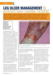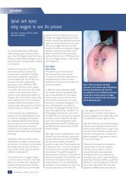
best practice guidelines: wound management in diabetic foot ulcers
best practice guidelines: wound management in diabetic foot ulcers
best practice guidelines: wound management in diabetic foot ulcers
- No tags were found...

Create successful ePaper yourself
Turn your PDF publications into a flip-book with our unique Google optimized e-Paper software.
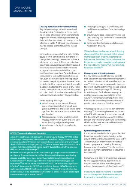
DFU WOUNDMANAGEMENTBOX 5: The use of advanced therapiesDress<strong>in</strong>g application and <strong>wound</strong> monitor<strong>in</strong>gRegularly review<strong>in</strong>g a patient's <strong>wound</strong> anddress<strong>in</strong>g is vital. For <strong>in</strong>fected or highly exud<strong>in</strong>g<strong>wound</strong>s, a healthcare professional should<strong>in</strong>spect the <strong>wound</strong> and change the dress<strong>in</strong>gdaily, and then every two or three days once the<strong>in</strong>fection is stable. A different type of dress<strong>in</strong>gmay be needed as the status of the <strong>wound</strong>changes.Some patients, especially those with mobilityissues or work commitments may prefer tochange their dress<strong>in</strong>gs themselves, or have arelative or carer to do it. These patients shouldbe advised about us<strong>in</strong>g aseptic technique andthe <strong>wound</strong> should cont<strong>in</strong>ue to be reviewedat regular <strong>in</strong>tervals by the MDFT or otherhealthcare team members. Patients should beencouraged to look out for signs of deterioration,such as <strong>in</strong>creased pa<strong>in</strong>, swell<strong>in</strong>g, odour,purulence or septic symptoms. In some cases(eg <strong>in</strong> the first few days of antibiotic therapy) itis a good idea to mark the extent of any cellulitiswith an <strong>in</strong>delible marker and tell the patientto contact the <strong>foot</strong>care team immediately if theredness moves substantially beyond the l<strong>in</strong>e.When apply<strong>in</strong>g dress<strong>in</strong>gs: Avoid bandag<strong>in</strong>g over toes as this maycause a tourniquet effect (<strong>in</strong>stead, layergauze over the toes and secure with a bandagefrom the metatarsal heads to a suitablepo<strong>in</strong>t on <strong>foot</strong>) Use appropriate techniques (eg avoid<strong>in</strong>gcreases and be<strong>in</strong>g too bulky) and take carewhen dress<strong>in</strong>g weight-bear<strong>in</strong>g areas Avoid strong adhesive tapes on fragilesk<strong>in</strong>Adjunctive treatments such as negative pressure <strong>wound</strong> therapy (NPWT), biologicaldress<strong>in</strong>gs, bioeng<strong>in</strong>eered sk<strong>in</strong> equivalents, hyperbaric oxygen therapy, plateletrich plasma and growth factors may be considered, if appropriate and where availablefor DFUs that are not progress<strong>in</strong>g 95 . These techniques require advanced cl<strong>in</strong>icaldecision mak<strong>in</strong>g and should be carried out only by practitioners with appropriateskills and anatomical knowledge 22 .However, such therapies represent considerable greater product cost than standardtherapy. These costs may be justified if they result <strong>in</strong> improved ulcer heal<strong>in</strong>g,reduced morbidity, fewer lower-extremity amputations and improved patientfunctional status 95 . There is a good level of evidence for some biological sk<strong>in</strong>equivalents 95 as well as for the use of NPWT <strong>in</strong> DFU patients without significant<strong>in</strong>fection 96 . More recently, NPWT with <strong>in</strong>stillation therapy (NPWTi) us<strong>in</strong>g antisepticagents (eg PHMB) has become available. Although there are limited dataon its benefits, it could be considered when there is a need for <strong>wound</strong> cleans<strong>in</strong>g ortreatment with topical antimicrobials 97 . Avoid tight bandag<strong>in</strong>g at the fifth toe andthe fifth metatarsal head (trim the bandageback) Ensure <strong>wound</strong> dead space is elim<strong>in</strong>ated (eguse a dress<strong>in</strong>g that conforms to the contoursof the <strong>wound</strong> bed) Remember that <strong>foot</strong>wear needs to accommodateany dress<strong>in</strong>g.Wounds should be cleansed at each dress<strong>in</strong>gchange and after debridement with a <strong>wound</strong>cleans<strong>in</strong>g solution or sal<strong>in</strong>e. Cleans<strong>in</strong>g canhelp remove devitalised tissue, re-balance thebioburden and reduce exudate to help preparethe <strong>wound</strong> bed for heal<strong>in</strong>g 98 . It may also helpto remove biofilms 88 .Manag<strong>in</strong>g pa<strong>in</strong> at dress<strong>in</strong>g changesIt is now acknowledged that many patients —even those with neuropathy or neuroischaemia— can feel pa<strong>in</strong> due to their <strong>wound</strong> or a procedure99 . It is important to <strong>in</strong>corporate strategiesto prevent trauma and m<strong>in</strong>imise <strong>wound</strong>-relatedpa<strong>in</strong> dur<strong>in</strong>g dress<strong>in</strong>g changes 100 . This may<strong>in</strong>clude the use of soft silicone dress<strong>in</strong>gs andavoid<strong>in</strong>g unnecessary manipulation of the<strong>wound</strong> 99 . Remember also that patients whohave lost the protective pa<strong>in</strong> sensation are atgreater risk of trauma at dress<strong>in</strong>g change 99 .When appropriate, use low- or non-adherentdress<strong>in</strong>gs 99 . If a dress<strong>in</strong>g becomes encrustedor is difficult to remove, it is important to soakthe dress<strong>in</strong>g with sal<strong>in</strong>e or a <strong>wound</strong> irrigationsolution and check the <strong>wound</strong> and surround<strong>in</strong>gsk<strong>in</strong> for evidence of trauma and <strong>in</strong>fection ondress<strong>in</strong>g removal 99 .Epithelial edge advancementIt is important to debride the edges of the ulcerto remove potential physical barriers to thegrowth of the epithelium across the ulcer bed 74 .The demarcation l<strong>in</strong>e between any necrotictissue or gangrene and healthy tissue maybecome a site of <strong>in</strong>fection 48 . Similar problemscan be seen when a gangrenous toe touches ahealthy toe 50 .Conversely, ‘die-back’ is an abnormal responseto over-aggressive sharp debridement. It<strong>in</strong>volves necrosis at the <strong>wound</strong> edge andextends through previously healthy tissue 50 .If the <strong>wound</strong> does not respond to standard<strong>wound</strong> <strong>management</strong> <strong>in</strong>terventions despitetreatment of the underly<strong>in</strong>g cause and316 BEST PRACTICEBEST PRACTICEGUIDELINESGUIDELINES:FOR SKINWOUNDAND WOUNDMANAGEMENTCARE ININEPIDERMOLYSISDIABETIC FOOTBULLOSAULCERS