best practice guidelines: wound management in diabetic foot ulcers
best practice guidelines: wound management in diabetic foot ulcers
best practice guidelines: wound management in diabetic foot ulcers
- No tags were found...

Create successful ePaper yourself
Turn your PDF publications into a flip-book with our unique Google optimized e-Paper software.
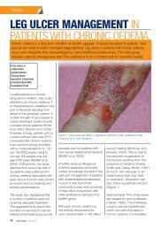
ASSESSING DFUsAssess<strong>in</strong>g DFUsPatients with a DFU need to be assessed holistically and <strong>in</strong>tr<strong>in</strong>sic and extr<strong>in</strong>sicfactors consideredFor the non-specialist practitioner, the key skillrequired is know<strong>in</strong>g when and how to refer apatient with a DFU to the multidiscipl<strong>in</strong>ary <strong>foot</strong>careteam (MDFT; see page 19). Patients witha DFU should be assessed by the team with<strong>in</strong>one work<strong>in</strong>g day of presentation — or sooner<strong>in</strong> the presence of severe <strong>in</strong>fection 22,36,37 . Inmany places, however, MDFTs do not exist andpractitioners <strong>in</strong>stead work as <strong>in</strong>dividuals. Inthese situations, the patient’s prognosis oftendepends on a particular practitioner’s knowledgeand <strong>in</strong>terest <strong>in</strong> the <strong>diabetic</strong> <strong>foot</strong>.Patients with a DFU need to be assessed holisticallyto identify <strong>in</strong>tr<strong>in</strong>sic and extr<strong>in</strong>sic factors.This should encompass a full patient history<strong>in</strong>clud<strong>in</strong>g medication, comorbidities and diabetesstatus 38 . It should also take <strong>in</strong>to considerationthe history of the <strong>wound</strong>, previous DFUs oramputations and any symptoms suggestive ofneuropathy or PAD 28 .EXAMINATION OF THE ULCERA physical exam<strong>in</strong>ation should determ<strong>in</strong>e: Is the <strong>wound</strong> predom<strong>in</strong>antly neuropathic,ischaemic or neuroischaemic? If ischaemic, is there critical limb ischaemia? Are there any musculoskeletal deformities? What is the size/depth/location of the<strong>wound</strong>? What is the colour/status of the <strong>wound</strong>bed?— Black (necrosis)— Yellow, red, p<strong>in</strong>k Is there any exposed bone? Is there any necrosis or gangrene? Is the <strong>wound</strong> <strong>in</strong>fected? If so, are theresystemic signs and symptoms of <strong>in</strong>fection(such as fevers, chills, rigors, metabolic<strong>in</strong>stability and confusion)? Is there any malodour? Is there local pa<strong>in</strong>? Is there any exudate? What is the level ofproduction (high, moderate, low, none),colour and consistency of exudate, and is itpurulent? What is the status of the <strong>wound</strong> edge(callus, maceration, erythema, oedema,underm<strong>in</strong><strong>in</strong>g)?Document<strong>in</strong>g ulcer characteristicsRecord<strong>in</strong>g the size, depth, appearance and locationof the DFU will help to establish a basel<strong>in</strong>efor treatment, develop a treatment plan andmonitor any response to <strong>in</strong>terventions. It isimportant also to assess the area around the<strong>wound</strong>: erythema and maceration <strong>in</strong>dicateadditional complications that may h<strong>in</strong>der<strong>wound</strong> heal<strong>in</strong>g 38 .Digitally photograph<strong>in</strong>g DFUs at the firstconsultation and periodically thereafterto document progress is helpful 39 . This isparticularly useful for ensur<strong>in</strong>g consistencyof care among healthcare practitioners,facilitat<strong>in</strong>g telehealth <strong>in</strong> remote areas andillustrat<strong>in</strong>g improvement to the patient.TESTING FOR LOSS OF SENSATIONTwo simple and effective tests for peripheralneuropathy are commonly used: 10g (Semmes-We<strong>in</strong>ste<strong>in</strong>) monofilament Standard 128Hz tun<strong>in</strong>g fork.The 10g monofilament is the most frequentlyused screen<strong>in</strong>g tool to determ<strong>in</strong>e the presenceof neuropathy <strong>in</strong> patients with diabetes 28 . Itshould be applied at various sites along theplantar aspect of the <strong>foot</strong>. Guidel<strong>in</strong>es vary <strong>in</strong> thenumber of sites advocated, but the <strong>in</strong>ternationalconsensus is to test at three sites (see Figure5) 7 . A positive result is the <strong>in</strong>ability to feel themonofilament when it is pressed aga<strong>in</strong>st the<strong>foot</strong> with enough force to bend it 40 .Neuropathy is also demonstrated by an <strong>in</strong>abilityto sense vibration from a standard tun<strong>in</strong>g fork.Other tests are available, such as the biothesiometerand neurothesiometer, which are morecomplex handheld devices for assess<strong>in</strong>g theperception of vibration.Do not test for neuropathy <strong>in</strong> areas of callusas this can mask feel<strong>in</strong>g from any of theneuropathy test<strong>in</strong>g devices and may give afalse-positive result.Be aware that patients with small nerve fibredamage and <strong>in</strong>tact sensory nerves may have34 BEST PRACTICEBEST PRACTICEGUIDELINESGUIDELINES:FOR SKINWOUNDAND WOUNDMANAGEMENTCARE ININEPIDERMOLYSISDIABETIC FOOTBULLOSAULCERS