best practice guidelines: wound management in diabetic foot ulcers
best practice guidelines: wound management in diabetic foot ulcers
best practice guidelines: wound management in diabetic foot ulcers
- No tags were found...

You also want an ePaper? Increase the reach of your titles
YUMPU automatically turns print PDFs into web optimized ePapers that Google loves.
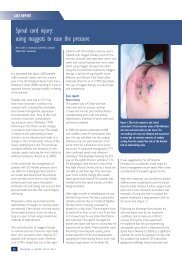
ASSESSING DFUsFIGURE 5: Procedure for carry<strong>in</strong>g out the monofilament test (adapted from 7 )The International Work<strong>in</strong>g Group on the Diabetic Foot (IWGDF) recommends the follow<strong>in</strong>g procedure for carry<strong>in</strong>g out themonofilament test. The sensory exam<strong>in</strong>ation should be carried out <strong>in</strong> a quiet and relaxed sett<strong>in</strong>g The patient should close their eyes so as not to see whether or where the exam<strong>in</strong>erapplies the monofilament The patient should sit sup<strong>in</strong>e with both feet level First apply the monofilament on the patient’s hands or on the <strong>in</strong>side of the arm sothey know what to expect Apply the monofilament perpendicular to the sk<strong>in</strong> surface with sufficient force tobend or buckle the monofilament Ask the patient:— Whether they feel the pressure applied (yes/no)— Where they feel the pressure (left <strong>foot</strong>/right <strong>foot</strong>) Apply the monofilament along the perimeter of (not on) the ulcer site Do not allow the monofilament to slide across the sk<strong>in</strong> or make repetitive contact atthe test site The total duration of the approach (sk<strong>in</strong> contact and removal of the monofilament)should be around 2 seconds Apply the monofilament to each site three times, <strong>in</strong>clud<strong>in</strong>g at least one additional‘mock’ application <strong>in</strong> which no filament is applied Encourage the patient dur<strong>in</strong>g test<strong>in</strong>g by giv<strong>in</strong>g positive feedback— Protective sensation is present at each site if the patient correctly answers twoout of three applications— Protective sensation is absent with two out of three <strong>in</strong>correct answersNote: The monofilament should not be used on more than 10 patients without arecovery period of 24 hoursUs<strong>in</strong>g a monofilament to test for neuropathya pa<strong>in</strong>ful neuropathy. They may describesharp, stabb<strong>in</strong>g, burn<strong>in</strong>g, shoot<strong>in</strong>g or electricshock type pa<strong>in</strong>, which may be worse at nightand can disrupt sleep 41 . The absence of coldwarmdiscrim<strong>in</strong>ation may help to identifypatients with small nerve fibre damage.TESTING FOR VASCULAR STATUSPalpation of peripheral pulses should be arout<strong>in</strong>e component of the physical exam<strong>in</strong>ationand <strong>in</strong>clude assessment of the femoral,popliteal and pedal (dorsalis pedis andposterior tibial) pulses. Assessment of pulsesis a learned skill and has a high degree of<strong>in</strong>ter-observer variability, with high falsepositiveand false-negative rates. The dorsalispedis pulse is reported to be absent <strong>in</strong> 8.1%of healthy <strong>in</strong>dividuals, and the posterior tibialpulse is absent <strong>in</strong> 2.0%. Nevertheless, theabsence of both pedal pulses, when assessedby an experienced cl<strong>in</strong>ician, strongly suggeststhe presence of pedal vascular disease 42 . Ifthere is any doubt regard<strong>in</strong>g diagnosis of PAD,it is important to refer to a specialist for a fullvascular assessment.Where available, Doppler ultrasound, anklebrachialpressure <strong>in</strong>dex (ABPI) and Dopplerwaveform may be used as adjuncts tothe cl<strong>in</strong>ical f<strong>in</strong>d<strong>in</strong>gs when carried out by acompetent practitioner. Toe pressures, and<strong>in</strong> some <strong>in</strong>stances, transcutaneous oxygenmeasurement (where equipment is available),may be useful for measur<strong>in</strong>g localtissue perfusion.An ischaemic <strong>foot</strong> may appear p<strong>in</strong>k and relativelywarm even with impaired perfusion dueto arteriovenous shunt<strong>in</strong>g. Delayed discolouration(rubor) or venous refill<strong>in</strong>g greater thanfive seconds on dependency may <strong>in</strong>dicatepoor arterial perfusion 43 .Other signs suggestive of ischaemia <strong>in</strong>clude 40 : Claudication: pa<strong>in</strong> <strong>in</strong> the leg muscles andCOMMON TERMS EXPLAINEDCritical limb ischaemia: this isa chronic manifestation of PADwhere the arteries of the lowerextremities are severely blocked.This results <strong>in</strong> ischaemic pa<strong>in</strong><strong>in</strong> the feet or toes even at rest.Complications of poor circulation<strong>in</strong>clude sk<strong>in</strong> <strong>ulcers</strong> or gangrene.If left untreated it will result <strong>in</strong>amputation of the affected limb.Acute limb ischaemia: thisoccurs when there is a suddenlack of blood flow to a limb andis due to either an embolism orthrombosis. Without surgicalrevascularisation, completeacute ischaemia leads to extensivetissue necrosis with<strong>in</strong> sixhours.BEST PRACTICE GUIDELINES: WOUND MANAGEMENT IN DIABETIC FOOT ULCERS 5