- Page 2:
MEDICAL RADIOLOGYRadiation Oncology
- Page 6:
Jiade J. Lu, MD, MBAAssociate Profe
- Page 10:
PrefaceNasopharyngeal cancer is a u
- Page 14:
Contents1 The Epidemiology of Nasop
- Page 18:
The Epidemiology of Nasopharyngeal
- Page 22:
The Epidemiology of Nasopharyngeal
- Page 26:
The Epidemiology of Nasopharyngeal
- Page 30:
The Epidemiology of Nasopharyngeal
- Page 34:
10 M-S. Zeng and Y-X. Zengenvironme
- Page 38:
12 M-S. Zeng and Y-X. Zengmedicinal
- Page 42:
14 M-S. Zeng and Y-X. Zengloss and
- Page 46:
16 M-S. Zeng and Y-X. ZengNPC sampl
- Page 50:
18 M-S. Zeng and Y-X. Zengthe basal
- Page 54:
20 M-S. Zeng and Y-X. Zengmultistep
- Page 58:
22 M-S. Zeng and Y-X. ZengHui AB, L
- Page 62:
24 M-S. Zeng and Y-X. ZengSnudden D
- Page 66:
Molecular Signaling Pathways 3in Na
- Page 70:
Molecular Signaling Pathways in Nas
- Page 74:
Molecular Signaling Pathways in Nas
- Page 78:
Molecular Signaling Pathways in Nas
- Page 82:
Molecular Signaling Pathways in Nas
- Page 86:
Molecular Signaling Pathways in Nas
- Page 90:
Molecular Signaling Pathways in Nas
- Page 94:
Natural History, Presenting Symptom
- Page 98:
Natural History, Presenting Symptom
- Page 102:
Natural History, Presenting Symptom
- Page 106:
Natural History, Presenting Symptom
- Page 110:
Natural History, Presenting Symptom
- Page 114:
Natural History, Presenting Symptom
- Page 118:
54 P-J. Lou, W-L. Hsu, Y-C. Chien e
- Page 122:
56 P-J. Lou, W-L. Hsu, Y-C. Chien e
- Page 126:
58 P-J. Lou, W-L. Hsu, Y-C. Chien e
- Page 130:
60 P-J. Lou, W-L. Hsu, Y-C. Chien e
- Page 134:
62 P-J. Lou, W-L. Hsu, Y-C. Chien e
- Page 138:
64 P-J. Lou, W-L. Hsu, Y-C. Chien e
- Page 142:
66 K. S. Lohobviously surmise that
- Page 146:
68 K. S. LohTable 6.2. Causal assoc
- Page 152:
Pathology of Nasopharyngeal Carcino
- Page 156:
Pathology of Nasopharyngeal Carcino
- Page 160:
Pathology of Nasopharyngeal Carcino
- Page 164:
Pathology of Nasopharyngeal Carcino
- Page 168:
Pathology of Nasopharyngeal Carcino
- Page 172:
Imaging in the Diagnosis and Stagin
- Page 176:
Imaging in the Diagnosis and Stagin
- Page 180:
Imaging in the Diagnosis and Stagin
- Page 184:
Imaging in the Diagnosis and Stagin
- Page 188:
Imaging in the Diagnosis and Stagin
- Page 192:
Imaging in the Diagnosis and Stagin
- Page 196:
Imaging in the Diagnosis and Stagin
- Page 200:
96 J-C. Linon the outcome. The aim
- Page 204:
98 J-C. LinTable 9.1. Summary of pr
- Page 208:
100 J-C. LinTable 9.2. Summary of p
- Page 212:
102 J-C. Lintumor volume was found
- Page 216:
104 J-C. Lin(Neel et al. 1984a; Nee
- Page 220:
106 J-C. Linradiotherapy became hig
- Page 224:
108 J-C. Linsis. However, the false
- Page 228:
110 J-C. LinTable 9.5. Summary of p
- Page 232:
112 J-C. LinTable 9.6. Summary of o
- Page 236:
114 J-C. LinTable 9.7. Summary of t
- Page 240:
116 J-C. LinTable 9.8. Summary of p
- Page 244:
118 J-C. Lincontaining 59 NPC patie
- Page 248:
120 J-C. Linpatients of three nonke
- Page 252:
122 J-C. Linfactor for metastasis-f
- Page 256:
124 J-C. LinTable 9.9. Prognostic i
- Page 260:
126 J-C. LinTable 9.10. Prognostic
- Page 264:
128 J-C. Linthat it may trigger the
- Page 268:
130 J-C. LinChen Y, Liu MZ, Liang S
- Page 272:
132 J-C. LinHwang CF, Cho CL, Huang
- Page 276:
134 J-C. LinMa BB, Leung SF, Hui EP
- Page 280:
136 J-C. LinWang CC (1991) Improved
- Page 284:
138 R. Ove, R. R. Allison, and J. J
- Page 288:
140 R. Ove, R. R. Allison, and J. J
- Page 292:
142 R. Ove, R. R. Allison, and J. J
- Page 296:
144 R. Ove, R. R. Allison, and J. J
- Page 300:
146 R. Ove, R. R. Allison, and J. J
- Page 304:
Drug Therapy for Nasopharyngeal Car
- Page 308:
Drug Therapy for Nasopharyngeal Car
- Page 312:
Drug Therapy for Nasopharyngeal Car
- Page 316:
Drug Therapy for Nasopharyngeal Car
- Page 320:
Drug Therapy for Nasopharyngeal Car
- Page 324:
Drug Therapy for Nasopharyngeal Car
- Page 328:
The Intergroup 0099 Trial for Nasop
- Page 332:
The Intergroup 0099 Trial for Nasop
- Page 336:
The Intergroup 0099 Trial for Nasop
- Page 340:
Concurrent Chemotherapy-Enhanced Ra
- Page 344:
Concurrent Chemotherapy-Enhanced Ra
- Page 348:
Concurrent Chemotherapy-Enhanced Ra
- Page 352:
Concurrent Chemotherapy-Enhanced Ra
- Page 356:
Concurrent Chemotherapy-Enhanced Ra
- Page 360:
Concurrent Chemotherapy-Enhanced Ra
- Page 364:
Concurrent Chemotherapy-Enhanced Ra
- Page 368:
Concurrent Chemotherapy-Enhanced Ra
- Page 372:
184 A. W. M. Lee14.2Nonrandomized S
- Page 376:
186 A. W. M. Leescheduled. With a m
- Page 380:
188 A. W. M. LeeTable 14.2. Phase I
- Page 384:
190 A. W. M. LeeLR-FFR was slightly
- Page 388:
192 A. W. M. LeeLee AW, Lau WH, Tun
- Page 392:
194 B. C. Gohthe similarly favorabl
- Page 396:
196 B. C. GohLi Zhang, Chong Zhao,
- Page 400:
198 L. Kong, J. J. Lu, and N. Leean
- Page 404:
200 L. Kong, J. J. Lu, and N. Leetr
- Page 408:
202 L. Kong, J. J. Lu, and N. LeeFi
- Page 412:
204 L. Kong, J. J. Lu, and N. LeeFi
- Page 416:
206 L. Kong, J. J. Lu, and N. LeeFi
- Page 420:
208 L. Kong, J. J. Lu, and N. LeeTa
- Page 424:
210 L. Kong, J. J. Lu, and N. Leevo
- Page 428:
Selection and Delineation of Target
- Page 432:
Selection and Delineation of Target
- Page 436:
Selection and Delineation of Target
- Page 440:
Selection and Delineation of Target
- Page 444:
Selection and Delineation of Target
- Page 448:
Selection and Delineation of Target
- Page 452:
Selection and Delineation of Target
- Page 456:
Selection and Delineation of Target
- Page 460:
Selection and Delineation of Target
- Page 464:
Selection and Delineation of Target
- Page 468:
Post-treatment Follow-Up of Patient
- Page 472:
Post-treatment Follow-Up of Patient
- Page 476:
Post-treatment Follow-Up of Patient
- Page 480:
Post-treatment Follow-Up of Patient
- Page 484:
Management of Patients with Failure
- Page 488:
Management of Patients with Failure
- Page 492:
Management of Patients with Failure
- Page 496:
Management of Patients with Failure
- Page 500:
Management of Patients with Failure
- Page 504:
Management of Patients with Failure
- Page 508:
254 W. I. Weimanagement of recurren
- Page 512:
256 W. I. Weicisplatin and 5-fluoro
- Page 516:
258 W. I. WeiTumourAdipose tissueFi
- Page 520:
260 W. I. Weiradiation dose is high
- Page 524:
262 W. I. Weiartery has to be prote
- Page 528:
264 W. I. WeiFig. 20.21. The mucope
- Page 532:
Systemic Treatment for Incurable Re
- Page 536:
Systemic Treatment for Incurable Re
- Page 540:
Systemic Treatment for Incurable Re
- Page 544:
Systemic Treatment for Incurable Re
- Page 548:
Long-Term Complications in the Trea
- Page 552:
Long-Term Complication in the Treat
- Page 556:
Long-Term Complication in the Treat
- Page 560:
Long-Term Complication in the Treat
- Page 564:
Long-Term Complication in the Treat
- Page 568:
Long-Term Complication in the Treat
- Page 572:
Long-Term Complication in the Treat
- Page 576: Long-Term Complication in the Treat
- Page 580: Long-Term Complication in the Treat
- Page 584: Long-Term Complication in the Treat
- Page 588: Nasopharyngeal Cancer in Pediatric
- Page 592: Nasopharyngeal Cancer in Pediatric
- Page 596: Nasopharyngeal Cancer in Pediatric
- Page 600: Nasopharyngeal Cancer in Pediatric
- Page 604: Nasopharyngeal Cancer in Pediatric
- Page 608: Nasopharyngeal Cancer in Pediatric
- Page 612: Nasopharyngeal Cancer in Pediatric
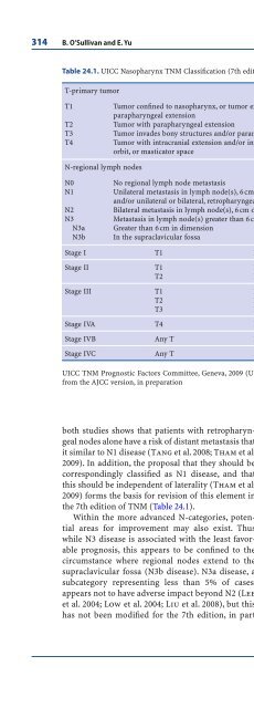
- Page 616: Staging of Nasopharyngeal Carcinoma
- Page 620: Staging of Nasopharyngeal carcinoma
- Page 624: Staging of Nasopharyngeal carcinoma
- Page 630: 316 B. O’Sullivan and E. YuFig. 2
- Page 634: 318 B. O’Sullivan and E. Yudefine
- Page 638: 320 B. O’Sullivan and E. YuFig. 2
- Page 642: 322 B. O’Sullivan and E. YuLee CC
- Page 646: 324 Subject Index- - hand-foot synd
- Page 650: 326 Subject IndexInverse planning (
- Page 654: 328 Subject IndexRadiosensitizer, 1
- Page 658: List of ContributorsRon R. Allison,
- Page 662: List of Contributors 333Shaojun Lin
- Page 666: Medical RadiologyDiagnostic Imaging
- Page 670: Medical RadiologyDiagnostic Imaging


