Program Book - Keck School of Medicine of USC - University of ...
Program Book - Keck School of Medicine of USC - University of ...
Program Book - Keck School of Medicine of USC - University of ...
- No tags were found...
Create successful ePaper yourself
Turn your PDF publications into a flip-book with our unique Google optimized e-Paper software.
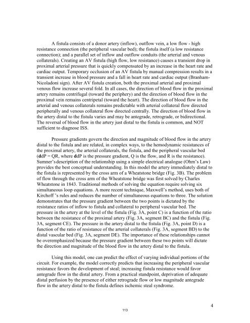
A fistula consists <strong>of</strong> a donor artery (inflow), outflow vein, a low flow - highresistance connection (the peripheral vascular bed); the fistula itself (a low resistanceconnection); and a parallel set <strong>of</strong> inflow and outflow conduits (the arterial and venouscollaterals). Creating an AV fistula (high flow, low resistance) causes a transient drop inproximal arterial pressure that is quickly compensated by an increase in the heart rate andcardiac output. Temporary occlusion <strong>of</strong> an AV fistula by manual compression results in atransient increase in blood pressure and a fall in heart rate and cardiac output (Branham-Nicoladoni sign). After AV fistula creation, both the proximal arterial and proximalvenous flow increase several fold. In all cases, the direction <strong>of</strong> blood flow in the proximalartery remains centrifugal (toward the periphery) and the direction <strong>of</strong> blood flow in theproximal vein remains centripetal (toward the heart). The direction <strong>of</strong> blood flow in thearterial and venous collaterals remains predictable with arterial collateral flow directedperipherally and venous collateral flow directed centrally. The direction <strong>of</strong> blood flow inthe artery distal to the fistula varies and may be antegrade, retrograde, or bidirectional.The reversal <strong>of</strong> blood flow in the artery just distal to the fistula is common, and NOTsufficient to diagnose ISS.Pressure gradients govern the direction and magnitude <strong>of</strong> blood flow in the arterydistal to the fistula and are related, in complex ways, to the hemodynamic resistances <strong>of</strong>the proximal artery, the arterial collaterals, the fistula, and the peripheral vascular bed(ddP = QR, where ddP is the pressure gradient, Q is the flow, and R is the resistance).Sumner’s description <strong>of</strong> the relationship using a simple electrical analogue (Ohm’s Law)provides the best conceptual understanding. In this model the artery immediately distal tothe fistula is represented by the cross arm <strong>of</strong> a Wheatstone bridge (Fig. 3B). The problem<strong>of</strong> flow through the cross arm <strong>of</strong> the Wheatstone bridge was first solved by CharlesWheatstone in 1843. Traditional methods <strong>of</strong> solving the equation require solving sixsimultaneous loop equations. A more recent technique, Maxwell’s method, uses both <strong>of</strong>Kirch<strong>of</strong>f ’s rules and reduces the number <strong>of</strong> simultaneous equations to three. The solutiondemonstrates that the pressure gradient between the two points is dictated by theresistance ratios <strong>of</strong> inflow to fistula and collateral to peripheral vascular bed. Thepressure in the artery at the level <strong>of</strong> the fistula (Fig. 3A, point C) is a function <strong>of</strong> the ratiobetween the resistance <strong>of</strong> the proximal artery (Fig. 3A, segment BC) and the fistula (Fig.3A, segment CE). The pressure in the artery distal to the fistula (Fig. 3A, point D) is afunction <strong>of</strong> the ratio <strong>of</strong> resistance <strong>of</strong> the arterial collaterals (Fig. 3A, segment BD) to thedistal vascular bed (Fig. 3A, segment DE). The importance <strong>of</strong> these relationships cannotbe overemphasized because the pressure gradient between these two points will dictatethe direction and magnitude <strong>of</strong> the blood flow in the artery distal to the fistula.Using this model, one can predict the effect <strong>of</strong> varying individual portions <strong>of</strong> thecircuit. For example, the model correctly predicts that increasing the peripheral vascularresistance favors the development <strong>of</strong> steal; increasing fistula resistance would favorantegrade flow in the distal artery. From a practical standpoint, deprivation <strong>of</strong> adequatedistal perfusion by the presence <strong>of</strong> either retrograde flow or low magnitude antegradeflow in the artery distal to the fistula defines ischemic steal syndrome.1134


