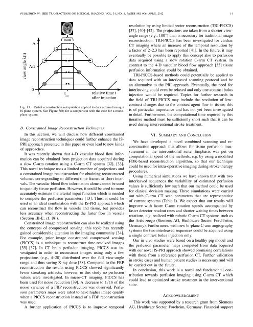
PUBLISHED IN: IEEE TRANSA<strong>CT</strong>IONS ON MEDICAL IMAGING, VOL. 31, NO. 4, PAGES 892–906, APRIL 2012 14Fig. 13. Partial reconstruction interpolation applied to data acquired using abi-plane system. See Figure 3(b) for a comparison with the case for a monoplanesystem.B. Constrained Image Reconstruction TechniquesIn this section, we will discuss how different constrainedimage reconstruction techniques could further enhance the IS-PRI approach presented in this paper or even lead to new kindsof approaches.It was recently shown that 4-D vascular blood flow informationcan be obtained from projection data acquired duringa slow C-arm rotation using a C-arm <strong>CT</strong> system [32], [33].This novel technique uses a limited number of projections anda constrained image reconstruction for obtaining reconstructedvolumes corresponding to different time frames at short intervals.The vascular blood flow information alone cannot be usedto quantify tissue perfusion. However, it could be used to moreaccurately estimate the arterial input function which is neededto compute the perfusion parameters [13]. Thus, it could beused in an ideal combination with the IS-PRI approach whichcan reconstruct the flow in vessels and tissue, but providesless accuracy when reconstructing the faster flow in vessels(Section III-E; cf. [8]).Constrained image reconstruction can also be realized usingthe concepts of compressed sensing; this topic has recentlygained considerable attention in the imaging community [34].For example, prior image constrained compressed sensing(PICCS) is a technique to reconstruct time-resolved images[35]–[37]. In <strong>CT</strong> brain perfusion imaging, PICCS was investigatedin order to reconstruct images using only a fewprojections (e.g., 4–20) distributed over the full view-anglerange and thus saving X-ray dose [38]. Compared to the FBPreconstruction the results using PICCS showed significantlyfewer streaking artifacts; however, in this study no perfusionvalues were investigated. In micro-<strong>CT</strong> imaging, PICCS hasbeen used for noise reduction [39]. A decrease to 1/16 of thenoise variance of a FBP reconstruction was observed. <strong>Perfusion</strong>parameters maps were rated to have higher image qualitywhen a PICCS reconstruction instead of a FBP reconstructionwas used.A further application of PICCS is to improve temporalresolution by using limited sector reconstruction (TRI-PICCS)[37], [40]–[42]. The projections are taken from a shorter viewanglerange (e.g., 100 ◦ ) than is necessary for traditional imagereconstruction. TRI-PICCS has been investigated for cardiac<strong>CT</strong> imaging where an increase of the temporal resolution bya factor of 2–2.3 has been reported [41]. In the future, it mayeventually be possible to apply this concept also to perfusiondata acquired using a slow rotation C-arm <strong>CT</strong> system. Incontrast to the 4-D vascular blood flow approach [33] tissueperfusion information could be obtained.TRI-PICCS-based methods could potentially be applied todata acquired with an interleaved scanning protocol and bean alternative to the PRI approach. Eventually, the need forinterleaving could even be relaxed and only one contrast bolusinjection would be required. Topics for further research inthe field of TRI-PICCS may include the resolution of lowcontrastchanges due to the contrast agent flow in tissue; thisis of particular importance and has not yet been investigatedin detail. Furthermore, the computational time required by thisiterative method must be sufficiently short such that it can beused during interventional stroke treatment.VI. SUMMARY AND CONCLUSIONWe have developed a novel combined scanning and reconstructionapproach that allows for tissue perfusion measurementin the interventional suite. Emphasis was put oncomputational speed of the methods, e.g. by using a modifiedFDK-based reconstruction algorithm, so that our techniquecould be used for intra-operative imaging during stroke therapyprocedures.<strong>Using</strong> numerical simulations we have shown that with twointerleaved sequences the variability of estimated perfusionvalues is sufficiently low such that our method could be usedfor clinical decision making. These simulations were carriedout with C-arm <strong>CT</strong> scan parameters that are representativeof current systems (Table I). We expect that our results willimprove with faster C-arm rotation speeds accompanied byfaster detector readout rates and shorter waiting times betweenrotations, e.g. realized with robotic C-arm <strong>CT</strong> systems such asthe Artis zeego (Siemens AG, Healthcare Sector, Forchheim,Germany). Furthermore, with new bi-plane C-arm angiographysystems the two interleaved sequences could be acquired usinga single contrast bolus injection only.Our in vivo studies were based on a healthy pig model andthe perfusion parameter maps computed from data acquiredwith our novel IS-PRI approach showed promising correlationswith those from a reference perfusion <strong>CT</strong>. Further validationin stroke cases and human patient studies is necessary and willbe carried out in the future.In conclusion, this work is a novel and fundamental contributiontowards perfusion imaging using C-arm <strong>CT</strong> whichcould lead to optimized stroke treatment in the interventionalsuite.ACKNOWLEDGMENTThis work was supported by a research grant from SiemensAG, Healthcare Sector, Forcheim, Germany. Financial support
PUBLISHED IN: IEEE TRANSA<strong>CT</strong>IONS ON MEDICAL IMAGING, VOL. 31, NO. 4, PAGES 892–906, APRIL 2012 15was also provided through NIH 1K99EB007676 and the LucasFoundation. The authors gratefully acknowledge funding ofthe Erlangen Graduate School in Advanced Optical Technologies(SAOT) by the German Research Foundation (DFG) inthe framework of the German excellence initiative. We thankGünter Lauritsch and Michael Manhart for helpful adviceduring preparation of the manuscript and Frank Dennerleinfor providing parts of the simulation framework. The experttechnical assistance of Wendy Baumgardner, Pamela Hertz andLior Molvin is gratefully acknowledged.REFERENCES[1] R. G. González, J. A. Hirsch, W. J. Koroshetz, M. H. Lev, and P. W.Schaefer, Eds., Acute Ischemic Stroke, 1st ed. Berlin, Germany:Springer, 2006.[2] A. Fieselmann, “<strong>Interventional</strong> perfusion imaging using C-arm computedtomography: Algorithms and clinical evaluation,” Ph.D. dissertation,Friedrich-Alexander University of Erlangen-Nuremberg, Germany, 2011.[3] N. Strobel, O. Meissner, J. Boese, T. Brunner, B. Heigl, M. Hoheisel,G. Lauritsch, M. Nagel, M. Pfister, E.-P. Rührnschopf, B. Scholz,B. Schreiber, M. Spahn, M. Zellerhoff, and K. Klingenbeck-Regn,Multislice <strong>CT</strong>, 3rd ed. Berlin, Germany: Springer, 2009, ch. 3D <strong>Imaging</strong>with Flat-Detector C-<strong>Arm</strong> Systems, pp. 33–51.[4] C. Neukirchen, M. Giordano, and S. Wiesner, “An iterative method fortomographic x-ray perfusion estimation in a decomposition model-basedapproach,” Medical Physics, vol. 37, no. 12, pp. 6125–6141, 2010.[5] L. A. Feldkamp, L. C. Davis, and J. W. Kress, “Practical cone-beamalgorithm,” Journal of the Optical Society of America, vol. A1, pp. 612–619, 1984.[6] C. Rohkohl, B. Keck, H. Hofmann, and J. Hornegger, “Rabbit<strong>CT</strong>– an open platform for benchmarking 3D cone-beam reconstructionalgorithms,” Medical Physics, vol. 36, no. 9, pp. 3940–3944, 2009.[7] P. Montes and G. Lauritsch, “A temporal interpolation approach fordynamic reconstruction in perfusion <strong>CT</strong>,” Medical Physics, vol. 34,no. 7, pp. 3077–3092, 2007.[8] A. Fieselmann, A. Ganguly, Y. Deuerling-Zheng, M. Zellerhoff,J. Boese, J. Hornegger, and R. Fahrig, “A dynamic reconstructionapproach for cerebral blood flow quantification with an interventionalC-arm <strong>CT</strong>,” in Proc. IEEE International Symposium on Biomedical<strong>Imaging</strong> 2010, Rotterdam, The Netherlands, 2010, pp. 53–56.[9] A. Ganguly, A. Fieselmann, M. Marks, J. Rosenberg, J. Boese,Y. Deuerling-Zheng, M. Straka, G. Zaharchuk, R. Bammer, andR. Fahrig, “Cerebral <strong>CT</strong> perfusion using an interventional C-arm imagingsystem: Cerebral blood flow measurements,” American Journal ofNeuroradiology, vol. 32, no. 8, pp. 1525–1531, 2011.[10] G. Lauritsch, J. Boese, L. Wigström, H. Kemeth, and R. Fahrig,“Towards cardiac C-arm computed tomography,” IEEE Transactions onMedical <strong>Imaging</strong>, vol. 25, no. 7, pp. 922–934, 2006.[11] M. Wintermark, W. S. Smith, N. U. Ko, M. Quist, P. Schnyder, and W. P.Dillon, “Dynamic perfusion <strong>CT</strong>: optimizing the temporal resolution andcontrast volume for calculation of perfusion <strong>CT</strong> parameters in strokepatients.” American Journal of Neuroradiology, vol. 25, no. 5, pp. 720–729, 2004.[12] M. Wiesmann, S. Berg, G. Bohner, R. Klingebiel, V. Schöpf, B. M.Stoeckelhuber, I. Yousry, J. Linn, and U. Missler, “Dose reduction indynamic perfusion <strong>CT</strong> of the brain: effects of the scan frequency onmeasurements of cerebral blood flow, cerebral blood volume, and meantransit time.” European Radiology, vol. 18, no. 12, pp. 2967–2974, 2008.[13] A. Fieselmann, M. Kowarschik, A. Ganguly, J. Hornegger, and R. Fahrig,“Deconvolution-based <strong>CT</strong> and MR brain perfusion measurement: Theoreticalmodel revisited and practical implementation details,” InternationalJournal of Biomedical <strong>Imaging</strong>, vol. 2011, article ID 467563, 20pages, 2011.[14] A. Fieselmann, F. Dennerlein, Y. Deuerling-Zheng, J. Boese, R. Fahrig,and J. Hornegger, “A model for filtered backprojection reconstructionartifacts due to time-varying attenuation values in perfusion C-arm <strong>CT</strong>,”Physics in Medicine and Biology, vol. 56, no. 12, pp. 3701–3717, 2011.[15] C. T. Badea, S. M. Johnston, E. Subashi, Y. Qi, L. W. Hedlund, andG. A. Johnson, “Lung perfusion imaging in small animals using 4Dmicro-<strong>CT</strong> at heartbeat temporal resolution,” Medical Physics, vol. 37,no. 1, pp. 54–62, 2010.[16] M. D. Silver, “A method for including redundant data in computedtomography,” Medical Physics, vol. 27, no. 4, pp. 773–774, 2000.[17] W. H. Press, S. A. Teukolsky, W. T. Vetterling, and B. P. Flannery,Numerical Recipes, 3rd ed. Cambridge, UK: Cambridge UniversityPress, 2007.[18] F. N. Fritsch and R. E. Carlson, “Monotone piecewise cubic interpolation,”SIAM Journal on Numerical Analysis, vol. 17, pp. 238–246, 1980.[19] M. D. Buhmann, Radial Basis Functions: Theory and Implementations,1st ed. Cambridge, UK: Cambridge University Press, 2003.[20] L. Østergaard, R. M. Weisskoff, D. A. Chesler, C. Gyldensted, andB. R. Rosen, “High resolution measurement of cerebral blood flow usingintravascular tracer bolus passages. part I: Mathematical approach andstatistical analysis,” Magnetic Resonance in Medicine, vol. 36, no. 5,pp. 715–725, 1996.[21] H. M. Silvennoinen, L. M. Hamberg, L. Valanne, and G. J. Hunter,“Increasing contrast agent concentration improves enhancement in firstpass<strong>CT</strong> perfusion,” American Journal of Neuroradiology, vol. 28, no. 7,pp. 1299–1303, 2007.[22] J. Bredno, M. E. Olszewski, and M. Wintermark, “Simulation model forcontrast agent dynamics in brain perfusion scans,” Magnetic Resonancein Medicine, vol. 6, no. 1, pp. 280–290, 2010.[23] A. Fieselmann, “DIPPO — a digital brain perfusion phantom,”www5.cs.fau.de/~fieselma/data/, accessed December 20, 2011.[24] A. Fieselmann, A. Ganguly, Y. Deuerling-Zheng, J. Boese, J. Hornegger,and R. Fahrig, “Automatic measurement of contrast bolus distributionin carotid arteries using a C-arm angiography system to support interventionalperfusion imaging,” in Proc. SPIE Medical <strong>Imaging</strong> 2011:Visualization, Image-Guided Procedures, and Modeling, vol. 7964, LakeBuena Vista, USA, 2011, pp. 79 641W1–6.[25] K. Kudo, M. Sasaki, K. Yamada, S. Momoshima, H. Utsunomiya,H. Shirato, and K. Ogasawara, “Differences in <strong>CT</strong> perfusion mapsgenerated by different commercial software: Quantitative analysis byusing identical source data of acute stroke patients,” Radiology, vol.254, no. 1, pp. 200–209, 2010.[26] F. Zanderigo, A. Bertoldo, G. Pillonetto, and C. Cobelli, “Nonlinearstochastic regularization to characterize tissue residue function in bolustrackingMRI: Assessment and comparison with SVD, block-circulantSVD, and Tikhonov,” IEEE Transactions on Biomedical Engineering,vol. 56, no. 5, pp. 1287–1297, 2009.[27] K. Kudo, S. Terae, C. Katoh, M. Oka, T. Shiga, N. Tamaki, andK. Miyasaka, “Quantitative cerebral blood flow measurement withdynamic perfusion <strong>CT</strong> using the vascular-pixel elimination method:Comparison with H 152 O positron emission tomography,” American Journalof Neuroradiology, vol. 24, no. 3, pp. 419–426, 2003.[28] P. Sauleaua, E. Lapoublea, D. Val-Lailleta, and C.-H. Malberta, “The pigmodel in brain imaging and neurosurgery,” The International Journal ofAnimal Biosciences, vol. 3, no. 8, pp. 1138–1151, 2009.[29] M. Mehra, N. Henninger, J. A. Hirsch, J. Chueh, A. K. Wakhloo, andM. J. Gounis, “Preclinical acute ischemic stroke modeling,” Journal ofNeuro<strong>Interventional</strong> Surgery, 2011, (in press).[30] D. Morhard, C. D. Wirth, G. Fesl, C. Schmidt, M. F. Reiser, C. R.Becker, and B. Ertl-Wagner, “Advantages of extended brain perfusioncomputed tomography: 9.6 cm coverage with time resolved computedtomography-angiography in comparison to standard stroke-computedtomography,” Investigative Radiology, vol. 45, no. 7, pp. 363–369, 2010.[31] R. Fahrig, R. Dixon, T. Payne, R. Morin, A. Ganguly, and N. Strobel,“Dose and image quality for a cone-beam C-arm <strong>CT</strong> system,” MedicalPhysics, vol. 33, no. 12, pp. 4541–50, 2006.[32] C. A. Mistretta, E. Oberstar, B. Davis, E. Brodsky, and C. M. Strother,“4D-DSA and 4D fluoroscopy: preliminary implementation,” in Proc.SPIE Medical <strong>Imaging</strong> 2010: Physics of Medical <strong>Imaging</strong>, vol. 7622,San Diego, USA, 2010, pp. 7 622 271–8.[33] C. A. Mistretta, “Sub-Nyquist acquisition and constrained reconstructionin time resolved angiography,” Medical Physics, vol. 38, no. 6, pp. 2975–2985, 2011.[34] M. Fornasier and H. Rauhut, Handbook of Mathematical Methods in<strong>Imaging</strong>, 1st ed. Berlin, Germany: Springer, 2011, ch. CompressiveSensing, pp. 187–228.[35] G.-H. Chen, J. Tang, and S. Leng, “Prior image constrained compressedsensing (PICCS),” Proc. SPIE Photons Plus Ultrasound: <strong>Imaging</strong> andSensing 2008, vol. 6856, p. 685618, 2008.[36] ——, “Prior image constrained compressed sensing (PICCS): a methodto accurately reconstruct dynamic <strong>CT</strong> images from highly undersampledprojection data sets,” Medical Physics, vol. 35, no. 2, pp. 660–663, 2008.[37] G.-H. Chen, J. Tang, B. Nett, Z. Qi, S. Leng, and T. Szczykutowicz,“Prior image constrained compressed sensing (PICCS) and applicationsin X-ray computed tomography,” Current Medical <strong>Imaging</strong> Reviews,vol. 6, no. 2, pp. 119–134, 2010.