2006 merck/merial - School of Veterinary Medicine - Louisiana State ...
2006 merck/merial - School of Veterinary Medicine - Louisiana State ...
2006 merck/merial - School of Veterinary Medicine - Louisiana State ...

Create successful ePaper yourself
Turn your PDF publications into a flip-book with our unique Google optimized e-Paper software.
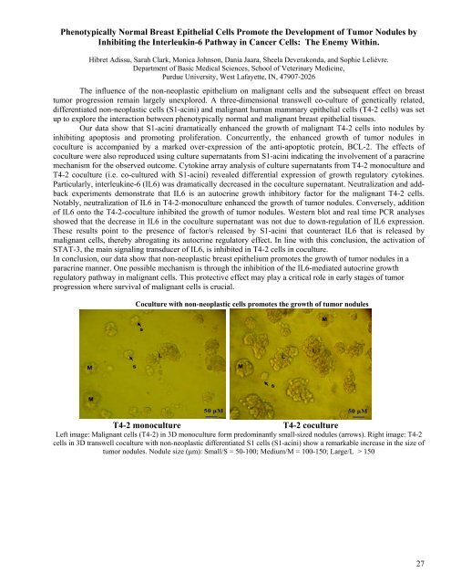
Phenotypically Normal Breast Epithelial Cells Promote the Development <strong>of</strong> Tumor Nodules byInhibiting the Interleukin-6 Pathway in Cancer Cells: The Enemy Within.Hibret Adissu, Sarah Clark, Monica Johnson, Dania Jaara, Sheela Deverakonda, and Sophie Lelièvre.Department <strong>of</strong> Basic Medical Sciences, <strong>School</strong> <strong>of</strong> <strong>Veterinary</strong> <strong>Medicine</strong>,Purdue University, West Lafayette, IN, 47907-2026The influence <strong>of</strong> the non-neoplastic epithelium on malignant cells and the subsequent effect on breasttumor progression remain largely unexplored. A three-dimensional transwell co-culture <strong>of</strong> genetically related,differentiated non-neoplastic cells (S1-acini) and malignant human mammary epithelial cells (T4-2 cells) was setup to explore the interaction between phenotypically normal and malignant breast epithelial tissues.Our data show that S1-acini dramatically enhanced the growth <strong>of</strong> malignant T4-2 cells into nodules byinhibiting apoptosis and promoting proliferation. Concurrently, the enhanced growth <strong>of</strong> tumor nodules incoculture is accompanied by a marked over-expression <strong>of</strong> the anti-apoptotic protein, BCL-2. The effects <strong>of</strong>coculture were also reproduced using culture supernatants from S1-acini indicating the involvement <strong>of</strong> a paracrinemechanism for the observed outcome. Cytokine array analysis <strong>of</strong> culture supernatants from T4-2 monoculture andT4-2 coculture (i.e. co-cultured with S1-acini) revealed differential expression <strong>of</strong> growth regulatory cytokines.Particularly, interleukine-6 (IL6) was dramatically decreased in the coculture supernatant. Neutralization and addbackexperiments demonstrate that IL6 is an autocrine growth inhibitory factor for the malignant T4-2 cells.Notably, neutralization <strong>of</strong> IL6 in T4-2-monoculture enhanced the growth <strong>of</strong> tumor nodules. Conversely, addition<strong>of</strong> IL6 onto the T4-2-coculture inhibited the growth <strong>of</strong> tumor nodules. Western blot and real time PCR analysesshowed that the decrease in IL6 in the coculture supernatant was not due to down-regulation <strong>of</strong> IL6 expression.These results point to the presence <strong>of</strong> factor/s released by S1-acini that counteract IL6 that is released bymalignant cells, thereby abrogating its autocrine regulatory effect. In line with this conclusion, the activation <strong>of</strong>STAT-3, the main signaling transducer <strong>of</strong> IL6, is inhibited in T4-2 cells in coculture.In conclusion, our data show that non-neoplastic breast epithelium promotes the growth <strong>of</strong> tumor nodules in aparacrine manner. One possible mechanism is through the inhibition <strong>of</strong> the IL6-mediated autocrine growthregulatory pathway in malignant cells. This protective effect may play a critical role in early stages <strong>of</strong> tumorprogression where survival <strong>of</strong> malignant cells is crucial.Coculture with non-neoplastic cells promotes the growth <strong>of</strong> tumor nodulessMMsLMLLsM50 μM 50 μMT4-2 monoculture T4-2 cocultureLeft image: Malignant cells (T4-2) in 3D monoculture form predominantly small-sized nodules (arrows). Right image: T4-2cells in 3D transwell coculture with non-neoplastic differentiated S1 cells (S1-acini) show a remarkable increase in the size <strong>of</strong>tumor nodules. Nodule size (μm): Small/S = 50-100; Medium/M = 100-150; Large/L > 15027