- Page 4 and 5:
DIAGNOSIS AND MANAGEMENT OFPITUITAR
- Page 6:
To my beloved Hazel, whose love and
- Page 10:
PrefacePituitary tumors represent a
- Page 14 and 15:
ContributorsCHARLES F. ABBOUD, MD,
- Page 16 and 17:
Color PlatesColor Plates appear aft
- Page 18 and 19:
2 RANDALL, SCHEITHAUER, AND KOVACSF
- Page 20 and 21:
4 RANDALL, SCHEITHAUER, AND KOVACSr
- Page 22 and 23:
6 RANDALL, SCHEITHAUER, AND KOVACSw
- Page 24 and 25:
8 RANDALL, SCHEITHAUER, AND KOVACSt
- Page 26 and 27:
10 RANDALL, SCHEITHAUER, AND KOVACS
- Page 28 and 29:
12 RANDALL, SCHEITHAUER, AND KOVACS
- Page 30 and 31:
14 RHOTONFigure 2-1 Osseous relatio
- Page 32 and 33:
16 RHOTONFigure 2-3 Septa in the sp
- Page 34 and 35:
18 RHOTONFigure 2-5 Stepwise dissec
- Page 36 and 37:
20 RHOTONFigure 2-7 (continued on n
- Page 38 and 39:
22 RHOTONFigure 2-8 (continued on n
- Page 40 and 41:
24 RHOTONFigure 2-9 Relationships i
- Page 42 and 43:
26 RHOTONFigure 2-11 Six sagittal s
- Page 44 and 45:
28 RHOTONFigure 2-13 (continued on
- Page 46 and 47:
30 RHOTONFigure 2-15 Anterosuperior
- Page 48 and 49:
32 RHOTONFigure 2-17 (continued on
- Page 50 and 51:
34 RHOTONFigure 2-18 Right anterior
- Page 52 and 53:
36 RHOTONOPHTHALMIC ARTERY The opht
- Page 54 and 55:
38 RHOTONFigure 2-21 Anterior cereb
- Page 56 and 57:
40 RHOTONcrosses the optic chiasm t
- Page 58 and 59:
42 GOTH, MAKARA, AND GERENDAIFigure
- Page 60 and 61:
44 GOTH, MAKARA, AND GERENDAImedial
- Page 62 and 63:
46 GOTH, MAKARA, AND GERENDAIplatel
- Page 64 and 65:
48 GOTH, MAKARA, AND GERENDAIcomple
- Page 66 and 67:
50 GOTH, MAKARA, AND GERENDAImembra
- Page 68 and 69:
52 GOTH, MAKARA, AND GERENDAITable
- Page 70 and 71:
54 GOTH, MAKARA, AND GERENDAITable
- Page 73 and 74:
CHAPTER 4 / EPIDEMIOLOGY OF PITUITA
- Page 75 and 76:
Taiwan-South part, 1983-1992 23.2%
- Page 77 and 78:
CHAPTER 4 / EPIDEMIOLOGY OF PITUITA
- Page 79 and 80:
CHAPTER 4 / EPIDEMIOLOGY OF PITUITA
- Page 81 and 82:
CHAPTER 4 / EPIDEMIOLOGY OF PITUITA
- Page 83 and 84:
CHAPTER 4 / EPIDEMIOLOGY OF PITUITA
- Page 85:
CHAPTER 4 / EPIDEMIOLOGY OF PITUITA
- Page 88 and 89:
72 SHIMON AND MELMEDTable 1G Protei
- Page 90 and 91:
74 SHIMON AND MELMEDnal allelic mut
- Page 92 and 93:
76 SHIMON AND MELMEDgrowth factor s
- Page 94 and 95:
78 SHIMON AND MELMED35. Lowy DR, Wi
- Page 97 and 98:
CHAPTER 6 / TUMORIGENESIS EXPERIMEN
- Page 99 and 100:
CHAPTER 6 / TUMORIGENESIS EXPERIMEN
- Page 101 and 102:
CHAPTER 6 / TUMORIGENESIS EXPERIMEN
- Page 103 and 104:
CHAPTER 6 / TUMORIGENESIS EXPERIMEN
- Page 105 and 106:
CHAPTER 6 / TUMORIGENESIS EXPERIMEN
- Page 107 and 108:
CHAPTER 7 / PITUITARY ADENOMAS AND
- Page 109 and 110:
CHAPTER 7 / PITUITARY ADENOMAS AND
- Page 111 and 112:
CHAPTER 7 / PITUITARY ADENOMAS AND
- Page 113 and 114:
CHAPTER 7 / PITUITARY ADENOMAS AND
- Page 115 and 116:
CHAPTER 7 / PITUITARY ADENOMAS AND
- Page 117 and 118:
CHAPTER 7 / PITUITARY ADENOMAS AND
- Page 119 and 120:
CHAPTER 7 / PITUITARY ADENOMAS AND
- Page 121 and 122:
CHAPTER 7 / PITUITARY ADENOMAS AND
- Page 123 and 124:
CHAPTER 7 / PITUITARY ADENOMAS AND
- Page 125 and 126:
CHAPTER 7 / PITUITARY ADENOMAS AND
- Page 127 and 128:
CHAPTER 7 / PITUITARY ADENOMAS AND
- Page 129 and 130:
CHAPTER 7 / PITUITARY ADENOMAS AND
- Page 131 and 132:
CHAPTER 7 / PITUITARY ADENOMAS AND
- Page 133 and 134:
CHAPTER 7 / PITUITARY ADENOMAS AND
- Page 135 and 136:
CHAPTER 7 / PITUITARY ADENOMAS AND
- Page 137 and 138:
CHAPTER 7 / PITUITARY ADENOMAS AND
- Page 139 and 140:
CHAPTER 7 / PITUITARY ADENOMAS AND
- Page 141 and 142:
CHAPTER 7 / PITUITARY ADENOMAS AND
- Page 143 and 144:
CHAPTER 7 / PITUITARY ADENOMAS AND
- Page 145 and 146:
CHAPTER 7 / PITUITARY ADENOMAS AND
- Page 147 and 148:
CHAPTER 7 / PITUITARY ADENOMAS AND
- Page 149 and 150:
CHAPTER 7 / PITUITARY ADENOMAS AND
- Page 151 and 152:
CHAPTER 7 / PITUITARY ADENOMAS AND
- Page 153 and 154:
CHAPTER 7 / PITUITARY ADENOMAS AND
- Page 155 and 156:
CHAPTER 7 / PITUITARY ADENOMAS AND
- Page 157 and 158:
CHAPTER 7 / PITUITARY ADENOMAS AND
- Page 159 and 160:
CHAPTER 7 / PITUITARY ADENOMAS AND
- Page 161 and 162:
CHAPTER 7 / PITUITARY ADENOMAS AND
- Page 163 and 164:
CHAPTER 7 / PITUITARY ADENOMAS AND
- Page 165 and 166:
CHAPTER 7 / PITUITARY ADENOMAS AND
- Page 167 and 168:
CHAPTER 7 / PITUITARY ADENOMAS AND
- Page 169 and 170:
CHAPTER 7 / PITUITARY ADENOMAS AND
- Page 171 and 172:
CHAPTER 8 / MOLECULAR PATHOLOGY 155
- Page 173 and 174:
CHAPTER 8 / MOLECULAR PATHOLOGY 157
- Page 175 and 176:
CHAPTER 8 / MOLECULAR PATHOLOGY 159
- Page 177 and 178:
CHAPTER 8 / MOLECULAR PATHOLOGY 161
- Page 179:
CHAPTER 8 / MOLECULAR PATHOLOGY 163
- Page 182 and 183:
166 VANCETable 1Lesions of the Sell
- Page 184 and 185:
168 VANCEFigure 9-1 Twenty-four-hou
- Page 186 and 187:
170 VANCEclude that every neurosurg
- Page 188 and 189:
172 VANCEgaly, prolactinoma). Pitui
- Page 190 and 191:
174 STIVER AND SHARPEFigure 10-1 Co
- Page 192 and 193:
176 STIVER AND SHARPEFigure 10-5 Cr
- Page 194 and 195:
178 STIVER AND SHARPEFigure 10-8 Fi
- Page 196 and 197:
180 STIVER AND SHARPETable 1Inciden
- Page 198 and 199:
182 STIVER AND SHARPEFigure 10-11 (
- Page 200 and 201:
184 STIVER AND SHARPEFigure 10-13 F
- Page 202 and 203:
186 STIVER AND SHARPEFigure 10-15 F
- Page 204 and 205:
188 STIVER AND SHARPEFigure 10-17 W
- Page 206 and 207:
190 STIVER AND SHARPEpituitary apop
- Page 208 and 209:
192 STIVER AND SHARPEin extrafoveal
- Page 210 and 211:
194 STIVER AND SHARPEinto the empty
- Page 212 and 213:
196 STIVER AND SHARPE5. Hoyt WF. Co
- Page 214 and 215:
198 STIVER AND SHARPE104. Wertenbak
- Page 216 and 217:
200 STIVER AND SHARPE204. Nakane T,
- Page 218 and 219:
202 EMERY AND KUCHARCZYKFigure 11-1
- Page 220 and 221:
204 EMERY AND KUCHARCZYKFigure 11-4
- Page 222 and 223:
206 EMERY AND KUCHARCZYKFigure 11-7
- Page 224 and 225:
208 EMERY AND KUCHARCZYKFigure 11-1
- Page 226 and 227:
210 EMERY AND KUCHARCZYKFigure 11-1
- Page 228 and 229:
212 EMERY AND KUCHARCZYKFigure 11-1
- Page 230 and 231:
214 EMERY AND KUCHARCZYKFigure 11-2
- Page 232 and 233:
216 EMERY AND KUCHARCZYK3. Kulkarni
- Page 235 and 236:
CHAPTER 12 / PET IN SELLAR TUMORS 2
- Page 237 and 238:
CHAPTER 12 / PET IN SELLAR TUMORS 2
- Page 239 and 240:
CHAPTER 12 / PET IN SELLAR TUMORS 2
- Page 241 and 242:
CHAPTER 13 / PITUITARY SURGERY 2251
- Page 243 and 244:
CHAPTER 13 / PITUITARY SURGERY 227F
- Page 245 and 246:
CHAPTER 13 / PITUITARY SURGERY 229F
- Page 247 and 248:
CHAPTER 13 / PITUITARY SURGERY 231F
- Page 249 and 250:
CHAPTER 13 / PITUITARY SURGERY 233F
- Page 251 and 252:
CHAPTER 13 / PITUITARY SURGERY 235T
- Page 253 and 254:
CHAPTER 13 / PITUITARY SURGERY 237h
- Page 255 and 256:
CHAPTER 13 / PITUITARY SURGERY 239t
- Page 257 and 258:
CHAPTER 13 / PITUITARY SURGERY 241T
- Page 259 and 260:
CHAPTER 13 / PITUITARY SURGERY 243T
- Page 261 and 262:
CHAPTER 13 / PITUITARY SURGERY 2451
- Page 263 and 264:
CHAPTER 14 / MEDICAL THERAPY 24714M
- Page 265 and 266:
CHAPTER 14 / MEDICAL THERAPY 249Tab
- Page 267 and 268:
CHAPTER 14 / MEDICAL THERAPY 251One
- Page 269 and 270:
CHAPTER 14 / MEDICAL THERAPY 253Fig
- Page 271 and 272:
CHAPTER 14 / MEDICAL THERAPY 255and
- Page 273 and 274:
CHAPTER 14 / MEDICAL THERAPY 257abs
- Page 275 and 276:
CHAPTER 14 / MEDICAL THERAPY 259but
- Page 277 and 278:
CHAPTER 14 / MEDICAL THERAPY 26124.
- Page 279 and 280:
CHAPTER 14 / MEDICAL THERAPY 263110
- Page 281 and 282:
CHAPTER 14 / MEDICAL THERAPY 265184
- Page 283:
CHAPTER 14 / MEDICAL THERAPY 267264
- Page 286 and 287:
270 MOOSE AND SHAWtherapy and compa
- Page 288 and 289:
272 MOOSE AND SHAWCUSHING’S SYNDR
- Page 290 and 291:
274 MOOSE AND SHAWFor patients with
- Page 292 and 293:
276 MOOSE AND SHAW19. Gomez F, Reye
- Page 295 and 296:
CHAPTER 16 / PROLACTINOMAS 27916Pro
- Page 297 and 298:
CHAPTER 16 / PROLACTINOMAS 281In th
- Page 299 and 300:
CHAPTER 16 / PROLACTINOMAS 283resol
- Page 301 and 302:
CHAPTER 16 / PROLACTINOMAS 285term
- Page 303 and 304:
CHAPTER 16 / PROLACTINOMAS 287Figur
- Page 305 and 306:
CHAPTER 16 / PROLACTINOMAS 289level
- Page 307 and 308:
CHAPTER 16 / PROLACTINOMAS 291Optio
- Page 309 and 310:
CHAPTER 16 / PROLACTINOMAS 29398. B
- Page 311 and 312:
CHAPTER 17 / ACROMEGALY 29517Somato
- Page 313 and 314:
CHAPTER 17 / ACROMEGALY 297Figure 1
- Page 315 and 316:
CHAPTER 17 / ACROMEGALY 299pedograp
- Page 317 and 318:
CHAPTER 17 / ACROMEGALY 301Table 1C
- Page 319 and 320:
CHAPTER 17 / ACROMEGALY 303In histo
- Page 321 and 322:
CHAPTER 17 / ACROMEGALY 305Table 2D
- Page 323 and 324:
CHAPTER 17 / ACROMEGALY 307must be
- Page 325 and 326:
CHAPTER 17 / ACROMEGALY 30973. Åst
- Page 327 and 328: CHAPTER 17 / ACROMEGALY 311143. Van
- Page 329 and 330: CHAPTER 17 / ACROMEGALY 313213b. Sh
- Page 331: CHAPTER 17 / ACROMEGALY 315266. Shi
- Page 334 and 335: 318 LO, TYRRELL, AND WILSONTable 1C
- Page 336 and 337: 320 LO, TYRRELL, AND WILSONFigure 1
- Page 338 and 339: 322 LO, TYRRELL, AND WILSONFigure 1
- Page 340 and 341: 324 LO, TYRRELL, AND WILSONterm cur
- Page 342 and 343: 326 LO, TYRRELL, AND WILSON(118). O
- Page 344 and 345: 328 LO, TYRRELL, AND WILSONREFERENC
- Page 346 and 347: 330 LO, TYRRELL, AND WILSON70. Mill
- Page 348 and 349: 332 LO, TYRRELL, AND WILSON157. Nel
- Page 350 and 351: 334 BUCHFELDER AND FAHLBUSCHsized o
- Page 352 and 353: 336 BUCHFELDER AND FAHLBUSCHimmunoh
- Page 354 and 355: 338 BUCHFELDER AND FAHLBUSCHFigure
- Page 356 and 357: 340 BUCHFELDER AND FAHLBUSCHFigure
- Page 358 and 359: 342 BUCHFELDER AND FAHLBUSCH39. Gri
- Page 360 and 361: 344 YOUNGFigure 20-1 Serum prolacti
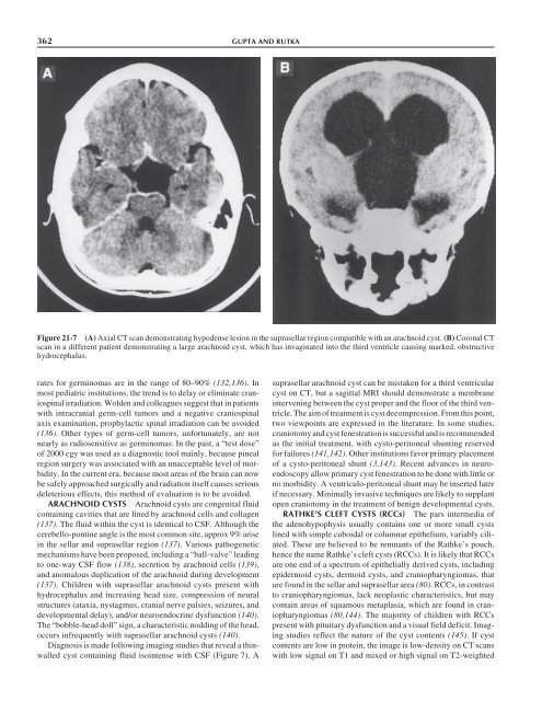
- Page 362 and 363: 346 YOUNGFigure 20-3 Images from a
- Page 364 and 365: 348 YOUNGFigure 20-5 Visual fields
- Page 366 and 367: 350 YOUNGFigure 20-7 Serial MRI fro
- Page 369 and 370: CHAPTER 21 / PEDIATRIC PITUITARY TU
- Page 371 and 372: CHAPTER 21 / PEDIATRIC PITUITARY TU
- Page 373 and 374: CHAPTER 21 / PEDIATRIC PITUITARY TU
- Page 375 and 376: CHAPTER 21 / PEDIATRIC PITUITARY TU
- Page 377: CHAPTER 21 / PEDIATRIC PITUITARY TU
- Page 381 and 382: CHAPTER 21 / PEDIATRIC PITUITARY TU
- Page 383: CHAPTER 21 / PEDIATRIC PITUITARY TU
- Page 386 and 387: 370 PERNICONE AND SCHEITHAUERFigure
- Page 388 and 389: 372 PERNICONE AND SCHEITHAUERFigure
- Page 390 and 391: 374 PERNICONE AND SCHEITHAUERFigure
- Page 392 and 393: 376 PERNICONE AND SCHEITHAUERFigure
- Page 394 and 395: 378 PERNICONE AND SCHEITHAUERFigure
- Page 396 and 397: 380 PERNICONE AND SCHEITHAUERFigure
- Page 398 and 399: 382 PERNICONE AND SCHEITHAUERFigure
- Page 400 and 401: 384 PERNICONE AND SCHEITHAUER32. Th
- Page 403 and 404: CHAPTER 23 / SELLAR TUMORS 38723Sel
- Page 405 and 406: CHAPTER 23 / SELLAR TUMORS 389mode
- Page 407 and 408: CHAPTER 23 / SELLAR TUMORS 391Figur
- Page 409 and 410: CHAPTER 23 / SELLAR TUMORS 393Figur
- Page 411 and 412: CHAPTER 23 / SELLAR TUMORS 395Figur
- Page 413 and 414: CHAPTER 23 / SELLAR TUMORS 397Figur
- Page 415 and 416: CHAPTER 23 / SELLAR TUMORS 399most
- Page 417 and 418: CHAPTER 23 / SELLAR TUMORS 401Figur
- Page 419 and 420: CHAPTER 23 / SELLAR TUMORS 403Figur
- Page 421 and 422: CHAPTER 23 / SELLAR TUMORS 405Figur
- Page 423 and 424: CHAPTER 23 / SELLAR TUMORS 407Figur
- Page 425 and 426: CHAPTER 23 / SELLAR TUMORS 409Figur
- Page 427 and 428: CHAPTER 23 / SELLAR TUMORS 411Figur
- Page 429 and 430:
CHAPTER 23 / SELLAR TUMORS 413Figur
- Page 431 and 432:
CHAPTER 23 / SELLAR TUMORS 4154.4.4
- Page 433 and 434:
therapy and, in some cases, radioth
- Page 435 and 436:
CHAPTER 23 / SELLAR TUMORS 419Figur
- Page 437 and 438:
CHAPTER 23 / SELLAR TUMORS 421Figur
- Page 439 and 440:
CHAPTER 23 / SELLAR TUMORS 423Figur
- Page 441 and 442:
CHAPTER 23 / SELLAR TUMORS 425the u
- Page 443 and 444:
CHAPTER 23 / SELLAR TUMORS 427Figur
- Page 445 and 446:
CHAPTER 23 / SELLAR TUMORS 4297.2.2
- Page 447 and 448:
CHAPTER 23 / SELLAR TUMORS 431Figur
- Page 449 and 450:
CHAPTER 23 / SELLAR TUMORS 43319. P
- Page 451 and 452:
CHAPTER 23 / SELLAR TUMORS 43520. N
- Page 453 and 454:
CHAPTER 23 / SELLAR TUMORS 43716. P
- Page 455 and 456:
CHAPTER 23 / SELLAR TUMORS 4393. Wi
- Page 457 and 458:
CHAPTER 23 / SELLAR TUMORS 4414.3.
- Page 459 and 460:
CHAPTER 23 / SELLAR TUMORS 44319. T
- Page 461 and 462:
CHAPTER 23 / SELLAR TUMORS 4457. Lo
- Page 463:
CHAPTER 23 / SELLAR TUMORS 44714. M
- Page 466 and 467:
450 SAEGERTermDiffuse hyperplasiaNo
- Page 468 and 469:
452 SAEGERFigure 24-3 ACTH cell hyp
- Page 470 and 471:
454 SAEGERFigure 24-9 Septic absces
- Page 472 and 473:
456 SAEGERabnormality of the arachn
- Page 474 and 475:
458 SAEGERFigure 24-15 Densely gran
- Page 476 and 477:
460 SAEGER76. Asa SL. Tumors of the
- Page 478 and 479:
462 MILLER, ZHANG, AND KLIBANSKItio
- Page 480 and 481:
464 MILLER, ZHANG, AND KLIBANSKItum
- Page 483 and 484:
INDEXABC peroxidase method, 94Acrom
- Page 485 and 486:
INDEX 469Chordomas (cont.) Cranioph
- Page 487 and 488:
INDEX 471Glycoprotein hormones/SV4O
- Page 489 and 490:
INDEX 473Invasion (cont.)Lymphomas
- Page 491 and 492:
INDEX 475Osteogenic sarcomas, 416-4
- Page 493 and 494:
INDEX 477Pro-opiomelanocortin (POMC
- Page 495:
INDEX 479Visual outcomes (cont.) Vi