Chapter 126

You also want an ePaper? Increase the reach of your titles
YUMPU automatically turns print PDFs into web optimized ePapers that Google loves.
<strong>126</strong><br />
CHAPTER<br />
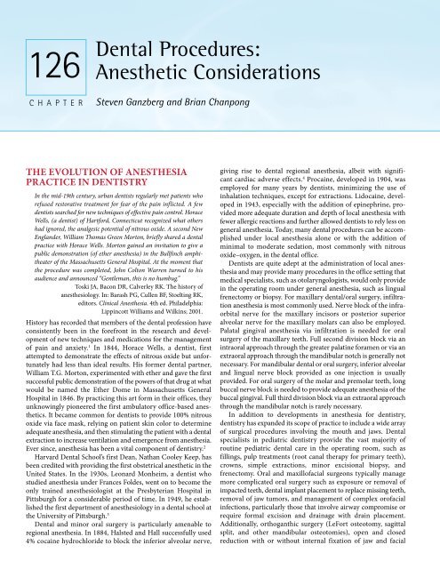
Dental Procedures:<br />
Anesthetic Considerations<br />
Steven Ganzberg and Brian Chanpong<br />
THE EVOLUTION OF ANESTHESIA<br />
PRACTICE IN DENTISTRY<br />
In the mid-19th century, urban dentists regularly met patients who<br />
refused restorative treatment for fear of the pain inflicted. A few<br />
dentists searched for new techniques of effective pain control. Horace<br />
Wells, (a dentist) of Hartford, Connecticut recognized what others<br />
had ignored, the analgesic potential of nitrous oxide. A second New<br />
Englander, William Thomas Green Morton, briefly shared a dental<br />
practice with Horace Wells. Morton gained an invitation to give a<br />
public demonstration (of ether anesthesia) in the Bullfinch amphitheater<br />
of the Massachusetts General Hospital. At the moment that<br />
the procedure was completed, John Colton Warren turned to his<br />
audience and announced “Gentleman, this is no humbug.”<br />
Toski JA, Bacon DR, Calverley RK. The history of<br />
anesthesiology. In: Barash PG, Cullen BF, Stoelting RK,<br />
editors. Clinical Anesthesia. 4th ed. Philadelphia:<br />
Lippincott Williams and Wilkins; 2001.<br />
History has recorded that members of the dental profession have<br />
consistently been in the forefront in the research and development<br />
of new techniques and medications for the management<br />
of pain and anxiety. 1 In 1844, Horace Wells, a dentist, first<br />
attempted to demonstrate the effects of nitrous oxide but unfortunately<br />
had less than ideal results. His former dental partner,<br />
William T.G. Morton, experimented with ether and gave the first<br />
successful public demonstration of the powers of that drug at what<br />
would be named the Ether Dome in Massachusetts General<br />
Hospital in 1846. By practicing this art form in their offices, they<br />
unknowingly pioneered the first ambulatory office-based anesthetics.<br />
It became common for dentists to provide 100% nitrous<br />
oxide via face mask, relying on patient skin color to determine<br />
adequate anesthesia, and then stimulating the patient with a dental<br />
extraction to increase ventilation and emergence from anesthesia.<br />
Ever since, anesthesia has been a vital component of dentistry. 2<br />
Harvard Dental School’s first Dean, Nathan Cooley Keep, has<br />
been credited with providing the first obstetrical anesthetic in the<br />
United States. In the 1930s, Leonard Monheim, a dentist who<br />
studied anesthesia under Frances Foldes, went on to become the<br />
only trained anesthesiologist at the Presbyterian Hospital in<br />
Pittsburgh for a considerable period of time. In 1949, he established<br />
the first department of anesthesiology in a dental school at<br />
the University of Pittsburgh. 3<br />
Dental and minor oral surgery is particularly amenable to<br />
regional anesthesia. In 1884, Halsted and Hall successfully used<br />
4% cocaine hydrochloride to block the inferior alveolar nerve,<br />
giving rise to dental regional anesthesia, albeit with significant<br />
cardiac adverse effects. 4 Procaine, developed in 1904, was<br />
employed for many years by dentists, minimizing the use of<br />
inhalation techniques, except for extractions. Lidocaine, developed<br />
in 1943, especially with the addition of epinephrine, provided<br />
more adequate duration and depth of local anesthesia with<br />
fewer allergic reactions and further allowed dentists to rely less on<br />
general anesthesia. Today, many dental procedures can be accomplished<br />
under local anesthesia alone or with the addition of<br />
minimal to moderate sedation, most commonly with nitrous<br />
oxide–oxygen, in the dental office.<br />
Dentists are quite adept at the administration of local anesthesia<br />
and may provide many procedures in the office setting that<br />
medical specialists, such as otolaryngologists, would only provide<br />
in the operating room under general anesthesia, such as lingual<br />
frenectomy or biopsy. For maxillary dental/oral surgery, infiltration<br />
anesthesia is most commonly used. Nerve block of the infraorbital<br />
nerve for the maxillary incisors or posterior superior<br />
alveolar nerve for the maxillary molars can also be employed.<br />
Palatal gingival anesthesia via infiltration is needed for oral<br />
surgery of the maxillary teeth. Full second division block via an<br />
intraoral approach through the greater palatine foramen or via an<br />
extraoral approach through the mandibular notch is generally not<br />
necessary. For mandibular dental or oral surgery, inferior alveolar<br />
and lingual nerve block provided as one injection is usually<br />
provided. For oral surgery of the molar and premolar teeth, long<br />
buccal nerve block is needed to provide adequate anesthesia of the<br />
buccal gingival. Full third division block via an extraoral approach<br />
through the mandibular notch is rarely necessary.<br />
In addition to developments in anesthesia for dentistry,<br />
dentistry has expanded its scope of practice to include a wide array<br />
of surgical procedures involving the mouth and jaws. Dental<br />
specialists in pediatric dentistry provide the vast majority of<br />
routine pediatric dental care in the operating room, such as<br />
fillings, pulp treatments (root canal therapy for primary teeth),<br />
crowns, simple extractions, minor excisional biopsy, and<br />
frenectomy. Oral and maxillofacial surgeons typically manage<br />
more complicated oral surgery such as exposure or removal of<br />
impacted teeth, dental implant placement to replace missing teeth,<br />
removal of jaw tumors, and management of complex orofacial<br />
infections, particularly those that involve airway compromise or<br />
require formal excision and drainage with drain placement.<br />
Additionally, orthoganthic surgery (LeFort osteotomy, sagittal<br />
split, and other mandibular osteotomies), open and closed<br />
reduction with or without internal fixation of jaw and facial
CHAPTER <strong>126</strong> ■ Dental Procedures: Anesthetic Considerations 2081<br />
fractures, and distraction osteogenesis of the jaw are commonly<br />
performed by oral and maxillofacial surgeons. Lastly, cleft palate<br />
repair, craniofacial anomaly reconstruction, facial plastic surgery,<br />
tracheotomy, temporomandibular joint (TMJ) surgery (at times<br />
with costochondral bone graft), jaw reconstructive surgery, usually<br />
in conjunction with cleft palate deformities or trauma and with<br />
iliac crest bone graft, are provided. Many of these latter procedures<br />
require overnight admission.<br />
In many situations, therefore, regional anesthesia is either<br />
ineffective or not possible. Oral sedation, intravenous sedation<br />
and general anesthesia are all used by dentists in a variety of<br />
settings to complete surgical goals.<br />
TRAINING OF DENTISTS IN SEDATION<br />
AND GENERAL ANESTHESIA<br />
Many dentists, through additional postgraduate residencies, are<br />
trained to provide sedation as well as general anesthesia. To ensure<br />
adequate training and competence, a dentist must legally posses a<br />
special permit from the state/provincial dental board to provide<br />
intravenous sedation or deep sedation/general anesthesia. Many<br />
states now require a special permit for pediatric or adult oral<br />
moderate sedation as well. No permit is generally required for<br />
minimal sedation or nitrous oxide–oxygen sedation. The postgraduate<br />
residencies that require extensive sedation or general<br />
anesthesia experience include pediatric dentistry, oral and maxillofacial<br />
surgery, and dental anesthesiology. Oral surgeons have<br />
consistently been in the forefront of office-based deep sedation/<br />
general anesthesia. However, with the elimination of operator–<br />
anesthetist deep sedation and general anesthesia in medicine, as<br />
well as the limitation of the oral surgeon to providing only<br />
extractions for dental concerns, specific residencies in anesthesiology<br />
for dentists were developed. The training and scope of<br />
practice of dentists involved in pediatric sedation and general<br />
anesthesia are described below.<br />
Pediatric Dentists<br />
U.S. training programs require a minimum of 20 cases of dental<br />
treatment under general anesthesia in the operating room and an<br />
additional one month rotation on the anesthesiology service itself<br />
to familiarize residents with the provision of general anesthesia<br />
and airway management. 5 Pediatric dentists are trained in oral<br />
moderate sedation and/or nitrous oxide–oxygen sedation for<br />
children that might have limited dental treatment needs and have<br />
some capability to cooperate but are anxious for care in the dental<br />
office. The most common oral sedation medications include<br />
midazolam or chloral hydrate with hydroxyzine but many<br />
different medications and combinations are used. There is an<br />
increasing use of general anesthesia for precooperative or uncooperative<br />
children, especially those with more than one, or<br />
possibly two, required short dental treatments. 6 Additionally,<br />
pediatric dentists are frequently called upon to treat adult mentally<br />
or physically challenged patients in the operating room.<br />
Oral and Maxillofacial Surgeons<br />
In the United States and Canada, oral surgeons must complete a<br />
4-month rotation on the anesthesia service and provide 90 deep<br />
sedations and 10 general anesthetics for clinic oral surgery patients<br />
as part of their program’s requirements. There is no specific<br />
requirement for anesthetizing children, defined as less than<br />
13 years old, except that the resident must be trained in the unique<br />
anatomic/pharmacologic/physiologic variations of the pediatric<br />
anesthesia patient. 7 Many U.S. and Canadian oral surgeons provide<br />
deep sedation for older teenaged pediatric patients with a limited<br />
number intravenously sedating preadolescent children in the<br />
dental office and rarely children younger than 6 years old. In this<br />
last remaining operator–anesthetist model, the oral surgeon<br />
provides both the deep sedation and the surgery in conjunction<br />
with a surgical dental assistant and another dental assistant to<br />
monitor the patient and provide airway support. In the United<br />
Kingdom, single drug intravenous moderate sedation is the only<br />
I.V. sedation regimen administered by dentists and may not be<br />
practical, especially for many younger pediatric patients. 8<br />
Dentist Anesthesiologists<br />
Dentist anesthesiologists are dentists in the United States and<br />
Canada who have completed 2 to 3 years of accredited training in<br />
anesthesiology, with a minimum of 1 year of operating room<br />
general anesthesia for all types of surgical procedures and additional<br />
specialized rotations in office-based anesthesia for dental,<br />
oral, and maxillofacial surgery. They provide the full range of<br />
anesthesia services for patients of all ages and medical complexity,<br />
from moderate sedation to intubated general anesthesia, for<br />
dental, oral, and maxillofacial procedures in hospitals, ambulatory<br />
surgery centers, and office settings. 9 Dentist anesthesiologists in<br />
the United States almost exclusively provide anesthesia services<br />
only. Most administer anesthesia in dental offices where they<br />
generally provide all required monitors, anesthetic drugs, and<br />
emergency drugs/equipment for the provision of safe anesthesia<br />
care while the dentist or oral surgeon operates. With the advent of<br />
newer, shorter-acting anesthetic agents and improved monitoring<br />
techniques, there is an increasing use of the office-based setting<br />
for general anesthesia for all types of procedures, medical or<br />
dental, particularly in North America. 10<br />
PREANESTHETIC ASSESSMENT<br />
Dental and oral surgery is frequently needed over a patient’s<br />
lifetime. This has particular implications for the mentally and<br />
physically challenged patient who may require general anesthesia<br />
every 1 to 3 years for routine dental care. Furthermore, as dental<br />
and minor oral surgery carries very low surgical risk, the administration<br />
of anesthesia may potentially be the most hazardous<br />
component of the perioperative experience. Therefore, the preanesthetic<br />
evaluation takes on increased importance.<br />
The vast majority of dental or oral surgical procedures are<br />
performed on an ambulatory, outpatient basis. The abbreviated<br />
time to complete the admission process poses issues common to<br />
all same-day surgical care, such as ensuring that patients receive<br />
complete preoperative medical evaluation and continue appropriate<br />
medications on the day of surgery. This is further complicated<br />
by the fact that pediatric dentists can not complete their own<br />
history and physical within 7 days of surgery, a Joint Commission<br />
requirement in the United States. This means that the history and<br />
physical must be performed by the patient’s pediatrician a few days<br />
before the procedure and forwarded to the anesthesia department<br />
for review. When this information is not available, the case must
2082 PART 5 ■ Anesthetic, Surgical, and Interventional Procedures: Considerations<br />
be cancelled. Good communication with the pediatric dentist is<br />
essential if optimal care is to be provided. In the United States and<br />
Canada, oral and maxillofacial surgeons may have only a dental<br />
(DDS, DMD) degree or are dually qualified with a medical degree<br />
(MD) and license. In the United Kingdom, most oral and<br />
maxillofacial surgeons are dually qualified. Regardless of degree,<br />
in most U.S. hospitals, oral surgeons can independently admit<br />
patients and provide admission history and physical examination,<br />
whereas pediatric dentists are not so trained.<br />
As nasotracheal intubation is likely planned, all patients should<br />
receive a careful airway examination with additional evaluation of<br />
the nares. If it is possible to determine the most patent nares<br />
preoperatively, this is ideal. A history of epistaxis and from which<br />
nares should be elicited and may lead to a decision to avoid nasal<br />
intubation. Chronic allergic rhinitis may predispose to nasal<br />
bleeding during intubation and on extubation. A history of snoring<br />
or frank obstructive sleep apnea should provoke additional caution<br />
especially with the possibly increased difficulty with nasal<br />
intubation. As with all pediatric patients, it is important to remember<br />
that the pediatric patient will be exfoliating their primary<br />
teeth from the age of 6–7 years until the age of 11–12 years. It is<br />
important to determine if there are currently any loose teeth in the<br />
patient’s mouth that may accidentally become avulsed during<br />
intubation.<br />
Since many pediatric patients receiving dental care under<br />
general anesthesia are precooperative (
CHAPTER <strong>126</strong> ■ Dental Procedures: Anesthetic Considerations 2083<br />
are sensorally challenged as well and find the feeling of local<br />
anesthesia of the oral cavity difficult to tolerate. The planned use<br />
of an intravenous nonsteroidal anti-inflammatory drug (NSAID)<br />
such as ketorolac or an opioid may be preferred over local anesthesia.<br />
Similarly, when planning for anesthetic maintenance and<br />
emergence, it may be preferable to consider a total intravenous<br />
propofol-based technique. or early discontinuation of inhalation<br />
agents near case completion with subsequent propofol infusion,<br />
to allow rapid and clear recovery. Planned extubation in the<br />
operating room may be preferable due to potential combativeness.<br />
Facial Cellulitis Patient<br />
The spread of dental infection to the fascial spaces of the face<br />
results in facial swelling that may or may not compromise the<br />
airway. Advanced Ludwig angina, with floor of mouth elevation,<br />
difficulty managing oral secretions, and body posturing to maintain<br />
airway patency, is rare in younger children, as this usually<br />
occurs with abscess spread from the second and third molars,<br />
which erupt after 12 years of age. When present, awake fiberoptic<br />
intubation is essential, because airway anatomy is frequently<br />
distorted, making direct laryngoscopy very difficult or impossible;<br />
loss of airway with sedation or general anesthesia is common, and<br />
there is concern of purulent discharge and risk of aspiration with<br />
airway instrumentation. More commonly in the pediatric patient,<br />
there is a buccal space swelling with or without trismus. Mouth<br />
opening may be severely limited but is usually due to muscle<br />
splinting secondary to pain, which resolves with unconsciousness<br />
and analgesia. Preoxygenation with slow inhalation induction to<br />
maintain spontaneous respiration or slow intravenous induction<br />
with assessment of mask ventilation can be planned, because<br />
usually, with increasing depth of inhalation anesthesia or paralysis,<br />
mouth opening can be accomplished to acceptable levels for<br />
endotracheal intubation. Appropriately sized oral and nasopharyngeal<br />
airways should be readily available. At times, a ratchet<br />
style mouth prop, used by the dentist to keep the mouth open<br />
under anesthesia, may be needed to increase mouth opening.<br />
Once the airway is secured, conventional anesthetic management<br />
is usually planned. If airway compromise was present at induction<br />
and fiberoptic intubation needed, transfer to the intensive care<br />
unit for continued post-operative intubation is generally necessary.<br />
Facial Trauma/TMJ Patient<br />
The patient with facial trauma presents several concerns for the<br />
anesthesiologist. Jaw fractures usually limit mouth opening due<br />
to muscle splinting secondary to pain. This presents a similar<br />
situation as with facial cellulitis, where a surgical depth of<br />
anesthesia usually allows mouth opening to be accomplished.<br />
However, depending on the type of fracture, forced mouth<br />
opening may worsen fracture separation. Preoperative consultation<br />
with the oral surgeon is important. If maxillomandibular<br />
fixation (wiring of the jaws together) is planned, nasotracheal<br />
intubation will be required. If intraoral bleeding has occurred, the<br />
patient should be considered to have a full stomach and appropriate<br />
precautions taken. There is the additional concern of TMJ<br />
trauma, even if no fracture is evident. If TMJ disc displacement<br />
has occurred, this may not allow full mouth opening despite a<br />
deep anesthetic plane. If mouth opening is limited, this should be<br />
discussed with the oral surgeon preoperatively to determine<br />
etiology and possible complications at anesthetic induction.<br />
Alternative intubation techniques may need to be considered. TMJ<br />
surgery itself is very rare for a patient younger than 18 years of<br />
age. Juvenile rheumatoid arthritis can affect the TMJ and, of<br />
course, jaw opening would need careful evaluation, as would<br />
cervical range of motion. Patients with skull base fracture should<br />
not receive nasal intubation. 19<br />
Post–Head and Neck Radiation<br />
The pediatric patient may have received radiation therapy to the<br />
head and neck for various cancers, typically for nasopharyngeal<br />
carcinoma. Fibrosis of the masticatory muscles is expected and<br />
mouth opening is usually quite limited. Dental care is almost<br />
always provided under general anesthesia due to severely<br />
restricted mouth opening. Depending on the degree of limited<br />
mouth opening, a variety of techniques can be considered based<br />
on patient age, including fiberoptic intubation, Bullard laryngoscope<br />
or blind nasal intubation. Extractions are commonly avoided<br />
in this population due to the risk of osteoradionecrosis, a form of<br />
severe osteomyelitis secondary to poor bone blood supply. Hyperbaric<br />
oxygen therapy before and after oral surgery is necessary to<br />
minimize this risk.<br />
ANTIBIOTIC PROPHYLAXIS<br />
Dental and oral surgery is conducted in an environment that is<br />
inherently inundated with bacteria. The need for antibiotics for<br />
these procedures is, however, quite limited. Surgical antibiotic<br />
prophylaxis is generally not provided for routine dental or minor<br />
oral surgical care. However, in the patient immunocompromised<br />
for whatever reason, including the cancer chemotherapy patient,<br />
surgical antibiotic prophylaxis is generally provided, using the<br />
same regimen as for infective endocarditis (IE) prophylaxis (Table<br />
<strong>126</strong>–1). With a functional neutrophil count of at least 1500 cells/<br />
mm 3 , antibiotic prophylaxis may not be necessary to help combat<br />
bacterial infection. Physician consultation is recommended.<br />
Infective endocarditis prophylaxis for dental procedures has<br />
been updated to reflect the fact that transient bacteremias likely<br />
occur more frequently with daily activities, such as tooth brushing<br />
and chewing, than with dental procedures themselves. Addition -<br />
ally, a risk stratification for those cardiac conditions with the<br />
highest morbidity and mortality from endocarditis were identified.<br />
A limited number of patients are now being recommended for IE<br />
antibiotic prophylaxis. Tables <strong>126</strong>–1 and <strong>126</strong>–2 describe the<br />
cardiac conditions for which IE antibiotic prophylaxis is now<br />
recommended and currently accepted regimens. 13 Any patient<br />
taken to the operating room for a dental procedure will experience<br />
a bacteremia and nasal intubation itself may also provoke<br />
bacteremia. What may not be well addressed in the newest guide -<br />
lines is the effect of duration of bacteremia as most office dental<br />
procedures, especially on children, are short resulting in a short<br />
duration bacteremia. When full mouth rehabilitation is provided<br />
in the operating room, high levels of bacteremia may be present<br />
for hours. Consultation between the cardiologist, anesthesiologist<br />
and dentist may lead to a decision to proceed with antibiotic<br />
prophylaxis until enough time has passed to assess the efficacy of<br />
the new prevention guidelines. This decision should be weighed<br />
against the risk of anaphylaxis from antibiotic administration as<br />
well as future increased antibiotic resistance.
2084 PART 5 ■ Anesthetic, Surgical, and Interventional Procedures: Considerations<br />
TABLE <strong>126</strong>-1. Indications for Preventive Antibiotics<br />
Prior to a Dental Procedure<br />
1. Artificial heart valves<br />
2. A history of infective endocarditis<br />
3. Certain specific, serious congenital (present from birth) heart<br />
conditions, including<br />
Paired or incompletely repaired cyanotic congenital heart<br />
disease, including palliative shunts and conduits<br />
A completely repaired congenital heart defect with prosthetic<br />
material or device, whether placed by surgery or by<br />
catheter intervention, during the first 6 months after the<br />
procedure<br />
Any repaired congenital heart defect with residual defect<br />
at the site or adjacent to the site of a prosthetic patch or<br />
a prosthetic device<br />
4. A cardiac transplant that develops a problem in a heart valve<br />
The decision to provide antibiotic prophylaxis for nonvalvular<br />
cardiac devices, ventriculoperitoneal and ventriculoatrial shunts,<br />
arteriovenous shunts, and central lines is controversial. Recommendations<br />
that suggest no prophylaxis for some of these devices<br />
in most circumstances have been made based on currently<br />
available evidence. 14 However, many physicians and surgeons still<br />
request antibiotic prophylaxis in these situations. Again, consultation<br />
between the physician, anesthesiologist and dentist may lead<br />
to a decision to proceed with antibiotic prophylaxis. This decision<br />
should also be weighed against the risk of anaphylaxis from<br />
antibiotic administration as well as future increased antibiotic<br />
resistance. The standard IE antibiotic prophylaxis regimen is<br />
commonly used.<br />
Although total joint replacement (TJR) is rare in the pediatric<br />
population, it should be remembered that the American Association<br />
of Orthopedic Surgeons and the American Dental Association<br />
have joint recommendations for antibiotic prophylaxis to<br />
prevent TJR infection following invasive dental procedures for<br />
2 years postinsertion, as well as at all times for any immunocompromised<br />
patient and for the hemophiliac. 15<br />
Except for these very specific situations, antibiotics are<br />
generally not administered for routine dental or oral surgical<br />
procedures.<br />
INTRAOPERATIVE CONSIDERATIONS<br />
Surgical Access<br />
The challenge of having to share the airway with the dentist or oral<br />
surgeon presents a challenge to the anesthesiologist. Although a<br />
nonintubated general anesthetic is possible with a dental patient,<br />
placing either an endotracheal tube (ETT) or a LMA to secure the<br />
airway may provide a higher level of comfort for the anesthesiologist.<br />
Either nasal or oral intubation can be considered, although<br />
dentists greatly prefer the nasal endotracheal approach. Using a<br />
flexible LMA or an oral ETT will interfere with the dentist taking<br />
mandibular dental radiographs or checking the occlusion (bite).<br />
A dentist must manually manipulate the mandible in order to<br />
evaluate the proper closure of the mouth, which cannot be done<br />
with the presence of an oral tube or LMA. For radiographs, the<br />
tube may need to be moved from side to side. Jaw fracture repair<br />
requiring maxillomandibular fixation or orthognathic surgery will<br />
exclude the use of a flexible LMA or an oral ETT and necessitates<br />
the use of a nasal intubation.<br />
Nasal Intubation Techniques<br />
A preformed nasal ETT is by far the most common form of airway<br />
management for dental or oral surgery. This allows clear access to<br />
the oral cavity for the dentist and allows for greatly increased ETT<br />
stability, minimizing the possibility of unplanned extubation<br />
during the procedure. Standard formulas can be used to determine<br />
ETT size, but for patients with low growth for age, an ETT 0.5 mm<br />
or more smaller than calculated may be needed to allow for<br />
atraumatic passage through the nasal structures. The relatively<br />
large leak with use of a smaller noncuffed ETT is offset by the use<br />
of a throat pack by the dentist which allows for good positive<br />
pressure ventilation intraoperatively. Postoperative nasal bleeding<br />
is the most common serious complication of nasal intubation,<br />
reported in 20 to 35% of cases. If it is possible to determine the<br />
most patent nares, this side is usually attempted first. If obstruction<br />
is encountered, the opposite nares should be tried. Adenoidectomy<br />
20 and turbinectomy 21 have been reported following<br />
nasotracheal intubation. Nasal tissue can also become dislodged<br />
during nasotracheal intubation and be inadvertently carried into<br />
the trachea by the tube. 22 Exfoliating teeth from ages 6 to 12 may<br />
TABLE <strong>126</strong>-2. Antibiotic Regimens<br />
Single Dose 30 to 60 min Before Procedure<br />
Situation Agent Adults Children<br />
Able to take oral medication Amoxicillin 2 g 50 mg/kg<br />
Unable to take oral medication Ampicillin 2 g I.M. or I.V. 50 mg/kg I.M. or I.V.<br />
Cefazolin or ceftriaxone 1 g I.M. or I.V. 50 mg/kg I.M. or I.V.<br />
Allergic to penicillins or ampicillin Cephalexin* † 2 g 50 mg/kg<br />
Clindamycin 600 mg 20 mg/kg<br />
Azithromycin or clarithromycin 500 mg 15 mg/kg<br />
Allergic to penicillins or ampicillin Cefazolin or ceftriaxone † 1 g I.M. or I.V. 50 mg/kg I.M. or I.V.<br />
and unable to take oral medication Clindamycin 600 mg I.M. or I.V. 20 mg/kg I.M. or I.V.<br />
I.M. = intramuscular; I.V. = intravenous.<br />
*Or other first- or second-generation oral cephalosporin in equivalent adult or pediatric dosage.<br />
Cephalosporins should not be used in an individual with a history of anaphylaxis, angioedema, or urticaria with penicillins or ampicillin.
CHAPTER <strong>126</strong> ■ Dental Procedures: Anesthetic Considerations 2085<br />
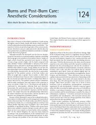
Figure <strong>126</strong>-1. Red rubber catheter placed over the distal end of<br />
the preformed nasotracheal tube.<br />
A<br />
become dislodged during laryngoscopy and potentially be<br />
aspirated. Methods of relatively atraumatic intubation can reduce<br />
these complications of nasal intubation. One of the more common<br />
difficulties encountered is once the ETT has passed the<br />
vocal cords, there is obstruction at the level of the cricoid cartilage.<br />
This is easily overcome by turning the tube at the nose to redirect<br />
the distal end. A variety of other techniques have been<br />
proposed to aide in atraumatic intubation. The following are some<br />
methods that have been suggested: Various tube guides, precurving<br />
the ETT, thermosoftening the ETT, and topical nasal<br />
vasoconstrictors.<br />
The use of a red rubber catheter as a guide for the nasotracheal<br />
tube was shown to reduce the severity of bleeding. 23 The flared<br />
end of the #10–12 French red rubber catheter is placed over the tip<br />
of the tube (Figure <strong>126</strong>–1). After lubricating the catheter with<br />
water-soluble lubricant, the distal end of the catheter is placed into<br />
the patient’s nasal cavity. The tip of the catheter is then retrieved<br />
from the oral cavity with a Magill forceps and disconnected from<br />
the nasotracheal tube, which is then advanced into the trachea. 24<br />
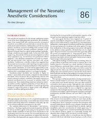
For nasal passages through which it is difficult to pass the<br />
nasotracheal tube, a suction catheter can be used as a guide. The<br />
suction catheter is placed inside the tube prior to insertion into<br />
the chosen nares (Figure <strong>126</strong>–2). The catheter will help navigate<br />
Figure <strong>126</strong>-2. Suction catheter placed inside a 5.0 nasal RAE tube.<br />
B<br />
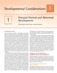
Figure <strong>126</strong>-3. Stylet placed in the distal end of the preformed<br />
nasotracheal tube to form a pigtail.<br />
through the nasal passage first and then the nasotracheal tube can<br />
follow over it similar to the Seldinger technique<br />
Precurving the nasal tube can also aid the intubation process.<br />
A stylet can be inserted into the end of a nasotracheal tube which<br />
can then be bent to resemble a pigtail 5 to 10 minutes before the<br />
beginning of the case (Figures <strong>126</strong>–3, <strong>126</strong>–4, and <strong>126</strong>–5). Just<br />
before using the tube, the stylet is withdrawn from the tube. As a<br />
result the tube will have a slight curve to it. This will help facilitate<br />
the passage of the tube as it is less likely to be caught up in the<br />
bulge of the posterior nasopharynx at the level of C2. Additionally,<br />
with the tip of the tube pointing ventrally, guiding the tube into the<br />
trachea may be accomplished without need for a Magill forceps,<br />
although one should always be available.<br />
One of the most common methods to allow atraumatic<br />
nasotracheal intubation is thermosoftening of the tube. When the<br />
distal 2 to 3 centimeters of the tube is placed in sterile water, heated<br />
to at least 450°C for several seconds immediately before insertion,<br />
ETT passage through the nasal cavity is similar to a soft nasophyarngeal<br />
tube, thus reducing the incidence of epistaxis. 25 It<br />
should be noted that heating of too much length of the ETT can<br />
make it difficult to “turn the tube” to allow passage past the cricoid<br />
cartilage, as the ETT may be so soft that it twists on itself.
2086 PART 5 ■ Anesthetic, Surgical, and Interventional Procedures: Considerations<br />
managed with an oral ETT or an LMA in order to avoid a nasal<br />
bleeding or other trauma or in the case of the patient with bilateral<br />
choanal atresia where there may not be enough space the nasal<br />
passages to pass an ETT.<br />
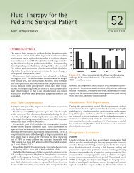
Figure <strong>126</strong>-4. Different view of the nasotracheal tube with the<br />
stylet placed to form a pig tail.<br />
Vasoconstrictors are often used, at times in conjunction with<br />
one of the other previously mentioned techniques. Oxymetazoline<br />
0.05% is probably most commonly used and has been shown to<br />
be as effective as 10% cocaine in reducing the incidence of<br />
bleeding during intubation. 26<br />
As an alternative to direct laryngoscopy, the use of a trachlight<br />
has been suggested. 27 Hung and Stewart suggested the use of a<br />
modified trachlight for nasotracheal intubation. With the rigid<br />
stylet removed, the trachlight is inserted into the nasotracheal<br />
tube. After placement into either nares, the combination tube and<br />
trachlight is advanced until a loss of resistance is felt. Once the<br />
operating room lights are dimmed, the light source is turned on to<br />
visualize the location of the tip. A jaw lift is required in order to<br />
raise the epiglottis. Once the light source is at its brightest and in<br />
the midline, the tube is slowly advanced. Once it enters the glottis<br />
the light should remain bright and should be seen just below the<br />
thyroid prominence. 27 The trachlight can then be removed and<br />
the placement of the tube verified.<br />
There are a number of conditions mentioned under “Preanesthetic<br />
Assessment” that describe relative contraindications to<br />
nasotracheal intubation. These include history of palatopharyngoplasty,<br />
frequent epistaxis, bilateral choanal atresia, coagulopathy,<br />
or a suspected basal skull fracture. These patients may best be<br />
Laryngeal Mask Airway and<br />
Oral Endotracheal Tube<br />
Since its approval by the Food and Drug Administration in 1991,<br />
LMAs have been used for dental restoration procedures 16 and for<br />
oral and maxillofacial surgical procedures 17 with great success. The<br />
original LMA tubing is stiff and has a large diameter; however, the<br />
development of the wire-reinforced LMA provides a flexible and<br />
smaller tubing that can be taped to either side of the oral cavity or<br />
down the midline of the chin. As can be seen in Figures <strong>126</strong>–6 and<br />
<strong>126</strong>–7, the maxillary arch and sometimes the mandibular arch can<br />
be fully isolated with a rubber dam without interfering with the<br />
LMA tubing. A throat pack will still need to be placed by the<br />
dentist or anesthesiologist.<br />
An oral endotracheal tube can also be used. A standard, preformed<br />
RAE or coiled ETT can be used. Securing the ETT is<br />
particularly important. If the mandibular anterior teeth are not to<br />
be restored, the tube can be secured in the midline, as for<br />
tonsillectomy, although a mouth gag is not used. Alternatively, the<br />
tube may pass posterior to the back teeth and taped to the side of<br />
the face. This will likely require moving the tube to the opposite<br />
side as needed. Placement of the laryngoscope allows the distal<br />
end of the tube to be securely moved in its entirely, rather than<br />
only the upper portion, which may predispose to extubation.<br />
Throat Pack<br />
When a dentist is working in the mouth, regardless of whether it<br />
is restorative dentistry or surgical extractions, a gauze throat pack<br />
Figure <strong>126</strong>-5. Shape of the nasotracheal tube with the stylet removed<br />
before intubation.<br />
Figure <strong>126</strong>-6. Flexible laryngeal mask airway with rubber dam<br />
placed for the maxillary arch.
CHAPTER <strong>126</strong> ■ Dental Procedures: Anesthetic Considerations 2087<br />
Figure <strong>126</strong>-8. Throat pack with a radio-opaque lining.<br />
Figure <strong>126</strong>-7. Flexible laryngeal mask airway with rubber dam<br />
placed for the mandibular arch.<br />
is generally placed in the throat or hypopharynx behind the<br />
tonsillar fauces to prevent any debris from migrating to the hypopharynx.<br />
Any debris left behind can potentially be aspirated by<br />
the patient upon extubation. However, complications have arisen<br />
when the throat pack was accidentally left behind, which has led<br />
to airway obstruction or ingestion 28 and has even resulted in<br />
death. 29 The anesthesiologist must also be cognizant of the throat<br />
pack should unanticipated extubation occur during the procedure.<br />
At this time, the dentist must remove all of his/her dental<br />
equipment and the anesthesiologist may attempt positive pressure<br />
mask ventilation prior to throat pack removal. Clearly, ventilation<br />
will be suboptimal. To minimize this occurrence, many institutions<br />
have devised methods to remind them of the intact throat<br />
pack. Some of the suggestions have been to: 29–31<br />
1. Leave part of the throat pack outside the mouth<br />
2. Tie a piece of dental floss to the pack and tape the floss to<br />
the face<br />
3. Tie part of the throat pack to the tracheal tube<br />
4. Tape a sign to the forehead<br />
5. Tape a sign to the ventilator switch<br />
6. Have the person responsible for placing the tube be the one<br />
who removes the pack<br />
7. Have all members of the team witness the removal of the pack<br />
8. Include the pack as part of the scrub nurse’s count<br />
9. Visually inspect the pharynx with the laryngoscope prior to<br />
extubation<br />
Using a throat pack with a radio-opaque lining (Figure <strong>126</strong>–8)<br />
would be advisable and can be helpful in cases where the pack has<br />
become unaccounted for and an x-ray is needed to determine<br />
whether it was left in the patient. 28<br />
Surgical Issues and Pain Management<br />
Typically, restorative dentistry on pediatric patients under general<br />
anesthesia does not require local anesthesia. Local anesthesia with<br />
epinephrine may allow improved hemostasis for extractions,<br />
however. The most stimulating procedures for the pediatric<br />
patient intraoperatively are placement of the rubber dam, where<br />
the oral cavity and tongue are stretched; removal of the dental pulp<br />
(pulpotomy); and extractions. It is helpful for the dentist to alert<br />
the anesthesiologist to these procedures.<br />
Regardless of whether or not extractions are performed<br />
during the dental procedure, the dental patient still requires<br />
postoperative pain management. Placement of stainless steel<br />
crowns can be very painful for the child, because the crowns are<br />
placed well under the gum line and can fit together very tightly.<br />
The dentist may use local anesthesia intraoperatively, which may<br />
provide acceptable pain control postoperatively. Many dentists,<br />
however, do not wish to give local anesthesia, particularly to<br />
younger children who may bite their lips, cheeks or tongue, as this<br />
can lead to severe mutilation of the oral soft tissues. NSAIDs are<br />
effective in controlling mild to moderate levels of dental pain 32<br />
and are a good choice for pain management in dental patients.<br />
Ketorolac tromethamine, a parenterally available NSAID, has been<br />
shown to be very effective in the management of dental pain.<br />
Purday et al. showed no difference between intravenous ketorolac<br />
and morphine for postoperative pain in children undergoing<br />
dental surgery, with the added benefit of reducing postoperative<br />
nausea and vomiting in the 24-hour postoperative period. 33 Dsida<br />
et al. showed that 0.5 mg/kg of I.V. ketorolac in children produced<br />
plasma blood levels similar to adults for therapeutic concentrations.<br />
34 Some pediatric hospitals allow 1 mg/kg of I.V. ketorolac<br />
as a single dose followed by acetaminophen for continued postoperative<br />
pain control. Opioids can also be considered, with the<br />
most commonly used being morphine sulfate at a dose of 0.05 to<br />
0.1 mg/kg.<br />
Extubation<br />
If oral bleeding or heavy secretions are expected at case completion,<br />
deep extubation may lead to an increased risk of<br />
laryngospasm during anesthetic emergence. Additionally, extubation<br />
of a nasotracheal tube may lead to nasal bleeding into the<br />
oral cavity, with increased risk of laryngospasm if deep extubation<br />
is planned. This can occur despite apparent atraumatic intubation.<br />
Epistaxis can also occur. Nasal bleeding may have an anterior or<br />
posterior component. The use of ketorolac tromethamine may
2088 PART 5 ■ Anesthetic, Surgical, and Interventional Procedures: Considerations<br />
exacerbate this adverse event. Frequently, placement of a soft<br />
nasopharyngeal airway can temporarily tamponade nasal bleeding<br />
and allow for hemostasis to take place. In rare cases, a nasal<br />
tampon (Merocel or Doyle sponge; Laubscher, Germany) may be<br />
needed. Uncontrolled posterior bleeding is usually controlled with<br />
a balloon catheter and provided by an otolaryngologist.<br />
In summary, the anesthesiologist should consult as needed with<br />
the dentist preoperatively regarding behavioral challenges, type of<br />
intubation needed, and any airway concerns that the anesthesiologist<br />
may recognize, as well as postoperative pain control choices<br />
to provide optimal patient care.<br />
POSTOPERATIVE CONSIDERATIONS<br />
Some unique postoperative considerations may be associated with<br />
anesthesia for dental or oral surgery in addition to typical postoperative<br />
complications. These include:<br />
1. Increased incidence of postoperative nausea and vomiting<br />
during oral and maxillofacial surgery due to passive ingestion<br />
of blood during the procedure or active swallowing of blood<br />
during recovery.<br />
2. Miscellaneous dental debris inadvertently left behind after the<br />
completion of the procedure and possibly aspirated during<br />
extubation. Dental debris left behind by the surgeon and/or<br />
assistant can include but are not limited to such items as cotton<br />
pellets, pieces of amalgam or other dental filling materials,<br />
stainless steel crowns, other small dental equipment and/or<br />
pieces of a tooth.<br />
3. Retained throat pack; this has the potential to be swallowed or<br />
to obstruct the patient in recovery.<br />
4. Although maxillomandibular fixation is not used as frequently<br />
as in the past for jaw fracture, if this has occurred, immediate<br />
availability of wire scissors and other instruments for<br />
immediate release of fixation should be available in the<br />
recovery room.<br />
AMBULATORY SURGERY IN THE<br />
OFFICE VS HOSPITAL/AMBULATORY<br />
SURGERY CENTER<br />
Office-based general anesthesia started with dentists, Horace<br />
Wells and William T.G. Morton, who discovered nitrous oxide and<br />
ether anesthesia, respectively. Later, with the introduction of<br />
intravenous barbiturates, oral surgeons were the leaders for many<br />
years in intravenous office-based anesthesia, promoting the<br />
methohexital bolus technique for short tooth extraction procedures<br />
long before the medical community was comfortable with<br />
office-based anesthesia and surgery. Today, with improved anesthetic<br />
medications and monitoring techniques, there is an<br />
increasing trend toward office-based surgery in both medicine and<br />
dentistry, paralleling the increase in overall outpatient surgery to<br />
greater than 75% of total surgical procedures. 35 In the United<br />
States, this is driven mainly by cost considerations, with unique<br />
circumstances for dentistry. Many U.S. medical insurance plans<br />
do not reimburse for anesthesia, facility fees, and additional<br />
routine hospital/ASC charges for needed dental procedures as they<br />
do for medical procedures. This is particularly problematic for the<br />
pediatric patient who is unable to cooperate with delicate dental<br />
treatment on very small oral structures. With the average global<br />
cost of a 2-hour operating room visit, excluding the dental charges,<br />
ranging from $8000 to $12,000, many parents are unable to<br />
afford this expense. The child frequently lives with chronic dental<br />
pain 36 or eventually may find an oral surgeon willing to provide<br />
operator–anesthetist general anesthesia or sedation for extraction<br />
of abscessed teeth, the only procedure the oral surgeon is trained<br />
to provide. In other countries, such as Canada and the United<br />
Kingdom, there may be long waits for treatment resulting in<br />
continued patient suffering, lost time from school, and in some<br />
cases, hospitalization due to facial cellulitis formation. 36<br />
With the increasing number of dentist anesthesiologists providing<br />
anesthesia services in the dental office, as well as physician<br />
anesthesiologists gradually making their way into the office venue,<br />
more parents and pediatric patients are able to afford dental care<br />
under general anesthesia as there are no facility fees. Anesthesia<br />
costs for a 2-hour case range from $750 to $1500 depending on<br />
the region of the United States and exclusive of dental surgical<br />
costs. For pediatric patients, these office procedures are usually<br />
for routine dental care and limited dentoalveolar surgery, such as<br />
extraction of impacted teeth.<br />
Anesthesiologists who provide treatment in dental offices many<br />
times do not have the “luxury” of an anesthesia machine. Most<br />
commonly, general anesthesia is provided via a total intravenous<br />
anesthesia (TIVA) technique with an unsecured airway but with a<br />
gauze throat partition to minimize the risk of small objects and<br />
fluids reaching the hypopharynx. Anesthesia for uncooperative<br />
pediatric patients, for whom obtaining I.V. access is challenging,<br />
is most commonly induced with 2 to 3 mg/kg or more of ketamine<br />
I.M., frequently with the addition of midazolam and possibly an<br />
anticholinergic. This allows for adequate dissociation within 3 to<br />
6 minutes in order to place monitors, establish I.V. access, position<br />
the patient with a shoulder roll to maintain airway patency, and<br />
allow incremental doses or a continuous infusion of propofol to be<br />
used. Local anesthesia of the oral cavity is used, except for very<br />
short or relatively nonstimulating procedures, minimizing anes -<br />
thetic requirements as surgical stimulation is kept to a minimum.<br />
Variations of this technique include the use of bolus opioids or<br />
remifentanil infusion in combination with propofol or ketamine<br />
with propofol. At times, a nasopharyngeal airway is used to help<br />
maintain airway patency and an LMA can be considered. Where<br />
I.V. access can be obtained, frequently with nitrous oxide–oxygen<br />
or oral midazolam premedication for younger children, with or<br />
without common topical skin anesthetics, I.V. induction can be<br />
performed.<br />
Because most dental offices have a nitrous oxide delivery<br />
system, an intubated, nontriggering, TIVA technique can also be<br />
provided, using this gas delivery system with a single limb circuit<br />
or with specialized disposable carbon dioxide absorbers, which<br />
allow a circle system. If nitrous oxide is used, proper scavenging<br />
must be maintained. Alternatively, a flexible LMA can be used,<br />
with or without nitrous oxide, to aid in securing the airway.<br />
As with all office-based surgery, patient selection, surgeon<br />
selection, and surgical procedure determine the anticipated safety<br />
of providing general anesthesia in this setting. Patients are<br />
generally American Society of Anesthesiologists (ASA) classification<br />
I or II, but select ASA III patients, generally without<br />
cardiopulmonary compromise, can also be considered. For<br />
instance, a patient with persistent seizures despite optimal treatment,<br />
but for whom the neurologist would not admit the patient<br />
after general anesthesia, may be quite acceptable for anesthesia in<br />
the office setting with an adequate postoperative observation<br />
period. Alternatively, a child with obstructive sleep apnea<br />
secondary to tonsillar hypertrophy who is ASA II may not be a
CHAPTER <strong>126</strong> ■ Dental Procedures: Anesthetic Considerations 2089<br />
good candidate for a an in-office nonintubated general anesthetic.<br />
Regarding surgeon/dentist selection, the nuances of working<br />
within a relatively unprotected airway requires some understanding<br />
and modification of surgical technique, such as minimal<br />
use of water spray to cool the tooth while drilling, which may<br />
migrate to the glottis and lead to laryngospasm. Additionally, there<br />
can be considerable differences in anesthesia time with slower<br />
versus faster surgeons. Lastly, some procedures, such as more complex<br />
maxillofacial procedures, may require intubation and preferably<br />
potent inhalation agents or mechanical ventilation, thus<br />
necessitating a formal operating room suite with full anesthesia<br />
capabilities. Regardless of the surgical procedure, provision should<br />
be made for duplicates of all critical pieces of equipment, both for<br />
anesthesia and surgery, should some device fail prior to procedure<br />
completion. Lastly, should a surgical or anesthetic complication<br />
arise which necessitates transfer to the hospital, arrangements<br />
should be made in advance for how and where this will be<br />
accomplished.<br />
SUMMARY<br />
General anesthesia for dental and oral surgery is frequently<br />
necessary for uncooperative patients, patients for whom local<br />
anesthesia is difficult to obtain, procedures of significant surgical<br />
complexity, or when local anesthesia cannot be obtained. Particularly<br />
for pediatric patients, there is an increasing use of hospital<br />
and office-based general anesthesia for dental procedures. The<br />
anesthesiologist is faced with the challenge of sharing the airway<br />
with the dentist/surgeon and the frequent need for nasotracheal<br />
intubation. Proper preoperative planning and consultation with<br />
the dentist/surgeon will enhance the safe and comfortable<br />
administration of anesthesia for the patient and dentist.<br />
REFERENCES<br />
1. Bankoff G. The Conquest of Pain: The Story of Anesthesia. London:<br />
MacDonald; 1946.<br />
2. Malamed SF. Medical Emergencies in the Dental Office. 6th ed. St. Louis:<br />
Mosby-Year Book; 2008.<br />
3. Weaver J. Changing of the guard in Pittsburgh: C. Richard Bennett, DDS,<br />
PhD, retires. Anesth Prog. 2006;53(3):77.<br />
4. Calatayud J, Gonzalez A. History of the development and evolution of local<br />
anesthesia since the coca leaf. Anesthesiology. 2003;98(6):1503–1508.<br />
5. American Dental Association. Accreditation standards for advanced<br />
specialty education programs in pediatric dentistry. Available at: http://<br />
www.ada.org/prof/ed/accred/standards/ped.pdf. Accessed September 3,<br />
2010.<br />
6. Bohaty B, Spencer P. Trends in dental treatment under general anesthesia,<br />
1978–1990. J Clin Pediatr Dent. 1992;16:222–224.<br />
7. American Society of Anesthesiologists. The medical specialty of anes -<br />
thesiology. Available at: http://www.asahq.org/patientEducation/specialty.<br />
htm. Accessed September 3, 2010.<br />
8. Postillo D. General anesthesia, sedation and resuscitation in dentistry.<br />
Report of an expert working party for the standing dental advisory<br />
committee. London: Department of Health; 1990.<br />
9. Flick WG, Katsnelson A, Alstrom H. Illinois Dental Anesthesia and<br />
Sedation Survey for 2006. Anesth Prog. 2007;54:52–58.<br />
10. Koch ME, Barinholtz D. Office-based anesthesia: an overview. Anesthesiol<br />
Clin North Am. 2003;21(2):417–443.<br />
11. Brennan LJ, Watson B. Modern day-case anaesthesia for children. Br J<br />
Anaesth. 1999:83–91.<br />
12. McGraw T, Kendrick A. Oral midazolam premedication and postoperative<br />
behaviour in children. Paediatr Anaesth. 1998;8(2):117–121.<br />
13. Wilson W, TK, Gewitz M, et al. Prevention of infective carditis. Guidelines<br />
from the American Heart Association. Circulation. 2007;116:1736–1754.<br />
14. Baddour LM, Wilson WR, Bayer AS, et al. Nonvalvular cardiovascular<br />
device-related infections. AHA Statement. Circulation. 2003;108:2015–<br />
2031.<br />
15. ADA/AAOS advisory statement. Antibiotic prophylaxis for dental<br />
patients with total joint replacements. The evidence base for the efficacy<br />
of antibiotic prophylaxis in dental practice. J Am Dent Assoc. 2007;<br />
138:458–474.<br />
16. Brimacombe J, BA. The laryngeal mask airway for dental surgery—a<br />
review. Aust Dent J. 1995;40(1):10–14.<br />
17. Bennett J, Petito A, Zandsberg S. Use of the laryngeal mask airway in oral<br />
and maxillofacial surgery. J Oral Maxillofac Surg. 1996;54(11):1346–1351.<br />
18. Hee HI, Conskunfirat ND. Airway management in a patient with a cleft<br />
palate after pharyngoplasty: a case report. Can J Anaesth. 2003;50(7):721–<br />
724.<br />
19. Marlow TJ, Goltra DD Jr, Schabel SI. Intracranial placement of a<br />
nasotracheal tube after facial fracture: a rare complication. J Emerg Med.<br />
1997;15(2):187–191.<br />
20. Harvey DC, Amorosa P. Traumatic nasotracheal intubation. Anaesthesia.<br />
1986;41(4):442.<br />
21. Williams AR, Burt N, Warren T. Accidental middle turbinectomy: a<br />
complication of nasal intubation. Anesthesiology. 1999;90(6):1782–1784.<br />
22. Knuth TE, Richards JR. Mainstem bronchial obstruction secondary to<br />
nasotracheal intubation: a case report and review of the literature. Anesth<br />
Analg. 1991;73(4):487–489.<br />
23. Elwood T, Stillions DM, Woo DW, et al. Nasotracheal intubation: a<br />
randomized trial of two methods. Anesthesiology. 2002;96(1):51–53.<br />
24. Elwood T, Parker S, Ramamoorthy C. Pediatric-nasotracheal intubation<br />
made atraumatic. Anesthesiology. 1998;89(2):550.<br />
25. Kim YC, Lee SH, Noh GJ, et al. Thermosoftening treatment of the<br />
nasotracheal tube before intubation can reduce epistaxis and nasal<br />
damage. Anesth Analg. 2000;91(3):698–701.<br />
26. Katz RI, Hovagim AR, Finkelstein HS, et al. A comparison of cocaine,<br />
lidocaine with epinephrine, and oxymetazoline for prevention of epistaxis<br />
on nasotracheal intubation. J Clin Anesth. 1990;2(1):16–20.<br />
27. Hung OR, Stewart RD. Lightwand intubation: I—a new lightwand device.<br />
Can J Anaesth. 1995;42(9):820–825.<br />
28. To EW, Tsang WM, Yiu F, et al. A missing throat pack. Anaesthesia.<br />
2001;56(4):383–384.<br />
29. Crawford BS. Prevention of retained throat pack. BMJ. 1977;2(6093):1029.<br />
30. Stone JP, Collyer J. Aide-memoir to pharyngeal pack removal. Anesth<br />
Analg. 2003;96(1):304.<br />
31. Knepila GJ, Blackburn CW. Retained throat packs: results of a national<br />
survey and the application of an organisational accident model. Br J Oral<br />
Maxillofac Surg. 2008;46:473–6.<br />
32. Haas DA. An update on analgesics for the management of acute<br />
postoperative dental pain. J Can Dent Assn. 2002;68(8):476–482.<br />
33. Purday JP, Reichert CC, Merrick PM. Comparative effects of three<br />
doses of intravenous ketorolac or morphine on emesis and analgesia<br />
for restorative dental surgery in children. Can J Anaesth. 1996;43(3):<br />
221–225.<br />
34. Dsida RM, Wheeler M, Birmingham PK, et al. Age-stratified pharmacoki -<br />
netics of ketorolac tromethamine in pediatric surgical patients. Anesth<br />
Analg. 2002;94(2):266–270.<br />
35. RK S. Office-based anesthesia growth provokes safety fears. Anesth Patient<br />
Safety Found Newsletter. 2000;15(1):1.<br />
36. North S, Davidson LE, Blinkhorn AS, Mackie IC. The effects of a long wait<br />
for children’s dental general anesthesia. Int J Paediatr. 2007;17:105–109.