Chapter 105

You also want an ePaper? Increase the reach of your titles
YUMPU automatically turns print PDFs into web optimized ePapers that Google loves.
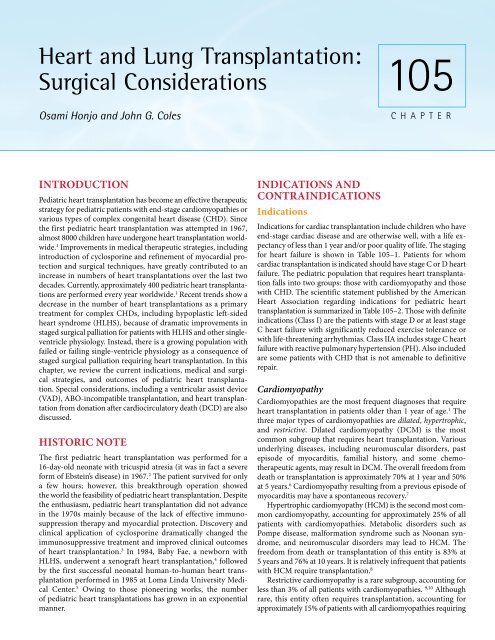
Heart and Lung Transplantation:<br />
Surgical Considerations<br />
Osami Honjo and John G. Coles<br />
<strong>105</strong><br />
CHAPTER<br />
INTRODUCTION<br />
Pediatric heart transplantation has become an effective therapeutic<br />
strategy for pediatric patients with end-stage cardiomyopathies or<br />
various types of complex congenital heart disease (CHD). Since<br />
the first pediatric heart transplantation was attempted in 1967,<br />
almost 8000 children have undergone heart transplantation world -<br />
wide. 1 Improvements in medical therapeutic strategies, including<br />
introduction of cyclosporine and refinement of myocardial pro -<br />
tection and surgical techniques, have greatly contributed to an<br />
increase in numbers of heart transplantations over the last two<br />
decades. Currently, approximately 400 pediatric heart transplanta -<br />
tions are performed every year worldwide. 1 Recent trends show a<br />
decrease in the number of heart transplantations as a primary<br />
treatment for complex CHDs, including hypoplastic left-sided<br />
heart syndrome (HLHS), because of dramatic improvements in<br />
staged surgical palliation for patients with HLHS and other singleventricle<br />
physiology. Instead, there is a growing population with<br />
failed or failing single-ventricle physiology as a consequence of<br />
staged surgical palliation requiring heart transplantation. In this<br />
chapter, we review the current indications, medical and surgical<br />
strategies, and outcomes of pediatric heart transplantation.<br />
Special considerations, including a ventricular assist device<br />
(VAD), ABO-incompatible transplantation, and heart transplan -<br />
tation from donation after cardiocirculatory death (DCD) are also<br />
discussed.<br />
HISTORIC NOTE<br />
The first pediatric heart transplantation was performed for a<br />
16-day-old neonate with tricuspid atresia (it was in fact a severe<br />
form of Ebstein’s disease) in 1967. 2 The patient survived for only<br />
a few hours; however, this breakthrough operation showed<br />
the world the feasibility of pediatric heart transplantation. Despite<br />
the enthusiasm, pediatric heart transplantation did not advance<br />
in the 1970s mainly because of the lack of effective immuno -<br />
suppression therapy and myocardial protection. Discovery and<br />
clinical appli cation of cyclosporine dramatically changed the<br />
immunosup pressive treatment and improved clinical outcomes<br />
of heart trans plantation. 3 In 1984, Baby Fae, a newborn with<br />
HLHS, under went a xenograft heart transplantation, 4 followed<br />
by the first successful neonatal human-to-human heart trans -<br />
plantation per formed in 1985 at Loma Linda University Medical<br />
Center. 5 Owing to those pioneering works, the number<br />
of pediatric heart trans plantations has grown in an exponential<br />
manner.<br />
INDICATIONS AND<br />
CONTRAINDICATIONS<br />
Indications<br />
Indications for cardiac transplantation include children who have<br />
end-stage cardiac disease and are otherwise well, with a life ex -<br />
pectancy of less than 1 year and/or poor quality of life. The staging<br />
for heart failure is shown in Table <strong>105</strong>–1. Patients for whom<br />
cardiac transplantation is indicated should have stage C or D heart<br />
failure. The pediatric population that requires heart transplanta -<br />
tion falls into two groups: those with cardiomyopathy and those<br />
with CHD. The scientific statement published by the American<br />
Heart Association regarding indications for pediatric heart<br />
transplantation is summarized in Table <strong>105</strong>–2. Those with definite<br />
indications (Class I) are the patients with stage D or at least stage<br />
C heart failure with significantly reduced exercise tolerance or<br />
with life-threatening arrhythmias. Class IIA includes stage C heart<br />
failure with reactive pulmonary hypertension (PH). Also included<br />
are some patients with CHD that is not amenable to definitive<br />
repair.<br />
Cardiomyopathy<br />
Cardiomyopathies are the most frequent diagnoses that require<br />
heart transplantation in patients older than 1 year of age. 1 The<br />
three major types of cardiomyopathies are dilated, hypertrophic,<br />
and restrictive. Dilated cardiomyopathy (DCM) is the most<br />
common subgroup that requires heart transplantation. Various<br />
underlying diseases, including neuromuscular disorders, past<br />
episode of myocarditis, familial history, and some chemotherapeutic<br />
agents, may result in DCM. The overall freedom from<br />
death or transplan tation is approximately 70% at 1 year and 50%<br />
at 5 years. 6 Cardio myopathy resulting from a previous episode of<br />
myocarditis may have a spontaneous recovery. 7<br />
Hypertrophic cardiomyopathy (HCM) is the second most com -<br />
mon cardiomyopathy, accounting for approximately 25% of all<br />
patients with cardiomyopathies. Metabolic disorders such as<br />
Pompe disease, malformation syndrome such as Noonan syn -<br />
drome, and neuromuscular disorders may lead to HCM. The<br />
freedom from death or transplantation of this entity is 83% at<br />
5 years and 76% at 10 years. It is relatively infrequent that patients<br />
with HCM require transplantation. 8<br />
Restrictive cardiomyopathy is a rare subgroup, accounting for<br />
less than 3% of all patients with cardiomyopathies. 9,10 Although<br />
rare, this entity often requires transplantation, accounting for<br />
approximately 15% of patients with all cardiomyopathies requiring
1782 PART 5 ■ Anesthetic, Surgical, and Interventional Procedures: Considerations<br />
TABLE <strong>105</strong>-1. Heart Failure Staging in Pediatric Heart Disease<br />
Stage Interpretation Clinical Examples<br />
A<br />
B<br />
C<br />
D<br />
At risk for developing heart failure<br />
Abnormal cardiac structure and/or function<br />
No symptoms of heart failure<br />
Abnormal cardiac structure and/or function<br />
Past or present symptoms of heart failure<br />
Abnormal cardiac structure and/or function<br />
Continuous infusion of intravenous inotropes or prostaglandin E 1<br />
Mechanical ventilatory and/or mechanical circulatory support<br />
Congenital heart defects<br />
Family history of cardiomyopathy<br />
Anthracycline exposure<br />
Univentricular hearts<br />
Asymptomatic cardiomyopathy<br />
Asymptomatic congenital heart disease<br />
Repaired or unrepaired congenital heart defects<br />
Cardiomyopathies<br />
Same as stage C<br />
TABLE <strong>105</strong>-2. Indications for Pediatric Heart Transplantation (AHA Scientific Statement 2007)<br />
Class I<br />
Class IIA<br />
Class IIB<br />
Class III<br />
Recommendations<br />
Stage D heart failure associated with systemic ventricular dysfunction in cardiomyopathies or congenital<br />
heart disease<br />
Stage C heart failure with severe exercise and activity limitations (maximal oxygen comsumption 6 Wood unit/m 2 and/or a transpulmonary pressure gradient >15 mmHg if reactive<br />
to inotropes or pulmonary vasodilators<br />
Stage C heart failure with reactive pulmonary hypertension and a potential risk of developing fixed,<br />
irreversible elevation of PVR<br />
Certain anatomic and physiologic conditions likely worsen the natural history of functional single<br />
ventricle, which can lead to use of heart transplantation as primary therapy:<br />
1) severe stenosis or atresia in proximal coronary arteries<br />
2) moderate to severe stenosis and/or insufficiency of the atrioventricular and/or semilunar valve(s)<br />
3) severe ventricular dysfunction<br />
Several anatomic and physiologic conditions likely worsen the natural history of previously repaired or<br />
palliated congenital heart disease with stage C heart failure:<br />
1) pulmonary hypertension and a potential risk of developing fixed, irreversible elevation of PVR<br />
2) severe aortic or systemic atrioventricular valve insufficiency not amenable to surgical correction<br />
3) severe arterial oxygen desaturation (cyanosis) not amenable to surgical correction<br />
4) persistent protein-losing enteropathy despite optimal medical-surgical therapy<br />
Efficacy of heart transplantation is not established in the following conditions:<br />
1) previous infection with hepatitis B or C or with HIV infection<br />
2) history of recent use of illicit drugs or tobacco or a recent history of alcohol abuse<br />
3) history of psychological, behavioral, or cognitive disorders; poor family support structure; or non -<br />
compliance with previous therapies<br />
Heart transplantation is not efficacious in the following conditions:<br />
1) Severe irreversible disease in other organ system in a part of a multisystemic disease process.<br />
Multiorgan transplantation may be considered<br />
2) Severe irreversible fixed elevation of PVR<br />
3) Presence of severe hypoplasia of the central branch pulmonary arteries and veins<br />
4) Limited supply of pediatric donors, especially infant donors<br />
Adopted from Canter et al. Circulation. 2007;115:658–676. 59 Class I, condition for which there is evidence and/or general agreement that heart transplantation is<br />
useful and effective; Class II, conflicting evidence or a divergence of opinion about usefulness/efficacy; Class IIA, weight of evidence/opinion is in favor of usefulness/<br />
efficacy; Class IIB, usefulness/efficacy is less well established by evidence/opinion.
CHAPTER <strong>105</strong> ■ Heart and Lung Transplantation: Surgical Considerations 1783<br />
transplantation. 11 This subgroup is frequently associated with<br />
secondary PH due to progressive increase in left ventricular enddiastolic<br />
pressure, which may complicate transplantation. The<br />
freedom from death or transplantation is 39% at 5 years and 20%<br />
at 10 years. 12 Less frequently, patients with left ventricular non -<br />
compaction or cardiac tumor may be indicated for transplan -<br />
tation.<br />
Congenital Heart Disease (CHD)<br />
Indications of heart transplantation for patients with CHD fall<br />
mainly into two categories: (1) heart transplantation as a primary<br />
therapy and (2) heart transplantation for previously repaired or<br />
palliated complex CHD. Diagnoses in heart transplant recipients<br />
older than 6 months of age with previously repaired or palliated<br />
CHD are shown in Table <strong>105</strong>–3. More than half of the candidates<br />
have a functional single ventricle. 13<br />
Primary Heart Transplantation for Unrepaired<br />
Complex Congenital Heart Disease<br />
Since Loma Linda University pioneered neonatal or infantile heart<br />
transplantation for patients with HLHS as a primary surgical<br />
therapy, HLHS has been a leading CHD in this category, from the<br />
1980s to the early 1990s. The 5-year survival with neonatal<br />
transplantation in patients with HLHS was 84%. 14 This was signi -<br />
ficantly superior to the results of staged surgical palliation for this<br />
entity, for which the early mortality was as high as 50% at that<br />
time. 15 Nonetheless, mortality during waiting for transplantation<br />
has also been substantial, as high as 20%. The paradigm has shifted<br />
from primary transplantation to staged surgical palliation as a<br />
result of significant improvement in management and subsequent<br />
survival in patients with HLHS who undergo the Norwood pro -<br />
cedure and subsequent Fontan operation. 16 Primary heart trans -<br />
plantation for patients with HLHS is currently indicated in most<br />
of the centers when patients have severely reduced ventricular<br />
function, systemic atrioventricular valve insufficiency, and/or<br />
pulmonary valve abnormalities.<br />
Other CHDs that can make a patient a candidate for primary<br />
heart transplantation include pulmonary atresia with intact ven -<br />
tricular septum that has major coronary artery abnormalities,<br />
especially if the right ventricle–dependent coronary circulation is<br />
TABLE <strong>105</strong>-3. Primary Diagnoses of Patients With<br />
Congenital Heart Disease Who Required Heart<br />
Transplantation<br />
Primary Diagnosis Number %<br />
Single ventricle not otherwise specified 22 21<br />
Tricuspid atresia 13 12<br />
HLHS/Sshone’s complex 12 11<br />
Double-inlet left ventricle 10 9<br />
D-Transposition of the great arteries 10 9<br />
Tetralogy of Fallot +/– pulmonary atresia 9 8<br />
Pulmonary atresia with intact ventricular 8 7<br />
septum<br />
L-Transposition of the great arteries 6 6<br />
Ebstein’s anomaly 3 3<br />
Other 13 12<br />
Adopted from Chen et al. Ann Thorac Surg. 2004;78:1252–1261. 13<br />
present. 17 Patients with heterotaxy syndrome, especially right<br />
isomerism, with a functional single ventricle and complex intra -<br />
cardiac and systemic and pulmonary venous abnormalities may<br />
be candidates for primary heart transplantation, considering that<br />
the outcomes of staged single-ventricle palliation have been ex -<br />
tremely poor. 18,19 Patients with any type of complex CHD with<br />
poor ventricular function and/or severe ventricular hypertrophy<br />
can be very high risks for corrective surgery and therefore might<br />
be candidates for primary heart transplantation.<br />
Heart Transplantation for Previously Repaired<br />
or Palliated Complex Congenital Heart Disease<br />
Patients who underwent two-ventricle repair for their CHD are<br />
less likely to be candidates for heart transplantation in the long<br />
term compared with patients who underwent staged singleventricle<br />
palliation. Some patients who have the right ventricle in<br />
the systemic position, that is, patients with D-transposition of the<br />
great arteries who underwent the atrial switch operation (Mustard<br />
or Senning procedure), or those with L-transposition of the great<br />
arteries who underwent physiologic repair, may have progressive<br />
right ventricular dysfunction and/or tricuspid valve regurgitation<br />
in the systemic circulation that may require heart transplan -<br />
tation. 20,21 Patients who received insufficient myocardial protection<br />
at the time of biventricular repair or had significant residual<br />
lesions that caused chronic volume and/or pressure overload to<br />
the systemic ventricle may be candidates for heart transplantation<br />
after two-ventricle repair.<br />
The Fontan operation or total cavopulmonary connection is<br />
the final physiologic status that patients with a functional single<br />
ventricle can possibly have. Major anatomic and physiologic<br />
factors that preclude the Fontan completion include poor ventri -<br />
cular function, significant systemic atrioventricular valve insuf -<br />
ficiency, increased fixed pulmonary vascular resistance (PVR),<br />
and unrepairable systemic and/or pulmonary venous abnormali -<br />
ties. If patients have such conditions during staged single-ventricle<br />
palliation, the Fontan operation is no longer indicated and heart<br />
transplantation is considered, although some conditions such as a<br />
significant increase in PVR may also preclude heart transplan -<br />
tation.<br />
Failing or Failed Fontan Physiology<br />
Since many patients have survived staged single-ventricle pallia -<br />
tion, a population that requires transplantation due to failing or<br />
failed Fontan physiology has been growing and has made up the<br />
largest single group of patients with CHD requiring heart trans -<br />
plantation. Even though patients achieved successful Fontan<br />
operations, there are several long-term issues that possibly com -<br />
promise Fontan physiology. Major indications for transplantation<br />
in this entity include progressive ventricular dysfunction,<br />
especially in patients with a right ventricle as a systemic chamber,<br />
elevated PVR, thromboembolism in the Fontan circuit, atrial<br />
and/or ventricular arrhythmias, protein-losing enteropathy, and<br />
persistent pleural effusion. Pretransplantation survival after listing<br />
is 78% at 6 months and 74% at 12 months. 22 This patient group<br />
may be the highest risk group for transplantation because of<br />
poor preoperative condition, potentially elevated PVR, previous<br />
multiple open-heart surgeries, and the need for a branch PA and/<br />
or aortic arch reconstruction.
1784 PART 5 ■ Anesthetic, Surgical, and Interventional Procedures: Considerations<br />
Retransplantation<br />
Retransplantation accounts for a small part of all the pediatric<br />
transplantations. It has made up approximately 1% of infant<br />
recipients and gradually increased in numbers to 7% of pediatric<br />
recipients. 1 Graft failure or posttransplant coronary vasculopathy<br />
is the major indication for pediatric retransplantation.<br />
Contraindications<br />
Absolute and relative contraindications to heart transplantation<br />
are listed in Table <strong>105</strong>–4. The presence of human immunode -<br />
ficiency virus (HIV) infection has been an absolute contraindica -<br />
tion for heart transplantation; however, recent antiviral drug<br />
therapy may alter the natural history of this infectious disease. In<br />
fact, successful heart transplantation for adult patients with HIV<br />
infection has been reported. 23,24 Nonetheless, most centers includ -<br />
ing our center generally consider that HIV infection contraindi -<br />
cates heart transplantation in pediatric population. Active<br />
malignancy is an absolute contraindication for transplantation.<br />
Some controversies exist on indications for transplantation in<br />
patients who have a history of malignancy. Recent clinical ex -<br />
perience showed that cancer recurrence among patients who<br />
underwent heart transplantation for anthracycline-related car -<br />
diomyopathy is rare and warranted the reduction of the 5-year<br />
disease-free waiting period. 25<br />
Increased PVR that is not reactive to pulmonary vasodilation<br />
therapy is an absolute contraindication for surgery. Pulmonary<br />
vascular resistance greater than 4 Wood units used to be an absolute<br />
contraindication. Because of recent developments in pulmonary<br />
vasodilation agents and preoperative optimization strategies,<br />
consensus now is to consider PVR greater than 8 Wood units as an<br />
absolute contraindication.<br />
Relative contraindications include a history of poor drug<br />
compliance, lack of family support, and significant chromosomal,<br />
genetic, or extracardiac disorders.<br />
TABLE <strong>105</strong>-4. Contraindications to Pediatric<br />
Heart Transplantation<br />
Absolute<br />
HIV infection<br />
Active malignancy<br />
Irreversible pulmonary hypertension<br />
Uncontrolled infection/sepsis<br />
Other organ failure<br />
Central nervous system dysfunction<br />
Significant neurodevelopmental disorder<br />
Severe stroke<br />
Significant psychiatric disorder<br />
Relative<br />
Poor compliance with medical regimen<br />
Prohibitive psychosocial circumstances (lack of family support)<br />
Chromosomal or genetic abnormalities<br />
Previous malignancy (
CHAPTER <strong>105</strong> ■ Heart and Lung Transplantation: Surgical Considerations 1785<br />
β-blockers may significantly improve cardiac function, allowing<br />
some children to be removed from the waiting list. 27<br />
Patients with severe acute heart failure or acutely decom -<br />
pensated heart failure should be treated more intensively. Intra -<br />
venous administration of inotropes, commonly dobutamine and/<br />
or milrinone, systemic vasodilation therapy, and optimization of<br />
ventilation by noninvasive positive airway ventilation are the<br />
essential components of treatment. Endotrachial intubation is<br />
necessary if noninvasive ventilation does not improve symptoms<br />
and oxygenation. The effectiveness of β-blockers on acutely de -<br />
compensated heart failure depends on the patient’s ventricular<br />
condition.<br />
Systemic anticoagulation may be necessary in patients with<br />
severely reduced left ventricular function to prevent thrombus<br />
formation and subsequent thromboembolic events. Intravenous<br />
heparin, low molecular weight heparin, or coumadin are the<br />
agents of choice.<br />
Cardiac resynchronization results in significant clinical im -<br />
provement in some patients who have moderate-to-severe heart<br />
failure and an intraventricular conduction delay in adults. 28 None -<br />
theless, it is uncertain whether cardiac resynchronization therapy<br />
would benefit patients who are listed for transplantation. Place -<br />
ment of implantable cardioverter-defibrillators may be necessary<br />
in patients with cardiomyopathy and sustained ventri cular arrhy -<br />
thmias to prevent sudden death while waiting for transplan -<br />
tation. 29<br />
Medical management for patients with HLHS and its variant<br />
during the waiting period is particularly challenging. Primary<br />
focus is to balance systemic and pulmonary blood flow in the<br />
setting of ‘in-parallel’ circulation of HLHS. Systemic vasodilation<br />
therapy and/or mechanical ventilation in order to control PVR<br />
may be necessary to stabilize hemodynamics and to optimize<br />
systemic oxygen delivery. Arterial duct patency must be secured<br />
by continuous infusion of prostaglandin E 1<br />
. Stenting the arterial<br />
duct may be necessary for progressively restricting the duct despite<br />
prostaglandin therapy. Bilateral PA banding may also be necessary<br />
to mechanically restrict pulmonary blood flow. 30 The newly<br />
emerging hybrid palliation, that is, duct stenting and bilateral PA<br />
banding, seems to be an effective procedure as a palliative measure<br />
for bridging the gap to transplantation. 31<br />
Overall, mortality while waiting for transplantation is 17%. A<br />
small fragment of the remainder of the population (8%) recovers<br />
and the rest (63%) subsequently undergo transplantation. 26 The<br />
use of extracorporeal membrane oxygenation (ECMO), ventila -<br />
tory support, listing status 1A, CHD, and dialysis support are the<br />
risk factors for death during the waiting period. Mortality while<br />
waiting is somewhat high in patients with HLHS (25%), who<br />
mainly die as a result of heart failure (50%). 32<br />
Mechanical Circulatory Support:<br />
Bridge to Transplantation<br />
Mechanical circulatory support is indicated when a patient’s syste -<br />
mic cardiac output cannot be maintained by maximal medical<br />
therapy, including intravenous inotropes and mechanical ventila -<br />
tion. Current choices of devices include ECMO, the Thoratec VAD<br />
(Thoratec Corp, Pleasanton, CA), the EXCOR Berlin Heart (Berlin<br />
Heart AG, Berlin, Germany), and the MEDOS VAD (MEDOS<br />
Medizintechnik, AG, Stolberg, Germany). 33 Only the latter two<br />
devices are designed exclusively for children.<br />
Extracorporeal membrane oxygenation has been the primary<br />
device of choice in the pediatric population that requires biventri -<br />
cular support, especially in infants and/or children with complex<br />
CHDs. It can be initiated promptly via neck vessel cannulation<br />
and can support both circulation and oxygenation. Nonetheless, it<br />
is only designed for a short-term support of up to 2 weeks, and<br />
long-term ECMO-related complications may preclude transplan -<br />
tation. 34 In the Hospital for Sick Children, ECMO was used as a<br />
primary means of bridge to transplantation from 1990 to 2005<br />
until EXCOR Berlin Heart became available. Forty-six patients<br />
had been supported by ECMO. Mortality while waiting was 34%<br />
(16 out of 46%). Post-transplant survival of the patients who were<br />
supported by ECMO was 67% and 52% at 1 and 5 years, respec -<br />
tively. 35 Extracorporeal membrane oxygenation will still be the<br />
first-line device for neonates or small infants and patients with<br />
complex CHDs, especially those with single-ventricle anatomy<br />
who are not amenable to be supported by VAD.<br />
The multi-institutional study showed that 4% (99 out of 2375<br />
patients) of the patients who were listed for heart transplantation<br />
from 1993 to 2003 were supported by VAD. Pretransplant mor -<br />
tality was 17%. There were high rates of complications, including<br />
stroke (19%), bleeding (35%), and infection (35%).<br />
EXCOR Berlin Heart Ventricular Assist Device<br />
The EXCOR Berlin Heart VAD is the paracorporeal pulsatile<br />
device designed exclusively for pediatric use. Stroke volume ranges<br />
from 10 to 80 mL and the circuits are heparin-coated. The report<br />
from Deutsches Herzzentrum Berlin showed the successful bridge<br />
to transplantation or recovery rate of 68% in the recent experi -<br />
ences. 36 In The Hospital for Sick Children, Toronto, the EXCOR<br />
Berlin Heart VAD has been used as the first-line device bridging<br />
to transplantation since 2004. We electively implant biventricular<br />
assist devices in all cases because many children who are<br />
initially supported with left VAD subsequently develop right<br />
ventricular failure requiring mechanical support. Sixteen patients<br />
have been supported by EXCOR VAD. Pretransplant mortality<br />
during VAD support was 12% (2 patients) due to thromboembo -<br />
lism and sepsis.<br />
After a median sternotomy and systemic heparinization,<br />
standard cardiopulmonary bypass (CPB) is established by aortic<br />
and bicaval cannulations. The left ventricular apex is exposed and<br />
3-0 or 4-0 polypropylene sutures with pledgets (Ethicon, Inc,<br />
Somerville, NJ) are placed around a proposed cannulation site. A<br />
coin-sized incision corresponding to the cannula size is made and<br />
a left ventricular cannula is inserted. A side-biting vascular clamp<br />
is placed on the ascending aorta and a round hole is made.<br />
Pledgeted 4-0 polypropylene sutures are placed around the<br />
incision and are sewn to the aortic cannula. The same technique<br />
is used for the main PA cannulation. Finally, pled geted 4-0<br />
polypropylene sutures are placed on the right atrium, and the<br />
cannula is placed on the right atrium. The cannulas are tunneled<br />
through the abdominal fasciae and are connected to the pumps<br />
(Figure <strong>105</strong>–1). Careful de-airing is performed before initiation<br />
of the pump support. Pump support is slowly started, and CPB is<br />
terminated. Heparin is carefully reversed by protamine administration.<br />
A Gore-Tex membrane (W.L. Gore & Associates,<br />
Flagstaff, AZ) is placed to cover the cannulas underneath the<br />
sternum to avoid cannula injury at chest reentry. The chest is<br />
routinely closed in the operating room. Anticoagulation, the<br />
continuous infusion of heparin, is started at 12 to 24 hours after<br />
operation once complete hemostasis is attained.
1786 PART 5 ■ Anesthetic, Surgical, and Interventional Procedures: Considerations<br />
TABLE <strong>105</strong>-6. Donor Inclusion and Exclusion Criteria<br />
Inclusion Criteria<br />
Declared brain death<br />
Freedom from active infection<br />
Heart:<br />
Structurally normal +/– minor abnormalities (e.g., patent<br />
foramen ovale)<br />
Reasonable cardiac function:<br />
left ventricular fraction shortening >25%<br />
left ventricular ejection fraction >40%<br />
Normal electrocardiogram<br />
ABO matched or compatible with potential recipient (recipient<br />
> 1 year old)<br />
Appropriately size-matched to potential recipient donor hearts<br />
up to three times greater in weight than that of the recipient<br />
Exclusion Criteria<br />
Figure <strong>105</strong>-1. Configuration of Berlin heart EXCOR biventricular<br />
support.<br />
DONOR SELECTION CRITERIA<br />
AND MANAGEMENT<br />
Donor Selection<br />
Organ donors should be patients who have suffered irreversible<br />
brain death due to various injury processes such as traumatic brain<br />
injury or subarachnoid hemorrhage. Determi nation of brain death<br />
should be made with absolute certainty using accepted criteria. 37,38<br />
Inclusion and exclusion criteria of donor hearts are listed in Table<br />
<strong>105</strong>–6. Donor hearts should have reason able function without<br />
any evidence of significant ischemic myo cardial injury or mitral<br />
regurgitation. Donor hearts up to three times greater in weight than<br />
the recipient’s are generally accep table. Caution must be exercised<br />
when using infant donor hearts smaller than those of the recipient.<br />
Duration of cardiac arrest generally does not preclude eligibility<br />
for donation as long as cardiac function is reasonable. Sudden<br />
infant death syndrome (SIDS) in the donor is not a contraindication<br />
to donation if cardiac function is satisfactory.<br />
Impact of Brain Death on<br />
Hemodynamics and Metabolism<br />
Major hemodynamic changes induced by brain death include<br />
systemic hypertension caused by an increase in intracranial pres -<br />
sure inducing “catecholamine storm.” This phenomenon is usually<br />
temporary but significantly increases afterload to the potential<br />
donor heart. Both ventricles are severely dilated with significant<br />
pressure and volume overload. 39 Sudden increase in afterload may<br />
result in arrhythmias, myocardial ischemia, left ventricular failure,<br />
and secondary pulmonary edema. This may eventually lead to<br />
persistent decrease in vascular tone and hypotension.<br />
Does not meet brain death criteria<br />
Anencephaly (unless brain death present and all other criteria<br />
are met)<br />
Cardiac malformation other than<br />
simple patent ductus arteriosus<br />
simple atrial septal defect<br />
trivial ventricular septal defect<br />
trivial semilunar valve abnormalities<br />
Evidence of severe myocardial ischemic injury poor ventricular<br />
function without improvement with volume replacement and<br />
inotropes and/or<br />
left ventricular ejection fraction < 40%<br />
left ventricular fraction shortening < 25%<br />
mitral regurgitation<br />
Evidence of significant infection<br />
uncontrolled bacterial sepsis<br />
HIV positivity<br />
hepatitis B surface antigenemia<br />
hepatitis C positivity<br />
ABO incompatibility with potential recipient (if recipient<br />
> 1 year old)<br />
Inappropriate size match<br />
Adapted from Pediatric Heart Transplantation Protocol, Loma Linda<br />
International Heart Institute. 60<br />
Following brain death, free triidothyronine (T 3<br />
), thyroxine (T 4<br />
),<br />
cortisol, and insulin levels are reduced. Secondary reduction in<br />
glucose, pyruvate, and palmitate utilization result in the accu -<br />
mulation of lactate and free fatty acids, inducing a shift from<br />
aerobic to anaerobic metabolism. This shift in metabolism can be<br />
reversed by administering T 3<br />
. 40<br />
Donor Management<br />
Damage of the donor myocardium should be minimized during<br />
the period from brain death to the time of organ procurement.<br />
Myocardial injury due to catecholamine storm and subsequent<br />
increase in afterload can be minimized by afterload reduction<br />
therapy using nitroprusside and/or milrinone. Bradycardia and<br />
asystole during herniation are not responsive to atropine but<br />
isoproterenol or epinephrine can be effective. Blood pressure
CHAPTER <strong>105</strong> ■ Heart and Lung Transplantation: Surgical Considerations 1787<br />
should be maintained normally for age, and normal arterial pH<br />
and oxygenation should be sustained. Hydration to optimize<br />
intravascular volume status is essential to correct hypovolemic<br />
hypotension due to the loss of vasoregulatory function. Urine out -<br />
put should be maintained at a reasonable level. Diabetes insipidus<br />
due to insufficient secretion of antidiuretic hormone may result<br />
in massive diuresis, hypokalemia, and hypernatremia. If excessive<br />
urine output becomes an issue, vasopressin or desmopressin<br />
should be administered. Prophylactic antibiotics should be given<br />
before the retrieval.<br />
SURGICAL TECHNIQUES<br />
Donor Organ Procurement<br />
A median sternotomy is performed and the pericardium is<br />
opened. The heart is examined for any anomalies that may have<br />
been missed at the preoperative echocardiogram. Heparin (300 U/<br />
kg) is administered intravenously. A cardioplegia cannula is<br />
inserted into the ascending aorta. Both ventricles should be totally<br />
decompressed before aortic cross-clamping. The inferior vena cava<br />
(IVC) is transected. The right pulmonary veins or left atrial<br />
appendage is incised as well. The aortic cross-clamp is placed and<br />
crystalloid cardioplegia (30 mg/kg, maximum 1 L) is administered<br />
through the aortic root. The myocardium is further protected by<br />
topical cooling. Care is taken to make sure that ventricles are not<br />
distended during cardioplegic administration. After completing<br />
cardioplegic administration, the heart is harvested. Depending on<br />
the recipient’s anatomy, the type of systemic venous anastomosis<br />
(biatrial or bicaval), and the reconstruction required (aortic arch,<br />
branch PAs), adequate margins of systemic and pulmonary veins<br />
and great vessels are secured. We routinely excise the superior vena<br />
cava (SVC) high up close to the innominate vein in order to create<br />
a large SVC anastomosis. If the recipient has a left SVC, the<br />
innominate vein should be harvested in situ in order to anasto -<br />
mose both SVCs. The main PA is excised just below the bifurca -<br />
tion. For the patients with HLHS or a failed Fontan procedure,<br />
branch PAs are harvested so that they can be used for branch PA<br />
reconstruction. The aorta is usually excised at the distal aortic<br />
arch. The neck vessels are transected about 1 cm from their origin.<br />
In cases of HLHS, the aortic arch is dissected and excised below<br />
the ductus ligamentum so that it can be used for aortic arch re -<br />
construction. If the lungs are supposed to be harvested, care is<br />
taken not to cut into the pulmonary veins or branch PAs and/or<br />
not to cut too close to the atrioventricular groove. The heart is<br />
stored in Ringer lactate in a series of sterile plastic bags and is<br />
placed in a protective plastic container to avoid mechanical injury.<br />
Unlike kidney and liver transplantation, graft function and<br />
survival following heart transplantation are generally considered<br />
to be decreased by an ischemic time of more than 4 to 5 hours.<br />
Nonetheless, recently clinical study showed that a prolonged<br />
ischemic time of more than 8 hours does not affect long-term graft<br />
survival. 41<br />
Recipient Operation<br />
A median sternotomy is performed. After systemic heparinization,<br />
CPB is initiated with ascending aortic and bicaval cannulations.<br />
Mild hypothermia is induced. Heart transplantation for complex<br />
CHDs often requires deep hypothermic circulatory arrest, for<br />
which further cooling is performed. The aorta is cross-clamped,<br />
and the heart is excised. The right atrium is incised and the<br />
incision is extended along the atrioventricular groove. The aorta<br />
and the PA are excised at their valves. If bicaval anastomosis is<br />
performed (our preferred approach), the right atrial wall is excised<br />
and the SVC and IVC are trimmed. Both atrial appendages are<br />
excised. The donor heart is inspected and trimmed. The atrial<br />
septum should be inspected before starting anastomosis. Patent<br />
ovale foramen should be closed if any. The donor heart is placed<br />
on the left side of the mediastinum. The left atrial anastomosis is<br />
first made starting at the base of the left atrial appendages using a<br />
long 4-0 or 5-0 polypropylene suture. A vent tube is placed in the<br />
left atrium via the suture line until releasing the aortic cross-clamp.<br />
The donor main PA is shortened as much as possible to avoid<br />
possible kinking. The PA anastomosis is made with a fine poly -<br />
propylene suture, such as a 6-0 suture. Care is taken not to pursestring<br />
the suture line, which potentially causes stenosis. The donor<br />
aorta is usually left relatively long, and the aortic anastomosis is<br />
made typically with a 5-0 polypropylene suture. The left atrial<br />
anastomotic suture is tied, and the venting tube is removed. After<br />
appropriate de-airing, the aorta is declamped. While the heart is<br />
beating, the IVC anastomosis is made using a relatively large<br />
suture, such as 4-0 polypropylene suture. Finally, the SVC<br />
anastomosis is made using a fine suture, such as 6-0 or 7-0<br />
polypropylene suture. Our preferred technique is to use a running<br />
suture technique on the back wall of the SVC and to use inter -<br />
rupted stitches on the anterior wall to minimize the risk of SVC<br />
stenosis. If the patient is a neonate or small infant, a biatrial<br />
anastomosis technique is used to minimize the risk of caval<br />
obstruction.<br />
Left Superior Vena Cava<br />
If the recipient has a left SVC, there are two techniques to handle<br />
this issue. If the left SVC drains into the coronary sinus, the<br />
recipient cardiectomy can be altered in a way that the left SVC<br />
continues to drain via the recipient coronary sinus into the new<br />
atrium. Subsequent biatrial anastomosis allows left SVC drainage<br />
to drain into the new right atrium via the coronary sinus.<br />
Alternatively, the donor innominate vein is used to reconstruct<br />
the left SVC. This technique allows bicaval anastomosis with an<br />
additional left SVC–innominate vein anastomosis.<br />
Hypoplastic Left-Sided Heart Syndrome<br />
The aortic arch, ductus arteriosus, and branch PAs are dissected<br />
out. The neck vessels are dissected and taped for circulatory arrest.<br />
Cardiopulmonary bypass is established with the main PA and<br />
bicaval cannulations. A single venous cannulation on the right<br />
atrium is applied when operating on neonates with small SVCs. A<br />
patient is cooled down to 18 degrees, typically for 20 minutes. The<br />
heart is excised (Figure <strong>105</strong>–2A). The left atrial anastomosis is<br />
made with a 5-0 polypropylene suture (Figure <strong>105</strong>–2B). Deep<br />
hypothermic circulatory arrest is commenced. The aortic incision<br />
is extended into the descending aorta beyond the duct-inserted<br />
site, that is, the coarctation site. The aortic arch reconstruction is<br />
made using the donor aortic arch using 7-0 or 6-0 polypropylene<br />
suture. Selective cerebral perfusion of approximately 30% of<br />
total pump flow is commenced if a bloodless field is secured. Pul -<br />
monary artery anastomosis is made in a standard manner unless<br />
the patient has had previous bilateral PA banding as a palliative<br />
procedure, for which bilateral branch PA plasty with donor branch
1788 PART 5 ■ Anesthetic, Surgical, and Interventional Procedures: Considerations<br />
A<br />
C<br />
B<br />
PAs may be required. The aorta is declamped and the patient is<br />
rewarmed. The right atrial anastomosis (our preferred approach<br />
for neonates and small infants) is performed in the beating state<br />
during rewarming, completing the anastomoses (Figure <strong>105</strong>–2C).<br />
The chest may have to be left open, especially when the donor<br />
heart is considerably larger than the recipient’s.<br />
Transplantation for Failed or Failing<br />
Fontan Procedure<br />
Heart transplantation for failed Fontan physiology involves exten -<br />
sive reconstruction of branch PAs and/or the aortic arch. Patients<br />
with failed Fontan physiology undergoing heart transplantation<br />
should be brought to the operating room earlier than standard<br />
cases because severe adhesions are expected as a result of multiple<br />
previous operations. After the dissection, CPB is initiated with<br />
ascending aortic and bicaval cannulation. Caval cannulations<br />
should be placed as distal as possible in cases requiring plasty of<br />
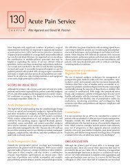
Figure <strong>105</strong>-2. Surgical techniques in heart transplantation<br />
for patients with hypoplastic left-sided heart syndrome. A:<br />
Anatomy and incisions. The ductal tissue is completely removed.<br />
The aortic arch is incised all the way down to the descending<br />
aorta beyond the duct-inserted site. The main pulmonary artery<br />
is excised just underneath the bifurcation of branch pulmonary<br />
arteries. B: The beginning of anastomosis. The left atrial anastomosis<br />
is started at the base of the left atrial appendage. C: Completion<br />
of anastomosis. The aortic arch is reconstructed with the<br />
donor aortic arch.<br />
caval anastomosis. Moderate to deep hypothermia is induced for<br />
possible deep hypothermic circulatory arrest for PA and/or aortic<br />
arch reconstruction. Failed Fontan patients frequently have<br />
multiple collateral vessels, which makes establishment of a<br />
bloodless field difficult. The heart is excised. The sequence of<br />
anastomosis is similar to that of standard transplantation. The<br />
branch PAs are typically reconstructed by the donor branch PAs.<br />
If the aorta is abnormally dilated (typically as a result of a previous<br />
Norwood type operation), the aortic arch reconstruction is<br />
sometimes necessary (techniques described in “Hypoplastic Left-<br />
Sided Heart Syndrome”).<br />
Intraoperative Medical Management<br />
Cardiopulmonary bypass is terminated with chronotropic (isopre -<br />
naline) and/or inotropic (epinephrine) supports. A phosphdieste -<br />
rase inhibitor (milrinone) is routinely used. Temporary pacing<br />
wires are placed and the heart is paced at an adequate heart rate.
CHAPTER <strong>105</strong> ■ Heart and Lung Transplantation: Surgical Considerations 1789<br />
Central venous pressure is maintained at less than 10 to 12 mmHg<br />
to avoid excessive volume overload to the right ventricle and<br />
subsequent right ventricular dilatation. Transesophageal echocar -<br />
diography is routinely performed. Biventricular function, especi -<br />
ally right ventricular function, estimated right ventricular systolic<br />
pressure, the presence or absence of semilunar valve and atrioven -<br />
tricular valve insufficiencies, and the presence or absence of<br />
anas tomotic stenosis are evaluated. Pulmonary hypertension is<br />
managed with nitric oxide inhalation. The chest is left open in<br />
neonates or infants with a large donor heart with or without right<br />
ventricular dysfunction. Postoperative management and immuno -<br />
suppression therapy are not discussed in this chapter.<br />
CLINICAL OUTCOMES<br />
A recent report from the Pediatric Heart Transplant Study Group<br />
showed that overall actuarial survival after transplantation for all<br />
age groups is 85% at 1 year and 75% at 5 years. Survival among<br />
infants less than 6 months old is 82% at 1 year and 66% at 10<br />
years. 42,43 There is a trend toward improving outcome in transplan -<br />
tation over time from 5-year survival of 72% for the era from 1993<br />
to 2000 to 77% for the years from 2001 to 2005. Survival after<br />
transplantation among the patients who had failed Fontan<br />
physiology is 76% at 1 year and 68% at 5 years. Protein-losing<br />
enteropathy in those patients can be resolved after transplantation.<br />
SPECIAL CONSIDERATIONS<br />
ABO-Incompatible Transplantation<br />
Neonates have an immature immune system and both humoral<br />
and cellular immunity are suppressed. Neonates and young infants<br />
have been described as presenting less aggressive immune reac -<br />
tions to foreign transplanted tissue until 3 months of age. Neonates<br />
also do not produce anti-A or anti-B antibodies until 5 to 6<br />
months of age, with titers gradually increasing up until 2 years of<br />
age. Antibody production may occur earlier if there is sufficient<br />
antigenic stimulation, such as the presence of A and/or B antigens<br />
on a heart graft. Antibody present in the serum from birth is<br />
maternally derived IgG, typically present in low titers. The im -<br />
maturity of the infantile immune system led us to initiate an ABOincompatible<br />
heart transplantation program.<br />
In The Hospital for Sick Children, since 1996, parents of fetuses<br />
and infants who are candidates for heart transplantation have been<br />
offered the heart from the first available donor of compatible size,<br />
regardless of blood type. 44 All patients on the transplant list are<br />
tested periodically for the presence of anti-A or anti-B antibodies.<br />
Once a potential donor is identified, serum antibody testing is<br />
repeated immediately. The recipient’s antibody titer level is<br />
quantified preoperatively. An exchange transfusion of the recipient<br />
is performed at the initiation of CPB to reduce the concentration<br />
of circulating antibody level to blood group antigens to an<br />
undetectable level. 45 The patient’s blood is drained into a separate<br />
bag while transfusing the primed volume at initiation of CPB. The<br />
volume of exchanged plasma is equal to three times the estimated<br />
blood volume (80 mL/kg). Red blood cells used in priming are<br />
ABO-compatible with the recipient. All plasma components do<br />
not contain anti-A or anti-B antibodies to donor or recipient<br />
(Table <strong>105</strong>–7). To ensure the effectiveness of exchange transfusion,<br />
anti-A and anti-B antibody titers are sent to the laboratory for level<br />
determination at 10 minutes on CPB, prior to aortic cross-clamp<br />
removal, and at the termination of CPB. Exchange transfusion is<br />
repeated as necessary to maintain low titers.<br />
All infants receive 20 mg/kg of methylprednisolone at the<br />
induction of anesthesia and before the release of the aortic crossclamp.<br />
An infusion of rabbit polyclonal antilymphocyte prepara -<br />
tion is started at induction and runs for 2 to 7 days, adjusted to<br />
yield a lymphocyte count of 200 to 400 per mm. 44 Primary im -<br />
munosuppression consists of a triple drug therapy: steroids,<br />
tacrolimus, and mycophenolate mofetil (MMF).<br />
The initial experience in the Hospital for Sick Children that<br />
consisted of 10 infants from 1996 to 2000 showed early survival of<br />
80%. Two early deaths were not related to ABO-incompatibility.<br />
The recent multi-institutional study based on the United Network<br />
for Organ Sharing database showed that 35 (6%) patients out of<br />
591 patients who were less than 1 year of age underwent ABOincompatible<br />
heart transplantation from 1999 to 2007. 46 There was<br />
no difference in survival between the groups (70% at 1 year for<br />
both groups) or incidence of hyperactive rejection (one in in -<br />
compatible group vs. two in compatible group). Another multiinstitutional<br />
study from England showed no hospital mortality<br />
among 21 patients who underwent ABO-incompatible heart<br />
transplantation from 2000 to 2006. 47<br />
Heart Transplantation from Donation<br />
After Cardiocirculatory Death<br />
Donation after cardiocirculatory death (DCD), which was used to<br />
refer to “non–heart-beating-donors,” has been proposed as a<br />
means to expand the donor pool for heart transplantation in the<br />
TABLE <strong>105</strong>-7. Blood Group Compatibility When Administering Blood Products During an ABO-Incompatible<br />
Heart Transplantation 44<br />
Donor’s Blood Group Recipient’s Blood Group Indicated Blood Group<br />
Plasma PRBC Platelets<br />
AB O AB O AB<br />
B O AB or B O AB or B<br />
A O AB or A O AB or A<br />
AB B AB O or B AB<br />
A B AB O or B AB<br />
AB A AB O or A AB<br />
B A AB O or A AB<br />
PRBC = packed red blood cells
1790 PART 5 ■ Anesthetic, Surgical, and Interventional Procedures: Considerations<br />
face of significant donor shortage and substantial mortality during<br />
waiting for transplantation, especially in infants. 26 In fact, the first<br />
successful heart transplantation in an adult involved a donor who<br />
died from cardiocirculatory death. 48 Although attractive, this stra -<br />
tegy has stayed within laboratory medicine up until very recently<br />
because of substantial hypoxic myocardial injury during the<br />
agonal period and reperfusion injury after a long warm ischemic<br />
period. The Loma Linda group has published a landmark animal<br />
study looking at the possibility of pediatric heart transplantation<br />
from DCD. 49 The study showed that the animals survived as long<br />
as 34 days after as long as 30 minutes of warm ischemia with<br />
reasonable left ventricular ejection fraction (mean 76%). 49 Nume -<br />
rous experimental studies have been performed on this particular<br />
subject focusing on myocardial protection; however, many of<br />
those studies involved multiple premedications before withdrawal<br />
of care, which limits clinical application of those strategies. 50 The<br />
Denver group recently published their experience of three pedi -<br />
atric heart transplantations from DCD. The protocol indicates that<br />
if death occurs within 30 minutes after extubation, the patient is<br />
considered to be a candidate for donation. The mean time of<br />
donors was 3.7 days. All donors suffered birth asphyxia as a cause<br />
of death. Extubation was performed after heparin (100 U/kg)<br />
administration and sedation and analgesia (fentanyl and loraze -<br />
pam). The mean time to death after withdrawal of life support was<br />
18 minutes (11 to 27 minutes). When cardiocirculatory function<br />
ceased, the patients was observed for 3 minutes (the first patient)<br />
or 75 minutes (the rest) and the organ donation process was<br />
initiated with the administration of cold cardioplegia into the<br />
aortic root through the long balloon catheter placed in the<br />
ascending aorta. The 6-month survival time was 100% compared<br />
to 84% in 17 control patients in the same period. There were no<br />
late deaths. These three patients had reasonable left ventricular<br />
systolic function at 6 months and a similar number of rejection<br />
episodes compared to controls (0.3 per patient versus 0.4 per<br />
patients in controls). The first clinical experience is indeed<br />
encouraging but still holds some medical and/or ethical issues to<br />
be overcome. One of the major issues is the duration between the<br />
declaration of cardiocirculatory death and organ retrieval. A 1997<br />
report from the Institute of Medicine suggested that 5 minutes<br />
should elapse between cardiocirculatory death and organ retrie -<br />
val. 51 The second report from the Institute of Medicine in 2000<br />
reassessed the time interval and stated that the empirical data<br />
available indicate that cardiopulmonary arrest becomes irrever -<br />
sible within a shorter time interval—60 seconds or less. 52 On the<br />
basis of this report, the Denver group used 75 seconds as the<br />
duration from death to retrieval; however, no scientific data have<br />
yet been elucidated to support this practice. Pediatric heart<br />
transplantation from DCD seems to be feasible, but graft preser -<br />
vation technique, long-term graft function, and ethical issues<br />
including time interval from declaration of death to retrieval<br />
should be well discussed and established before regular clinical<br />
application.<br />
Management of Highly Sensitized Patients<br />
Undergoing Heart Transplantation<br />
Some patients awaiting heart transplantation have circulating<br />
antibodies against human leukocyte antigens (HLA). The process<br />
by which antibodies are formed is called sensitization. Sensitiza -<br />
tion may result from previous blood transfusion, 53 homograft<br />
materials used for reconstruction in congenital heart surgery, 54 or<br />
use of mechanical circulatory assist devices. 55 Patients who require<br />
retransplantation often have allosentization. 56 There has been an<br />
increase in heart transplant candidates who have been allosensi -<br />
tized to HLA antigens over the years. The recent study showed<br />
that panel-reactive antibody (PRA) higher than 25% is associated<br />
with poor survival after heart transplantation. 57 Recent experience<br />
showed that 13 (8%) out of 167 patients who had undergone trans -<br />
plantation from 1990 to 2006 met the criteria for being allosensi -<br />
tized before heart transplantation, characterized by a PRA greater<br />
than 10%. 58 Nine (69%) were infants who had had previous<br />
palliation for CHD. Antibody-mediated rejection occurred in<br />
9 (69%) patients and acute cellular rejection (>ISHLT Grade 2 R)<br />
occurred in 7 (53%) patients, which seems more frequent than a<br />
regular transplant group. The actuarial survival at 1 year was 71%.<br />
Pretransplant treatment includes weekly intravenous administra -<br />
tion of immune globulin or an oral low dose of MMF (20 mg/<br />
kg/d) in an attempt to reduce circulating alloantibodies. Perio -<br />
perative management includes plasma exchange during transplan -<br />
tation as described above and induction of thymoglobulin. Most<br />
recently, rituximab, an anti-CD20 monoclonal antibody that<br />
rapidly causes destruction of CD20 positive cells, has been used<br />
empirically perioperatively. Postoperative management includes<br />
induction therapy with thymoglobin (1.5 mg/kg/day) for 2 to 7<br />
days and standard triple immunosuppression with tacrolimus,<br />
MMF, and steroid.<br />
In summary, current practice in pediatric heart transplantation<br />
has attained reasonable early and long-term survival and graft<br />
function in all subsets of patients with end-stage heart failure.<br />
Ventricular assist device as a means of bridge to transplantation,<br />
ABO-incompatible transplantation, and possibly transplantation<br />
from DCD are the key practices to improve overall outcomes by<br />
reducing mortality while awaiting transplantation or by improving<br />
the preoperative condition of such patients. High pretransplant<br />
mortality, management of the growing number of transplantations<br />
for failed Fontan procedure patients, and the sensitization issue<br />
have to be overcome.<br />
REFERENCES<br />
1. Kirk R, Edwards LB, Aurora P, et al. Registry of the International Society<br />
for Heart and Lung Transplantation: eleventh official pediatric heart<br />
transplantation report–2008. J Heart Lung Transplant. 2008;27:970–977.<br />
2. Kantrowitz A, Haller JD, Joos H, et al. Transplantation of the heart in an<br />
infant and an adult. Am J Cardiol. 1968;22:782–790.<br />
3. Borel JF. History of the discovery of cyclosporin and of its early<br />
pharmacological development. Wien Klin Wochenschr. 2002;114:433–437.<br />
4. Bailey LL, Nehlsen-Cannarella SL, Concepcion W, Jolley WB. Baboonto-human<br />
cardiac xenotransplantation in a neonate. JAMA. 1985;254:<br />
3321–3329.<br />
5. Bailey LL, Nehlsen-Cannarella SL, Doroshow RW, et al. Cardiac allotrans -<br />
plantation in newborns as therapy for hypoplastic left heart syndrome.<br />
N Engl J Med. 1986;315:949–951.<br />
6. Tsirka AE, Trinkaus K, Chen SC, et al. Improved outcomes of pediatric<br />
dilated cardiomyopathy with utilization of heart transplantation. J Am<br />
Coll Cardiol. 2004;44:391–397.<br />
7. Lee KJ, McCrindle BW, Bohn DJ, et al. Clinical outcomes of acute myo -<br />
carditis in childhood. Heart. 1999;82:226–233.<br />
8. Nugent AW, Daubeney PE, Chondros P, et al. Clinical features and<br />
outcomes of childhood hypertrophic cardiomyopathy: results from a<br />
national population-based study. Circulation. 2005;112:1332–1338.<br />
9. Nugent AW, Daubeney PE, Chondros P, et al. The epidemiology of<br />
child hood cardiomyopathy in Australia. N Engl J Med. 2003;348:<br />
1639–1646.
CHAPTER <strong>105</strong> ■ Heart and Lung Transplantation: Surgical Considerations 1791<br />
10. Lipshultz SE, Sleeper LA, Towbin JA, et al. The incidence of pediatric<br />
cardiomyopathy in two regions of the United States. N Engl J Med. 2003;<br />
348:1647–1655.<br />
11. Dellgren G, Koirala B, Sakopoulus A, et al. Pediatric heart transplantation:<br />
improving results in high-risk patients. J Thorac Cardiovasc Surg. 2001;<br />
121:782–791.<br />
12. Russo LM, Webber SA. Idiopathic restrictive cardiomyopathy in children.<br />
Heart. 2005;91:1199–1202.<br />
13. Chen JM, Davies RR, Mital SR, et al. Trends and outcomes in trans -<br />
plantation for complex congenital heart disease: 1984 to 2004. Ann Thorac<br />
Surg. 2004;78:1352–1361; discussion: 61.<br />
14. Bailey LL, Gundry SR, Razzouk AJ, et al. Bless the babies: one hundred<br />
fifteen late survivors of heart transplantation during the first year of life.<br />
The Loma Linda University Pediatric Heart Transplant Group. J Thorac<br />
Cardiovasc Surg. 1993;<strong>105</strong>:805–814; discussion 14–5.<br />
15. Helton JG, Aglira BA, Chin AJ, et al. Analysis of potential anatomic or<br />
physiologic determinants of outcome of palliative surgery for hypoplastic<br />
left heart syndrome. Circulation. 1986;74:I70–6.<br />
16. Mahle WT, Spray TL, Wernovsky G, et al. Survival after reconstructive<br />
surgery for hypoplastic left heart syndrome: A 15-year experience from a<br />
single institution. Circulation. 2000;102:III136–41.<br />
17. Guleserian KJ, Armsby LB, Thiagarajan RR, et al. Natural history of<br />
pulmonary atresia with intact ventricular septum and right-ventricledependent<br />
coronary circulation managed by the single-ventricle ap -<br />
proach. Ann Thorac Surg. 2006;81:2250–2257; discussion 8.<br />
18. Lim JS, McCrindle BW, Smallhorn JF, et al. Clinical features, management,<br />
and outcome of children with fetal and postnatal diagnoses of isomerism<br />
syndromes. Circulation. 2005;112:2454–2461.<br />
19. Yun TJ, Al-Radi OO, Adatia I, et al. Contemporary management of right<br />
atrial isomerism: effect of evolving therapeutic strategies. J Thorac<br />
Cardiovasc Surg. 2006;131:1108–1113.<br />
20. Chang AC, Wernovsky G, Wessel DL, et al. Surgical management of late<br />
right ventricular failure after Mustard or Senning repair. Circulation.<br />
1992;86:II140–9.<br />
21. Blanche C, Valenza M, Czer LS, Trento A. Heart transplantation in cor -<br />
rected transposition of the great arteries. J Heart Lung Transplant. 1994;<br />
13:631–634.<br />
22. Bernstein D, Naftel D, Chin C, et al. Outcome of listing for cardiac trans -<br />
plantation for failed Fontan: a multi-institutional study. Circulation.<br />
2006;114:273–280.<br />
23. Bisleri G, Morgan JA, Deng MC, et al. Should HIV-positive recipients<br />
undergo heart transplantation? J Thorac Cardiovasc Surg. 2003;126:1639–<br />
1640.<br />
24. Calabrese LH, Albrecht M, Young J, et al. Successful cardiac transplanta -<br />
tion in an HIV-1-infected patient with advanced disease. N Engl J Med.<br />
2003;348:2323–2328.<br />
25. Ward KM, Binns H, Chin C, et al. Pediatric heart transplantation for<br />
anthracycline cardiomyopathy: cancer recurrence is rare. J Heart Lung<br />
Transplant. 2004;23:1040–1045.<br />
26. Almond CS, Thiagarajan RR, Piercey GE, et al. Waiting list mortality<br />
among children listed for heart transplantation in the United States.<br />
Circulation. 2009;119:717–727.<br />
27. Azeka E, Franchini Ramires JA, Valler C, Alcides Bocchi E. Delisting of<br />
infants and children from the heart transplantation waiting list after<br />
carvedilol treatment. J Am Coll Cardiol. 2002;40:2034–2038.<br />
28. Abraham WT, Fisher WG, Smith AL, et al. Cardiac resynchronization in<br />
chronic heart failure. N Engl J Med. 2002;346:1845–1853.<br />
29. Rhee EK, Canter CE, Basile S, et al. Sudden death prior to pediatric heart<br />
transplantation: would implantable defibrillators improve outcome?<br />
J Heart Lung Transplant. 2007;26:447–452.<br />
30. Mitchell MB, Campbell DN, Boucek MM, et al. Mechanical limitation of<br />
pulmonary blood flow facilitates heart transplantation in older infants<br />
with hypoplastic left heart syndrome. Eur J Cardiothorac Surg. 2003;23:<br />
735–742.<br />
31. Caldarone CA, Benson L, Holtby H, et al. Initial experience with hybrid<br />
palliation for neonates with single-ventricle physiology. Ann Thorac Surg.<br />
2007;84:1294–1300.<br />
32. Chrisant MR, Naftel DC, Drummond-Webb J, et al. Fate of infants<br />
with hypoplastic left heart syndrome listed for cardiac transplantation:<br />
a multicenter study. J Heart Lung Transplant. 2005;24:576–<br />
582.<br />
33. Chang AC, McKenzie ED. Mechanical cardiopulmonary support in<br />
children and young adults: extracorporeal membrane oxygenation,<br />
ventricular assist devices, and long-term support devices. Pediatr Cardiol.<br />
2005;26:2–28.<br />
34. Delius RE, Zwischenberger JB, Cilley R, et al. Prolonged extracorporeal<br />
life support of pediatric and adolescent cardiac transplant patients. Ann<br />
Thorac Surg. 1990;50:791–795.<br />
35. BarZiv SM, McCrindle BW, West LJ, et al. Outcomes of pediatric patients<br />
bridged to heart transplantation from extracorporeal membrane oxygena -<br />
tion support. ASAIO J. 2007;53:97–102.<br />
36. Hetzer R, Potapov EV, Stiller B, et al. Improvement in survival after<br />
mechanical circulatory support with pneumatic pulsatile ventricular assist<br />
devices in pediatric patients. Ann Thorac Surg. 2006;82:917–924; discus -<br />
sion 24–25.<br />
37. Report of Special Task Force. Guidelines for the determination of brain<br />
death in children. American Academy of Pediatrics Task Force on Brain<br />
Death in Children. Pediatrics. 1987;80:298–300.<br />
38. Guidelines for the determination of death. Report of the medical<br />
consultants on the diagnosis of death to the President’s Commission for<br />
the Study of Ethical Problems in Medicine and Biomedical and Behavioral<br />
Research. JAMA. 1981;246:2184–2186.<br />
39. Osaki S, Ishino K, Kotani Y, et al. Circulatory load during hypoxia impairs<br />
post-transplant myocardial functional recovery in donation after cardiac<br />
death. J Heart Lung Transplant. 2009;28:266–272.<br />
40. Cooper DK, Novitzky D, Wicomb WN, et al. A review of studies relating<br />
to thyroid hormone therapy in brain-dead organ donors. Front Biosci.<br />
2009;14:3750–3770.<br />
41. Scheule AM, Zimmerman GJ, Johnston JK, et al. Duration of graft cold<br />
ischemia does not affect outcomes in pediatric heart transplant recipients.<br />
Circulation. 2002;106:I163–7.<br />
42. Hsu DT, Naftel DC, Webber SA, et al. Lessons learned from the pediatric<br />
heart transplant study. Congenit Heart Dis. 2006;1:54–62.<br />
43. Morrow WR. Outcomes following heart transplantation in children. Progr<br />
Pediatr Cardiol. 2009;26:39–46.<br />
44. West LJ, Pollock-Barziv SM, Dipchand AI, et al. ABO-incompatible heart<br />
transplantation in infants. N Engl J Med. 2001;344:793–800.<br />
45. Foreman C, Gruenwald C, West L. ABO-incompatible heart transplan -<br />
tation: a perfusion strategy. Perfusion. 2004;19:69–72.<br />
46. Patel ND, Weiss ES, Scheel J, et al. ABO-incompatible heart transplan -<br />
tation in infants: analysis of the united network for organ sharing<br />
database. J Heart Lung Transplant. 2008;27:1085–1089.<br />
47. Roche SL, Burch M, O’Sullivan J, et al. Multicenter experience of ABOin<br />
compatible pediatric cardiac transplantation. Am J Transplant. 2008;8:<br />
208–215.<br />
48. Barnard CN. The operation. A human cardiac transplant: an interim<br />
report of a successful operation performed at Groote Schuur Hospital,<br />
Cape Town. S Afr Med J. 1967;41:1271–1274.<br />
49. Gundry SR, Fukushima N, Eke CC, et al. Successful survival of primates<br />
receiving transplantation with “dead,” nonbeating donor hearts. J Thorac<br />
Cardiovasc Surg. 1995;109:1097–2010; discussion 101–102.<br />
50. Osaki S, Ishino K, Kotani Y, et al. Resuscitation of non-beating<br />
donor hearts using continuous myocardial perfusion: the importance<br />
of controlled initial reperfusion. Ann Thorac Surg. 2006;81:<br />
2167–2171.<br />
51. Potts J. Non-heart-beating organ transplantation: medical and ethical<br />
issues in procurement. Division of Health Care Services. Institute of<br />
Medicine. National Academy Press. Washington, DC; 1997.<br />
52. Schwab P. Non-heart-beating organ transplantation: practice and<br />
protocols. Institute of Medicine National Academy Press. Washington,<br />
DC; 2000.<br />
53. Mehra MR, Uber PA, Uber WE, et al. Allosensitization in heart transplan -<br />
tation: implications and management strategies. Curr Opin Cardiol. 2003;<br />
18:153–158.<br />
54. Shaddy RE, Hawkins JA. Immunology and failure of valved allografts in<br />
children. Ann Thorac Surg. 2002;74:1271–1275.<br />
55. McKenna DH, Jr., Eastlund T, Segall M, et al. HLA alloimmunization in<br />
patients requiring ventricular assist device support. J Heart Lung Trans -<br />
plant. 2002;21:1218–1224.<br />
56. Shaddy RE, Fuller TC. The sensitized pediatric heart transplant candidate:<br />
causes, consequences, and treatment options. Pediatr Transplant. 2005;9:<br />
208–214.<br />
57. Nwakanma LU, Williams JA, Weiss ES, et al. Influence of pretransplant<br />
panel-reactive antibody on outcomes in 8,160 heart transplant<br />
recipients in recent era. Ann Thorac Surg. 2007;84:1556–1562; discussion<br />
62–3.
1792 PART 5 ■ Anesthetic, Surgical, and Interventional Procedures: Considerations<br />
58. Pollock-BarZiv SM, den Hollander N, Ngan BY, et al. Pediatric heart<br />
transplantation in human leukocyte antigen sensitized patients: evolving<br />
management and assessment of intermediate-term outcomes in a highrisk<br />
population. Circulation. 2007;116:I172–8.<br />
59. Canter CE, Shaddy RE, Bernstein D, et al. Indications for heart trans -<br />
plantation in pediatric heart disease: a scientific statement from the<br />
American Heart Association Council on Cardiovascular Disease in the<br />
Young; the Councils on Clinical Cardiology, Cardiovascular Nursing, and<br />
Cardiovascular Surgery and Anesthesia; and the Quality of Care and<br />
Outcomes Research Interdisciplinary Working Group. Circulation. 2007;<br />
115:658–676.<br />
60. Transplantation protocol: Loma Linda University Medical Center<br />
2002. http://www.llu.edu/ihi/pedproto.pdf, accessed on May 10,<br />
2009.