MEDICINSKI GLASNIK - Aktuelno Ljekarska komora ZE - DO kantona
MEDICINSKI GLASNIK - Aktuelno Ljekarska komora ZE - DO kantona
MEDICINSKI GLASNIK - Aktuelno Ljekarska komora ZE - DO kantona

You also want an ePaper? Increase the reach of your titles
YUMPU automatically turns print PDFs into web optimized ePapers that Google loves.
22<br />
Medicinski Glasnik, Volumen 8, Number 1, February 2011<br />
Figure 2. Distribution of the nuchal translucency values (NT) in<br />
relation to the crown-rump length (CRL) in the total sample.<br />
Distribution of NT values in relation to the median<br />
in the control group (1.90 mm) was 135:301,<br />
or 55.15% above the median, or 44.85% below<br />
the median (Figure 2,3).<br />
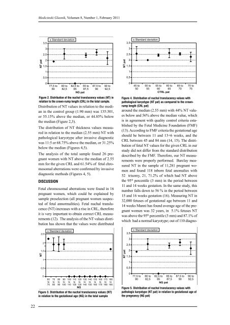
The distribution of NT thickness values measured<br />
in relation to the median (2.55 mm) NT with<br />
pathological karyotype after invasive diagnostic<br />
was 11:5 or 68.75% above the median, or 31.25%<br />
below the median (Figures 4,5).<br />
The analysis of the total sample found 26 pregnant<br />
women with NT above the median of 2.55<br />
mm for the given CRL and 61.54% of fetal chromosomal<br />
aberrations were confirmed by invasive<br />
diagnostic methods (Figures 4, 5).<br />
DISCUSSION<br />
Fetal chromosomal aberrations were found in 16<br />
pregnant women, which could be explained by<br />
sample preselection (all pregnant women suspected<br />
of fetal annormalities). Fetal nuchal translucence<br />
(NT) increases with a rise in CRL, therefore<br />
it is very important to obtain correct CRL measurements<br />
(12). The analysis of the NT values distribution<br />
has shown that the values were distributed<br />
Figure 3. Distribution of the nuchal translucency values (NT)<br />
in relation to the gestational age (NG) in the total sample<br />
Figure 4. Distribution of nuchal translucency values with<br />
pathological karyotype (NT pat) as compared to the crownrump<br />
length (CRL pat)<br />
around the median (2.55 mm) with 44% NT values<br />
below and 56% above the median value, which<br />
is in agreement with quality control criteria established<br />
by the Fetal Medicine Foundation (FMF)<br />
(13). According to FMF criteria the gestational age<br />
should be between 11 and 13+6 weeks, and the<br />
CRL between 45 and 84 mm (14, 15). The distribution<br />
of fetal NT values for the given CRL in our<br />
study did not differ from the standard distribution<br />
described by the FMF. Therefore, our NT measurements<br />
were properly performed. Bаrclay measured<br />
NT in the sample of 11,281 pregnant women<br />
and found 118 inborn fetal anomalies with<br />
52 trisomy, 21, 71.2% of which had NT above<br />
the 95th percentile (3 mm) in the period between<br />
11 and 14 weeks gestation. In the same study, this<br />
number falls down to 56 % in the period between<br />
15 and 16 weeks gestation (16). Measuring NT in<br />
32,000 fetuses of gestational age between 11 and<br />
14 weeks Mаnni has found average age of the pregnant<br />
women was 32 years, in 5.1% fetuses NT<br />
was above the 95th percentile (3 mm) and 87.1% of<br />
which had a normal karyotype; out of 110 diagno-<br />
Figure 5. Distribution of nuchal translucency values with<br />
pathologic karyotype (NT pat) in relation to gestational age of<br />
the pregnancy (NG pat)