MEDICINSKI GLASNIK
MEDICINSKI GLASNIK
MEDICINSKI GLASNIK

Create successful ePaper yourself
Turn your PDF publications into a flip-book with our unique Google optimized e-Paper software.
<strong>MEDICINSKI</strong> <strong>GLASNIK</strong><br />
Official publication of the Medical Association of Zenica-Doboj Canton, Bosnia and Herzegovina<br />
Volume 6 Number 2, August 2009.<br />
ISSN 1840-0132
Published and copyright by: Medical Assotiation of Zenica-Doboj Canton; Address: Zenica, 72000, Bulevar kralja Tvrtka I 4, Bosnia and Herzegovina;<br />
tel./fax: +387 32 444 270; Email: ljkozedo@bih.net.ba, web site: http//www.ljkzedo.com.ba<br />
For ordering information please contact: Tatjana Žilo, ljkozedo@bih.net.ba; Access to this journal is available free online trough: www.ljkzedo.ba<br />
The Journal is indexed by EMBASE (Exerpta Medica), Scopus, Science Citation Index Expanded (SciSearch ® ), and Journal Citation Reports/Science Edition,<br />
EBSCO; ISSN 1840-0132<br />
Printed by: EG - ING & Graphic and web design studio “B Panel” Zenica, Armije BiH 2, E-mail: info@bpanel.ba, tel. +387 32 441 290, 441 291
Medicinski Glasnik<br />
Official Publication of the Medical Association of<br />
Zenica-Doboj Canton<br />
Bosnia and Herzegovina<br />
Editorial Board<br />
Editor-in-chiEf<br />
Selma Uzunović-Kamberović<br />
Zenica, Bosnia and Herzegovina<br />
tEchnical Editor<br />
Harun Drljević<br />
Zenica, Bosnia and Herzegovina<br />
Editors<br />
Adem Balić, Tuzla, Bosnia and Herzegovina<br />
Dubravka Bartolek, Zagreb, Croatia<br />
Branka Bedenić, Zagreb, Croatia<br />
Asja Čelebić, Zagreb, Croatia<br />
Josip Čulig, Zagreb, Croatia<br />
Filip Čulo, Mostar, Bosnia and Herzegovina<br />
Jordan Dimanovski, Zagreb, Croatia<br />
Branko Dmitrović, Osijek, Croatia<br />
Davorin Đanić, Slavonski Brod, Croatia<br />
Lejla Ibrahimagić-Šeper, Zenica, Bosnia and Herzegovina<br />
Tatjana Ille, Belgrade, Serbia<br />
Vjekoslav Jerolimov, Zagreb, Croatia<br />
Mirko Šamija, Zagreb, Croatia<br />
Ines Drenjančević-Perić, Osijek, Croatia<br />
Sven Kurbel, Osijek, Croatia<br />
Snježana Pejičić, Banja Luka, Bosnia and Herzegovina<br />
Belma Pojskić, Zenica, Bosnia and Herzegovina<br />
Asja Prohić, Sarajevo, Bosnia and Herzegovina<br />
Velimir Profozić, Zagreb, Croatia<br />
Zlatko Puvačić, Sarajevo, Bosnia and Herzegovina<br />
Radivoje Radić, Osijek, Croatia<br />
Amira Redžić, Sarajevo, Bosnia and Herzegovina<br />
Suad Sivić, Zenica, Bosnia and Herzegovina<br />
Sonja Smole-Možina, Ljubljana, Slovenia<br />
Vladimir Šimunović, Mostar, Bosnia and Herzegovina<br />
Adrijana Vince, Zagreb, Croatia<br />
Jasmina Vraneš, Zagreb, Croatia<br />
Živojin Žagar, Zagreb, Croatia<br />
Secretary: Tatjana Žilo;<br />
Proofreaders: Aras Borić (Bosnian, Croatian, Serbian),<br />
Glorija Alić (English),<br />
Cover: Jasmin Kukavica
<strong>MEDICINSKI</strong> <strong>GLASNIK</strong><br />
Official Publication of the Medical Association of Zenica-Doboj Canton, Bosnia and Herzegovina<br />
Volume 6, Number 2, August 2009<br />
Free full-text online at: www.ljkzedo.com.ba, and www.doaj.org (DOAJ, Directory of Open Access<br />
Journals)<br />
Review 147 Značenje nastanka mikrobnog biofilma u patogenezi i liječenju kroničnih<br />
infekcija<br />
Significance of microbial biofilm occurence in the pathogenesis and treatment<br />
of chronic infections<br />
Jasmina Vraneš, Vladimira Leskovar<br />
Original<br />
article<br />
166 Effect of inoculum size of Enterobacteriaceae producing SHV and<br />
CTX-M extended-spectrum β-lactamases on the susceptibility to β-lactam<br />
combinations with inhibitors and carbapenems<br />
Branka Bedenić, Jasmina Vraneš, Nataša Beader, Ines Jajić-Benčić, Vanda<br />
Plečko, Selma Uzunović-Kamberović, Smilja Kalenić<br />
173 Comparison of the frequency and the occurrence of antimicrobial<br />
resistance among C. jejuni and C. coli isolated from human infections,<br />
retail poultry meat and poultry in Zenica-Doboj Canton, Bosnia and<br />
Herzegovina<br />
Selma Uzunović-Kamberović, Tina Zorman, Ingrid Berce, Lieve Herman,<br />
Sonja Smole Možina<br />
181 Atelektaza pluća i infekcije donjih dišnih puteva u djece na odjelu za<br />
intenzivno liječenje<br />
Lung atelectasis and lower respiratory tract infections in children in the<br />
intensive care unit<br />
Nada Mladina, Devleta Hadžić, Amela Selimović<br />
188 Evaluacija dijagnostičke vrijednosti Interleukina-6 i C-reaktivnog<br />
proteina iz krvi pupčanika u prepoznavanju rane infekcije terminske<br />
novorođenčadi male porođajne mase<br />
Diagnostic value of Interleukin 6 and C-reactive protein from umbilical cord<br />
blood in recognition of early infection in term newborns with low birth weight<br />
Almira Ćosićkić, Fahrija Skokić, Selmira Brkić<br />
197 Alterations in body weight and biochemistry in patient treated with different<br />
psychotropic drugs in a clinic in Istanbul<br />
Aliye Ozenoglu, Serdal Ugurlu, Huriye Balci, Gunay Can, Funda Elmacıoglu,<br />
Yeltekin Demirel, Engin Eker<br />
203 Klinička revizija lipidnog statusa kod tipa 2 dijabetesa na nivou timova<br />
obiteljske medicine u općini Zenica, Bosna i Hercegovina<br />
Audit of lipids control level for Diabetes Mellitus type 2 patients done by<br />
Family Medicine Teams in Zenica, Bosnia and Hercegovina<br />
Larisa Gavran, Selmira Brkić<br />
211 Učestalost pušenja i nikotinska ovisnost kod medicinskih radnika<br />
Incidence of smoking and nicotine depedence among medical workers<br />
Željko Martinović, Cvita Martinović, Mladen Čuturić<br />
218 Smoking is the most frequent risk factor for cardiovascular diseases in<br />
Croatian Western region: findings of the Croatian health survey 2003.<br />
Đulija Malatestinić, Nena Rončević, 1 Henrietta Benčević-Striehl, Suzana<br />
Janković, Vladimir Mićović
Case<br />
report<br />
Erratum 284<br />
227 Influence of different glass fiber reinforcements on denture base polymer<br />
strength (Fiber reinforcements of dental polymer)<br />
Denis Vojvodić, Dragutin Komar, Zdravko Schauperl, Asja Čelebić, Ketij<br />
Mehulić, Domagoj Žabarović<br />
235 Influence of cast surface finishing process on metal-ceramic bond strength<br />
Ketij Mehulić, Martina Lauš-Šošić , Zdravko Schauperl, Denis Vojvodić, Sanja<br />
Štefančić<br />
243 Use of digital photography in the reconstruction of the occlusal plane<br />
orientation<br />
Nikola Petričević, Marko Guberina, Robert Ćelić, Ketij Mehulić, Marko<br />
Krajnović, Robert Antonić, Josipa Borčić, Asja Čelebić<br />
249 A three-dimensional evaluation of microleakage of the class V cavities<br />
restored with flowables<br />
Paris Simeon, Silvana Jukić-Krmek, Goranka Prpić-Mehičić, Ivica Smojver,<br />
Ivica Anić, Ivica Pelivan<br />
256 Oral health of the Croatian army recruits in 2001<br />
Tomislav Badel, Jadranka Keros, Vjekoslav Jerolimov, Nikša Dulčić, Snježana<br />
Restek Despotušić<br />
261 Security perception of a portable PC user (The difference between medical<br />
doctors and engineers): a pilot study<br />
Krešimir Šolić, Vesna Ilakovac<br />
265 Akutna bruceloza udružena sa Coombs-pozitivnom autoimunosnom<br />
hemolitičkom anemijom i diseminiranom intravaskularnom koagulacijom<br />
Acute brucellosis associated with Coombs-positive autoimmune hemolytic<br />
anemia and disseminated intravascular coagulation (DIC)<br />
Nerma Mušić<br />
268 Pneumolabirint<br />
Pneumolabyrinth<br />
Đenad Hodžić<br />
271 Sphenochoanal polyposis<br />
Ivana Pajić-Penavić, Davorin Đanić, Ljubica Fuštar-Preradović<br />
274 Primary extranodal Natural Killer/T-cell lymphoma of the ethmoid sinus<br />
masquerading as orbital cellulites<br />
Davorin Đanić, Ana Đanić Hadžibegović, Ivana Mahovne<br />
277 Knee disarticulation<br />
Ognjen Živković, Antun Muljačić, Renata Poljak-Guberina<br />
280 Izvanmaternična trudnoća izliječena metrotreksatom<br />
Extrauterine pregnancy treated with Metrotrexat<br />
Ljiljana Bilobrk Josipović, Branka Lovrinović, Anton Galić<br />
Medicinski Glasnik is indexed by EMBASE (Exerpta Medica), Scopus, Science Citation Index Expanded<br />
(SciSearch ® ), Journal Citation Reports/Science Edition and EBSCO
REVIEW<br />
Značenje nastanka mikrobnog biofilma u patogenezi i liječenju<br />
kroničnih infekcija<br />
Jasmina Vraneš 1, 2 , Vladimira Leskovar 2<br />
1 2 Katedra za medicinsku mikrobiologiju, Medicinski fakultet Sveučilišta u Zagrebu, Služba za mikrobiologiju, Zavod za javno zdravstvo<br />
“Dr. Andrija Štampar“; Zagreb, Hrvatska<br />
Corresponding author:<br />
Jasmina Vraneš,<br />
Zavod za javno zdravstvo “Dr. Andrija<br />
Štampar“,<br />
Mirogojska cesta 16, 10 000 Zagreb,<br />
Hrvatska<br />
Phone: +385 1 4696 197;<br />
Fax: +385 1 4678 006;<br />
E-mail: jasmina.vranes@stampar.hr<br />
Originalna prijava:<br />
06. april 2009.;<br />
Korigirana verzija:<br />
08. april 2009.;<br />
Prihvaćeno:<br />
03. maj 2009.<br />
Med Glas 2009; 6(2):147-165<br />
SAŽETAK<br />
Sposobnost adherencije bakterija na biotičke i abiotičke površine,<br />
funkcioniranje kao zajednica, te međusobna komunikacija bakterijskih<br />
stanica, od posebne su važnosti u nastanku kroničnih infektivnih<br />
bolesti. Sesilna zajednica mikroorganizama, danas poznata<br />
kao biofilm, povezuje se s brojnim bakterijskim infekcijama.<br />
Ključne karakteristike biofilm-infekcija jesu perzistencija infekcije,<br />
te rezistencija na antimikrobne lijekove i obranu imunološkog<br />
sustava domaćina. Napretkom tehnologije i primjenom novih mikroskopskih<br />
i molekularnih metoda u proučavanju ultrastrukture i<br />
funkcionalnim odnosima unutar biofilma, nastoji se pronaći novi<br />
terapijski pristup u kontroli biofilm-infekcija, koje su jedan od<br />
najvećih izazova 21. stoljeća.<br />
Ključne riječi: biofilm, kronične infekcije, patogeneza, liječenje<br />
147
148<br />
Medicinski Glasnik, Volumen 6, Number 2, August 2009<br />
UVOD<br />
U prirodi mikroorganizmi mogu egzistirati<br />
kao planktonski organizmi - individualne stanice<br />
koje slobodno plivaju u tekućem mediju; ili<br />
u obliku sesilne zajednice - biofilma. Biofilm je<br />
izrazito rasprostranjen način života u okolišu,<br />
gotovo na svakoj granici vode i zraka, te zemlje<br />
i vode (1, 2). U protekla dva desetljeća definicija<br />
biofilma se neprestano mijenjala jer svako<br />
novo istraživanje nadograđuje postojeće znanje<br />
o stvaranju, strukturi, sazrijevanju, te rezistenciji<br />
biofilma. Objedinjenjem spoznaja o već<br />
poznatim karakteristikama, te novootkrivenim<br />
fiziološkim osobinama, biofilm je danas definiran<br />
kao sesilna zajednica mikroorganizama čije<br />
su stanice ireverzibilno povezane sa supstratom i<br />
međusobno, te uklopljene u izvanstanični matriks<br />
polisaharidnih polimera koji su same stvorile, a<br />
ispoljavaju izmijenjen fenotip uslijed promijenjene<br />
brzine razmnožavanja i transkripcije gena<br />
koje ne uočavamo u planktonskih organizama (3).<br />
Formiranje biofilma započinje kondicioniranjem<br />
neke površine polimerima iz vodenog okoliša što<br />
omogućuje adherenciju mikroorganizama (2).<br />
Površine na kojima se može naći biofilm jesu,<br />
primjerice, metal, plastika, kamen, čestice zemlje,<br />
medicinski implantati, te tkiva (2). Nakon<br />
početnog, reverzibilnog vezivanja planktonskih<br />
stanica za površinu supstrata, slijedi stvaranje stabilne<br />
veze posredovane adhezinima na staničnoj<br />
stijenki bakterija, a potom proliferacija, te nakupljanje<br />
bakterijskih stanica u višeslojne stanične<br />
nakupine i stvaranje izvanstaničnog polisaharidnog<br />
matriksa. Održavanje takve višestanične zajednice<br />
bilo bi teško bez postojanja međustanične<br />
komunikacije bakterijskih stanica, posredovane<br />
malim signalnim molekulama sa sposobnošću<br />
difuzije u izvanstanični okoliš (4). Nakupljanje<br />
signalnih molekula u okolišu omogućuje svakoj<br />
pojedinoj bakterijskoj stanici procjenu stanične<br />
gustoće, odnosno ukupnog broja bakterija, a ta se<br />
pojava naziva detekcija kvoruma (engl. quorum<br />
sensing). Signalne molekule u gram-negativnih<br />
bakterija jesu N-acil-homoserinski laktoni (AHL),<br />
a u gram-pozitivnih su mali ili modificirani peptidi<br />
(Slika 1) (4). Koncentracija signalnih mole-<br />
kula postignuta pri točno određenoj, kritičnoj<br />
gustoći stanica, postaje dovoljna za aktivaciju<br />
gena uključenih u sintezu faktora virulencije ili<br />
sekundarnih metabolita (4, 5). Maturacija biofilma<br />
odvija se u pet razvojnih stadija (6). Prvi stadij<br />
je reverzibilno povezivanje, u kojem se planktonske<br />
stance tek kratkotrajno povezuju sa supstratom,<br />
a nakon toga slijedi drugi stadij, ireverzibilno<br />
povezivanje, u kojem se planktonske stanice<br />
čvrsto vežu na površinu supstrata i gube svojstvo<br />
pokretljivosti (6). Treći stadij jeste maturacija I za<br />
koji je karakteristično stvaranje izvanstaničnog<br />
matriksa, te povećavanje i višeslojnost mikrokolonijâ<br />
(6). Mikrokolonije, najčešće gljivolikog ili<br />
stupićastog izgleda, svoju maksimalnu veličinu<br />
dosežu u stadiju maturacije II (6). Proces maturacije<br />
biofilma završava petim stadijem, disperzijom,<br />
u kojem se između mikrokolonija formiraju<br />
vodeni kanalići, a same mikrokolonije mijenjaju<br />
svoj oblik u školjkasti, uslijed izdvajanja bakterijskih<br />
stanica smještenih u središnjem dijelu<br />
mikrokolonija u potrazi za novim i boljim izvorom<br />
hranjivih tvari (6). Biofilm može činiti samo<br />
jedna bakterijska vrsta, no češće se sastoji od više<br />
vrsta bakterija, ili bakterija i gljiva (1, 3). Jednom<br />
pričvršćeni za površinu, mikroorganizmi biofilma<br />
mogu, svojim aktivnostima, doprinositi ili štetiti<br />
zbivanjima u okolišu, ovisno o uvjetima koji u<br />
njemu vladaju. Poznato je da ove sesilne zajednice<br />
sudjeluju u razgradnji brojnih onečišćivača<br />
okoliša, stvaranju i razgradnji organske tvari, te<br />
u kruženju dušika, sumpora i drugih elemenata u<br />
prirodi (7-10). Prije saznanja o postojanju, strukturi<br />
i karakteristikama biofilma, biotehnolozi su<br />
već koristili prednosti biofilma u sustavima za<br />
pročišćavanje voda, pri uklanjanju opasnih tvari<br />
koje su kontaminirale zemlju i podzemne vode,<br />
te pri ekstrakciji plemenitih metala iz rudače (11-<br />
13). No, pozitivna djelovanja biofilma daleko su<br />
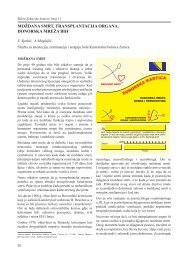
Slika 1. Stvaranje biofilma (Alma Šimunec Jović, 2007.)
jeđa od štetnih koja uzrokuju ogromne ekonomske<br />
gubitke u agrokulturi i industriji, te mnogobrojne<br />
probleme u medicini. Biofilm se povezuje<br />
sa stafilokoknim mastitisom u goveda (14), biokorozijom<br />
(deterioracijom metalnih materijala<br />
u prisutnosti biofilma) vodenih sustava u industriji<br />
(15), te naftnih cjevovoda (16), kao i problemima<br />
u industriji prehrambenih i papirnatih<br />
artikala (17-18). U medicini biofilm se povezuje<br />
s brojnim kroničnim infekcijama (1), te različitim<br />
infekcijama biomaterijala poput kateterâ, protezâ,<br />
implantatâ, te drugih medicinskih naprava<br />
od metala ili plastičnih polimera koji se nalaze u<br />
ljudskom organizmu (1, 19). Prema procjenama<br />
Centra za kontrolu bolesti i prevenciju iz Atlante,<br />
biofilm se povezuje s gotovo dvije trećine bakterijskih<br />
infekcija (3). Brojnost, kroničan tijek, te<br />
rezistencija na antimikrobnu terapiju, tek su neke<br />
od značajki biofilm-infekcija, koje su jedan od<br />
najvećih izazova medicine 21. stoljeća.<br />
REZISTENCIJA BIOFILMA<br />
Perzistencija infekcija uzrokovanih mikrobnim<br />
biofilmom kontinuirano motivira istraživače<br />
u potrazi za jedinstvenim mehanizmom rezistencije<br />
biofilma, kako na antimikrobne lijekove,<br />
tako i na dezinfekcijska sredstva (3). Poznato<br />
je da su bakterije, unutar biofilma, 10-1.000<br />
puta rezistentnije na djelovanje antimikrobnih<br />
lijekova nego planktonske stanice, što govori u<br />
prilog postojanja mehanizma rezistencije kojeg<br />
potencijalno posjeduju svi patogeni, no koji<br />
se ispoljava samo pri tvorbi biofilma (20, 21).<br />
Proučavanjem biofilma takav mehanizam još nije<br />
utvrđen, niti su otkriveni mutanti u planktonskim<br />
kulturama koji bi govorili tomu u prilog (21),<br />
pa se trenutno rezistencija biofilma objašnjava<br />
tek hipotezama. Hipoteze starijeg datuma, kao<br />
moguće mehanizme rezistencije, navode oslabljenu<br />
difuziju antimikrobnih lijekova u biofilm<br />
(20, 22) ili promjene u mikrookolišu biofilma<br />
koje utječu na brzinu razmnožavanja mikroorganizama,<br />
odnosno djelotvornost antimikrobnih<br />
lijekova u dubini biofilma (20, 22). Oslabljena<br />
difuzija antimikrobnih lijekova objašnjava se<br />
dvojako; s jedne strane, interakcijom molekula<br />
Vraneš et al Biofilm i kronične infekcije<br />
s egzopolisaharidnim matriksom biofilma, koji<br />
djeluje poput ‘’molekularnog sita’’ na velike<br />
molekule ili kao ionski izmjenjivač na hidrofilne,<br />
pozitivno nabijene molekule antimikrobnih<br />
lijekova (1, 22, 23); te, s druge strane, enzimskom<br />
razgradnjom, kojom se biofilm štiti od<br />
molekula, kao, primjerice, vodikovog peroksida<br />
ili β-laktamskih antimikrobnih lijekova (20, 23).<br />
Kao što je vidljivo, primjenjivost ove hipoteze<br />
ovisna je o prirodi samog lijeka, no nisu svi antimikrobni<br />
lijekovi visokoreaktivne molekule<br />
poput, primjerice, aminoglikozida, pa ih većina,<br />
vjerojatno, ipak penetrira u biofilm (23). Biofilm<br />
je prostorno heterogeničan (24). S porastom debljine<br />
slojeva stanica u biofilmu, mijenjaju se<br />
i uvjeti mikrookoliša, poput dostupnosti hranjivih<br />
tvari, prisutnosti kisika ili kiselih otpadnih<br />
metaboličkih tvari (20, 22, 25). Sve to utječe<br />
na brzinu razmnožavanja bakterijskih stanica,<br />
koje, u takvim uvjetima, prelaze u stacionarnu<br />
fazu u kojoj su manje osjetljive na baktericidno<br />
djelovanje antimikrobnih lijekova, osobito onih<br />
čije je ciljno mjesto djelovanja sinteza makromolekula<br />
(26). Novija hipoteza jeste hipoteza<br />
perzistera. Većina stanica biofilma, kao i planktonske<br />
stanice koje se oslobađaju s površine biofilma,<br />
osjetljive su na antimikrobnu terapiju (23,<br />
27). Naprotiv, samo mala frakcija stanica (0,1-<br />
10% svih stanica biofilma) neosjetljiva je na<br />
produženu izloženost ili više koncentracije antimikrobnih<br />
lijekova, te perzistira usprkos terapiji<br />
(26). Premda su perzisteri otkriveni još sredinom<br />
20. stoljeća u planktonskim kulturama (21),<br />
o njihovoj se prirodi još uvijek malo zna. Za sada<br />
je poznato da perzisteri nisu mutanti (21), te da<br />
ne predstavljaju poseban stadij staničnog ciklusa<br />
bakterija (23). Pretpostavlja se da je riječ o fenotipskoj<br />
varijanti koja je, iz divljeg tipa, spontano<br />
ušla u stanje promjenjivih fenotipskih obilježja<br />
(26). Mehanizam nastanka perzistera još je manje<br />
poznat. Do sada je utvrđeno da ove stanice ne<br />
nastaju kao odgovor na antimikrobnu terapiju<br />
(21, 27), te da bakterijske signalne molekule,<br />
kojima se detektira stanična gustoća, nemaju<br />
nikakva utjecaja na stvaranje perzistera (21, 27).<br />
U novije vrijeme pretpostavlja se da u kontroli<br />
stvaranja perzistera sudjeluju dva procesa. Jedan<br />
149
150<br />
Medicinski Glasnik, Volumen 6, Number 2, August 2009<br />
je promjena razine specifičnih perzister-proteina<br />
(27), ovisno o fazi razvitka biofilma (21), a<br />
drugi je kontroliranje razine ekspresije tih proteina,<br />
koja ovisi o gustoći stanica (27), te o faktorima<br />
mikrookoliša, poput niske koncentracije<br />
supstrata (26). Misli se da perzister-proteini induciraju<br />
prelazak bakterija u stanje mirovanja,<br />
tzv. dormant fazu (27), što rezultira tolerancijom<br />
na antimikrobne lijekove uslijed utišane<br />
ekspresije gena u biosintetskim putevima ovih<br />
stanica, a time i onemogućavanjem djelovanja<br />
lijekova na njihova ciljna mjesta (21, 27). Ako<br />
znamo da perzisteri postoje i u planktonskim<br />
kulturama (23, 27), onda se nameće pitanje<br />
kako perzisteri biofilma doprinose njegovoj visokoj<br />
rezistenciji. Odgovor leži u sinergističkom<br />
djelovanju imunološkog sustava i antimikrobnih<br />
lijekova. Dok imunološki sustav uništava i<br />
uklanja zaostale planktonske perzistere (21, 23,<br />
27), spram perzistera biofilma je nemoćan jer<br />
su ovi zaštićeni egzopolisaharidnim matriksom<br />
(23, 27) i nakon što se smanji koncentracija lijeka<br />
(21, 27) ili prekine terapija uslijed nestanka<br />
simptoma bolesti (23), perzisteri obnavljaju biofilm,<br />
oslobađaju se nove planktonske stanice i<br />
simptomi bolesti se vraćaju (21, 23).<br />
ULOGA BIOFILMA U NASTANKU KRONIČNIH<br />
INFEKCIJA U LJUDI<br />
Akutne bakterijske infekcije, koje izazivaju<br />
planktonski oblici patogenih bakterija, stoljećima<br />
su odnosile ljudske živote. Razvoj antimikrobnih<br />
lijekova i cjepiva omogućio je stjecanje kontrole<br />
nad ovim akutnim bolestima, te se može reći da<br />
one sada, uglavnom, pripadaju prošlosti. Sredinom<br />
prošlog stoljeća započela je postantibiotska<br />
era, odnosno era biofilm-infekcija. Ove infekcije<br />
perzistiraju mjesecima i godinama, izmjenjuju se<br />
periodi odsutnosti kliničkih simptoma infekcije s<br />
akutnim egzacerbacijama, manje su agresivne od<br />
akutnih infekcija, te pogađaju umjereno kompromitirane<br />
osobe (28). Uzročnici biofilm-infekcija<br />
jesu bakterijske vrste koje se nalaze u okolišu ili<br />
koje nalazimo kao komenzale ljudskog organizma<br />
(3, 29), a koje već stoljećima, u svom prirodnom<br />
okolišu, postoje u obliku biofilm-zajednice.<br />
Biofilm se, u ljudskom organizmu, razvija na<br />
vitalnom ili nekrotičnom tkivu, te na inertnim<br />
površinama različitih biomaterijala (29). Bez<br />
obzira radi li se o kroničnoj infekciji ili o infekciji<br />
biomaterijala, ove biofilm-infekcije imaju<br />
iste kliničke karakteristike, a to su spor nastanak<br />
na jednom ili više mjesta u organizmu, te kasna<br />
pojava simptoma bolesti (29). Nekoliko karakteristika<br />
biofilm čini jedinstvenim u procesu<br />
nastanka infekcije. Prva od njih jeste mogućnost<br />
disperzije stanica ili agregata biofilma, što može<br />
rezultirati nastankom embolusa, ili pak infekcijom<br />
mokraćnog ili krvožilnog sustava (2, 30).<br />
Disperzija pojedinih stanica nastaje uslijed odvajanja<br />
stanica-kćeri iz aktivno-razmnožavajućih<br />
stanica, te kao posljedica promjene dostupnosti<br />
hranjivih tvari ili detekcije kvoruma (2), a<br />
odvajanje manjih dijelova biofilma nastaje pod<br />
utjecajem brzine protoka tekućine u neposrednoj<br />
blizini površine biofilma (2, 30). Dok pojedine<br />
stanice, ubrzo nakon odvajanja, poprimaju<br />
fenotip planktonskih stanica, čini se da čestice<br />
biofilma zadržavaju određene karakteristike biofilma,<br />
poput antimikrobne rezistencije (2, 30).<br />
Antimikrobnom terapijom eradiciraju se planktonske<br />
stanice oslobođene iz biofilma, što rezultira<br />
povlačenjem simptoma akutne egzacerbacije<br />
bolesti, no ne uspjeva se uništiti sesilna zajednica<br />
(2, 29, 30). Rekurirajući simptomi biofilm-infekcije<br />
u konačnici će nestati tek nakon uklanjanja<br />
biofilma kirurškim zahvatom ili odstranjenjem<br />
inficiranog biomaterijala (29, 30). Slijedeća jedinstvena<br />
karakteristika biofilm- infekcija jeste rezistencija<br />
biofilma na stanični i humoralni odgovor<br />
imunološkog sustava domaćina. U početku se<br />
smatralo da rezistenciji pridonosi interferencija<br />
egzopolisaharidnog matriksa biofilma s kemotaksijom<br />
polimorfonuklearnih leukocita (PMN)<br />
(31), te s prodorom baktericidnih i opsonizirajućih<br />
protutijela (28, 31). Danas je poznato da aktivirani<br />
PMN penetriraju u biofilm, ulaze u vodene<br />
kanaliće, te penetriraju u mikrokolonije, no<br />
tamo ne uspijevaju fagocitirati stanice biofilma<br />
koje se nalaze u njihovoj neposrednoj blizini<br />
(32). Mehanizam ove neuspješne fagocitoze tek<br />
predstoji objasniti (32). Opsonizirajuća protutijela,<br />
dio humoralne obrane, u slučaju biofilm-
infekcije, dijele sudbinu polimorfonuklearnih<br />
leukocita. Iako prodiru u dubinu biofilma (33),<br />
u matriks ulaze u interakciju s antigenima koji<br />
su tamo u suvišku, te uslijed toga ne dospijevaju<br />
do antigena na površini stanica biofilma, što<br />
onemogućuje njihovo opsonizirajuće djelovanje<br />
i posljedično uništavanje stanica biofilma (33).<br />
Perzistencija kroničnih infekcija, nemogućnost<br />
detekcije mikrobnog uzročnika standardnim<br />
mikrobiološkim tehnikama kultivacije u većini<br />
slučajeva, te neuspjeh antimikrobne terapije<br />
i imunološkog sustava domaćina u eradikaciji<br />
uzročnika, naveli su neke znanstvenike na<br />
pomisao da kronične infekcije uzrokuje biofilm.<br />
Ovu tezu valjalo je potkrijepiti istraživanjem<br />
sesilnih zajednica in situ, što, uslijed nedostatka<br />
raspoloživih metoda, nije bilo ostvarivo sve do<br />
unazad dvadesetak godina. Razvoj i primjena<br />
konfokalnog skenirajućeg laserskog mikroskopa<br />
(engl. confocal scanning laser microscope,<br />
CSLM) omogućila je istraživanje ultrastrukture<br />
biofilma (1-3), a nove molekularne tehnologije,<br />
poput primjene fluorescentnih rRNK-usmjerenih<br />
proba, te fluorescentne in situ hibridiza-<br />
Kronična infekcija<br />
Chronic infection<br />
Endokarditis nativnih valvula<br />
Native valve endocarditis<br />
Periodontitis<br />
Periodontitis<br />
Cistična fibroza<br />
Cystic fibrosis<br />
Kronični bakterijski prostatitis<br />
Chronic bacterial prostatitis<br />
Kronični cistitis<br />
Chronic cystitis<br />
Inficirani bubrežni kamenci<br />
Infected kidney stones<br />
Kronični otitis media<br />
Chronic otitis media<br />
Mišićno-koštane infekcije<br />
Musculoskeletal infections<br />
Nekrotizirajući fasciitis<br />
Necrotizing fasciitis<br />
Infekcije bilijarnog sustava<br />
Biliary tract infection<br />
Osteomijelitis<br />
Osteomyelitis<br />
Meloidoza<br />
Meloidosis<br />
Kronične rane<br />
Chronic wounds<br />
cije (FISH), doprinijele su identifikaciji i kvantifikaciji<br />
uzročnikâ biofilma, te razumijevanju<br />
njihovih međusobnih funkcionalnih odnosa (1).<br />
Mogućnost vizualizacije i proučavanja biofilma<br />
na biomaterijalima, te u humanim uzorcima, rezultirala<br />
je intenzivnim istraživanjima brojnih<br />
infekcija i njihove povezanosti sa stvaranjem<br />
biofilma (13). Danas se biofilm, uz infekcije biomaterijala,<br />
povezuje s brojnim kroničnim infekcijama.<br />
Najčešće kronične infekcije, te njihove<br />
uzročnike, prikazuje Tablica 1.<br />
PERIODONTITIS<br />
U periodontalne bolesti ubraja se cijeli<br />
niz poremećaja potpornog tkiva zubi, od kojih<br />
pobolijeva gotovo 90% svjetske populacije.<br />
Najblaži oblik periodontalne bolesti je gingivitis,<br />
reverzibilna upala gingive (desni), a najteži,<br />
kronični periodontitis, kronična destrukcija periodontalnog<br />
tkiva (gingive, periodontalnog ligamenta<br />
i alveolarne kosti) koji može rezultirati<br />
gubitkom zubi (3). Primarno mjesto periodontalne<br />
infekcije jeste prostor između korijena zuba<br />
Uobičajen bakterijski patogen biofilma<br />
Common biofilm bacterial pathogen<br />
Streptokoki viridans grupe<br />
Viridans group streptococci<br />
Gram-negativne anaerobne bakterije<br />
Gram-negative anaerobic bacteria<br />
Pseudomonas aeruginosa<br />
Pseudomonas aeruginosa<br />
Gram-negativne enterobakterije<br />
Gram-negative enterobacteria<br />
Uropatogena Escherichia coli<br />
Uropathogenic Escherichia coli<br />
Ureaza-producirajuće enterobakterije<br />
Ureasa-producing enterobacteria<br />
Haemophylus influenzae, Streptococcus pneumoniae<br />
Haemophylus influenzae, Streptococcus pneumoniae<br />
Gram-pozitivni aerobni koki<br />
Gram-positive aerobic cocci<br />
Streptococcus pyogenes<br />
Enterobacteriaceae<br />
Vraneš et al Biofilm i kronične infekcije<br />
Tablica 1. Najčešći uzročnici kroničnih infekcija povezanih s biofilmom (The most common etiology of chronic infections involving<br />
biofilm)<br />
Različite bakterijske i gljivične vrste<br />
Various bacterial and fungal species<br />
Pseudomonas pseudomallei<br />
Pseudomonas pseudomallei<br />
Različite aerobne i anaerobne bakterijske vrste<br />
Various aerobic and anaerobic bacterial species<br />
151
152<br />
Medicinski Glasnik, Volumen 6, Number 2, August 2009<br />
i gingive, koji se naziva subgingivalna pukotina,<br />
a koji se, s progresijom bolesti, može produbiti u<br />
periodontalni ‘’džep’’. Inače, smatra se da periodontitis<br />
nastaje kao posljedica poremećaja ekvilibrija<br />
u dentalnom plaku (34). Dentalni plak je<br />
mikrobni biofilm kojeg sačinjavaju mikroorganizmi<br />
normalne flore usne šupljine (34). Iako je<br />
dentalni plak najproširenija sesilna zajednica u<br />
ljudi, klinički se znakovi periodontitisa javljaju<br />
samo u onih kod kojih kronično prisustvo dentalnog<br />
plaka izaziva pojavu imunološkog odgovora<br />
i upale (35). To je uvjetovano osjetljivošću<br />
domaćina ili promjenom uvjeta u mikrookolišu<br />
usne šupljine (36). Osjetljivost domaćina<br />
određuju genetski i okolišni faktori, te stečene<br />
navike poput pušenja (35). Iako je upalni odgovor<br />
domaćina protektivan, destrukcija gingivalnog<br />
i periodontalnog tkiva može nastati ukoliko<br />
je preslabo ili prejako izražen (35). Destrukciji<br />
tkiva doprinosi i ekspresija faktora virulencije<br />
mikroorganizama u trenutku kada se, uslijed<br />
povećanog protoka tekućine u gingivalnim pukotinama<br />
i dotoka hranjivih tvari, te povišenja<br />
pH, mijenja mikrookoliš u oralnoj šupljini (34,<br />
36). Nastanak dentalnog plaka odvija se u tri<br />
koraka. Neposredno, nakon čišćenja površine<br />
zubi, izloženi dijelovi cakline bivaju obloženi<br />
proteinskim kondicionirajućim filmom, koji<br />
služi kao supstrat ranim bakterijskim kolonizatorima<br />
(1, 2, 37). Predominantni kolonizatori ranog<br />
biofilma su streptokoki (38), poput bakterija<br />
Streptococcus gordonii, S. sanguis, S. oralis, S.<br />
parasanguis i brojnih drugih (1, 38). Čimbenici<br />
koji favoriziraju streptokoke, kao inicijalne<br />
kolonizatore, jesu ekspresija adhezinâ koji prepoznaju<br />
receptore na kondicionirajućem filmu;<br />
sposobnost da, kao jedine izvore hranjivih tvari,<br />
metaboliziraju komponentne sline; te izrazita<br />
sposobnost intra- i inter-generičke koagregacije<br />
(38, 39). Procesom koagregacije, međustaničnog<br />
prepoznavanja i adherencije genetički različitih<br />
bakterija, te metaboličkim interakcijama drugih<br />
bakterija sa streptokokima, rani biofilm ubrzo<br />
postaje multigenerička mikrozajednica koju čine<br />
aerobne i aerotoleratne bakterijske vrste rodova<br />
Gemella, Actinomyces, Veillonella, Haemophilus,<br />
te Neisseria (38, 40). Skenirajućim elektronskim<br />
mikroskopom uspješno je prikazana dinamika<br />
stvaranja oralnog biofilma (39). Pojedinačne i<br />
manje nakupine stanica uočavaju se već nakon<br />
četiri sata, mikrokolonije nakon osam sati, a nakon<br />
12 sati na caklini se može uočiti monosloj<br />
bakterija. Ključnim posrednikom procesa koagregacije<br />
između ranih i kasnih kolonizatora u<br />
dentalnom plaku smatra se bakterija Fusobacterium<br />
nucleatum (41). Također se smatra kako<br />
ova gram-negativna bakterija promovira stvaranje<br />
anaerobnog mikrookoliša koji striktnoanaerobnim,<br />
kasnim kolonizatorima, omogućuje<br />
preživljavanje u aerobnoj atmosferi (41). Kasni<br />
kolonizatori su gram-negativne anaerobne bakterije,<br />
poput Porphyromonas gingivalis, Tanerella<br />
forsythensis i Treponema denticola (42).<br />
Apikalnom progresijom supragingivalnog plaka<br />
nastaje subgingivalni plak, a promjenom integriteta<br />
spojnog epitela dolazi do postepene kolonizacije<br />
površine zuba i stvaranja periodontalnog<br />
‘’džepa’’. U studijama bakterijske etiologije<br />
subgingivalnog biofilma, a time i periodontitisa,<br />
najčešće su bile korištene klasične metode kultivacije<br />
i molekularne identifikacijske metode,<br />
koje su, kao periodontalne patogene, identificirale<br />
proteolitičke vrste bakterija, koje su već<br />
navedene kao kasni kolonizatori dentalnog plaka<br />
(42). Kloniranjem i sekvencioniranjem gena<br />
bakterijske 16S rRNA u uzorcima subgingivalnog<br />
plaka osoba s periodontitisom, otkrilo se<br />
kako bakterije P. gingivalis, T. forsythensis i T.<br />
denticola čine tek manji dio zajednice, a kako<br />
dominiraju bakterije Peptostreptococcus sp. i<br />
Filifactor sp. Prevencija nastanka periodontitisa<br />
bazira se na kontroli nastanka oralnog biofilma,<br />
no za sada su se mehaničko odstranjivanje<br />
i kemijska kontrola pokazali neuspješnim (34).<br />
Nove strategije prevencije nastanka oralnog biofilma,<br />
poput interferencije transdukcije signala<br />
u biofilmu, modifikacije površine zubi, zamjena<br />
potencijalno patogenih s genetski modificiranim,<br />
manje virulentnim mikroorganizmima, te imunizacija,<br />
tek su početak nastojanja kontrole oralnog<br />
biofilma (34). Koja će od ovih strategija biti<br />
uspješna u prevenciji i kontroli oralnog biofilma<br />
pokazat će budućnost.
KRONIČNI OTITIS MEDIA<br />
Nakon obične prehlade, otitis media (OM)<br />
ili upala srednjeg uha, najčešći je razlog posjeta<br />
liječniku u dječjoj dobi. Do svoje treće godine<br />
života, gotovo 75% dječje populacije doživjet će<br />
najmanje jednu akutnu epizodu OM-a. Kronični<br />
OM dijeli se u dva podtipa - rekurentni (ROM) i<br />
eksudativni otitis media (eng. otitis media with<br />
effusion, OME) (43, 44). Dijagnoza rekurentnog<br />
OM-a postavlja se nakon tri ili više epizoda<br />
OM-a, tijekom šest mjeseci, između kojih nema<br />
kliničkih znakova bolesti (43, 44), a eksudativnog,<br />
ukoliko sekrecija perzistira duže od tri<br />
mjeseca (43). Kronični OM najčešći je uzrok<br />
provodnog oštećenja sluha u dječjoj dobi, što<br />
može imati za posljedicu otežan razvoj govora,<br />
a potom i socijalizacije djeteta (44). Primjena<br />
antimikrobnih lijekova u liječenju kroničnog<br />
OM-a, vrlo često rezultira terapijskim neuspjehom.<br />
Ukoliko eksudat u srednjem uhu perzistira<br />
dulje od šest tjedana, kirurškim se zahvatom<br />
u bubnjište postavlja cjevčica za kontinuiranu<br />
drenažu. Dugo vremena kronični OM smatrao se<br />
isključivo upalnim procesom usmjerenim spram<br />
zaostalih bakterijskih metabolita, a eksudat sterilnim<br />
(45). Tek od 1958. godine, otkrićem bakterija<br />
u eksudatu (43), patogeneza kroničnog OM-a<br />
objašnjava se bakterijskom infekcijom (46).<br />
Klasičnim metodama kultivacije u aspiratu eksudata<br />
srednjeg uha, tek u 20-40% slučajeva, može<br />
se dokazati bakterijski uzročnik kroničnog OM-a<br />
(43). Prema učestalosti, to su bakterije Haemophylus<br />
influenzae (8-20%), Streptococcus pneumoniae<br />
(4-10%) i Moraxella catarralis (2-8%),<br />
te manje zastupljeni patogeni, poput bakterija<br />
Staphylococcus aureus, Staphylococcus epidermidis,<br />
Streptococcus pyogenes i Pseudomonas<br />
aeruginosa (43, 46). Primjena visokoosjetljivih<br />
tehnika molekularne biologije, kao što je reakcija<br />
lančane polimeraze (engl. polymerase chain<br />
reaction, PCR), u detekciji bakterijske DNK tri<br />
najčešća patogena u aspiratu eksudata, rezultirala<br />
je pozitivnim nalazom u 80% slučajeva<br />
kroničnog OM-a (43, 44). Isprva velika razlika<br />
u detekciji uzročnika kroničnog OM-a klasičnim<br />
i PCR metodama, objašnjavala se činjenicom da<br />
Vraneš et al Biofilm i kronične infekcije<br />
početnice (engl. primer) korištene u PCR tehnologiji<br />
umnožavaju male bakterijske DNK fragmente,<br />
koji ne moraju nužno značiti prisutnost<br />
viabilnih mikroorganizama, već predstavljaju<br />
samo intaktne fragmente DNK uništenih bakterija<br />
ili ostatke patogena u uzorku (43, 47, 48).<br />
Ubrzo su uslijedila istraživanja koja su to opovrgla.<br />
Post i suradnici dokazali su, na animalnom<br />
modelu OM, da se pročišćena bakterijska DNK,<br />
te DNK toplinom inaktiviranih bakterija ne može<br />
detektirati dulje od tri dana u eksudatu bubnjišta<br />
(49). U istom je istraživanju uočeno da ubrzo,<br />
nakon primjene antimikrobnih lijekova, više nije<br />
moguće dokazati prisutnost uzročnika klasičnim<br />
metodama kultivacije, no PCR tehnologijom<br />
DNK patogena u uzorku može se detektirati i<br />
do tri tjedna nakon terapije (49). Slijedeći korak<br />
u istraživanjima bio je dokazivanje prisutnosti<br />
uzročnika u uzorcima. To je ostvareno detekcijom<br />
bakterijske glasničke ribonukleinske kiseline<br />
(mRNK) bakterije H. influenzae u uzorcima eksudata<br />
bubnjišta bolesnika s dijagnozom OME (50).<br />
Imajući na umu da je mRNK uzročnika infekcije<br />
molekula, čiji se životni vijek mjeri u sekundama<br />
i minutama, te da se sintetizira isključivo u intaknom<br />
mikroorganizmu, zaključeno je kako se u<br />
uzorku nalazi metabolički aktivan patogen (50).<br />
Nemogućnost uzgoja uzročnika klasičnim metodama<br />
kultivacije, te rezistencija pri primjeni antimikrobnih<br />
lijekova, rezultirala je postavljanjem<br />
hipoteze o bakterijskom biofilmu kao mogućem<br />
etiološkom faktoru u kroničnom OM-u. Koncept<br />
mukoznog biofilma zaživio je tek vizualizacijom<br />
sesilne zajednice na sluznici srednjeg<br />
uha, prvo na animalnim modelima (45, 51), a<br />
potom i na bioptatima sluznice djece s kroničnim<br />
OM-om (44). Istraživački tim američkog Centra<br />
za genomske znanosti (Center for Genomic Sciences,<br />
Pittsburgh, Pennsylvania), 2001. i 2002.<br />
godine, primjenom skenirajućeg elektronskog<br />
mikroskopa (SEM), detektirao je prisutnost bakterijskog<br />
biofilma na sluznici srednjeg uha, u prvom<br />
animalnom modelu, upotrijebivši u pokusu<br />
činčile (45, 51). Skenirajući uzorke elektronskim<br />
mikroskopom, pojava prvih mikrokolonija<br />
zamijećena je već 24 sata nakon indukcije eksperimentalnog<br />
OM-a, injiciranjem bakterije H.<br />
153
154<br />
Medicinski Glasnik, Volumen 6, Number 2, August 2009<br />
influenzae u bubnjište činčila (45). Prisutnost biofilma<br />
detektirala se i 21. dan nakon inokulacije,<br />
a viabilnost bakterija, unutar biofilma, potvrdila<br />
primjenom CLSM-a i diferencijalnih vitalnih boja<br />
(45). Ubrzo je detektirano stvaranje biofilma, na<br />
životinjskom modelu, i pri injiciranju bakterije<br />
P. aeruginosa u bubnjište makaki majmuna (52).<br />
Nakon studija na animalnim modelima, 2006.<br />
godine, primjenom CLSM-a, dokazan je mukozni<br />
biofilm u 46 od 50 bioptičkih uzoraka sluznice<br />
srednjeg uha, djece s OME-om i rekurentnim<br />
OM-om (44). To je bio ključni dokaz da kronična<br />
infekcija srednjeg uha nije rezultat sterilnog upalnog<br />
procesa, već indolentne bakterijske infekcije,<br />
te da se neuspjeh antimikrobne terapije i obrane<br />
imunološkog sustava domaćina, može objasniti<br />
postojanjem biofilma u patogenezi ove bolesti.<br />
KRONIČNA PLUĆNA INFEKCIJA U BOLESNIKA S<br />
CISTIČNOM FIBROZOM<br />
Cistična fibroza (CF) je kronična, genetski<br />
uvjetovana, bolest donjeg respiratornog sustava.<br />
Godine 1989. otkriven je gen za cističnu fibrozu,<br />
a njegov produkt označen je kraticom CFTR<br />
(regulatorni protein transmembranskog prijenosa<br />
u cističnoj fibrozi). Ovaj regulatorni protein djeluje<br />
kao kloridni kanal u epitelnim stanicama respiratornog<br />
sustava. U bolesnika oboljelih od<br />
cistične fibroze, izrazito je oštećen transport kloridnih<br />
iona, što za posljedicu ima dehidraciju i<br />
zgušnjavanje sluzi koja normalno prekriva respiratorni<br />
epitel, te oštećenje mukocilijarnog sustava<br />
koji pomaže u odstranjivanju inhaliranih čestica.<br />
Oštećenje ovog primarnog neupalnog obrambenog<br />
mehanizma, dovodi do začepljenja malih<br />
dišnih puteva hiperviskoznom sluzi, sekundarne<br />
akutne ili kronične bakterijske infekcije, te aktivacije<br />
upalnog obrambenog mehanizma pluća<br />
(53, 54). U slučaju perzistencije infekcije, aktivacija<br />
PMN i oslobađanje njihove DNA, doprinosi<br />
viskoznosti sekreta u dišnim putevima, a<br />
stvaranje IgG protutijela rezultira nastankom<br />
imunokompleksa, koji oštećuju plućno tkivo (53,<br />
54). U najranijoj životnoj dobi, u ovih bolesnika,<br />
kao prvi kolonizatori dišnih puteva, dominiraju<br />
sojevi bakterija S. aureus i H. influenzae koji se,<br />
uz oštećeni mukocilijarni klirens, te povećanu<br />
ekspresiju receptora za bakterije na površini<br />
epitelnih stanica, smatraju predisponirajućim<br />
faktorima za kolonizaciju sojevima P. aeruginosa<br />
(3, 55). Prevalencija kolonizacije ovim oportunističkim<br />
patogenom, kod bolesnika odrasle<br />
dobi, gotovo je 80% (55). Dišni sustav, sinusi, te<br />
probavni trakt (pretpostavlja se uslijed gutanja<br />
sputuma), inicijalno se koloniziraju nemukoidnim<br />
sojevima P. aeruginosa, koji se tipično nalaze<br />
u okolišu (55). Interferencijom faktora virulencije<br />
(posebno toksina elastaze i alkalne proteaze)<br />
bakterije P. aeruginosa s nespecifičnom i<br />
specifičnom imunološkom obranom domaćina,<br />
početna intermitentna prelazi u perzistentnu kolonizaciju<br />
(55). Tijekom perzistentne P. aeruginosa<br />
kolonizacije, na bakterije djeluju dehidrirani<br />
i visokoosmolarni mikrookoliš u dišnom sustavu,<br />
te slobodni radikali kisika, nastali uslijed PMNodgovora<br />
na kolonizaciju, što rezultira promjenom<br />
nemukoidnog fenotipa sojeva u mukoidni, uslijed<br />
prekomjerne produkcije alginata (53-56). Alginat,<br />
nerazgranati, linearni egzopolisaharid, koji<br />
se sastoji od monomera manuronske i guluronske<br />
kiseline, glavna je komponenta matriksa<br />
mikrokolonija koje P. aeruginosa stvara u dišnim<br />
putevima, te jedini antigen ovog patogena, koji<br />
se izravno povezuje s lošom kliničkom prognozom<br />
kod ovih bolesnika (53, 55). Tranzicija sojeva<br />
u mukoidni fenotip povezuje se s indukcijom<br />
transkripcije algC, ključnog gena za<br />
biosintezu alginata, te inaktivacijskom mutacijom<br />
mucA gena, koji kodira antisigma faktor algT<br />
gena (1, 56). Posljedica mutacije mucA gena jeste<br />
porast razine sigma faktora, produkta algT gena,<br />
koji, za sada, nepoznatim mehanizmom, utječe<br />
na regulaciju sinteze flagela, što rezultira<br />
izostankom pokretljivosti bakterije (1, 56). Pojava<br />
mukoidnog fenotipa korelira s povećanim<br />
stvaranjem protutijela na gotovo sve antigene<br />
(uključujući i alginat) i toksine P. aeruginosa, te<br />
lošom kliničkom prognozom, što se povezuje s<br />
kroničnom upalnom reakcijom u kojoj dominiraju<br />
PMN, aktivacija komplementa i lokalna<br />
produkcija proupalnih citokina, te stvaranje imunokompleksa<br />
u kojima je glavni antigen lipopolisaharid<br />
(LPS) bakterijske stijenke (53, 55). U
prilog hipotezi da kronični endobronhiolitis, u<br />
bolesnika s CF-om, uzrokuje bakterija P. aeruginosa,<br />
koja stvara biofilm, govore elektronskomikroskopski<br />
prikazi mikrokolonija u sputumu<br />
bolesnika i uzorcima plućnog tkiva, uzetim prigodom<br />
autopsije (53, 56), te detekcija homoserinlaktonskih<br />
signalnih molekula u sputumu bolesnika<br />
(58). U proteklih desetak godina, bakterija P.<br />
aeruginosa postala je ogledni mikroorganizam za<br />
istraživanje biofilma i uočeno je kako formiranje<br />
biofilma varira s obzirom na raspoložive nutritivne<br />
izvore, te uvjete okoliša. Iako je model heterogenog<br />
biofilma trenutno prihvaćen kao model<br />
nastanka P. aeruginosa biofilma, sve je više<br />
istraživanja koja upućuju na potrebu nadopune<br />
ovog modela (58). Prema navedenom modelu,<br />
koji je uočen pri korištenju glukoze kao izvora<br />
ugljika, u adherenciji bakterija i stvaranju<br />
pojedinačnog sloja bakterija sudjeluju flagele,<br />
dok tip IV fimbrije omogućavaju kretanje bakterije<br />
po površini, te agregaciju stanica i stvaranje<br />
mikrokolonija, a na stvaranje gljivolikih<br />
višestaničnih zajednica, u procesu maturacije,<br />
utječe detekcija kvoruma (58). Korištenjem citrata,<br />
kao izvora ugljika, uočeno je da P. aeruginosa<br />
stvara ravan biofilm, koji se bitno razlikuje<br />
od prethodno opisanog heterogenog modela (58).<br />
U ovom alternativnom modelu, flagele nisu sudjelovale<br />
u adherenciji bakterija za supstrat,<br />
mikrokolonije su nastale klonalnim rastom jedne<br />
stanice, a kretanje posredovano tip IV fimbrijama<br />
rezultiralo je širenjem bakterija duž supstrata, uz<br />
izostanak većih mikrokolonijskih struktura (58).<br />
Nedavno je iz soja 57RP P. aeruginosa biofilma,<br />
izolirana i visokoadherentna, mala fenotipska<br />
varijanta, sa prekomjerno izraženim fimbrijama,<br />
koja ima povećane sposobnosti stvaranja biofilma<br />
(59). Premda se u kronično inficiranih CF<br />
bolesnika susreće jedan ili tek nekoliko genotipova<br />
P. aeruginosa, značajne fenotipske varijabilnosti<br />
izolata rezultat su učestalih promjena<br />
okoliša u plućima bolesnika, te odgovora ove<br />
bakterije na te promjene fenotipskom adaptacijom<br />
(59, 60). Otkrivanje mehanizama koji<br />
omogućuju fenotipsku adaptaciju, te daljnje<br />
istraživanje stvaranja biofilma u kronično inficiranih<br />
bolesnika, povezuje se s uspješnijom<br />
Vraneš et al Biofilm i kronične infekcije<br />
kontrolom i eradikacijom infekcije u oboljelih. U<br />
kronično inficiranih bolesnika s CF-om, trajna<br />
eradikacija P. aeruginosa gotovo je nedostižan<br />
cilj, pa su potrebne druge kratkotrajne i prijelazne<br />
terapijske mjere s ciljem smanjenja broja bakterija<br />
u sputumu, poboljšanja funkcije pluća, te<br />
odgađanja nepovratnih oštećenja plućnog tkiva<br />
(55). Dugotrajna perzistencija P. aeruginosa, te<br />
rezistencija na primjenu antimikrobnih lijekova u<br />
bolesnika s CF-om, pripisuje se mutacijama bakterija<br />
i izmjeni genskog materijala koji doprinose<br />
rezistenciji na lijekove, smanjenoj djelotvornosti<br />
lijekova u anaerobnim uvjetima, te biofilmspecifičnim<br />
mehanizmima rezistencije (61).<br />
Nemogućnost trajne eradikacije i rezistencija patogena<br />
naglašavaju potrebu za prevencijom ili<br />
odgodom uspostavljanja kronične infekcije već u<br />
fazi intermitentne kolonizacije ranom i agresivnom<br />
antimikrobnom terapijom (55). Terapijske<br />
mjere, poduzete nakon inicijalnog izoliranja<br />
P. aeruginosa, poput inhalacija kolistina ili tobramicina,<br />
te oralne primjene ciprofloksacina, uz<br />
kohortnu izolaciju bolesnika, pokazale su se<br />
uspješnim u prevenciji i epidemiologiji kronične<br />
P. aeruginosa infekcije (62). Nove spoznaje rezultirale<br />
su promjenom terapijskog pristupa u<br />
kroničnoj infekciji, pa je prethodna terapija samo<br />
akutnih egzacerbacija kronične infekcije zamijenjena<br />
kroničnom supresivnom kemoterapijom<br />
kojom se nastoji smanjiti broj i aktivnost bakterije<br />
P. aeruginosa u plućima bolesnika s ciljem<br />
poboljšanja plućne funkcije (53). Iako mehanizam<br />
djelovanja antimikrobnih lijekova u<br />
kroničnoj infekciji povezanoj s biofilmom nije<br />
razjašnjen, in vitro se primjenom kombinacije<br />
β-laktama i aminoglikozida, najčešće korištenih<br />
antipseudomonasnih lijekova, uočilo smanjenje<br />
broja bakterija u sesilnoj zajednici na svega 20%,<br />
a pri primjeni pojedinih antibiotika u subinhibicijskim<br />
koncentracijama (subMIK), supresija<br />
stvaranja proteaze, fosfolipaze C, te alginata u P.<br />
aeruginosa (53, 63). Antimikrobna terapija rezultira<br />
smanjenjem prisutnosti bakterijskih antigena,<br />
a time i imunološki uzrokovanog oštećenja<br />
plućnog tkiva (53). Rezultati novijih istraživanja<br />
pokazali su djelotvornost makrolida u biofilm infekcijama<br />
uzrokovanih bakterijom P. aeruginosa,<br />
155
156<br />
Medicinski Glasnik, Volumen 6, Number 2, August 2009<br />
što se povezuje s njihovom protuupalnom<br />
aktivnošću, te subMIK učinkom, poput inhibicije<br />
adherencije, pokretljivosti bakterije, te detekcije<br />
kvoruma (64-66). Istraživanjima kronične P. aeruginosa<br />
infekcije u bolesnika s cističnom fibrozom,<br />
nastoji se otkriti učinkovita terapija koja bi<br />
prevenirala nastanak ili omogućila uništenje biofilma<br />
u ovih bolesnika.<br />
KRONIČNI CISTITIS<br />
Infekcije mokraćnog sustava (IMS), nakon<br />
respiratornih, druge su po učestalosti bakterijske<br />
infekcije. Većina se ovih infekcija javlja kod<br />
mladih i zdravih žena. Gotovo 80% žena svjetske<br />
populacije, tijekom svoga života, ima najmanje<br />
jednu IMS, a 27-44% ovih bolesnica, unutar<br />
slijedećih šest mjeseci, ima i rekurentnu<br />
epizodu bolesti, unatoč antimikrobnoj terapiji<br />
(67, 68). U gotovo 80% slučajeva IMS-a kao<br />
uzročnik se izolira uropatogena Escherichia coli<br />
(UPEC) (68, 69). Bakterijski biofilm ima važnu<br />
ulogu u patogenezi, perzistenciji i terapiji infekcija<br />
mokraćnog sustava. Naime, stvaranje biofilma<br />
u mokraćnom sustavu povezuje se s<br />
kroničnim cistitisom, te struvitnom urolitijazom.<br />
U posljednje tri godine intenzivno se proučava<br />
povezanost progresije IMS-a u perzistentnu UP-<br />
EC-induciranu infekciju, u nekoliko in vitro i<br />
animalnih in vivo modela (68, 70-72). Kao<br />
ključni faktor uspostavljanja biofilma uropatogene<br />
E. coli na abiotičkim površinama, u stacionarnim<br />
uvjetima i uvjetima hidrodinamičkog<br />
protoka, te luminalnoj površini mišjeg mokraćnog<br />
mjehura, smatra se FimH adhezin, koji se nalazi<br />
na vrhu tipa 1, manoza-senzitivnih fimbrija (71,<br />
72). Ovaj adhezin veže manozilirane ostatke na<br />
integralnim membranskim proteinima, uroplakinima<br />
(UP), koji su izraženi na luminalnoj strani<br />
superficijalnih epitelnih stanica mokraćnog mjehura<br />
(70). Udruživanjem četiri poznata UP molekula<br />
(UP Ia, Ib, II i III) nastaju heksamerni prsteni,<br />
a organizacijom prstena nastaju kristalni<br />
plakovi, koji prekrivaju gotovo cijelu luminalnu<br />
površinu mokraćnog mjehura sa zadaćom<br />
očuvanja integriteta epitelne membrane, te stvaranja<br />
kemijski nepropusnog sloja za difuziju u<br />
epitel (70). U mišjem modelu cistitisa, primjenom<br />
skenirajućeg i transmisijskog elektronskog<br />
mikroskopa, zamijećeno je da, jedan do tri sata<br />
nakon adheriranja bakterije na površinu epitelnih<br />
stanica, dolazi do brzog prodora UPEC-a u<br />
epitelne stanice. Prodor bakterije u stanicu posredovan<br />
je FimH adhezinom koji otpočinje signalnu<br />
kaskadu u domaćinovim epitelnim stanicama,<br />
što rezultira lokaliziranim preslagivanjem<br />
aktina, te uvlačenjem adherirane bakterije u stanicu<br />
zatvaranjem membrane oko mikroorganizma<br />
(68, 70, 72). Nakon invazije u superficijalne<br />
epitelne stanice mokraćnog mjehura, UPEC se<br />
ubrzano dijeli u citoplazmi, te u narednih osam<br />
sati, stvara slabo povezane, male nakupine bakterija,<br />
koje zadržavaju tipičnu morfologiju<br />
štapića (68, 70, 71). Uspostavljanjem ovih ranih<br />
intracelularnih bakterijskih zajednica (IBZ),<br />
bakterija stvara protektivni okoliš u kojem je<br />
zaštićena od djelovanja antimikrobnih lijekova,<br />
te domaćinova imunološkog odgovora (68, 70).<br />
Šest do osam sati nakon infekcije, dolazi do<br />
dramatične fenotipske promjene IBZ-a, te rana<br />
IBZ sazrijeva u zajednicu nalik na biofilm (70,<br />
71). Bakterije u biofilmu poprimaju kokoidni<br />
oblik i ispunjavaju cijelu superficijalnu epitelnu<br />
stanicu; brzina rasta bakterija usporava na više<br />
od jednog sata, male nakupine bakterija povezuju<br />
se u gustu organiziranu zajednicu globularnog<br />
izgleda (70, 71). U ovom stadiju, bakterije na<br />
svojoj površini imaju izražena brojna amiloidna<br />
vlakna, koja omogućavaju stvaranje individualnog<br />
odjeljka za svaku bakteriju, te površinske<br />
molekule, poput tip 1 fimbrija i antigena 43, koje<br />
doprinose interakcijama, tijekom stvaranja<br />
mikrokolonija (68, 70). Ključni proces maturacije<br />
intracelularnog biofilma, koji tvori E. coli,<br />
jeste sekrecija kolanske kiseline, egzopolisaharida<br />
koji stvara polisaharidni matriks oko bakterijskih<br />
stanica (68, 70). Vizualizacijom ovog stadija<br />
infekcije, na površini inficiranog mišjeg<br />
mokraćnog mjehura, uočene su brojne protruzije,<br />
nastale uslijed utjecaja mase bakterijskog biofilma<br />
na staničnu membranu superficijalnih<br />
epitelnih stanica (68, 71). Dvanaest sati nakon<br />
infekcije, kokoidne bakterije, na površini IBZ-a,<br />
ponovo poprimaju morfologiju štapića, postaju
izrazito pokretne, te se odvajaju iz bakterijskog<br />
biofilma (71). U trenutku kada stignu do ruba<br />
stanice domaćina, ove bakterije izbijaju kroz<br />
staničnu membranu, te prodiru u lumen<br />
mokraćnog mjehura procesom koji se naziva<br />
izlijevanje (71). Narušavanjem integriteta<br />
domaćinove stanične membrane, ubrzo se, nakon<br />
ovih pojedinačnih stanica, u lumenu<br />
mokraćnog mjehura pojavljuju bakterije iz<br />
mnogobrojnih IBZ-a, što rezultira oslobađanjem<br />
miliona UPEC, pojave bakteriurije, te širenja i<br />
kolonizacije novih superficijalnih epitelnih stanica<br />
(70, 71). U lumenu mokraćnog mjehura,<br />
bakterije, oslobođene iz biofilma, diferenciraju<br />
se u 70 µm duge filamentozne tvorbe (71). Iako<br />
je mehanizam i svrha nastanka filamenata do<br />
danas nepoznata, pretpostavlja se da, na taj<br />
način, UPEC preživljava napad imunološkog<br />
sustava domaćina dovoljno dugo da septacijom<br />
filamenata nastane nova populacija bakterija,<br />
koja započinje slijedeći ciklus invazije i stvaranja<br />
IBZ-a u do tada neinficiranim superficijalnim<br />
epitelnim stanicama (68, 71). U nekoliko<br />
slijedećih postinokulacijskih dana, bakterije<br />
prolaze kroz višestruke cikluse stvaranja IBZ-a,<br />
no, sa svakim novim ciklusom, vrijeme koje je<br />
potrebno da IBZ prođe kroz sva četiri fenotipa<br />
intracelularnog biofilma, sve je sporije i sporije<br />
(68). U konačnici se, unutar superficijalnih<br />
epitelnih stanica, uočavaju samo male nakupine<br />
štapićastih bakterija, koje ulaze u stanje mirovanja,<br />
tzv. dormant fazu (68). U fazi mirovanja,<br />
UPEC može perzistirati u mokraćnom mjehuru<br />
mjesecima nakon inicijalne infekcije (68).<br />
Zahvaljujući IBZ koja je nalik na biofilm, UPEC<br />
uspješno odolijeva nespecifičnom imunološkom<br />
odgovoru domaćina (70, 71). Naime, lipopolisaharidi<br />
stanične stijenke UPEC-a aktiviraju Tollnalik<br />
receptore tipa 4 na membrani superficijalnih<br />
epitelnih stanica, što rezultira oslobađanjem<br />
upalnih citokina i masivnom infiltracijom<br />
mokraćnog mjehura polimorfonuklearnim leukocitima<br />
(PMN) (68). Iako u mokraćnom mjehuru,<br />
u fazi nastanka rane IBZ, polimorfonukleari<br />
migriraju prema inficiranim stanicama i<br />
pokušavaju prodrijeti u intracelularni biofilm, a<br />
kasnije pokušavaju uništiti filamentozne oblike<br />
Vraneš et al Biofilm i kronične infekcije<br />
bakterija, njihovo djelovanje na UPEC je bez uspjeha<br />
(68, 71). Osim aktivacije PMN, infekcija<br />
inducira i masivnu eksfolijaciju superficijalnih<br />
epitelnih stanica, no odljuštenje stanica u lumen<br />
mokraćnog mjehura rezultira samo smanjenjem<br />
broja bakterija, a ne i uništenjem IBZ (70). Ako<br />
odolijevanju imunološkom odgovoru domaćina<br />
pridodamo i rezistenciju biofilma na antimikrobnu<br />
terapiju, jasno je da sposobnost stvaranja<br />
IBZ-a omogućuje uropatogenoj E. coli iznimno<br />
dugu perzistenciju u mokraćnom mjehuru, a time<br />
i mogućnost nastanka kroničnih rekurentnih infekcija<br />
(68). Nakon mišjeg modela infekcije, kojim<br />
je pokazano značenje biofilma u patogenezi<br />
IMS-a, slijedećim studijama predstoji dokazati<br />
postojanje IBZ-a u bolesnika s rekurentnim cistitisom.<br />
Uz kronični cistitis, bakterijski se biofilm<br />
povezuje i s patogenezom stuvitne uro/nefrolitijaze.<br />
Struvitni kamenci nastaju kao<br />
posljedica bakterijske IMS, te premda čine svega<br />
10-20% svih kamenaca u mokraćnom sustavu,<br />
predstavljaju pravi izazov u liječenju, uslijed<br />
brzog rasta i rekurencije koja se javlja u gotovo<br />
50% slučajeva ove bolesti (73). Ključni faktori<br />
virulencije bakterija, uključeni u stvaranje kamenaca,<br />
jesu produkcija metaloenzima ureaze i<br />
bakterijski egzopolisaharidi (73, 74). Enzim ureaza<br />
hidrolizira ureu, koja je u urinu prisutna u<br />
velikim količinama, pri čemu nastaje amonijak i<br />
ugljični dioksid. Tako stvoreni amonijak podiže<br />
pH urina, uslijed čega topivi ioni magnezija i kalcija<br />
precipitiraju, te nastaju kamenci koji sadrže<br />
magnezij amonij fosfat (struvit) i kalcij fosfat<br />
(apatit) (74). Bakterijski egzopolisaharidi doprinose<br />
stvaranju kamenaca ubrzavanjem supersaturacije<br />
i kristalizacije iona kalcija i magnezija<br />
elektrostatskim interakcijama, te vezivanjem<br />
ovih iona za anionske grupe na njihovoj površini<br />
(73). Uzročnik koji, uz ostale, posjeduje oba<br />
navedena faktora virulencije i koji se najčešće<br />
povezuje s nastankom stuvitnih kamenaca jeste<br />
bakterija Proteus mirabilis. Pretpostavljalo se<br />
da, u procesu nukleacije kristala i nastanka kamenaca,<br />
ključnu ulogu ima sposobnost bakterija<br />
da stvaraju biofilm, pri čemu bi izvanstanični<br />
polisaharidni matriks biofilma promovirao<br />
vezivanje kristala (75). To je potaklo istraživače<br />
157
158<br />
Medicinski Glasnik, Volumen 6, Number 2, August 2009<br />
da u studijama povezanosti bakterija sa struvitnim<br />
kamencima, primjene morfološke tehnike<br />
moderne mikrobiologije, poput skenirajućeg i<br />
transmisijskog elektronskog mikroskopa. Ovim<br />
je tehnikama tako dokazana prisutnost bakterija<br />
u obliku mikrokolonija, unutar glikokaliksa intersticija,<br />
jezgre i površine struvitnih kamenaca,<br />
kirurški odstranjenih iz bolesnika (76), te infekcijom<br />
induciranih u animalnim modelima (štakor,<br />
miš) (74, 77). Odolijevanje djelovanju antimikrobnih<br />
lijekova i obrani imunološkog sustava<br />
domaćina objašnjava se pozicioniranjem<br />
bakterija duboko u matriksu kamenca, odnosno<br />
stvaranjem sesilne zajednice, ukoliko se nalaze<br />
na njegovoj površini (74). Sve to doprinosi perzistenciji<br />
infekcije, daljnjem rastu struvitnih kamenaca,<br />
čestim rekurentnim infekcijama, te<br />
teškim oštećenjima, osobito bubrega. Danas se u<br />
terapiji struvitnih kamenaca upotrebljava mrvljenje<br />
kamenaca udarnim valovima ili perkutana<br />
nefrolitotomija, odnosno ureteroskopija, no u<br />
budućnosti se očekuje primjena lijekova koji<br />
penetriraju u biofilm kamenaca i uništavaju ga,<br />
te primjena inhibitora ureaze (78).<br />
KRONIČNI PROSTATITIS<br />
Prostatitis, upala prostate, najčešći je urološki<br />
problem kod muškaraca mlađih od 50 godina, te<br />
treći po učestalosti klinički entitet kod muškaraca<br />
starije životne dobi. Primjenom indeksa simptoma<br />
kroničnog prostatitisa (engl. Chronic Prostatitis<br />
Symptom Index, CPSI), razvijenog od<br />
strane američkog Nacionalnog instituta za zdravlje<br />
(NIH), kod muškaraca, u dobi od 20. do 74.<br />
godine, nađena je 10% prevalencija simptoma<br />
prostatitisa (79). Sve do 1995. godine, kada je u<br />
Bethesdi, pod pokroviteljstvom NIH-a, donešena<br />
nova klasifikacija (80), prostatitis se klasificirao<br />
u četiri klinička entiteta: akutni i kronični bakterijski<br />
prostatitis, nebakterijski prostatitis i prostatodinija.<br />
U novoj su klasifikaciji prve dvije<br />
kategorije ostale iste, no preostala dva klinička<br />
entiteta objedinjena su u kategoriju III – kronični<br />
abakterijski prostatitis/kronična bol u zdjelici<br />
(81). Ova kategorija, s obzirom na prisutnost ili<br />
odsutnost upalnih stanica u uzorcima specifičnim<br />
za prostatu (eksprimat prostate i prvi mlaz urina<br />
nakon masaže prostate) (80), dodatno se dijeli<br />
na upalnu (IIIA) i neupalnu (IIIB) potkategoriju.<br />
Novost spram stare klasifikacije jeste i kategorija<br />
IV, u koju ulaze bolesnici bez simptoma i dijagnoze<br />
prostatitisa, a kod kojih je biopsijom prostate<br />
ili u specifičnim uzorcima detektirana prisutnost<br />
upale i/ili mikroorganizama (80). Kronični<br />
bakterijski prostatitis karakteriziraju rekurentne<br />
infekcije mokraćnog sustava, s nespecifičnim<br />
simptomima između simptomatskih infekcija,<br />
te perzistencija bakterija u prostati usprkos<br />
višestruko provedenim antimikrobnim terapijama<br />
(82). Najčešći uzročnici prostatitisa jesu dobro<br />
poznati gram-negativni uropatogeni poput bakterija<br />
E. coli (u gotovo 80% slučajeva), Klebsiella<br />
sp., Serratia sp., Enterobacter sp. i P. aeruginosa.<br />
Gram-pozitivne bakterije, izuzev bakterije Enterococcus<br />
faecalis, koja se smatra uzročnikom<br />
kroničnog bakterijskog prostatitisa, imaju vrlo<br />
upitnu ulogu u mikrobnoj etiologiji prostatitisa<br />
(80). Kada uzročnici uđu u kanaliće i acinuse<br />
prostate, ascenzijom iz mokraćne cijevi i/ili povratom<br />
urina u prostatične kanaliće (75), ondje se<br />
ubrzano razmnožavaju i izazivaju akutni upalni<br />
odgovor domaćina. Vitalne i umiruće bakterije, i<br />
stanice akutne upale, deskvamirane epitelne stanice<br />
i stanični detritus, ispunjavaju kanaliće, no sve<br />
dok je infekcija u akutnoj fazi, odgovarajućom<br />
antimikrobnom terapijom, moguće je eradicirati<br />
planktonske uzročnike, te suzbiti upalni proces<br />
(75). Pri subakutnoj upali ili perzistenciji bakterija<br />
unutar opstruiranih prostatičnih kanalića, bakterije<br />
mogu adherirati na epitel stijenki kanalića i<br />
ubrzo stvoriti sporadične mikrokolonije biofilma,<br />
što može dovesti do perzistentne stimulacije<br />
imunološkog sustava i posljedično kronične upale<br />
(75). Premda je klasična kvantitativna kultivacija<br />
uzoraka urina i eksprimata prostate ključna<br />
za dijagnozu kroničnog prostatitisa, kada se radi<br />
o mikrobiološkoj dijagnostici kliničkih entiteta u<br />
kategoriji III i IV, to je onda pravi klinički izazov<br />
(75). Neuspjeh detekcije bakterija kultivacijom<br />
u ovih bolesnika objašnjava se prisutnošću/<br />
odsutnošću inhibitornih tvari u sekretima prostate,<br />
poput visoke koncentracije cinka i prostatičnog<br />
antibakterijskog faktora, te planktonskih bak-
terija u uzorcima (80, 83). Naime, planktonske<br />
se stanice teško odvajaju od sesilne zajednice i<br />
bivaju eradicirane primjenom empirijske antimikrobne<br />
terapije (80). Da antimikrobna terapija<br />
ne utječe na biofilm, te da bakterijske stanice<br />
perzistiraju u prostati, ubrzo je potvrđeno detekcijom<br />
mikroorganizama u bioptičkim uzorcima<br />
prostate u bolesnika, te elektronsko-mikroskopskim<br />
prikazom egzopolisaharidom prekrivenih<br />
mikrokolonija čvrsto adheriranih bakterija za<br />
stijenke kanalića i acinusa (84). Novi koncept<br />
mikrobiološke etiologije kroničnog prostatisa<br />
rezultirao je primjenom molekularnih tehnika u<br />
studijama prostatitisa. Primjenom PCR metode,<br />
detektirana je prisutnost 16S rRNA bakterija u<br />
77% bioptičkih uzoraka prostate (83), a potom u<br />
65% uzoraka eksprimata prostate u oboljelih od<br />
kroničnog prostatitisa (85). Ubrzo se postavilo<br />
pitanje jesu li detektirani mikroorganizmi doista<br />
i uzročnici kroničnog prostatitisa. Prvi dokaz dobiven<br />
je usporedbom PCR rezultata dobivenih<br />
iz uzoraka prostatičnog tkiva zdravih donora<br />
prostate i bolesnika koji su bili podvrgnuti prostatektomiji<br />
(86). Dok su u grupi zdravih donora,<br />
u svim uzorcima PCR-a, rezultati bili negativni,<br />
te znakovi upale odsutni, u grupi oboljelih od<br />
karcinoma prostate i benigne hipertrofije prostate<br />
(BHP), u 70% uzoraka, dokazana je prisutnost<br />
upale i dobiven pozitivan PCR rezultat. Imajući<br />
na umu da gotovo 90% bolesnika s BHP-om ima<br />
prostatitis (86), te da prisutnost upalnih stanica u<br />
uzorku bioptata prostate dobro korelira s detekcijom<br />
bakterijskih genskih sekvenci (83), može<br />
se zaključiti da su detektirani mikroorganizmi<br />
ujedno i uzročnici prostatitisa. Bakterijski biofilm<br />
utječe i na rezultate primjene antimikrobne<br />
terapije u sindromu kroničnog prostatitisa. Iako<br />
se u početku smatralo da kronični upalni proces<br />
u prostati mijenja farmakokinetska svojstva antimikrobnih<br />
lijekova (87) i utječe na terapijski<br />
uspjeh, studijama na animalnom modelu to nije<br />
potvrđeno. Danas se nemogućnost eradikacije<br />
bakterija u kroničnom prostatitisu objašnjava<br />
sesilnim načinom života bakterija u prostati, te<br />
intrizičnom rezistencijom biofilma (75). Primjena<br />
molekularnih tehnika u detekciji i elektronskog<br />
mikroskopa u vizualizaciji, omogućili su pov-<br />
Vraneš et al Biofilm i kronične infekcije<br />
ezivanje biofilma s etiologijom kroničnog prostatitisa,<br />
a rezultati daljnjih istraživanja trebali bi<br />
unaprijediti terapijski pristup ovom sindromu.<br />
KRONIČNE INFEKCIJE RANA<br />
Vrlo općenito, rane, prema njihovoj etiologiji,<br />
dijele se na akutne i kronične. Dok akutne<br />
rane uzrokuje eksterno oštećenje intaktne kože,<br />
kronične rane nastaju endogenim mehanizmima<br />
koji su povezani s predisponirajućim stanjima,<br />
poput metaboličke bolesti ili oštećenja arterijskog,<br />
odnosno venskog protoka, uslijed čega dolazi do<br />
oštećenja integriteta dermalnog i epidermalnog<br />
tkiva (88). Osim s kolonizacijom, prisutnost bakterija<br />
u akutnim i kroničnim ranama, povezuje se<br />
s razvojem upale, a time i procesom cijeljenja,<br />
invazivnim infekcijama i sepsom, zatajenjem organa,<br />
pa i smrtnim ishodom. Premda se u brojnim<br />
studijama mikroorganizmi, poput bakterija S. aureus,<br />
P. aeruginosa, te β-hemolitički streptokoki,<br />
najčešće povezuju s odgođenim cijeljenjem i infekcijom<br />
rana, u većini rana nalazi se polimikrobna,<br />
aerobna i anaerobna flora (88, 89). Zbog perzistencije<br />
infekcije, te rezistencije na sistemske i<br />
topičke antimikrobne lijekove, posljednjih godina<br />
pretpostavlja se kako bakterije koje koloniziraju<br />
kronične rane tvore sesilnu zajednicu, odnosno<br />
biofilm (90, 91). Sklonost bakterijskoj kontaminaciji,<br />
dostupnost supstrata, te izrazito velika<br />
površina koja podržava adherenciju bakterija,<br />
čini kronične rane gotovo idealnim okolišom za<br />
stvaranje biofilma (90). No, direktni dokazi koji bi<br />
podržali značenje biofilma u patogenezi kroničnih<br />
rana za sada su rijetki. Prvi pokušaji vizualizacije<br />
formiranja biofilma u kroničnim ranama temeljili<br />
su se na primjeni modificirane Kongo-red tehnike<br />
bojanja za prikaz polisaharida. Primjenom<br />
navedene tehnike u in vitro modelu P. aeruginosa<br />
biofilma, uočeno je kako je bakterijama, iz uzoraka<br />
rana ljudi s opeklinama, dovoljno tek sedam<br />
do deset sati za stvaranje zrelog biofilma (92).<br />
Nakon in vitro modela, ista grupa autora dokazala<br />
je postojanje biofilma i u ranama svinjskog<br />
in vivo modela (90). Uslijedio je i prikaz S. aureus<br />
biofilma u akutnim ranama pomoću skenirajuće<br />
elektronske mikroskopije (93), a naposljetku<br />
159
160<br />
Medicinski Glasnik, Volumen 6, Number 2, August 2009<br />
istraživači američkog Centra za inžinjering biofilma<br />
Sveučilišta u Montani dokazali su i prisutnost<br />
biofilma u 60% uzoraka kroničnih rana<br />
(94). Upravo se stvaranje biofilma u kroničnim<br />
ranama smatra najboljim objašnjenjem zatajenja<br />
procesa cijeljenja takvih rana. Na cijeljenje rane<br />
i tijek infekcije utječe bakterijska sposobnost<br />
stvaranja stabilne, sesilne zajednice, te sposobnost<br />
domaćinova imunološkog sustava da kontrolira<br />
infekciju (91). Zajednice mikroorganizama<br />
mogu na proces cijeljenja utjecati direktno,<br />
produkcijom destruktivnih enzima i toksina, ili<br />
indirektno, promoviranjem stanja kronične upale<br />
u kojem oslobađanje slobodnih radikala i brojnih<br />
litičkih enzima negativno utječe na proliferaciju<br />
stanica, a time i na proces cijeljenja rane (91).<br />
Nadalje, djelovanje biofilma na proces cijeljenja<br />
objašnjava se njegovom smanjenom osjetljivošću<br />
na imunološki obrambeni odgovor domaćina.<br />
Poznato je da matriks biofilma inhibira kemotaksiju<br />
i degranulaciju PMN-a i makrofaga, te da PMN<br />
ne uspijevaju fagocitirati bakterije unutar biofilma.<br />
To PMN potiče na oslobađanje velike količine<br />
proupalnih enzima i citokina, te metaloproteaza<br />
matriksa (95, 96). Posljedice povišene razine endogenih<br />
metaloproteaza matriksa, te nestanka njihovih<br />
prirodnih inaktivatora u kroničnim ranama,<br />
jesu mnogobrojne. Proteolitičkim djelovanjem<br />
ovih enzima smanjuje se razina faktora rasta i<br />
citokina koji reguliraju pokretanje i rast stanica<br />
poput keratinocita, fibroblasta i stanica endotela,<br />
a dolazi i do nekontrolirane destrukcije ekstracelularnog<br />
matriksa koji, osim što koži daje snagu i<br />
otpornost, čini osnovu za prerastanje epidermisa<br />
i rast krvnih žila i živaca (96). Kako svaki od<br />
navedenih faktora sudjeluje u procesu cijeljenja<br />
u točno određenom trenutku, izostanak normalna<br />
slijeda događaja rezultira zaustavljanjem kronične<br />
rane u fazi upale i stvaranja granulacijskog tkiva.<br />
Dokazana prisutnost biofilma u kroničnim<br />
ranama i njegova povezanost s procesom cijeljenja<br />
motivirala je istraživače na razmatranje<br />
novih strategija kontrole biofilma. Jedna od ideja<br />
temelji se na prevenciji razvoja bakterijskog biofilma<br />
posredstvom komponente nespecifične imunosti<br />
– laktoferina (97, 98). Laktoferin je glikoprotein<br />
sa sposobnošću vezivanja željeza, koji,<br />
pri koncentracijama nižim od onih potrebnih za<br />
uništenje ili prevenciju rasta bakterija, stimulira<br />
specijalni oblik kretanja bakterije na površini supstrata,<br />
te na taj način onemogućuje adherenciju i<br />
stvaranje mikrokolonija i nakupljanje (97, 98).<br />
Stoga se pretpostavlja da bi preparati laktoferina<br />
mogli razoriti već postojeći biofilm, što bi utjecalo<br />
na zaštitu kronične rane od infekcije. Druga<br />
mogućnost kontrole stvaranja biofilma jeste interferencija<br />
s detekcijom kvoruma. Poznato je<br />
da gram-negativne bakterije, kao signalne molekule,<br />
koriste AHL, a gram-pozitivne cikličke peptide<br />
(4). Proučavanjem strukture ovih signalnih<br />
molekula, te stvaranja, širenja i primanja signala,<br />
istraživači su, u potrazi za mogućnostima inhibicije<br />
detekcije kvoruma, pronašli brojne molekule<br />
s agonističkim, antagonističkim ili enzimskim<br />
djelovanjem na AHL, kao što su supstance<br />
nekih biljaka i gljiva koje interferiraju s AHLposredovanom<br />
komunikacijom, te halogenirane<br />
tvari furanona koje utječu na ekspresiju gena koji<br />
kontroliraju detekciju kvoruma i povećavaju osjetljivost<br />
biofilma na antimikrobne lijekove (4).<br />
Smatra se kako farmakološka inhibicija detekcije<br />
kvoruma pruža novu nadu u kontroli bakterijskog<br />
biofilma brojnih infekcija, pa tako i one u<br />
kroničnim ranama.<br />
Zaključak<br />
Svakako, nužni su novi terapijski pristupi<br />
koji bi omogućili uspješno liječenje biofilminfekcija.<br />
Potencijalna terapija, uz primjenu<br />
inhibitora detekcije kvoruma i činitelja virulencije<br />
bakterija, uključuje i enzimsku razgradnju ili<br />
oštećenje izvanstaničnog matriksa biofilma, što<br />
bi za posljedicu imalo disperziju biofilma, a time<br />
i smanjenje njegove rezistencije na djelovanje<br />
imunološkog sustava domaćina i antimikrobnih<br />
lijekova (99). Johansen i suradnici dokazali<br />
su baktericidnu aktivnost oksidoreduktaza, te<br />
učinak primjene enzima koji hidroliziraju polisaharide<br />
na uništenje bakterijskog biofilma koji<br />
tvore bakterije S. aureus, S. epidermidis, P. aeruginosa<br />
i Pseudomonas fluorescens sa abiotičkih<br />
supstrata (100). Nedavno je francuska grupa<br />
istraživača uočila uspjeh u primjeni disperzina B,
enzima koji hidrolizira poli-N-acetilglukozamin,<br />
u kombinaciji s proteazama u eradikaciji biofilma<br />
različitih stafilokoknih sojeva (101). Liječenje<br />
kroničnih infekcija mora počivati na spoznajama<br />
LITERATURA<br />
1. Davey ME, O’Toole GA. Microbial biofilms:<br />
from ecology to molecular genetics. Microbiol<br />
Mol Biol Rev 2000; 64:847-67.<br />
2. Donlan RM. Biofilms: microbial life on surfaces.<br />
Emerg Infect Dis 2002; 8:881-90.<br />
3. Donlan RM, Costerton JW. Biofilms: survival<br />
mechanisms of clinically relevant microorganisms.<br />
Clin Microbial Rev 2002; 15:167-93.<br />
4. Hentzer M, Givskov M. Pharmacological inhibition<br />
of quorum sensing for the treatment of<br />
chronic bacterial infections. J Clin Invest 2003;<br />
112:1300-7.<br />
5. Greenberg EP. Bacterial communication and<br />
group behavior. J Clin Invest 2003; 112:1288-90.<br />
6. Marić S, Vraneš J. Characteristics and significance<br />
of microbial biofilm formation. Period Biolog<br />
2007; 109:115-21.<br />
7. Gieseke A, Purkhold U, Wagner M, Amann R,<br />
Schramm A. Community structure and activity<br />
dynamics of nitrifying bacteria in a phospateremoving<br />
biofilm. Appl Environ Microbiol 2001;<br />
67:1351-62.<br />
8. Macedo AJ, Kuhlicke U, Neu TR, Timmis KN.<br />
Abraham WR. Three stages of a biofilm community<br />
developing at the liquid-liquid interface between<br />
polychlorinated biphenyls and water. Appl<br />
Environ Microbiol 2005; 71:7301-9.<br />
9. Chenier MR, Beaumier D, Roy R, Driscoll BT,<br />
Lawrence JR, Greer CW. Influence of nutrients,<br />
hexadecane, and temporal variations on nitrification<br />
and exopolysaccharide composition of river<br />
biofilms. Can J Microbiol 2006; 52:786-97.<br />
10. Chenier MR, Beaumier D, Fortin N, Roy R, Driscoll<br />
BT, Lawrence JR, Greer CW. Influence of<br />
nutrient inputs, hexadecane, and temporal variations<br />
on denitrification and community composition<br />
of river biofilms. Appl Environ Microbiol<br />
2006; 72:575-84.<br />
11. Satoh H, Yamakawa T, Kindaichi T, Ito T, Okabe<br />
S. Community structures and activities of nitrifying<br />
and denitrifying bacteria in industrial wastewater-treating<br />
biofilms. Biotechnol Bioeng 2006;<br />
94:762-72.<br />
12. Vinarov A, Robysheva Z, Sokolov D, Smirnov<br />
V. Examination of the long-term process for purifying<br />
gaseous discharges in industrial biofilter.<br />
Commun Agric Appl Biol Sci 2003; 68:211-3.<br />
Vraneš et al Biofilm i kronične infekcije<br />
o karakteristikama, organizaciji i funkcioniranju<br />
biofilma, kao cjelovite multicelularne strukture u<br />
određenoj kroničnoj infekciji, a ne biti usmjereno<br />
na pojedinačne mikroorganizme kao do sada.<br />
13. Valenzuela L, Chi A, Beard S, Orell A, Guiliani<br />
N, Shabanovitz J, Hunt DF, Jerez CA. Genomics,<br />
metagenomics and proteomics in biominimg<br />
microorganisms. Biotechnol Adv 2006; 24:197-<br />
211.<br />
14. Olson ME, Ceri H, Morck DW, Buret AG, Read<br />
RR. Biofilm bacteria: formation and comparative<br />
susceptibility to antibiotics. Can J Vet Res 2002;<br />
66:86-92.<br />
15. Coetser SE, Cloete TE. Biofouling and biocorrosion<br />
in industrial water systems. Crit Rev Microbiol<br />
2005; 31:213-32.<br />
16. Neria-Gonzales I, Wang ET, Ramirez F, Romero<br />
JM, Hernandez-Rodriguez C. Characterization of<br />
bacterial community associated to biofilms of corroded<br />
oil pipelines from the southeast of Mexico.<br />
Anaerobe 2006; 12:122-33.<br />
17. Smith JL, Fratamico PM, Novak JS. Quorum<br />
sensing: a primer for food microbiologists. J Food<br />
Prot 2004; 67:1053-70.<br />
18. Ekman J, Kosonen M, Jokela S, Kolari M, Korhonen<br />
P, Salkinoja-Salonen M. Detection and<br />
quantification of colored deposit-forming Meiothermus<br />
spp. in paper industry processes and<br />
end products. J Ind Microbiol Biotechnol 2007;<br />
34:203-11.<br />
19. Beech IB, Sunner JA, Arciola CR, Cristiani P.<br />
Microbially-influenced corrosion: damage to<br />
prostheses, delight for bacteria. Int J Artif Organs<br />
2004; 29:443-52.<br />
20. Mah TF, O’Toole GA. Mechanisms of biofilm<br />
resistance to antimicrobial agents. Trends Microbiol<br />
2001; 9:34-9.<br />
21. Lewis K. Persister cells and the riddle of biofilm<br />
survival. Biochemistry (Mosc) 2005; 70:267-74.<br />
22. Stewart PS, Costerton JW. Antibiotic resistance<br />
of bacteria in biofilms. Lancet 2001; 358:135-8.<br />
23. Lewis K. Riddle of biofilm resistance. Antimicrob<br />
Agents Chemother 2001; 45:999-1007.<br />
24. Xu KD, McFeters GA, Stewart PS. Biofilm resistance<br />
to antimicrobial agents. Microbiology 2000;<br />
146:547-9.<br />
25. Ehrlich GD, Hu FZ, Shen K, Stoodey P, Post JC.<br />
Bacterial plurality as a general mechanism driving<br />
persistance in chronic infections. Clin Orthop<br />
Relat Res 2005; 437:20-4.<br />
161
162<br />
Medicinski Glasnik, Volumen 6, Number 2, August 2009<br />
26. Roberts ME, Stewart PS. Modelling protection<br />
from antimicrobial agents in biofilms through the<br />
formation of persister cells. Microbiology 2005;<br />
151:75-80.<br />
27. Lewis K. Persister cells, dormancy and infectious<br />
disease. Nat Rev Microbiol 2007; 5:48-56.<br />
28. Costerton W, Veeh R, Shirtliff M, Pasmore M,<br />
Post C, Ehrlich G. The application of biofilm science<br />
to the study and control of chronic bacterial<br />
infections. J Clin Invest 2003; 112:1466-77.<br />
29. Costerton JW, Stewart PS, Greenberg EP. Bacterial<br />
biofilms: a common cause of persistent infections.<br />
Science 1999; 284:1318-22.<br />
30. Prakash B, Veeregowda BM, Krishnappa G. Biofilms:<br />
a survival strategy of bacteria. Curr Sci<br />
2003; 85:1299-307.<br />
31. Jensen ET, Kharazmi A, Lam K, Costerton JW,<br />
Hoiby N. Human polymorphonuclear leukocyte<br />
response to Pseudomonas aeruginosa grown in<br />
biofilms. Infect Immun 1990; 58:2383-5.<br />
32. Leid JG, Shirtliff ME, Costerton JW, Stoodley P.<br />
Human leukocytes adhere to, penetrate, and respond<br />
to Staphylococcus aureus biofilms. Infect<br />
Immun 2002; 70:6339-45.<br />
33. Cerca N, Jefferson KK, Oliveira R, Pier GB, Azeredo<br />
J. Comparative antibody-mediated phagocytosis<br />
of Staphylococcus epidermidis cells<br />
grown in a biofilm or in the planktonic state. Infect<br />
Immun 2006; 74:4849-55.<br />
34. Scheie AA, Petersen FC. The biofilm concept:<br />
consequences for future prophylaxis of oral diseases?<br />
Crit Rev Oral Biol Med 2004;15:4-12.<br />
35. Preshaw PM, Seymour RA, Heasman PA. Current<br />
concepts in periodontal pathogenesis. Dent<br />
Update 2004; 31:570-2, 574-8.<br />
36. Sbordone L, Bortolaia C. Oral microbial biofilms<br />
and plaque-related diseases:microbial communities<br />
and their role in the shift from oral health to<br />
disease. Clin Oral Investig 2003; 7:181-8.<br />
37. Marsh PD. Dental plaque as a microbial biofilm.<br />
Caries Res 2004;38:204-11.<br />
38. Diaz PI, Chalmers NI, Rickard AH i sur. Molecular<br />
characteriazation of subject-specific oral<br />
microflora during initial colonization of enamel.<br />
Appl Environ Microbiol 2006; 72:2837-48.<br />
39. Palmer RJ Jr, Gordon SM, Cisar JO, Kolenbrander<br />
PE. Coaggregation-mediated interactions<br />
of streptococci and actinomyces detected<br />
in initial human dental plaque. J Bacteriol 2003;<br />
185:3400-9.<br />
40. Kolenbrander PE, Ganeshkumar N, Cassels FJ,<br />
Hughes CV. Coaggregation: specific adherence<br />
among human oral plaque bacteria. FASEB J<br />
1993; 7:406-13.<br />
41. Kolenbrander PE. Oral microbial communities:<br />
biofilms, interactions, and generic systems. Annu<br />
Rev Microbiol 2000; 54:413-37.<br />
42. Kumar PS, Griffen AL, Moeschberger ML, Leys<br />
EJ. Identification of candidate periodontal pathogens<br />
and beneficial species by quantitative 16S<br />
clonal analysis. J Clin Microbiol 2005; 43:3944-<br />
55.<br />
43. Pereira MB, Pereira MR, Cantarelli V, Costa<br />
SS. Prevalence of bacteria in children with otitis<br />
media with effusion. J Pediatr (Rio J) 2004;<br />
80:41-8.<br />
44. Hall-Stoodley L, Hu FZ, Gieseke A, Nistico L,<br />
Nguyen D, Hayes J, Forbes M, Greenberg DP,<br />
Dice B, Burrows A, Wackym PA, Stoodly P, Post<br />
JC, Ehrlich GD, KerschnerJE. Direct detection of<br />
bacterial biofilms on the middle-ear mucosa of<br />
children with chronic otitis media. JAMA 2006;<br />
296:202-11.<br />
45. Ehrlich GD, Veeh R, Wang X, Costerton JW,<br />
Hayes JD, Hu FZ, Daigle BJ, Ehrlich MD, Post<br />
JC. Mucosal biofilm formation on middle-ear<br />
mucosa in the chinchilla model of otitis media.<br />
JAMA 2002; 287:1710-5.<br />
46. Park CW, Han JH, Jeong JH, Cho SH, Kang MJ,<br />
Tae K, Lee SH. Detection rates of bacteria in<br />
chronic otitis media with effusion in children. J<br />
Korean Med Sci 2004; 19:735-8.<br />
47. Cantekin EI. Bacterial DNA fragments in otitis<br />
media with effusion. JAMA 1996; 275:186.<br />
48. Weckx LLM. Presence or absence of bacteria<br />
in otitis media with effusion? J Pediatr 2004;<br />
80:5-6.<br />
49. Post JC, Aul JJ, White GJ, Wadowsky RM, Zavoral<br />
T, Tabari R, Kerber B, Doyle WJ, Ehrlich<br />
GD. PCR-based detection of bacterial DNA after<br />
antimicrobial treatment is indicative of persistent,<br />
viable bacteria in the chinchilla model of otitis<br />
media. Am J Otolaryngol 1996; 17:106-11.<br />
50. Rayner MG, Zhang Y, Gorry MC, Chen Y, Post<br />
JC, Ehrlich GD. Evidence of bacterial metabolic<br />
activity in culture-negative otitis media with effusion.<br />
JAMA 1998; 279:296-9.<br />
51. Post JC. Direct evidence of bacterial biofilms in<br />
otitis media. Laryngoscope 2001; 111:2083-94.<br />
52. Dohar JE, Hebda PA, Veeh R, Awad M, Costerton<br />
JW, Hayes J, Ehrlich GD. Mucosal biofilm formation<br />
on middle-ear mucosa in a nonhuman primate<br />
model of chronic suppurative otitis media.<br />
Laryngoscope 2005; 115:1469-72.<br />
53. Hoiby N, Krogh Johansen H, Moser C, Song Z,<br />
Ciofu O, Kharazmi A. Pseudomonas aeruginosa<br />
and the in vitro and in vivo biofilm mode of<br />
growth. Microbes Infect 2001; 3:23-35.
54. Tješić-Drinković D, Tješić-Drinković D, Vraneš<br />
J, Votava-Raić A, Kelečić J, Gagro A. Imunološki<br />
aspekt plućne bolesti u cističnoj fibrozi. Paediatr<br />
Croat 2005; (Supl 1):139-44.<br />
55. Hoiby N. Pseudomonas in cystic fibrosis: past,<br />
present, future. 1998. http://www.ecfsoc.org/<br />
pa_review/nh_lect.html (Last accessed 16 April<br />
2007.)<br />
56. Hentzer M, Teitzel GM, Balzer GJ, Heydorn<br />
A, Molin S, Givskov M, Parsek MR. Alginate<br />
overproduction affects Pseudomonas aeruginosa<br />
biofilm structure and function. J Bacteriol<br />
2001;183:5395-401.<br />
57. Singh PK, Schaefer AL, Parsek MR, Moninger<br />
TO, Welsh MJ, Greenberg EP. Quorum-sensing<br />
signals indicate that cystic fibrosis lungs are<br />
infected with bacterial biofilms. Nature 2000;<br />
407:762-4.<br />
58. Klausen M, Heydorn A, Ragas P, Lambertsen L,<br />
Aaes-Jorgensen A, Molin S, Tolker-Nielsen T.<br />
Biofilm formation by Pseudomonas aeruginosa<br />
wild type, flagella and type IV pili mutants. Mol<br />
Microbiol 2003; 48:1511-24.<br />
59. Haussler S, Ziegle I, Lottel A, von Gotz, Rohde<br />
M, Wehmhohner D, Saravanamuthu S, Tummler<br />
B, Steinmetz I. Highly adherent small-colony<br />
variants of Pseudomonas aeruginosa in cystic<br />
fibrosis lung infection. J Med Microbiol 2003;<br />
52:295-301.<br />
60. Vranes J, Bedenic B, Tjesic-Drinkovic D, Jarza-<br />
Davila N. In vitro effect of subMICs of antibiotics<br />
on the antigenic structure of Pseudomonas aeruginosa<br />
strains isolated from patients with cystic<br />
fibrosis. Clin Microbiol Infect 2005; 11(Suppl.<br />
2):394-5.<br />
61. Hill D, Rose B, Pajkos A, Robinson M, Bye P,<br />
Bell S, Elkins M, Thompson B, MacLeod C,<br />
Aaron SD, Harbur C. Antibiotic susceptibilities<br />
of Pseudomonas aeruginosa isolates derived<br />
form patients with cystic fibrosis under aerobic,<br />
anaerobic, and biofilm conditions. J Clin Microbiol<br />
2005; 43:5085-90.<br />
62. Frederiksen B, Koch C, Hoiby N. Changing epidemiology<br />
of Pseudomonas aeruginosa infection<br />
in Danish cystic fibrosis patients (1974-1995).<br />
Pediatr Pulmonol 1999; 28:159-66.<br />
63. Vraneš J. Effect of subminimal inhibitory concentrations<br />
of azithromycin on adherence of Pseudomonas<br />
aeruginosa to polystyrene. J Chemother 2000;<br />
12:280-85.<br />
64. Hoiby N. New antimicrobials in the menagement<br />
of cystic fibrosis. J Antimicrob Chemother 2002;<br />
49:235-8.<br />
65. Schultz MJ. Macrolide activities beyond their<br />
antimicrobial effects: macrolides in diffuse panbronchiolitis<br />
and cystic fibrosis. J Antimicrob<br />
Chemother 2004; 54:21-8.<br />
Vraneš et al Biofilm i kronične infekcije<br />
66. Vraneš J, Tješić-Drinković D, Žulj I, Kružić V,<br />
Turković B, Marić S. In vitro adherence ability<br />
of Pseudomonas aeruginosa strains isolated from<br />
patients with cystic fibrosis. Coll Antropol 2004;<br />
28:675-80.<br />
67. Gupta K, Stamm WE. Urinary tract infections. 2005.<br />
http://www.medscape.com/viewarticle/505095<br />
(6 March 2007.)<br />
68. Anderson GG, Dodson KW, Hooton TM, Hultgren<br />
SJ. Intracellular bacterial communities of<br />
uropathogenic Eschericia coli in urinary tract<br />
pathogenesis. Trends Microbiol 2004;12:424-30.<br />
69. Vraneš J, Kružić V, Schoenwald S. Virulence<br />
characteristics of Escherichia coli strains causing<br />
asymptomatic bacteriuria. Infection 2003;<br />
31:216-20.<br />
70. Anderson GG, Martin SM, Hultgren SJ. Host<br />
subversion by formation of intracellular bacterial<br />
communities in the urinary tract. Microbes Infect<br />
2004; 6:1094-101.<br />
71. Justice SS, Hung C, Theriot JA, Fletcher DA,<br />
Anderson GG, Footer MJ, Hultgren SJ. Differentiation<br />
and developmental pathways of uropathogenic<br />
Escherichia coli in urinary tract pathogenesis.<br />
Proc Natl Acad Sci USA 2004; 101:1333-8.<br />
72. Schembri MA, Klemm P. Biofilm formation in<br />
a hydrodynamic enviroment by novel fimh variants<br />
and ramification for virulence. Infect Immun<br />
2001; 69:1322-8.<br />
73. Torzewska A, Staczek P, Rozalski A. Crystallization<br />
of urine mineral components may depend on<br />
the chemical nature of Proteus endotoxin polysaccharides.<br />
J Med Microbiol 2003; 52:471-7.<br />
74. Li X, Zhao H, Lockatell CV, Drachenberg CB,<br />
Johnson DE, Mobley HL. Visualisation of Proteus<br />
mirabilis within the matrix of urease-induced<br />
bladder stones during experimental urinary tract<br />
infection. Infect Immun 2002;70:389-94.<br />
75. Nickel JC, McLean RJC. Bacterial biofilms in<br />
urology. Infect Urol 1998; 11:169-75.<br />
76. Nickel JC, Emtage J, Costerton JW. Ultrastructural<br />
microbial ecology of infection-induced urinary<br />
stones. J Urol 1985; 133:622-7.<br />
77. Nickel JC, Olson M, McLean RJ, Grant SK,<br />
Costerton JW. An ecological study of infected urinary<br />
stone genesis in an animal model. Br J Urol<br />
1987; 59:21-30.<br />
78. Cohen TD, Preminger GM. Struvite calculi. Semin<br />
Nephrol 1996; 16:425-34.<br />
79. Ahuja SK. Chronic bacterial prostatitis.<br />
2006. http://www.emedicine.medscape.com/<br />
80.<br />
article/458391-overview (02.05.2009)<br />
Nickel JC. Chronic prostatitis: an infectious disease?<br />
Infect Urol 2000; 13:31-8.<br />
163
164<br />
Medicinski Glasnik, Volumen 6, Number 2, August 2009<br />
81. Ahuja SK. Nonbacterial prostatitis. 2006. http://<br />
www.emedicine.medscape.com/article/456165overview<br />
(Last accessed 2 May 2009.)<br />
82. Domingue GJ Sr, Hellstrom WJ. Prostatitis. Clin<br />
Microbiol Rev 1998; 11:604-13.<br />
83. Krieger JN, Riley DE, Roberts MC, Berger RE.<br />
Prokaryotic DNA sequences in patients with<br />
chronic idiopathic prostatitis. J Clin Microbiol<br />
1996; 34:3120-8.<br />
84. Nickel JC, Costerton JW. Bacterial localization in<br />
antibiotic-refractory chronic bacterial prostatitis.<br />
Prostate 1993; 23:107-14.<br />
85. Tanner MA, Shoskes D, Shahed A, Pace NR. Prevalence<br />
of corynebacterial 16S rRNA sequences in<br />
patients with bacterial and “nonbacterial“ prostatitis.<br />
J Clin Microbiol 1999; 37:1863-70.<br />
86. Hochreiter WW, Duncan JL, Schaeffer AJ. Evaluation<br />
of the bacterial flora of the prostate using a<br />
16S rRNA gene based polymerase chain reaction.<br />
J Urol 2000;163:127-30.<br />
87. Nickel JC, Downey J, Clark J, Ceri H, Olson M.<br />
Antibiotic pharmacokinetics in the inflamed prostate.<br />
J Urol 1995; 153:527-9.<br />
88. Bowler PG, Duerden BI, Armstrong DG. Wound<br />
microbiology and associated approaches to<br />
wound management. Clin Microbiol Rev 2001;<br />
14:244-69.<br />
89. Vraneš J. Etiopatogeneza i dijagnostika infekcije<br />
dekubitalnih vrijedova. U: Hančević J, i sur. Dekubitus,<br />
etiologija, profilaksa i liječenje. Zagreb:<br />
Medicinska naklada, 2003: 89-105.<br />
90. Serralta VW, Harrison-Balestra C, Cazzaniga<br />
AL, Davis SC, Mertz PM. Life styles of bacteria<br />
in wounds: presence of biofilms? Wounds<br />
2001;13:29-34.<br />
91. Percival S, Bowler P. Understanding the effects<br />
of bacterial communities and biofilms on wound<br />
healing. 2004. http://www.worldwidewounds.<br />
com/2004/july/Percival/Community-Interactions-Wounds.html<br />
(Last accessed 2 May 2009)<br />
92. Harrison-Balestra C, Cazzaniga AL, Davis SC,<br />
Mertz PM. A wound-isolated Pseudomonas aeruginosa<br />
grows a biofilm in vitro within 10 hours<br />
and is visualized by light microscopy. Dermatol<br />
Surg 2003; 29:631-5.<br />
93. Mertz PM. Cuteaneus biofilms:friend or foe?<br />
Wounds 2003; 15:129-32.<br />
94. Center for Biofilm Engineering. Module 7, Section<br />
4: Biofilms in chronic wounds. http://www.<br />
erc.montana.edu/biofilm-book/MODULE_07/<br />
Mod07_S04-2_Blue.htm (Last accessed 2. May<br />
2009)<br />
95. Percival SL, Bowler PG. Biofilms and their potencial<br />
role in wound healing. Wounds 2004;<br />
16:234-40.<br />
96. Moore K. Compromised wound healing: a scintific<br />
approach to treatment. Br J Community Nurs<br />
2003; 8:274-8.<br />
97. Singh PK, Parsek MR, Greenberg EP, Welsh MJ. A<br />
component of innate immunity prevents bacterial<br />
biofilm development. Nature 2002; 417:552–5.<br />
98. Rogan MP, Geraghty P, Greene CM, O’Neill SJ,<br />
Taggart CC, McElvaney NG. Antimicrobial proteins<br />
and polypeptides in pulmonary innate defence.<br />
Respir Res 2006; 7: 29.<br />
99. Cegelski L, Marshall GR, Eldridge GR, Hultgren<br />
SJ. The biology and future prospects of antivirulence<br />
therapies. Nat Rev Microbiol 2008; 6:17-<br />
27.<br />
100. Johansen C, Falholt P, Gram L. Enzymatic removal<br />
and disinfection of bacterial biofilms. Appl<br />
Environ Microbiol 1997; 63:3724-8.<br />
101. Chaignon P, Sadovskaya I, Ragunah C, Ramasubbu<br />
N, Kaplan JB, Jabbouri S. Susceptibility of<br />
staphylococcal biofilms to enzymatic treatments<br />
depends on their chemical composition. Appl<br />
Microbiol Biotechnol 2007; 75:125-32.
Significance of microbial biofilm occurence in the pathogenesis<br />
and treatment of chronic infections<br />
Jasmina Vraneš 1, 2 , Vladimira Leskovar 2<br />
1 Zagreb University Medical School, 2 Institute of Public Health ‘’Dr. Andrija Štampar“, Zagreb; Zagreb, Croatia<br />
ABSTRACT<br />
The ability of bacterial cells to adhere to biotic and abiotic surfaces, to function as a group and to mutually<br />
communicate is crucial in the development of chronic infectious diseases. Sessile communities<br />
of microorganisms today known as biofilm are involved in a number of chronic bacterial infections.<br />
Key characteristics of biofilm-infections are the persistence of infection and resistance to antimicrobial<br />
agents and host defenses. Advances in the technology and the application of new microscopic and molecular<br />
methods while studing ultrastructure and functional relationships inside the biofilm give hope<br />
that a new therapeutic way to control biofilm-infections, one of the greatest challenges of 21 st century,<br />
will be found.<br />
Key words: biofilm, chronic infections, pathogenesis, therapy<br />
Original submission:<br />
06 April 2009;<br />
Revised submission:<br />
08 April 2009;<br />
Accepted:<br />
03 May 2009.<br />
Vraneš et al Biofilm i kronične infekcije<br />
165
166<br />
ORIGINAL ARTICLE<br />
Effect of inoculum size of Enterobacteriaceae producing SHV and<br />
CTX-M extended-spectrum ß-lactamases on the susceptibility to<br />
ß-lactam combinations with inhibitors and carbapenems<br />
Branka Bedenić 1,2 , Jasmina Vraneš 1,3 , Nataša Beader 1,2 , Ines Jajić-Benčić 4 , Vanda Plečko 1,2 , Selma<br />
Uzunović-Kamberović 5 , Smilja Kalenić 1,2<br />
1 2 Department of Microbiology, School of Medicine, University of Zagreb; Department of Clinical and Molecular Microbiology, Clinical<br />
Hospital Center Zagreb; 3Department of Microbiology, Zagreb Institute of Public Health; 4Department of Microbiology, Sisters of Mercy<br />
Hospital; Zagreb, Croatia; 5Laboratory for Diagnostics, Cantonal Public Health Institution Zenica, Bosnia and Herzegovina<br />
Corresponding author:<br />
Branka Bedenić<br />
Department of Microbiology, School of<br />
Medicine, University of Zagreb,<br />
Department of Clinical and Molecular<br />
Microbiology, Clinical Hospital Center<br />
Zagreb,<br />
Kišpatić Street 12, 10 000 Zagreb, Croatia<br />
Phone: +385 1 23 67 304;<br />
Fax: +385 1 45 90 130;<br />
E-mail: branka.bedenic@zg.htnet.hr<br />
Original submission:<br />
03 June 2008;<br />
Revised submission:<br />
14 August 2008;<br />
Accepted:<br />
15 September 2008.<br />
Med Glas 2009; 6(2): 166-172<br />
ABSTRACT<br />
Aim Many extended-spectrum β-lactamases (ESBL) producing<br />
isolates of E. coli and K. pneumoniae are susceptible in vitro to<br />
amoxycillin-clavulanate (AMC), ceftazidime-clavulanate (CAZ/<br />
cl), and piperacillin-tazobactam (TZP), but MICs increase substantially<br />
when higher inoculum is applied. The aim of this study<br />
was to determine the effect of inoculum size on the susceptibility<br />
of E. coli and K. pneumoniae isolates with well characterized ES-<br />
BLs, to amoxycillin (AMX), AMC - amoxycilin + clavulanate,<br />
ceftazidime (CAZ), CAZ/cl - ceftazidime + clavulanate, piperacillin<br />
(PIP), TZP - tazobactam + piperacilin, imipenem (IMI) and<br />
meropenem (MEM).<br />
Methods Minimum inhibitory concentrations (MIC) were determined<br />
by broth microdilution method using inocula that differed<br />
100 fold in density.<br />
Results Inoculum effect for CAZ/cl was detected in 52% of SHV-2<br />
producing K. pneumoniae strains followed by AMC (43%) and<br />
TZP (38%). SHV-5 producing K. pneumoniae strains showed the<br />
most pronounced inoculum effect with CAZ/cl and AMC and to<br />
lesser extent with TZP. Inoculum effect was observed for AMC,<br />
CAZ/cl and TZP in 71% of SHV-12 producers. E. coli producing<br />
SHV-5 β-lactamase showed the most pronounced inoculum<br />
effect with AMC, followed by CAZ/cl and TZP. Strains producing<br />
CTX-M β-lactamases had a marked inoculum effect with CAZ/cl,<br />
AMC and TZP. Carbapenems did not show inoculum effect with<br />
any type of ESBLs.<br />
Conclusion According to the results of this study, carbapenems<br />
remain the antibiotics of choice for the treatment of infections<br />
caused by ESBL-producing Enterobacteriaceae.<br />
Key words: inoculum effect, extended spectrum β-lactamases,<br />
carbapenems, β-lactam/inhibitor combinations
INTRODUCTION<br />
Extended-spectrum β-lactamases (ESBL)<br />
are enzymes capable of hydrolyzing oxyiminocephalosporins<br />
and aztreonam. They are produced<br />
by a variety of Gram-negative bacilli (1).<br />
A major problem with ESBLs is their capacity<br />
to cause therapeutic failures with cephalosporins<br />
and monobactams even when the causative agent<br />
appears susceptible in the laboratory tests (2-4).<br />
In response to this problem CLSI (Clinical and<br />
Laboratory Standard Institution, former NCCLS)<br />
recommends that laboratories should report<br />
ESBL producing isolates of K. pneumoniae and<br />
E. coli as resistant to penicillins, cephalosporins<br />
and monobactams regardless of the results of in<br />
vitro testing (5). There are also questions whether<br />
β-lactamase inhibitor combinations should<br />
be used for the therapy of infections caused by<br />
ESBL pathogens. Many ESBL producing isolates<br />
of E. coli and K. pneumoniae are susceptible in<br />
vitro to amoxycillin-clavulanate (AMC), ceftazidime-clavulanate<br />
(CAZ/cl) and piperacillin-tazobactam<br />
(TZP), but MICs (minimum inhibitory<br />
concentration) increases substantially when higher<br />
inoculum is applied (6-7). Efficacy has been<br />
reported in animal models but clinical failures<br />
were reported in patients. Previous studies have<br />
shown moderate inoculum effect of β-lactamase/<br />
inhibitor combinations against Enterobacteriaceae<br />
in general (6), but there are only few reports so<br />
far for ESBL positive enteric bacteria with well<br />
defined resistance mechanisms (7-9) .<br />
The aim of this study was to determine the<br />
effect of inoculum size on the susceptibility of E.<br />
coli and K. pneumoniae isolates with well characterized<br />
ESBLs to amoxycillin (AMX), amoxycillin/clavulanate<br />
(AMC), ceftazidime (CAZ),<br />
ceftazidime/clavulante (CAZ/cl), piperacillin<br />
(PIP), piperacillin/tazobactam (TZP) and carbapenems<br />
in comparison with ESBL negative<br />
strains. Inoculum effect was determined according<br />
to the type of ESBL in order to determine<br />
if there are differences in its magnitude between<br />
different types of ESBLs.<br />
Bedenić et al Inoculum effect of β-lactam antibiotics<br />
MATERIAL AND METHODS<br />
The experiments were performed on the set<br />
of K. pneumoniae and E. coli isolates with well<br />
defined resistance mehanisms: 52 K. pneumoniae<br />
strains producing SHV-5 β-lactamase, 21 K. pneumoniae<br />
with SHV-2 β-lactamase, seven K. pneumonia<br />
strains possessing SHV-12 β-lactamase,<br />
41 E. coli strains producing SHV-5 β-lactamase<br />
(10-12) and fourteen E. coli strains positive for<br />
CTX-M β-lactamases (nine CTX-M-3 and five-<br />
CTX-M-15) (Bedenić B, unpublished data).<br />
Twenty six ESBL negative isolates of K. pneumoniae<br />
were used as the control group. The<br />
β-lactamases were characterized by isoelectric<br />
focusing, PCR and sequencing of bla ESBL genes.<br />
Minimum inhibitory concentrations (MIC) of<br />
amoxycillin (AMX), amoxycillin/clavulanate<br />
(AMC), ceftazidime (CAZ), ceftazidime/clavulanic<br />
acid (CAZ/cl), piperacillin (PIP), piperacillin/tazobactam<br />
(TZP), imipenem (IMI) and<br />
meropenem (MEM) were determined by broth<br />
microdilution method using inocula that differed<br />
100 fold in density according to CLSI (5). The<br />
inocula contained 10 5 CFU/ml and 10 7 CFU/<br />
ml approximately in Mueller-Hinton broth. The<br />
MIC (minimum inhibitory concentration) was<br />
defined as the lowest antibiotic concentration<br />
that prevented the visible growth of bacteria after<br />
incubation at 37°C for 18 h. Inoculum effect was<br />
defined as at least eightfold increase in antibiotic<br />
MIC in the presence of high inoculum compared<br />
to standard inoculum size (7).<br />
RESULTS<br />
Inoculum effect for CAZ/cl was detected in<br />
52% of SHV-2 producing K. pneumoniae strains<br />
followed by AMC (43%) and TZP (38%). Two<br />
strains showed inoculum effect with imipenem.<br />
SHV-5 producing K. pneumoniae strains showed<br />
the most pronounced inoculum effect with CAZ/<br />
cl (57%) and AMC (55%) and to lesser extent<br />
with TZP (44%). Two strains showed inoculum<br />
effect with meropenem (Table 1).<br />
167
168<br />
Medicinski Glasnik, Volumen 6, Number 2, August 2009<br />
Table 1. Percentage of strains showing inoculum effect with ß-lactam/inhibitor combinations and carbapenems, according to the<br />
type of ESBL.<br />
Type of β-lactamase<br />
K. pneumoniae<br />
AMC CAZ/cl<br />
Antibiotic*<br />
TZP IMI MEM<br />
ESBL-negative 1/26 (3.8%) 0/26 (0%) 1/26 (3.8%) 0/26 (0%) 0/26 (0%)<br />
SHV-2 9/21 (43%) 11/21 (52%) 8/21 (38%) 2/21 (9.5%) 0/21 (0%)<br />
SHV-5 29/52 (55%) 30/52 (57%) 23/52 (44%) 0/52 (0 %) 2/52 (3.8 %)<br />
SHV-12<br />
E. coli<br />
5/7 (71%) 5/7 (71%) 5/7 (71%) 0/7 (0%) 0/7 (0%)<br />
SHV-5 25/41 (61%) 21/41 (51%) 9/41 (22%) 0/41 (0%) 0/41 (0%)<br />
CTX-M 8/14 (57%) 10/14 (71%) 7/14 (50) 0/14 (0%) 1/14 (7.1%)<br />
*AMC, amoxycillin/clavulanate, CAZ/cl, ceftazidime/clavulanate; TZP, piperacillin/tazobactam; IMI, imipenem; MEM, meropenem;<br />
Inoculum effect was observed for AMC,<br />
CAZ/cl and TZP in 71% of SHV-12 producers.<br />
None of the strains showed inoculum effect for<br />
the carbapenems (Table 1).<br />
E. coli producing SHV-5 β-lactamase showed<br />
the most pronounced inoculum effect with AMC<br />
(61%) followed by CAZ/cl (51%) (Table 1). TZP<br />
had the least inoculum effect (22%). Carbapenems<br />
were not affected.<br />
Strains producing CTX-M β-lactamases had<br />
a marked inoculum effect with CAZ/cl (71%),<br />
AMC (57%) and TZP (50%). One strain exhibited<br />
inoculum effect with meropenem (Table 1).<br />
The concentration necessary to inhibit 90%<br />
of the SHV-2 producing K. pneumoniae rose<br />
from 128 to ≥1024 mg/l for AMC and TZP, from<br />
256 to ≥1024 mg/L for CAZ, and from 4 to 32<br />
for CAZ/cl when high inoculum was applied as<br />
Table 2. MIC range, cumulative MIC values and percentage of resistant strains at standard and high inoculum testing, for Klebsiella<br />
pneumoniae strains producing SHV-ESBLs<br />
Standard inoculum High inoculum<br />
Antibiotic MIC range MIC 50 MIC 90 %R MIC range MIC 50 MIC 90 %R<br />
K. pneumoniae SHV-2 (n=21)<br />
amoxycillin ≥1024-≥1024 ≥1024 ≥1024 100 ≥1024-≥1024 ≥1024 ≥1024 100<br />
amoxycillin/clavulanate 1->1024 16 128 42.8 8->1024 128 ≥1024 90.5<br />
ceftazidime 4-512 32 256 76.2 16->1024 128 ≥1024 95.2<br />
ceftazidime/clavulanate 0.25-4 1 4 0 0.5-64 8 32 9.5<br />
piperacillin ≥1024-≥1024 ≥1024 ≥1024 100 ≥1024-≥1024 ≥1024 ≥1024 100<br />
piperacillin/tazobactam 4-256 32 128 19 32-≥1024 128 ≥1024 57<br />
imipenem ≤0.016-1 0.12 1 0 0.06-4 0.25 4 0<br />
meropenem ≤0.016-0.25 0.06 0.25 0 0.03-1 0.12 0.5 0<br />
K. pneumoniae SHV-5 (n=52)<br />
amoxycillin ≥1024-≥1024 ≥1024 ≥1024 100 ≥1024-≥1024 ≥1024 ≥1024 100<br />
amoxycillin/clavulanate 1-≥1024 64 128 80.7 8->1024 128 ≥1024 98<br />
ceftazidime 1-≥1024 ≥1024 ≥1024 98 128->1024 ≥1024 ≥1024 100<br />
ceftazidime/clavulanate 0.12-16 0.25 4 0 0.25-64 8 32 38.4<br />
piperacillin ≥1024-≥1024 ≥1024 ≥1024 100 ≥1024-≥1024 ≥1024 ≥1024 100<br />
piperacillin/tazobactam 16-256 16 256 38.4 32-≥1024 128 ≥1024 75<br />
imipenem 0.06-1 0.25 0.5 0 0.06-4 0.25 1 0<br />
meropenem 0.016-2 0.03 025 0.25 0.03-2 0.12 0.5 0<br />
K. pneumoniae SHV-12 (n=7)<br />
amoxycillin ≥1024-≥1024 ≥1024 ≥1024 100 ≥1024-≥1024 ≥1024 ≥1024 100<br />
amoxycillin/clavulanate 32->1024 128 ≥1024 100 256-≥1024 512 ≥1024 100<br />
ceftazidime 64-≥1024 128 ≥1024 100 256-≥1024 512 ≥1024 100<br />
ceftazidime/clavulanate 0.25-4 1 4 0 1-32 8 32 14.1<br />
piperacillin ≥1024-≥1024 ≥1024 ≥1024 100 ≥1024-≥1024 ≥1024 ≥1024 100<br />
piperacillin/tazobactam 32-128 128 128 57.1 128-≥1024 128 ≥1024 100<br />
imipenem 0.25-1 0.5 1 0 0.25-4 1 2 0<br />
meropenem 0.06-0.25 0.12 0.25 0 0.12-2 0.12 1 0
compared to the standard. Meropenem and imipenem<br />
were not affected by inoculum size with<br />
MIC 90 values of 0.25 and 1 mg/L respectively<br />
when standard inoculum was applied and MIC 90<br />
of 0.5 and 4 mg/L in high inoculum testing respectively.<br />
With increased inoculum the percentage<br />
of SHV-2 producers resistant to AMC rose<br />
from 43 to 90%, to CAZ from 76% to 95% and to<br />
TZP from 19% to 57%. At the standard inoculum<br />
testing none of the SHV-2 producers were resistant<br />
to CAZ/cl whereas at high inoculum 9.5% of<br />
the strains became resistant (Table 2).<br />
With SHV-5 producing K. pneumoniae the<br />
highest increase in MIC 90 due to inoculum effect<br />
was observed for CAZ/cl (4 to 32 mg/L) and AMC<br />
(128 to >1024 mg/L) followed by TZP (256 to<br />
≥1024 mg/L) whereas carbapenems showed only<br />
slight increase of the concentration necessary to<br />
inhibit 90% of the strains (0.5 to 1 mg/L for imipenem<br />
and 0.25 to 0.5 mg/L for meropenem). At<br />
the standard inoculum testing none of the SHV-5<br />
producers was resistant to CAZ/cl while at high<br />
inoculum 38% of the strains showed resistance<br />
(Table 2). The percentage of resistant strains was<br />
also significantly increased due to inoculum effect<br />
for TZP (38% to 75%) and AMC (81 to 98%).<br />
MIC 90 for SHV-12 producers at standard inoculum<br />
size was ≥1024 for AMX, for TZP 128<br />
mg/L, for CAZ/cl 4 mg/l, for IMI 1 mg/L and for<br />
MEM 0.25 mg/L whereas at high inoculum size it<br />
reached ≥1024 mg/l for AMX, AMC, CAZ, PIP<br />
and TZP, 32 mg/l for CAZ/cl, 2 mg/L for IMI and<br />
Bedenić et al Inoculum effect of β-lactam antibiotics<br />
Table 3. MIC range, cumulative MIC values and percentage of resistant strains at standard and high inoculum testing, for<br />
Escherihia coli strains producing SHV and CTX-M-ESBLs.<br />
Standard inoculum High inoculum<br />
Antibiotic MIC range MIC50 MIC90 %R MIC range<br />
E. coli - SHV-5 (n=41)<br />
MIC50 MIC90 %R<br />
amoxycillin 512≥1024 ≥1024 ≥1024 100 512≥1024 ≥1024 ≥1024 100<br />
amoxycillin/clavulanate 4-64 8 32 14.6 16-128 >128 128 90<br />
ceftazidime 16≥1024 256 ≥1024 95.1 32->1024 ≥1024 ≥1024 100<br />
ceftazidime/clavulanate 0.06-8 0.5 4 0 0.06-64 4 32 7.3<br />
piperacillin 512≥1024 ≥1024 ≥1024 100 512≥1024 ≥1024 ≥1024 100<br />
piperacillin/tazobactam 8-128 32 128 9.7 32-512 128 512 53.6<br />
imipenem 0.06-0.25 0.12 0.5 0 0.06-2 0.25 2 0<br />
meropenem 0.016 0.03 0.03 0 0.016-0.12 0.03 0.12 0<br />
E. coli - CTX-M (n=14)<br />
amoxycillin 64-≥1024 ≥1024 ≥1024 100 ≥1024-≥1024 ≥1024 ≥1024 100<br />
amoxycillin/clavulanate 1-64 16 64 42.8 8-512 128 512 92.8<br />
ceftazidime 0.25-32 4 32 35.1 2-512 32 512 57.1<br />
ceftazidime/clavulanate 0.06-2 0.5 1 0 1-16 4 8 0<br />
piperacillin 64-≥1024 ≥1024 ≥1024 100 ≥1024-≥1024 ≥1024 ≥1024 100<br />
piperacillin/tazobactam 2-128 15 128 42.8 16-≥1024 256 ≥1024 64.2<br />
imipenem 0.06-1 0.25 1 0 0.12-4 0.5 4 0<br />
meropenem 0.03-0.25 0.12 0.25 0 0.06-2 0.25 1 0<br />
1 mg/L for MEM. At the standard inoculum testing<br />
all strains were resistant to AMX, AMC, PIP<br />
and CAZ, whereas 57% were resistant to TZP.<br />
No resistance to CAZ/cl, IMI and MEM was observed.<br />
At high inoculum percentage of strains<br />
resistant to TZP rose to 100 % and to CAZ/cl to<br />
14.1% but the susceptibility IMI and MEM was<br />
maintained in spite of slightly higher MIC values<br />
for particular strains (Table 2).<br />
The concentration necessary to inhibit 90%<br />
of the SHV-5 producing E. coli strains rose for<br />
two dilutions with increased inoculum for AMC<br />
(32 →128 mg/L), TZP (128→512 mg/L), imipenem<br />
(0.5→2 mg/L) and meropenem (0.03→0.12<br />
mg/L) and for three dilutions in case of CAZ/cl<br />
(4→32 mg/L). When the inoculum was increased<br />
100 fold, resistance increased from 14 to 90%<br />
for AMC, from 10 to 53% for TZP and from 0 to<br />
7% for CAZ/cl (Table 3).<br />
The significant increase in the concentration<br />
that inhibited 90% of the CTX-M producers due<br />
to inoculum effect was obtained with AMC (64<br />
→512 mg/L), CAZ/cl (1→8 mg/L) TZP (128<br />
→≥1024 mg/L), where MICs of carbapenems did<br />
not have a marked increase in MIC 90 at high inoculum<br />
testing (two dilutions). When the inoculum<br />
was increased 100 fold resistance of CTX-M<br />
positive E. coli strains was increased from 43 to<br />
93% for AMC, from 35 to 57% for CAZ and from<br />
43 to 64% for TZP. All CTX-M producers maintained<br />
susceptibility to CAZ/cl and carbapenems<br />
even with high inoculum testing (Table 3).<br />
169
170<br />
Medicinski Glasnik, Volumen 6, Number 2, August 2009<br />
ESBL negative strains did not display in-<br />
oculum effect with any antibiotic tested. MIC 90<br />
of AMX, AMC, CAZ, CAZ/cl, PIP, TZP, IMI,<br />
and MEM was 8, 2, 0.5, 0.25, 8, 4, 0.12 and 0.06<br />
mg/L respectively in the presence of the standard<br />
inoculum while the values in the presence of high<br />
inoculum were 32, 8, 2, 1, 16, 8, 0.5 and 0.12<br />
mg/L respectively (Table 4). All ESBL negative<br />
strains were susceptible to all tested antibiotics at<br />
the standard inoculum testing. When testing was<br />
performed with high inoculum 19% of the ESBL<br />
negative strains were resistant to AMX, but no<br />
resistance to any other antibiotic was observed<br />
(Table 4).<br />
AMC and CAZ/cl were associated with inoculum<br />
effect against all type of ESBL producers:<br />
SHV-2, SHV-5, SHV-12 and CTX-M. TZP was<br />
less affected by the inoculum size then AMC,<br />
and CAZ/cl particularly with CTX-M producers.<br />
It was not possible to determine inoculum<br />
effect for AMX, PIP and CAZ alone because of<br />
the predominantly off- scale MIC values which<br />
exceeded 1024 mg/l even when tested with the<br />
standard inoculum size.<br />
DISCUSSION<br />
Clinicians rely on the results of in vitro<br />
susceptibility testing to choose appropriate antimicrobial<br />
agent for the therapy. Results of in<br />
vitro testing depend on many factors including<br />
inoculum effect (6). Inoculum effect was previously<br />
described for ceftazidime, cefotaxime,<br />
cefepime and other cephalosporins (13-15), but<br />
there are only few reports of inoculum effect<br />
with β-lactam/inhibitor combinations (6). Previous<br />
studies have shown small inoculum effect of<br />
β-lactamase/inhibitor combinations on Enterobacteriaceae<br />
in general (6), but in this research we<br />
studied the inoculum effect of these compounds<br />
in enteric bacteria with well defined resistance<br />
mechanisms. The studies on animal models have<br />
shown failures of ceftriaxone/sulbactam combination<br />
in experimental rabbit endocarditis due<br />
to the high density of K. pneumoniae producing<br />
TEM-3 β-lactamase (8) and E. coli producing<br />
SHV-2 β-lactamase in the cardiac vegetations (9).<br />
According to the results of this study, inoculum<br />
effect for all tested compounds was more pronounced<br />
for ESBL positive strains in comparison<br />
with ESBL negative. This is in concordance with<br />
previous reports which found the inoculum effect<br />
to be more significant if the antibiotic is susceptible<br />
to hydrolysis by a certain β-lactamase (7,13-<br />
14). It can lead to therapeutic failures if infections<br />
caused by ESBL producing microorganisms are<br />
treated with expanded-spectrum cephalosporins.<br />
Inoculum effect occurs when a bacterium produces<br />
enzyme capable of hydrolyzing an antibiotic<br />
(7). There are two explanations for the inoculum<br />
effect: antibiotic destruction by β-lactamases<br />
and filamentous transformations with continued<br />
growth (6). Susceptibility to AMC and CAZ/cl<br />
was more affected by inoculum size than TZP.<br />
There were slight differences observed in the<br />
magnitude of the inoculum effect with different<br />
types of ESBLs. The activity of TZP was mostly<br />
compromised in the presence of high density of<br />
SHV-5 producing K. pneumoniae. The fact that<br />
SHV-5 and SHV-12 producers showed the higher<br />
increase in the percentage of resistant strains for<br />
CAZ/cl in comparison with SHV-2 and CTX-M<br />
producers due to inoculum effect could be explained<br />
with higher hydrolysis rate of ceftazidime<br />
by SHV-5 and SHV-12 β-lactamase. Car-<br />
Table 4. MIC range, cumulative MIC values and percentage of resistant strains at standard and high inoculum testing, for ESBL<br />
negative Klebsiella pneumoniae strains.<br />
ESBL negative K. pneumoniae (n=26)<br />
Standard inoculum High inoculum<br />
Antibiotic<br />
amoxycillin<br />
MIC range<br />
0.5-16<br />
MIC50 2<br />
MIC90 8<br />
%R<br />
0<br />
MIC range<br />
0.5-64<br />
MIC50 4<br />
MIC90 32<br />
%R<br />
19.2<br />
amoxycillin/clavulanate 0.5-4 2 2 0 1-16 2 8 0<br />
ceftazidime 0.03-0.5 0.12 0.5 0 0.06-4 0.25 2 0<br />
ceftazidime/clavulanate 0.03-0.5 0.12 0.25 0 0.06-1 0.25 1 0<br />
piperacillin 0.5-8 4 8 0 1-32 4 16 0<br />
piperacillin/tazobactam 0.5-4 2 4 0 1-16 2 8 0<br />
imipenem
apenems were the most stable to inoculum efect<br />
regardless of the type of ESBL. This observation<br />
is in concordance with previous reports (16-17).<br />
For that reason carbapenems which are stable in<br />
the presence of high inoculum should be antibiotics<br />
of choice for the treatment of infections<br />
caused by ESBL producing Enterobacteriaceae.<br />
β-lactamase/inhibitor combinations should be<br />
avoided in the therapy because of the inoculum<br />
effect and development of hyperproducing variants<br />
during treatment which are not sufficiently<br />
inhibited with therapeutic concentrations of clavulanic<br />
acid or sulbactam (18). AMC could be<br />
used for the treatment of urinary tract infections<br />
caused by ESBL producing Enterobacteriaceae<br />
due to its high concentrations in urine which<br />
REFERENCES<br />
1. Bradford PA. Extended-spectrum β-lactamases in<br />
the 21st century: characterization, epidemiology,<br />
and detection of this important resistance threat.<br />
Clin Microbiol Rev 2001; 14:933-51.<br />
2. Paterson DL. Recommendation for treatment of<br />
severe infections caused by Enterobacteriaceae<br />
producing extended-spectrum β-lactamases (ES-<br />
BLs). Clin Microbiol Infect 2000; 6:460-3.<br />
3. Paterson DL, Ko WC, von Gottberg A, Casellas<br />
JM, Mulazimoglu L, Klugman KP, Bonomo<br />
RA, Rice L, McCormack J, Yu V. Outcome of<br />
cephalosporin treatment for serious infections<br />
due to apparently susceptible organisms producing<br />
extended-spectrum β-lactamases: implication<br />
for the clinical microbiology laboratory. J Clin<br />
Microbiol 2001; 39:2206-12.<br />
4. Karas JA, Pillay DG, Muckart D, Sturm W.<br />
Treatment failure due to extended-spectrum<br />
β-lactamase. J Antimicrob Chemother 1996;<br />
37:203-4.<br />
5. CLSI. Performance Standards for Antimicrobial<br />
Susceptibility Testing. Methods for dilution antimicrobial<br />
susceptibility tests for bacteria that<br />
grow aerobically. Eighteen Informational Supplement.<br />
CLSI document M100-S18. Wayne,<br />
PA: Clinical and Laboratory Standards Institute;<br />
2008.<br />
Bedenić et al Inoculum effect of β-lactam antibiotics<br />
overwhelm the inoculum effect and prevent development<br />
of hyperproducing mutants. Combinations<br />
of expanded-spectrum cephalosporins<br />
with β-lactamase inhibitors are not available at<br />
Croatian market but combination of cefoperazone<br />
with sulbactam is registered in France (18).<br />
The other important drawback of β-lactamase/inhibitor<br />
combinations is the selection of inhibitor<br />
resistant β-lactamases (18).<br />
ACKNOWLEDGEMENT/DISCLOSURE<br />
This research was supported by Croatian<br />
Ministry of Science, Education and Sport (Grant<br />
No 108-1080114-0015).<br />
6.<br />
7.<br />
8.<br />
9.<br />
Competing interests: none decleared.<br />
Goldstein EJ, Citron DM, Cherubin CE. Comparison<br />
of the inoculum effects of members of the<br />
family Enterobacteriaceae on cefoxitin and other<br />
cephalosporins, β-lactamase inhibitor combinations,<br />
and the penicillin-derived component of<br />
these combinations. Antimicrob Agents Chemother<br />
1991; 35:560-6.<br />
Thomson KS, Smith-Molland E. Cefepime,<br />
piperacillin:tazobactam, and the inoculum effect<br />
in tests with extended-spectrum β-lactamase producing<br />
Enterobacteriaceae. Antimicrob Agents<br />
Chemother 2001; 45: 3548-54.<br />
Caron F, Gutmann L, Bure A, Pangon B, Vallois<br />
JM, Pechinot A, Carbon C. Ceftriaxonesulbactam<br />
combinations in rabbit endocarditis<br />
caused by a strain of Klebsiella pneumoniae producing<br />
extended-spectrum TEM-3 β-lactamase.<br />
Antimicrob Agents Chemother 1990; 34: 2070-4.<br />
Fantin B, Pangon B, Potel G, Caron F, Vallee E,<br />
Vallois JM, Mohler J, Bure A, Philippon A, Carbon<br />
C. Activity of sulbactam in combinations with<br />
ceftriaxone in vitro and in experimental endocarditis<br />
caused by Escherichia coli producing SHV-2<br />
likw β-lactamase. Antimicrob Agents Chemother<br />
1990; 14. 581-6.<br />
171
172<br />
Medicinski Glasnik, Volumen 6, Number 2, August 2009<br />
10. Bedenić B, Žagar Ž. Extended-spectrum<br />
11.<br />
β-lactamases in clinical isolates of Klebsiella<br />
pneumoniae from Zagreb, Croatia. J Chemother<br />
1998;10:449-59.<br />
Bedenić B, Randegger CC, Stobberingh E,<br />
Hachler H. Molecular epidemiology of extendedspectrum<br />
β-lactamases from Klebsiella pneumoniae<br />
strains isolated in Zagreb, Croatia. Eur J Clin<br />
Microbiol Infect Dis 2001; 20:505-8.<br />
12. Bedenić B, Schmidt H, Herold S, Monaco M,<br />
Plečko V, Kalenić S, Skrlin J. Spread of Klebsiella<br />
pneumoniae producing SHV-5 β-lactamase<br />
in Dubrava University Hospital, Zagreb. J Chemother<br />
2005; 17:367-75.<br />
13. Queenan AM; Foleno B, Gownley C, Wira E,<br />
Bush K. Effects of inoculum and β-lactamase<br />
activity in AmpC and extended-spectrum<br />
β-lactamase (ESBL)-producing Escherichia coli<br />
and Klebsiella pneumoniae clinical isolates tested<br />
by using NCCLS ESBL methodology. J Clin<br />
Microbiol 2004; 42:269-75.<br />
14. Kang CI, Pai, H, Kim SB, Kim HB, Kim EC, Oh<br />
MD, Choe KW. Cefepime and the inoculum effect<br />
in tests with Klebsiella pneumoniae producing<br />
plasmid-mediated AmpC-type β-lactamase. J<br />
Antimicrob Chemother 2004; 54:1130-3.<br />
15. Bedenić B, Beader N, Žagar Ž. Effect of inoculum<br />
size on the antibacterial activity of cefpirome and<br />
cefepime against Klebsiella pneumoniae strains<br />
producing SHV extended-spectrum β-lactamases.<br />
Clin Microbiol Infect 2001; 7: 626-35.<br />
16. Wiseman LR, Wagstaff AJ, Brogden RN, Bryson<br />
HM: Meropenem: A Review of its antibacterial<br />
activity, pharmacokinetic properties and clinical<br />
efficacy. Drugs 1995; 50:73-101.<br />
17. Betriu C, Salso S, Sanchez A, Culebras E,<br />
Gomez M, Rodrigez-Avial I, Picazo JJ. Comparative<br />
in vitro activity and the inoculum effect of<br />
ertapenem against Enterobacteriaceae resistant to<br />
extended-spectrum cephalosporins. Int J Antimicrob<br />
Agents 2006; 28:1-5.<br />
18. Amyes SGB, Miles RS. Extended-spectrum<br />
β-lactamases: the role of inhibitors in the therapy<br />
1998; 42:415-7.
ORIGINAL ARTICLE<br />
Comparison of the frequency and the occurrence of antimicrobial<br />
resistance among C. jejuni and C. coli isolated from human infections,<br />
retail poultry meat and poultry in Zenica-Doboj Canton,<br />
Bosnia and Herzegovina<br />
Selma Uzunović-Kamberović 1 , Tina Zorman 2 , Ingrid Berce 3 , Lieve Herman 4 , Sonja Smole Možina 2<br />
1 2 Cantonal Public Health Institute Zenica, Laboratory for Clinical and Sanitary Microbiology, Zenica, Bosnia and Herzegovina; University<br />
of Ljubljana, Biotechnical Faculty, Department for Food science and Technology, Ljubljana, Slovenia; 3Institute of Public Health<br />
Nova Gorica, Nova Gorica, Slovenia; 4Agricultural Research Center-Ghent, Department for Animal Product Quality and Transformation<br />
Technology, Melle, Belgium;<br />
Corresponding author:<br />
Selma Uzunović-Kamberović,<br />
Cantonal Public Health Institute Zenica,<br />
Laboratory for Clinical and Sanitary<br />
Microbiology,<br />
Zenica, Bosnia and Herzegovina<br />
Phone: +387 32 443 580;<br />
Fax.: +387 32 443 530;<br />
Email: selma_kamb@yahoo.com<br />
Original submission:<br />
06 January 2009;<br />
Revised submission:<br />
17 March 2009;<br />
Accepted:<br />
06 April 2009.<br />
Med Glas 2009; 6(2): 173-180<br />
ABSTRACT<br />
Aim To compare the frequency of isolation and occurrence of antimicrobial<br />
resistance among C. jejuni and C. coli isolated in humans,<br />
retail poultry meat and poultry.<br />
Methods Fifty-three human, 52 retail poultry meat and 15 poultry<br />
Campylobacter jejuni/coli isolates were investigated for antibiotic<br />
susceptibility to eight antimicrobials by disk-diffusion method.<br />
Erythromycin and ciprofloxacin susceptibility were further determined<br />
by E-test, and additionally the MICs of erythromycin<br />
and ciprofloxacin were determined using the broth microdilution<br />
method.<br />
Results Prevalence of C. coli in humans, retail poultry meat and<br />
poultry was 28.3%, 56.9% and 53.3%, respectively. No significant<br />
differences were found in the overall resistance rates between C.<br />
jejuni and C. coli isolated from all three sources (p>0.05). Erythromycin<br />
and ciprofloxacin resistance was high and similar in humans,<br />
retail poultry meat and poultry (26.4%, 35.3%, 26.7%, and<br />
32.1%, 23.5%, 26.7%, respectively) (p>0.05). C. jejuni displayed<br />
higher prevalence of resistance to erythromycin than C. coli in<br />
all investigated sources (p>0.05). All ciprofloxacin and 94.4% of<br />
erythromycin positive isolates were highly resistant (≥ 32 μg/mL<br />
and ≥128 μg/mL, respectively).<br />
Conclusion The high prevalence of C. coli isolates from humans,<br />
poultry meat and poultry and higher both overall and erythromycin-resistance<br />
in C. jejuni than in C. coli isolates suggests that there<br />
may be a common source in the environment, which might be absent<br />
in other geographical regions. Further studies are required<br />
to determine the role of efflux mechanism in erythromycin- and<br />
ciprofloxacin-resistance related to the level of resistance.<br />
Key words: ciprofloxacin-resistance, erythromycin-resistance,<br />
Bosnia and Herzegovina, Campylobacter jejuni, Campylobacter<br />
coli<br />
173
174<br />
Medicinski Glasnik, Volumen 6, Number 2, August 2009<br />
INTRODUCTION<br />
Campylobacter species are one of the most<br />
common inducers of bacterial diarrhoea in humans<br />
worldwide (1). Campylobacters is considered<br />
a zoonotic disease, and occur in the intestine<br />
of domestic, production and wild animals. Transmission<br />
to humans occurs via food, drinking water<br />
and pets (2,3). The major route of infection<br />
in humans is thought to be the consumption of<br />
contaminated poultry meat, probably because of<br />
the high prevalence of contamination of chicken<br />
carcasses with Campylobacter and the frequency<br />
of poultry consumption (2, 4-7). Although most<br />
reports based on molecular typing have shown a<br />
major contribution of poultry to human Campylobacter<br />
infections, its epidemiology is still not<br />
completely defined (8,9).<br />
Erythromycin and fluoroquinolones are considered<br />
the drugs of choice for the treatment of<br />
gastrointestinal infections. An increase of antibiotic<br />
resistance of human Campylobacter isolates,<br />
especially to fluoroquinolones has been reported<br />
in many countries, but resistance to erythromycin<br />
and other antimicrobials has also been observed<br />
(10-12). As Campylobacter may be transferred<br />
from animals to humans, the possible development<br />
of antimicrobial resistance in Campylobacter<br />
spp., due to the use of antimicrobial agents in food<br />
animals, is a matter of concern (10,13).<br />
C. jejuni isolated from clinical infections is<br />
generally susceptible to erythromycin, whereas<br />
a much higher level of erythromycin resistance<br />
among C. coli isolates has been reported (10,14).<br />
Until recently, the fact that the majority of C.<br />
coli isolates were usually obtained from pigs,<br />
suggested that they were the most probable food<br />
source of human infections with C. coli (14).<br />
Since 1991 in Zenica-Doboj Canton, Bosnia<br />
and Herzegovina region, a significant proportion<br />
of C. coli in human infections has been<br />
reported as compared to other countries in both<br />
asymptomatic carriers and diarrhoeic patients<br />
comprising 36% and 26.5% of thermo tolerant<br />
Camyplobacter isolates (15,16). Moreover, C.<br />
coli isolates showed no difference to erythromy-<br />
cin-resistance as compared to C. jejuni isolates in<br />
this region (11).<br />
The majority of C. coli isolates analyzed in<br />
previous studies was obtained from pigs, suggesting<br />
that pigs were the most probable source of human<br />
infections with C. coli, rather than chicken<br />
and cattle (17). If pigs were the source of human<br />
C. coli infections, it would be surprising given<br />
that the ethnic structure of the Zenica-Doboj Canton<br />
population has changed due to mass migration<br />
during the wartime, with Muslims accounting<br />
for 82.3% of the post-war population (Statistical<br />
yearbook of R/F B&H, Sarajevo, 2000, 2004).<br />
For Muslims the consumption of pork is almost<br />
nonexistent and for that reason in the Zenica city<br />
there was no pork available in the markets before<br />
2004. This suggested that the primary source of<br />
C. coli infections might be other than pigs.<br />
The aim of this study was the comparison of<br />
antimicrobial resistance among C. jejuni and C.<br />
coli isolated from human infections, retail poultry<br />
meat and poultry in Zenica-Doboj Canton,<br />
Bosnia and Herzegovina.<br />
MATERIALS AND METHODS<br />
The Laboratory for Sanitary and Clinical<br />
Microbiology of the Cantonal Public Health Institute<br />
in Zenica serves a total population of 331,229<br />
inhabitants in Zenica-Doboj Canton: 149,053 in<br />
the urban area and 182,176 in the rural area. During<br />
the entire year of 2002, stool specimens were<br />
received from 2,491 consecutive outpatients with<br />
sporadic diarrhoea. There were 1,557 specimens<br />
taken from children (0-6 years of age), 331 from<br />
elementary school students (7-14 years of age),<br />
204 from high school students (15-19 years of<br />
age), 311 and 88 from adults (20-64 and >64<br />
years of age, respectively). The samples were<br />
cultured on modified Preston medium (OXOID,<br />
Basingstoke, United Kingdom) and incubated in<br />
a microaerophilic atmosphere (CampyGen, OX-<br />
OID) at 42 °C for 48 h.<br />
A total of 147 samples of retail poultry meat<br />
products (25 samples of chicken liver and 122
samples of poultry leg skin) from 53 different<br />
markets in the central zone of the Zenica city<br />
were examined for the presence of C. jejuni and<br />
C. coli during thirteen sampling visits between<br />
May and October 2002. Sampling was done by<br />
laboratory technicians with a permission of market<br />
managers. All poultry samples found in the<br />
markets which originated from different countries<br />
were sampled. Poultry meat samples came<br />
from 14 national (81 samples) and 7 international<br />
chicken meat producers (66 samples) imported<br />
from Croatia and Germany (20 samples from<br />
each country), Slovenia (22 samples), Turkey<br />
and Holland (2 samples from each country). For<br />
the isolation of Campylobacter from poultry<br />
meat, the standardized procedure recommended<br />
by ISO 10272 was followed (9,18). Chicken liver<br />
or skin from legs (5 g) were enriched in 45 mL of<br />
selective Preston broth (Oxoid), containing 5%<br />
of horse blood (SR 048C, Oxoid) and incubated<br />
in a microaerophilic atmosphere for 18 hours at<br />
42 °C (CampyGen, Oxoid). One loopful of enrichment<br />
broth was streaked on modified Preston<br />
medium (Oxoid) and incubated in a microaerophilic<br />
atmosphere at 42°C for 3 days.<br />
We also isolated 15 C. jejuni C. coli strains<br />
from 23 cloacal samples of chicken collected<br />
from the biggest local farm with conventional<br />
housing, and production of 750 kg/month. Sampling<br />
was done by farm staff with farm owner’s<br />
permission. A questionnaire about the contents of<br />
food and antibiotics given to poultry (duration of<br />
breeding before slaughtering, indication for antibiotic<br />
distribution), monthly production, a place<br />
of one-day storage of chickens was filled by a<br />
farm owner. The single sampling took place in<br />
July 2001. Isolates were stored at -70°C in a medium<br />
consisting of nutrient broth No. 2 (CM0271<br />
Oxoid) (32 g), agar (1.2 g), glycerol (150 mL)<br />
and distilled water (up to 1000 mL), supplemented<br />
with two vials of Campylobacter growth supplement<br />
(SR0232E, Oxoid, Basingstoke, United<br />
Kingdom).<br />
After original isolation, the isolates were<br />
long-term stored at -80°C in Biotechnical Faculty<br />
in Ljubljana (Slovenia) for further studies.<br />
Uzunović-Kamberović et al Antimicrobial resistance C. jejuni and C. coli<br />
C. jejuni and C. coli were identified using<br />
standard microbiological methods and multiplex<br />
PCR (5,19).<br />
The disc diffusion method was performed to<br />
test the resistance to eight antimicrobials used in<br />
human and veterinary medicine (Oxoid): ampicillin<br />
(10 µg); erythromycin (15 µg); gentamicin<br />
(10 µg), tetracycline (30 µg), chloramphenicol<br />
(30 µg) nalidixic acid (30 µg), ciprofloxacin (5<br />
µg); nitrofurantoin (300 µg). The inocula were<br />
adjusted to turbidity of a 0.5 McFarland standard<br />
and plated on Mueller-Hinton agar (Oxoid,<br />
Basingstoke, UK) supplemented with 5% sheep<br />
blood. The plates were incubated at 37°C for 48<br />
hrs in a microaerobic atmosphere. The applied<br />
susceptibility criteria were in accordance with<br />
CLSI (NCCLS) [20]. The following cutoff values<br />
were used: ampicillin, ≤ 13 mm, erythromycin, ≤<br />
13 mm, gentamicin, ≤ 12 mm, tetracycline, ≤ 14<br />
mm, chloramphenicol, ≤ 12 mm, nalidixic acid,<br />
≤ 13 mm, ciprofloxacin, ≤ 15 mm, nitrofurantoin,<br />
≤ 14 mm.<br />
C. jejuni ATCC 3356, E. coli ATCC 25922<br />
and Staphylococcus aureus ATCC 25923 control<br />
strains were used.<br />
Erythromycin and ciprofloxacin susceptibility<br />
were further determined by Etest (AB Biodisc,<br />
Solna, Sweden) as described previously (21).<br />
The MICs (minimal inhibitory concentration)<br />
of erythromycin and ciprofloxacin (Sigma<br />
Aldrich, Saint Louis, USA) in Campylobacter<br />
isolates were determined using the broth microdilution<br />
method as described previously (20). For<br />
erythromycin the method was slightly modified:<br />
two-fold serial dilutions of erythromycin were<br />
used at the concentrations from 0.015 to 512 μg/<br />
mL. The MICs lowest concentration where no<br />
growth was observed was determined on the base<br />
of fluorescent signal measured by Microplate<br />
Reader (Tecan, Mannedorf/Zurich, Switzerland)<br />
after adding CellTiter-Blue Reagent ® following<br />
manufacturer’s instructions (Promega Corporation,<br />
Madison, USA) to culture the media. Isolates<br />
were considered resistant to erythromycin<br />
when MIC ≥ 32 mg/L, and resistant to cipro-<br />
175
176<br />
Medicinski Glasnik, Volumen 6, Number 2, August 2009<br />
floxacin when MIC ≥ 4 mg/L. The strains that<br />
repeatedly presented the MIC of erythromycin<br />
and ciprofloxacin of
etween them (p>0.05). The resistance rates to<br />
erythromycin and ciprofloxacin of clinical isolates<br />
were higher in C. jejuni, 29.7% and 35.1%,<br />
respectively, than in C. coli, 20.0% and 26.7%,<br />
respectively (p>0.05). It is true for retail poultry<br />
and poultry isolates too. All ciprofloxacin positive<br />
isolates had a high-level resistance (≥ 32 μg/mL),<br />
and 94.4 % of erythromycin positive isolates had<br />
high-level resistance (≥128 μg/mL) (Table 2).<br />
Significantly higher prevalence of resistance<br />
for both erythromycin and ciprofloxacin was observed<br />
in the isolates from the age group of 20-64<br />
(53.8% for both antibiotics) as compared to the<br />
age group of 0-6 (23.3% for ciprofloxacin and<br />
22.3% for erythromycin) (data not shown).<br />
DISCUSSION<br />
Resistance rates to one or more antibiotics<br />
as found in this study for Campylobacter strains<br />
have surpassed the highest previously reported<br />
resistance rates for human and poultry meat isolates<br />
(4,23,24). Reportedly, multidrug resistant<br />
(MDR) isolates are usually present in humans to<br />
a much lesser extent (0.8% to 21%) compared to<br />
the results from this study (23-25).<br />
A significant increase in ciprofloxacin resistance<br />
of human isolates in this region between<br />
1998 (14%) and 2002 (32.1%) was observed<br />
(1). Several studies have shown that food animals<br />
can be a substantial source of human infections<br />
and that the same serotypes and genotypes<br />
can be isolated from humans and food animals<br />
(10,14). The consumption of imported chickens<br />
was identified as a possible risk factor for the<br />
acquisition of fluoroquinolone-resistant strains<br />
(10,13). Widespread use of antimicrobials in veterinary<br />
medicine has resulted in the emergence<br />
Table 2. Number of strains according to level of resistance to erythromycin and ciprofloxacin*<br />
Origin of isolates<br />
(No)<br />
Species (No)<br />
of strains of Campylobacter displaying a MDR<br />
phenotype (26). These strains are transmitted to<br />
humans usually through the consumption of undercooked<br />
contaminated food, particularly poultry.<br />
Of concern to public health is the increase in<br />
strains resistant to fluoroquinolone and macrolide<br />
drugs, important antibiotics used in the front-line<br />
treatment of campylobacteriosis (27).<br />
The results of our study do not show a significant<br />
difference in ciprofloxacin-resistance<br />
between domestic and imported poultry meat<br />
samples. We have got a confirmation of the examined<br />
farm owner that no antibiotics were applied<br />
to animal food, but still there is a possibility<br />
for an increase in their use for therapeutic purposes<br />
instead (28). The fact that the decrease of<br />
fluoroquinolone-resistant campylobacters had<br />
been observed before and during the use of antimicrobial<br />
growth promoters suggests that other<br />
factors might be involved (29).<br />
The reports about the connection between<br />
individual fluoroquinolone used in humans and<br />
development of fluoroquinolone-resistance in<br />
Campylobacter isolates are still controversial<br />
(4,10,12). It is worth mentioning that 56.6% of<br />
isolates from this study and 67% isolates from<br />
the previous study (11), all from the Zenica-<br />
Doboj region, originated from children up to 6<br />
years old. High ciprofloxacin-resistance in this<br />
age group (23.3%) was found and due to the fact<br />
that the use of quinolones is restricted for children,<br />
the high ciprofloxacin-resistance is probably<br />
not influenced by overuse of this drug. It is<br />
unlikely that the human use of fluoroquinolones<br />
alone could be responsible for high-resistance<br />
rates of human Campylobacters to fluoroquinolones<br />
observed in many countries (4,10,12). Our<br />
results indicate the resistance of an animal origin<br />
No of strains inhibited at MIC(µg/mL) of erythromycin:<br />
No of strains inhibited at<br />
MIC(µg/mL) of ciprofloxacin:<br />
512 32<br />
Human (52) C. jejuni (37) 23 3 2 3 3 3 24 12 1<br />
C. coli (15) 12 2 1 11 4<br />
Retail poultry (51) C. jejuni (22) 12 1 1 3 4 15 1 6<br />
C. coli (29) 19 1 1 3 3 2 24 1 4<br />
Poultry (15) C. jejuni (7) 4 3 4 3<br />
C. coli (8) 7 1 6 1<br />
*cutoff values: erythromycin MIC ≥ 32 mg/L; ciprofloxacin MIC ≥ 4mg/L;<br />
Uzunović-Kamberović et al Antimicrobial resistance C. jejuni and C. coli<br />
177
178<br />
Medicinski Glasnik, Volumen 6, Number 2, August 2009<br />
rather than the overuse of these drugs in humans<br />
as proposed previously (12). Antibiotic-sensitive<br />
bacteria that are gradually exposed to increasing<br />
concentrations of a given antibiotic develop increasing<br />
resistance to that antibiotic and that induces<br />
the resistance increase to other unrelated<br />
antibiotics. The development of a MDR phenotype<br />
suggests that the development of multidrug<br />
resistance in Gram-negative bacteria in patients<br />
treated with subinhibitory doses of the antibiotic<br />
occurs via the same mechanism (30).<br />
The prevalence of erythromycin-resistance for<br />
human isolates (32.1%) reported in this study has<br />
surpassed the usually reported rates (
and Science of Bosnia and Herzegovina, as well<br />
as the Ministry of the Flemish Community for the<br />
financial support of the project and authors bilateral<br />
cooperation.<br />
This study was presented as part of: Uzunovic-Kamberovic<br />
S, Zorman T, Herman L,<br />
Berce I, Smole-Mozina S. Campylobacter jejuni<br />
REFERENCES<br />
1. Tauxe RV. Emerging foodborne pathogens. Internat<br />
J Food Microbiol 2002; 78:31-41.<br />
2. Workman SN, Mathison GE, Lavoie MC. Pet<br />
dogs and chicken meat as reservoirs of Campylobacter<br />
spp. in Barbados. J Clin Microbiol 2005;<br />
43:2642-50.<br />
3. Sacks JJ, Lieb S, Baldy LM, Berta S, Patton CM,<br />
White MC, Bigler WJ, Witte JJ. Epidemic campylobacteriosis<br />
associated with a community water<br />
supply. Am J Public Health 1986; 76:424-8.<br />
4. Gallay A, Prouzet-Mauléon V, Kempf I, Lehours<br />
P, Labadi L, Camou C, Denis M, de Valk H, Desenclos<br />
JC, Mégraud F. Campylobacter antimicrobial<br />
drug resistance among humans, broiler<br />
chickens, and pigs, France. Emerg Infect Dis<br />
2007; 13:259-66.<br />
5. Zorman T, Smole Mozina S. Classical and molecular<br />
identification of thermotolerant campylobacters<br />
from poultry meat. Food Technol Biotechnol<br />
2002; 40:177-84.<br />
6. Corry JEL, Atabay HI. Poultry as a source of<br />
Campylobacter and related organisms. J Appl<br />
Microbiol 2001; 90:90S-114S.<br />
7. Stafford RJ, Schluter PJ, Wilson AJ, Kirk MD,<br />
Hall G, Unicomb L; OzFoodNet Working Group.<br />
Population-attributable risk estimates for risk factors<br />
associated with Campylobacter infection,<br />
Australia. Emerg Infect Dis 2008; 14:895-901.<br />
8. Hopkins KL, Desai M, Frost JA, Stanley J, Logan<br />
JM. Fluorescent amplified fragment length polymorphism<br />
genotyping of Campylobacter jejuni<br />
and Campylobacter coli strains and its relationship<br />
with host specificity, serotyping, and phage<br />
typing. J Clin Microbiol 2004; 42:229-35.<br />
9. Uzunovic-Kamberovic S, Zorman T, Heyndrickx<br />
M, Smole Mozina S. Role of poultry meat in sporadic<br />
Campylobacter infections in Bosnia and<br />
Herzegovina: laboratory-based study. Croat Med<br />
J 2007; 48:844-53.<br />
10. Smith KE, Besser JM, Hedberg CW, Leano FT,<br />
Bender JB, Wicklund JH, Johnson BP, Moore<br />
KA, Osterholm MT. Quinolone-resistant Campylobacter<br />
jejuni infections in Minnesota, 1992-<br />
1998. New Engl J Medicine 1999; 340:1525-32.<br />
Uzunović-Kamberović et al Antimicrobial resistance C. jejuni and C. coli<br />
and C. coli resistance in humans, poultry products<br />
and farm chickens: an epidemiological and<br />
laboratory study. Proceeding of the 14 th European<br />
Congress of Clinical Microbiology and Infectious<br />
Diseases, Prague, Chek, May 1-4, 2004.<br />
Clin Microbiol Infect 2004; 10 (Suppl 3):195.<br />
Competing interests: none decleared<br />
11. Uzunovic-Kamberovic S: Antibiotic susceptibility<br />
of Campylobacter jejuni and Campylobacter<br />
coli human isolates from Bosnia and Herzegovina.<br />
J Antimicrob Chemother 2003; 51:1049-51.<br />
12. Gupta A, Nelson JM, Barrett TJ, Tauxe RV, Rossiter<br />
SP, Friedman CR, Joyce KW, Smith KE,<br />
Jones TF, Hawkins MA, Shiferaw B, Beebe JL,<br />
Vugia DJ, Rabatsky-Ehr T, Benson JA, Root TP,<br />
Angulo FJ; NARMS Working Group. Antimicrobial<br />
resistance among Campylobacter strains,<br />
United States, 1997-2001. Emerg Infect Dis 2004;<br />
10:1102-9.<br />
13. Van Looveran M, Daube G, De Zutter L, Dumont<br />
J-M, Lammens C, Wijdooghe M, Vandamme P,<br />
Jouret M, Cornelis M, Goossens H. Antimicrobial<br />
susceptibilities of Campylobacter strains isolated<br />
from food animals in Belgium. J Antimicrob<br />
Chemother 2001; 48:235-40.<br />
14. Aarestrup MF, Nielsen EM, Madsen M, Engberg<br />
J. Antimicrobial susceptibility patterns of<br />
thermophilic Campylobacter spp. from humans,<br />
pigs, cattle and broilers in Denmark. Antimicrob<br />
Agents Chemother 1997; 41:2244-50.<br />
15. Uzunovic-Kamberovic S. Changes in Campylobacter<br />
jejuni / Campylobacter coli carriage rates<br />
in the Zenica region of Bosnia and Herzegovina<br />
in the pre- and postwar periods. J Clin Microbiol<br />
2001; 39:2036.<br />
16. Uzunovic-Kamberovic S. Some epidemiologic<br />
features of Campylobacter jejuni/coli infections<br />
in Bosnia and Herzegovina after the war. Clin<br />
Microbiol Infect 2003; 9: 458-60.<br />
17. Nielsen EM, Engberg J, Madsen M. Distribution<br />
of serotypes of Campylobacter jejuni and C. coli<br />
from Danish patients, poultry, cattle and swine.<br />
FEMS Immunol Med Microbiol 1997; 19:47-56.<br />
18. Zorman T, Heyndrickx M, Uzunovic-Kamberovic<br />
S, Smole Mozina S. Genotyping of Campylobacter<br />
coli and C. jejuni from retail chicken meat<br />
and humans with campylobacteriosis in Slovenia<br />
and Bosnia and Herzergovina. Intern J Food<br />
Microbiol 2006; 110:24-33.<br />
19. Smibert, RM: Genus Campylobacter Sebald and<br />
Véron 1963. In Bergey`s Manual of Systematic<br />
Bacteriology. Krieg NR, Holt JG, eds. Baltimore:<br />
Williams & Wilkins; 1984:11-118.<br />
179
180<br />
Medicinski Glasnik, Volumen 6, Number 2, August 2009<br />
20. Clinical and Laboratory Standards Institute.<br />
Methods for antimicrobial dilution and disk susceptibility<br />
testing of infrequently isolated bacteria.<br />
Approved guideline M45-A. Philadelphia:<br />
CLSI, Wayne, 2006.<br />
21. Kurincic M, Berce I, Zorman T, Smole Mozina S.<br />
The prevalence of multiple antibiotic resistance<br />
in Campylobacter spp. from retail poultry meat.<br />
Food Technol Biotechnol 2002; 43:157-163.<br />
22. Luber P, Bartelt E, Genschow E, Wagner J, Hahn<br />
H. Comparision of broth microdilution, E-test and<br />
agar dilution methods for antibiotic susceptibility<br />
testing of Campylobacter jejuni and Campylobacter<br />
coli. J Clin Microbiol 2003; 41:1062-8.<br />
23. Thwaites RT, Frost JA. Drug resistance in Campylobacter<br />
jejuni, C. coli, and C. lari isolated from<br />
humans in North West England and Wales, 1997.<br />
J Clin Pathol 1999; 52:812-4.<br />
24. Oza AN, McKenna JP, McDowell SWJ, Menzies<br />
FD, Neill SD. Antimicrobial susceptibility of<br />
Campylobacter spp. isolated from broiler chickens<br />
in Nothern Ireland. J Antimicrob Chemoth<br />
2003; 52:220-3.<br />
25. Wittwer M, Keller J, Wassenaar TM, Stephan R,<br />
Howald D, Regula G, Bissig-Choisat B. Genetic<br />
diversity and antibiotic resistance patterns in a<br />
Campylobacter population isolated from poultry<br />
farms in Switzerland. Appl Environ Microbiol<br />
2005; 71:2840-7.<br />
26. Aarestrup FM, Engberg J. Antimicrobial resistance<br />
of thermophilic Campylobacter. Vet Res<br />
2001; 32:311-21.<br />
27. Quinn T, Bolla J-M, Pages J-M, Fanning S. Antibiotic-resistant<br />
Campylobacter: could efflux<br />
pump inhibitors control infection. J Antimicrob<br />
Chemother 2007; 59:1230-6.<br />
28. Soonthornchaikul N, Garelick H, Jones H, Jacobs<br />
J, Ball D, Choudhury M. Resistance to three antimicrobial<br />
agents of Campylobacter isolated<br />
from organically- and intesively-reared chickens<br />
purchased from retail outlets. Int J Antimicrob<br />
Agents Chemother 2006; 27:125-30.<br />
29. Evans MC, Wegener H. Antimicrobial growth<br />
promoters and Salmonella spp., Campylobacter<br />
spp. in poultry and swine, Denmark. Emerg Infect<br />
Dis 2003; 9:489-92.<br />
30. Martins A, Couto I, Aagaard L, Martins M,<br />
Viveiros M, Kristiansen JE, Amaral L. Prolonged<br />
exposure of methicillin-resistant Staphylococcus<br />
aureus (MRSA) COL strain to increasing concentartion<br />
of oxacillin results in a multidrug-resistant<br />
phenotype. Int J Antimicrob Agents 2007;<br />
29:302-5.<br />
31. Engberg J, Aarestrup FM, Taylor DE, Gomer-<br />
Schmidt P, Nachamkin I. Quinolone and macrolide<br />
resistance in Campylobacter jejuni and<br />
Campylobacter coli: resistance mechanisms and<br />
trends in human isolates. Emerg Infect Dis 2001;<br />
7:24-34.<br />
32. Rao D, Rao JR, Crothers E, McMulan R, McDowell<br />
D, McMahon A, Rooney PJ, Millar BC, Moore<br />
JE. Increased erythromycin resistance in clinical<br />
Campylobacter in Northern Ireland-an update. J<br />
Antimicrob Chemother 2005; 55:395-6.<br />
33. Senok A, Yousif A, Mazi W, Tocque K, O’Brien<br />
S, Regan M, Elnima el-A, Botta G. Pattern of<br />
antibiotic susceptibility in Camyplobacter jejuni<br />
isolates of human and poultry origin. Jpn J Infect<br />
Dis 2007; 60:1-4.<br />
34. Unicomb L, Ferguson J, Stafford RJ, Ashbolt<br />
R, Kirk MD, Becker NG, Patel MS, Gilbert GL,<br />
Valcanis M, Mickan L; Australian Campylobacter<br />
Subtyping Study Group. Low-level fluoroquinolone<br />
resistance among Campylobacter<br />
jejuni isolates in Australia. Clin Infect Dis 2006;<br />
42:1368-74.<br />
35. Centers for Disease Control and Prevention<br />
(CDC). Outbreak of Campylobacter jejuni infections<br />
associated with drinking unpasteurized<br />
milk procured through a cow-leasing program –<br />
Wiskonsin, 2001. Morb Mortal Wkly Rep 2002;<br />
51: 548-9.<br />
36. Parisi A, Lanzilotta SG, Addante N, Normanno<br />
G, Di Modugno G. Prevalence, molecular characterization<br />
and antimicrobial resistance of thermophilic<br />
Campylobacter isolates from cattle,<br />
hens, broilers and broiler meat in south-eastern<br />
Italy. Wet Res Comm 2007; 31:113-23.<br />
37. Cardinale E, Dromigny J-A, Tall F, Ndiaye M,<br />
Konte M, Perrier-Gros-Claude JD. Fluoroquinolone<br />
susceptibility of Camyplobacter strains,<br />
Senegal. Emerg Infect Dis 2003; 9:1479-81.<br />
38. Cagliero C, Cloix L, Cloeckeart A, Payot S. High<br />
genetic variation in the multidrug transporter<br />
cmeB gene in Campylobacter jejuni and Campylobacter<br />
coli. J Antimicrob Chemother 2006;<br />
58:168-72.<br />
39. Kurinčić M, Botteldoom N, Herman L. Mechanisms<br />
of erythromycin resistance of Campylobacter<br />
spp. isolated from food, animals and humans.<br />
Int J Food Microbiol 2007; 30:186-90.
ORIGINAL ARTICLE<br />
Atelektaza pluća i infekcije donjih dišnih puteva u djece na odjelu<br />
za intenzivno liječenje<br />
Nada Mladina¹, Devleta Hadžić¹, Amela Selimović²<br />
¹Klinika za dječije bolesti, ²Klinika za ginekologiju i akušerstvo; Univerzitetski klinički centar Tuzla, Bosna i Hercegovina<br />
Corresponding author:<br />
Nada Mladina,<br />
Klinika za dječije bolesti,<br />
Univerzitetski klinički centar Tuzla,<br />
Trnovac bb, 75000 Tuzla,<br />
Bosna i Hercegovina<br />
Phone: ++387 35 303 713<br />
E-mail: nada.m@bih.net.ba<br />
Originalna prijava:<br />
22. juli 2008.;<br />
Korigirana verzija:<br />
25. oktobar 2008.;<br />
Prihvaćeno:<br />
08. novembar 2008.<br />
Med Glas 2009; 6(2): 181-187<br />
SAŽETAK<br />
Cilj rada Prikazati etiološke, kliničke i radiološke karakteristike<br />
atelektaze pluća kod djece na intenzivnom tretmanu u Odjeljenju<br />
intenzivne njege i terapije Klinike za dječije bolesti Tuzla u<br />
jednogodišnjem periodu.<br />
Metode Uzorak je obuhvatio 31 dijete sa infekcijom donjih dišnih<br />
puteva i atelektazom pluća. Prosječna dob djece iznosila je 3,6 ±<br />
3,9 g. Analizirane su etiološke, kliničke i radiološke karakteristike<br />
atelektaze pluća kod djece sa infekcijom donjih dišnih puteva.<br />
Rezultati U promatranom jednogodišnjem periodu zbog infekcija<br />
donjih dišnih puteva, bronhitisa i pneumonije, liječeno je ukupno<br />
332 pacijenta, od čega 208 dječaka (62,7%) i 124 djevojčice<br />
(37,3%). Kod 224 (67,5%) pacijenta radiološki nalaz je pokazao<br />
pneumoniju, dok je kod 31 (9,3%), uz pneumoniju, opisana<br />
i atelektaza pluća. Najčešća je bila desnostrana atelektaza (20 ili<br />
64,5%), dok je u 10 ili 32,3% registrovana lijevostrana, a kod<br />
jednog pacijenta (3,2%) obostrana. Općenito osnovno oboljenje<br />
bila je infekcija donjih dišnih puteva (30 ili 96,8%), dok je kod<br />
samo jednog pacijenta to bio medijastinalni ekspanzivni proces.<br />
Klinički znaci, nalazi gasnih analiza i pulsne oksimetrije, bili su u<br />
korelaciji i u smislu hipoksemijskog tipa respiratorne insuficijencije.<br />
Najčešći uzrok atelektazi pluća bila je staza gustog sekreta,<br />
koja je dovela do smetnji ventilacije. Kontinuiranu oksigenoterapiju<br />
zahtijevalo je svih 31 pacijenata, najmanje 24 sata, dok je<br />
monitoring vitalnih parametara kod 12 ili 38,7% pacijenata bio<br />
potreban duže od 24 sata. Prosječna dužina intenzivnog liječenja<br />
bila je 4,3 ± 2,7 dana (od 1 do 15 dana).<br />
Zaključak Atelektaze pluća kod djece sa infekcijama donjih<br />
dišnih puteva u odjeljenju intenzivne njege nisu rijetke. One predstavljaju<br />
dodatni faktor rizika za ozbiljne poremećaje plućne ventilacije,<br />
naročito u dojenačkoj dobi.<br />
Ključne riječi: atelektaza pluća, donji dišni putevi, dijete<br />
181
182<br />
Medicinski Glasnik, Volumen 6, Number 2, August 2009<br />
UVOD<br />
Atelektaza pluća predstavlja poremećaj plućne<br />
ventilacije u kojem manji ili veći dio plućnog<br />
parenhima nije u stanju da se ispuni vazduhom.<br />
Ovaj poremećaj može da se manifestuje odmah<br />
poslije rođenja, kao urođena totalna ili parcijalna<br />
atelektaza, ili kasnije, tokom života, kao stečena<br />
atelektaza (1). Atelektaza nije bolest za sebe već<br />
posljedični poremećaj ventilacije pluća nastao<br />
zbog niza bolesti i poremećaja. Uzroci atelektaze<br />
su brojni. Najčešće je to opstrukcija bronha koja<br />
može biti intraluminalna, muralna i ekstramuralna<br />
(2). Neke bolesti, kao što su astma, cistična<br />
fibroza, neuromuskularne bolesti i dr., uključuju i<br />
razvoj atelektaze. (3, 4). Poremećaji plućne ventilacije<br />
naročito su česti kod djece mlađe od pet<br />
godina, s obzirom da i minimalne promjene u disajnim<br />
putevima u tom uzrastu mogu da dovedu do<br />
opstrukcije disajnog puta (1, 2). Atelektaza može<br />
da protiče asimptomatski i da se otkrije tokom rutinskog<br />
fizikalnog ili radiološkog pregleda, a može<br />
biti i vrlo dramatična, naročito kod aspiracije stranog<br />
tijela u disajnim putevima (1, 2). Klinička<br />
slika zavisi od veličine atelektaze, a fizikalni nalaz<br />
i od primarne bolesti koja je doprinijela njenom<br />
razvoju. Rentgenski snimak pluća je ‘’zlatni<br />
standard’’ za dijagnozu atelektaze (5, 6). Fleksibilna<br />
bronhoskopija korisna je u dijagnostici i u<br />
liječenju atelektaze (7). Atelektaza može nastati<br />
i posredstvom refleksnog bronhospazma uslijed<br />
bola, prijeloma rebara, hirurških intervencija na<br />
abdomenu i toraksu, te dijagnostičkih i drugih procedura,<br />
kao što su bronhoskopija, bronhografija,<br />
anestezija i drugo (8-10). Prognoza i tretman zavise<br />
od uzroka atelektaze. Dijagnostika i liječenje<br />
moraju biti uzročno usmjereni (2, 6, 8).<br />
Cilj ovog istraživanja bila je analiza<br />
učestalosti, te etioloških, kliničkih i radioloških<br />
karakteristika atelektaza pluća kod djece na intenzivnom<br />
tretmanu radi infekcija donjih dišnih<br />
puteva u jednogodišnjem periodu.<br />
ISPITANICI I METODE<br />
Retrospektivnim istraživanjem analizirani<br />
su svi pacijenati liječeni na Odjeljenju intenzivne<br />
njege i terapije Klinike za dječije bolesti u Tuzli,<br />
sa radiološki i klinički potvrđenom atelektazom<br />
pluća, u periodu 1. 01. 2006. do 31. 12. 2006. godine.<br />
Izvor podataka bio je protokol Odjeljenja<br />
intenzivne njege Klinike za dječije bolesti, kao i<br />
istorije bolesti liječenih pacijenata. Analizirani su<br />
anamnestički podaci, klinički i laboratorijski nalazi,<br />
terapijski postupci, dužina boravka u Odjeljenju<br />
intenzivne njege i terapije, te ishod liječenja. Od<br />
anamnestičkih podataka analiza je obuhvatala<br />
mjesto stanovanja, broj hospitalizacijâ, sezonsku<br />
distribuciju hospitalizacije, te vodeće kliničke<br />
simptome. Od kliničkog nalaza analizirani su<br />
uzrast, spol djeteta, opći pedijatrijski nalaz, lokalni<br />
auskultatorni nalaz, vitalni parametri (frekvenca<br />
pulsa, broj respiracija, tjelesna temperatura, saturacija<br />
krvi kisikom, mjerena pulsnim oksimetrom).<br />
Od laboratorijskih parametara analizirani su biohemijski<br />
nalazi: sedimentacija eritrocita, kompletna<br />
krvna slika i hematokrit, C-reaktivni protein, gasne<br />
analize, radiološki nalazi i mikrobiološke analize.<br />
Sve navedene pretrage rađene su na Poliklinici za<br />
laboratorijsku dijagnostiku, Zavodu za radiologiju<br />
i Zavodu za mikrobiologiju Univerzitetskog kliničkog<br />
centra Tuzla. Od terapijskih postupaka<br />
posebno je analizirana primjena i vremenski period<br />
oksigenoterapije sa visokim protokom kisika, inhalatorne<br />
terapije bronhodilatatorima, antibiotske terapije,<br />
sistemske primjene kortikosteroida, potrebe<br />
za primjenom kardiotonika i diuretika, rehidratacije<br />
kristaloidima, otvaranja dišnog puta, upotrebe<br />
balona sa maskom, primjene mehaničke ventilacije,<br />
dužine liječenja u Odjeljenju intenzivne njege i<br />
terapije, kao i ishodi liječenja.<br />
U statističkoj obradi rezultata korištene su<br />
standardne metode deskriptivne statistike.<br />
REZULTATI<br />
U periodu od 1. 01. 2006. do 31. 12. 2006. u<br />
Odjeljenju intenzivne njege i terapije Klinike za<br />
dječije bolesti Tuzla liječeno je ukupno 767 djece.<br />
Zbog respiratornih bolesti liječeno je 332 djece,<br />
od čega 208 dječaka (62,7% ) i 124 djevojčice<br />
(37,3%). Radiološki nalaz bio je uredan kod 108<br />
pacijenata (32,5%). Kod 224 pacijenta (67,5%)
Tabela 1. Dob pacijenata liječenih zbog atelektaze pluća<br />
Dob (godine)<br />
Ukupno<br />
( n = 31)<br />
Dječaci<br />
( n = 18)<br />
Djevojčice<br />
( n = 13)<br />
0-1 13 (41,9) 6 7<br />
1-3 5 (16,1) 2 3<br />
3-7 6 (19,4) 4 2<br />
7-14 7 (22,6) 6 1<br />
radiološki je opisana pneumonija, od toga kod 100<br />
pacijenata (44,6%) desnostrana, kod 22 (9,8 %) lijevostrana,<br />
a kod 102 pacijenta (45,5%) obostrana<br />
pneumonija. Atelektaza pluća radiološki je verificirana<br />
kod 31 pacijenta, uzrasta od 2,5 mjeseca<br />
do 12,9 godina. Prosječna dob te djece iznosila je<br />
3,60 ± 3,96 godine. Odnos dječaka i djevojčica<br />
iznosio je 1,4 : 1. Najveći broj pacijenata bio je<br />
dojenačke dobi u kojoj je distribucija po spolu<br />
bila ujednačena, dok su u starijim dobnim skupinama<br />
dominirali dječaci (Tabela 1).<br />
Prvu hospitalizaciju zbog respiratornog<br />
oboljenja imalo je 18 pacijenata (58,1%), a njih<br />
13 (41,9%) sa radiološki verificiranom atelektazom<br />
pluća imalo je više rehospitalizacija zbog<br />
respiratorne bolesti. Najveći broj pacijenata<br />
sa radiološki verificiranom atelektazom pluća<br />
zabilježen je u mjesecu decembru (12; 38,7%),<br />
potom u mjesecima maju i junu (po 5 pacijenata;<br />
16,1%). Najviše liječenih pacijenata bilo je iz Tuzle,<br />
(12; 38,7%), potom iz Lukavca i Banovića<br />
(po 5 pacijenata; 16,1%). U Tabeli 2. prikazane<br />
su prateće bolesti i stanja koja su dijagnosticirana<br />
kod pacijenata liječenih zbog atelektaze pluća.<br />
Uz atelektazu pluća, najčešće su zabilježene<br />
infekcije donjih dišnih puteva, bronhitis i pneumonija<br />
(kod 30 pacijenata), dok je jedan pacijent<br />
imao ekspanzivni medijastinalni proces. Pacijenti<br />
sa ponavljanim hospitalizacijama zbog respiratornih<br />
bolesti imali su obično prateća hronična<br />
oboljenja, najčešće imunodeficijenciju, anemiju,<br />
urođene srčane greške, neuromišićne i neurorazvojne<br />
bolesti. Lokalizacija atelektaze bila je<br />
Tabela 3. Vitalni parametri kod djece sa atelektazom pluća<br />
Parametar<br />
Mean ±<br />
SD*<br />
Minimum<br />
Maksimum<br />
Median<br />
Puls 134 ± 15 100 160 136<br />
Broj respiracija 42 ± 13 20 65 42<br />
Tjelesna temperatura<br />
(°C)<br />
37,6 ± 0,74 36,4 39,4 37,5<br />
Pulsna<br />
oksimetrija (%)<br />
93 ± 3 85 95 95<br />
*aritmetička sredina ± standardna devijacija<br />
Mladina et al Atelektaza pluća i infekcije donjih dišnih puteva u djece<br />
Tabela 2. Prateće bolesti i stanja dijagnosticirana kod pacijenata<br />
liječenih zbog atelektaze pluća<br />
Prateće bolesti i stanja Broj pacijenata* (%)<br />
Bronhitis 20 (43,5)<br />
Pneumonija 10 (21,7)<br />
Imunodeficijencija 5 (10,9)<br />
Anemija 4 (8,7)<br />
Urođene srčane greške 2 (4,3)<br />
Neuromišićne bolesti 2 (4,3)<br />
Neurorazvojne bolesti i poremećaji 2 (4,3)<br />
Limfom 1 (2,2)<br />
*neki pacijenti imali su više od jednog favorizirajućeg faktora za<br />
nastanak atelektaze pluća<br />
najčešće u desnom plućnom krilu (kod 20 pacijenata;<br />
65%), u lijevom (kod 10 pacijenata; 32%), a<br />
samo kod jednog pacijenta bila je prisutna obostrana<br />
atelektaza pluća.<br />
Klinički atelektaza pluća kod naših ispitanika<br />
karakterizirala se znacima dispneje i povećanog<br />
rada pluća (tahipneja, tahikardija), hipoksemijom<br />
potvrđenom sniženim vrijednostima saturacije<br />
krvi kisikom, te znacima prijeteće respiratorne insuficijencije.<br />
Vitalni parametri pacijenata sa atelektazom<br />
u našem uzorku prikazani su u Tabeli 3.<br />
Vitalni parametri (puls i broj respiracija) pacijenata<br />
sa atelektazom, u našem uzorku, pokazali<br />
su odstupanje i bili većih vrijednosti u odnosu na<br />
normalne vrijednosti za dječiju dob. U Tabeli 4<br />
prikazane su vrijednosti pulsa i broja respiracija<br />
pacijenata s atelektazom pluća po pojedinim<br />
dobnim skupinama i u poređenju sa normalnim<br />
vrijednostima za dob.<br />
Tabela 4. Vitalni parametri pacijenata s atelektazom pluća<br />
po pojedinim dobnim skupinama<br />
Dob (godine) 0-1 1-3 3-7 7-14<br />
Puls<br />
(u min.)<br />
Respiracije<br />
(u min.)<br />
minimalno<br />
maksimalno<br />
126 120 120 100<br />
160 150 136 144<br />
mean 146,7 136 124,3 120,6<br />
normalno<br />
minimalno<br />
maksimalno<br />
110-160 100-140 95-120 60-110<br />
26 42 24 10<br />
65 60 52 48<br />
mean 49,3 47,6 36 29,4<br />
normalno<br />
30-40 25-30 20-25 15-20<br />
183
184<br />
Medicinski Glasnik, Volumen 6, Number 2, August 2009<br />
U Tabeli 5 prikazani su parametri acidobaznog<br />
statusa i gasnih analiza pacijenata s atelektazom<br />
pluća.<br />
Rezultati osnovnih laboratorijskih parametara<br />
krvne slike, sedimentacije eritrocita i nalaza<br />
C-reaktivnog proteina prikazani su u Tabeli 6.<br />
U 20 pacijenata (64,5%) vrijednost Creaktivnog<br />
proteina kod prijema bila je iznad 10<br />
mg/L. Broj leukocita kod prijema bio je u 14<br />
pacijenata (45,2%) iznad 15 x 10 9 /L, a 13 je pacijenata<br />
(41,9%) bilo febrilno kod prijema, sa izmjerenom<br />
tjelesnom temperaturom iznad 38 °C.<br />
Mikrobiološka analiza pokazala je pozitivne<br />
kulture brisa ždrijela i nosa kod 9 (29,0%), odnosno<br />
kod 3 (9,7%) pacijenta. Najčešće su bili izolirani<br />
Staphylococcus aureus (5 izolata), Klebsiella<br />
spp. (4 izolata), te Pseudomonas aeruginosa, Candida<br />
albicans i Mycobacterium tuberculosis (po 1<br />
izolat). Nije bilo pozitivnih nalaza hemokulture.<br />
Oksigenoterapija tokom najmanje 24 sata,<br />
primijenjena je kod svih (31) pacijenata. Monitoring<br />
vitalnih parametara duži od 24 sata, zahtijevalo<br />
je 12 pacijenata (38,7%). Antibiotska<br />
parenteralna terapija, terapija bronhodilatatorima<br />
i intenzivna respiratorna fizikalna terapija<br />
s ciljem drenaže bronhalnog sekreta, primijenjena<br />
je kod svih (31) pacijenata. Kardiotonike,<br />
u okviru terapijskog tretmana, zahtijevalo je 13<br />
pacijenata (41,9%). Najveći broj pacijenata, koji<br />
su imali manifestne znake srčanog popuštanja i<br />
zahtijevali u terapiji kardiotonike i diuretike, bila<br />
su dojenčad; osam pacijenata (61,5%). U jednog<br />
pacijenta bila je indicirana terapijska bronhoskopija<br />
i drenaža. Pacijent sa dijagnosticiranim malignim<br />
procesom u medijastinumu podvrgnut<br />
Tabela 5. Parametri acidobaznog statusa pacijenata s<br />
atelektazom<br />
Parametar Mean ± SD*<br />
Minimum<br />
Maksimum<br />
Median<br />
pH 7,35 ± 0,07 7,08 7,47 7,35<br />
pCO2 (kPa) 4,99 ± 0,96 2,64 7,49 4,9<br />
pO2 (kPa) 6,62 ± 1,27 3,59 8,6 6,85<br />
Bazni višak -2,4 ± 4 -14 2,5 -1,4<br />
Bikarbonati<br />
(mmol/ℓ)<br />
21 ± 3,6 10 28 21,25<br />
Saturacija<br />
kisikom (%)<br />
78 ± 12 45,2 90 80<br />
*aritmetička sredina ± standardna devijacija<br />
je odgovarajućoj terapiji prema usvojenim protokolima.<br />
Prosječna dužina liječenja u Odjeljenju intenzivne<br />
njege bila je 4,3 ± 2,7 dana (od 1 do 15<br />
dana), a prosječna dužina ukupnog bolničkog tretmana<br />
bila je 14,6 ± 11,4 dana (od 5 do 57 dana).<br />
DISKUSIJA<br />
Dobna distribucija pacijenata sa atelektazom<br />
pluća u našem istraživanju pratila je dobnu<br />
distribuciju ukupnog uzorka pacijenata sa respiratornim<br />
bolestima, što se uglavnom slaže sa<br />
podatkom da su, uz atelektazu pluća, najčešće<br />
zabilježene infekcije donjih dišnih puteva, bronhitis<br />
i pneumonija, kao i sa podacima iz literature<br />
(1-3). Distribucija po spolu pacijenata sa<br />
atelektazom u našem istraživanju razlikovala se<br />
u odnosu na ukupni uzorak pacijenata sa respiratornim<br />
bolestima. Nasuprot prednosti dječaka u<br />
ukupnom uzorku respiratornih bolesti, koja je u<br />
dojenačkom periodu najizrazitija, u našem uzorku<br />
pacijenata sa atelektazom pluća ustanovljena je<br />
minimalna prednost djevojčica u dojenačkom<br />
periodu. Ovu razliku teško je objasniti, čak i kad<br />
se uzmu u obzir i prateće bolesti i stanja koja su<br />
zabilježena kod naših pacijenata sa atelektazom.<br />
Mogući razlog mogao bi biti i relativno kratak<br />
period istraživanja.<br />
Pacijenti sa ponavljanim hospitalizacijama<br />
zbog respiratornih bolesti, u našem istraživanju<br />
imali su obično prateća hronična oboljenja,<br />
najčešće imunodeficijenciju, anemiju, urođene<br />
srčane greške, neuromišićne i neurorazvojne<br />
Tabela 6. Laboratorijski pokazatelji pacijenata sa atelektazom<br />
Parametar<br />
Sedimentacija<br />
eritrocita<br />
C-reaktivni<br />
prot. (mg/L)<br />
Hemoglobin<br />
(g/L)<br />
Broj eritrocita<br />
(x1012/L)<br />
Broj leukocita<br />
(x109/L)<br />
Hematokrit<br />
(L/L)<br />
Mean ±<br />
SD*<br />
Minimum<br />
Maksimum<br />
Median<br />
33 ± 27 5 110 30<br />
37 ± 56 0,1 249 17,5<br />
116 ± 16 75 148 119<br />
4,23 ± 0,52 3,2 5,4 4,2<br />
14,4 ± 5,4 5,2 27 13,7<br />
0,34 ± 0,04 0,27 0,4 0,34
olesti. Sva ova stanja obično podrazumijevaju<br />
teškoće sa disanjem i hranjenjem, što donekle<br />
može objasniti češću zastupljenost pacijenata sa<br />
ovim hroničnim bolestima i poremećajima među<br />
pacijentima sa atelektazom pluća. Ovo se uglavnom<br />
slaže sa rezultatima sličnih istraživanja<br />
(3, 4, 6). Sezonska distribucija pacijenata sa atelektazom<br />
pratila je sezonsku distribuciju ukupnog<br />
uzorka pacijenata sa respiratornim bolestima.<br />
Klinička slika zavisi od veličine područja<br />
zahvaćenog atelektazom (1-3). U našem<br />
istraživanju, kliničke karakteristike pacijenata sa<br />
atelektazom pluća uglavnom su bili znaci dispneje<br />
i povećanog rada pluća (tahipneja, tahikardija),<br />
što se slaže sa činjenicom da je u našem uzorku<br />
najčešći uzrok atelektazi pluća (kod 96,8%) najvjerovatnije<br />
bila staza sekreta u pacijenata sa<br />
bronhitisom i pneumonijom, dok je kod jednog<br />
pacijenta (3,2%) atelektaza bila uzrokovana kompresijom<br />
tumorskih masa iz medijastinuma. Podaci<br />
iz literature značajno se razlikuju od ustanove do<br />
ustanove (2-7) i uglavnom su ovisni o patologiji<br />
koju određeni zdravstveni centar zbrinjava. Našim<br />
istraživanjem nisu obuhvaćeni pacijenti sa stranim<br />
tijelom u bronhima jer se oni, po organizaciji posla,<br />
liječe u Klinici za otorinolaringologiju.<br />
Fizikalni nalaz, također, zavisi od primarne<br />
bolesti koja je doprinijela razvoju atelektaze<br />
(1-3). Mada je atelektaza uglavnom mehaničkog<br />
porijekla, vrlo često postoje znaci bakterijske<br />
infekcije, što najviše zavisi od primarnog<br />
plućnog poremećaja (9, 15-19). Atelektaze su<br />
vrlo podložne i sekundarnim bakterijskim infekcijama<br />
(9, 14). Nespecifični markeri upale<br />
(sedimentacija eritrocita, C-reaktivni protein i<br />
leukociti) imaju ograničeno značenje pri dijagnosticiranju<br />
bakterijskih infekcija kod djece s<br />
atelektazom. Ipak, značajno je istaći da se vrlo<br />
visoke vrijednosti pojedinih upalnih markera rijetko<br />
viđaju pri virusnim infekcijama (15-17).<br />
Lala i saradnici (18) ustanovili su kako se kod<br />
djece sa pneumonijom nalaz CRP-a veći od 10<br />
mg/l signifikantno češće javlja u grupi djece s<br />
bakterijskim pneumonijama u odnosu na ostale<br />
uzročnike. Slične rezultate objavili su Bircan i<br />
saradnici (19). U našem istraživanju našli smo<br />
Mladina et al Atelektaza pluća i infekcije donjih dišnih puteva u djece<br />
umjereno povišene biohemijske parametre bakterijske<br />
infekcije. Najčešće izolovani bakterijski<br />
uzročnici u našem istraživanju bili su Staphylococcus<br />
aureus i Klebsiella spp. što govori o populaciji<br />
našeg uzorka, s obzirom da ovi uzročnici<br />
više odgovaraju etiologiji bolničkih pneumonija<br />
i pneumonija kod imunodeficijentnih pacijenata.<br />
Podaci iz literature su različiti, a rezultati slični<br />
našima, objavljeni su u nekoliko studija (14, 20).<br />
Liječenje pacijenata sa atelektazom pluća zavisi<br />
od njezinog uzroka (6). U slučaju aspiracije i<br />
stranog tijela u disajnim putevima, terapija izbora<br />
je bronhoskopska evaluacija, evakuacija stranog<br />
sadržaja i drenaža. Fizikalna terapija općenito je od<br />
posebnog značaja i koristi. Drenaža sekreta je osnovna<br />
mjera i cilj terapije bilo koje endobronhalne<br />
opstrukcije (20). Smatra se da je kod atelektaze,<br />
koja traje duže od godinu dana, proces na plućima<br />
ireverzibilan i da je u tom slučaju indicirano<br />
hirurško liječenje (22). U našem istraživanju,<br />
tokom jednogodišnjeg perioda, u jednog pacijenta<br />
bila je indicirana terapijska bronhoskopija i<br />
drenaža, dok mehaničku potporu disanju i hirurško<br />
liječenje nije bilo neophodno primijeniti ni kod<br />
jednog djeteta sa atelektazom pluća.<br />
Analiza učestalosti atelektaze pluća kod<br />
djece sa infekcijama donjih dišnih puteva, koja su<br />
zahtijevala intenzivni nadzor i tretman, pokazala<br />
je da je učešće atelektaza kao precipitirajućeg<br />
faktora poremećaja plućne ventilacije značajno,<br />
naročito u dojenačkoj populaciji. Atelektaza<br />
pluća kod ovih pacijenata klinički je odgovarala<br />
slici osnovnog oboljenja. Blagovremena dijagnostika,<br />
kao i energične mjere liječenja infekcije<br />
donjih dišnih puteva, uz adekvatan fizikalni tretman,<br />
kod većine pacijenata u našem istraživanju<br />
bile su dovoljne za uspješno terapijsko rješavanje<br />
nastalog poremećaja plućne ventilacije. Kod<br />
tumačenja ovih rezultata svakako treba uzeti u<br />
obzir i kratak period istraživanja, te su potrebna<br />
daljnja prospektivna istraživanja koja će obuhvatiti<br />
veći broj pacijenata.<br />
ZAHVALE / IZJAVE<br />
Komercijalni ili potencijalni dvostruki interes<br />
ne postoji.<br />
185
186<br />
Medicinski Glasnik, Volumen 6, Number 2, August 2009<br />
LITERATURA<br />
1. Šićević S. Atelektaza pluća. U: Plućne bolesti u<br />
dece. Savremena administracija. Beograd 1990;<br />
312-1.<br />
2. Raos M, Klancir SB, Dodig S, Kovač K. Atelektaze<br />
pluća u dječjoj dobi. Paediatr Croat 1999;<br />
43: 95-99.<br />
3. Peroni DG, Boner AL. Atelectasis: mechanisms,<br />
diagnosis and management. Pediatr Respir Rev<br />
2000; 1: 274-8.<br />
4. Birnkrant DJ. The assessment and management<br />
of the respiratory complications of pediatric neuromuscular<br />
diseases. Clin Pediatr (Phila) 2002;<br />
41: 301-8.<br />
5. Ashizawa K, Hayashi K, Aso N, Minami K. Br J<br />
Radiol 2001; 74: 89-97.<br />
6. Schindler MB. Treatment of atelectasis: where is<br />
the evidence? Crit Care 2005; 9: 341-2.<br />
7. Holmgren NL, Cordova M, Ortuzar P, Sanchez I.<br />
Role of flexible bronchoscopy in the re-expansion<br />
of persistent atelectasis in children. Arch Bronconeumol<br />
2002; 38: 367-71.<br />
8. Hendriks T, de Hoog M, Lequin MH, Devos AS,<br />
Merkus PJ. Crit Care 2005; 9: 351-6.<br />
9. Riccetto AG, Zambom MP, Pereira IC, Morcillo<br />
AM. Influence of socioeconomic and nutritional<br />
factors on the evolution to complications in children<br />
hospitalized with pneumonia. Rev Assoc<br />
Med Bras 2003; 49: 191-5.<br />
10. Krause MF, von Bismarck P, Oppermann HC,<br />
Ankerman T. Bronchoscopic surfactant administration<br />
in pediatric patients with persistent lobar<br />
atelectasis. Respiration 2008; 75: 100-4.<br />
11. Toyoshima M, Maeoka Y, Kawahare H, Maegaki<br />
Y, Ohno K. Pulmonary atelectasis in patients with<br />
neurological or muscular disease; gravity-related<br />
lung compression by the heart and intra-abdominal<br />
organs on persistent supine position. No To<br />
Hattatsu 2006; 38: 419-24.<br />
12. Lutterbey G, Wattjes MP, Doerr D, Fischer NJ,<br />
Gieseke J Jr, Schild HH. Atelectasis in children<br />
undergoing either propofol infusion or positive<br />
pressure ventilation anesthesis for magnetic resonance<br />
imaging. Pediatr Anesth 2007; 17: 121-5.<br />
13. Blitman NM, Lee HK, Jain VR, Vicencio AG,<br />
Girshin M, Haramati LB. Pulmonary atelectasis<br />
in children anesthetized for cardiothoracic MR:<br />
evaluation of risk factors. J Comput Assist Tomogr<br />
2007; 31: 789-94.<br />
14. Wu KH, Lin CF, Huang CJ, Chen CC. Rigid ventilation<br />
bronchoscopy under general anesthesia<br />
for treatment of pediatric pulmonary atelectasis<br />
caused by pneumonia: A review of 33 cases. In<br />
Surgery 2006; 91: 291-4.<br />
15. Tumgor G, Celik U, Alabaz D, Cetiner S, Yaman<br />
A, Yildizdas D, Alhan E. Aetiological agenst,<br />
interleukin-6, interleukin-8 and CRP concentrations<br />
in children with communitiy-acquired pneumonia.<br />
Ann Trop Pediatr 2006; 26:285-91.<br />
16. Almirall J, Bolibar I, Toran P, Pera G, Boquet X,<br />
Balanzo X, Sauca G. Contribution of C-reactive<br />
protein to the diagnosis and assesment of severity<br />
of community-acquired pneumonia. Chest 2004;<br />
125:1335-42.<br />
17. Lagerstrom F, Engfeldt P, Holmberg H. C-reactive<br />
protein in diagnosis of comunity-acquired<br />
pneumonia in patient in primary care. Scand Infect<br />
Dis 2006; 38:964-9.<br />
18. Lala SG, Madhi SA, Pettifor JM. The discriminative<br />
value of C-reactive protein levels in distinguishing<br />
betwen community-acquired bacteriaemic<br />
and respiratory virus-associated lower<br />
respiratory tract infections in HIV-1-infected<br />
and uninfected children. Ann Trop Pediatr 2002;<br />
22:271-9.<br />
19. Bircan A, Kaya O, Gokirmak M, Ozturk O, Sahin<br />
U, Akkaya A. C-reactive protein, leukocyte count<br />
and ESR in the assesment of severity of community-acquuired<br />
pneumonia. Tuberk Toraks 2006;<br />
54:22-9.<br />
20. Schechter MS. Airway clearence applications in<br />
infants and children. Respir Care 2007; 52: 1382-<br />
90.<br />
21. Bar-Zohar D, Sivan Y. The yield of flexible<br />
fiberoptic bronchoscopy in pediatric intensive<br />
care patients. Chest 2004; 126: 1353-9.<br />
22. Choudhury SR, Chadha R, Mishra A, Kumar V,<br />
Singh V, Dubey NK. Lung resections in children<br />
for congenital and acquired lesions. Pediatr Surg<br />
2007; 23: 851-9.
Lung atelectasis and lower respiratory tract infections in chil-<br />
dren in the intensive care unit<br />
Nada Mladina¹, Devleta Hadžić¹, Amela Selimović²<br />
¹Pediatrics Clinic, ²Gynecology and Obstetrition Clinic; University and Clinical Center Tuzla, Bosnia and Herzegovina<br />
ABSTRACT<br />
Aim To determine etiologic, clinical and radiological findings of lung atelectasis in children undergoing<br />
intensive treatment in the Department of Intensive Care Unit at the Pediatric Clinic Tuzla in one-year<br />
period.<br />
Methods We analyzed a group of 31 children with lower respiratory tract infections and pulmonary<br />
atelectasis. Their average age was 3,6 ± 3,9 years. We analyzed etiologic, clinical and radiological findings<br />
of pulmonary atelectasis among children with lower respiratory tract infections.<br />
Results In one year period we treated 332 patients due to lower respiratory tract infections, bronchitis<br />
and pneumonia, 208 boys (62, 7%) and 124 girls (37, 3%). In 224 (67,5%) patients radiologic findings<br />
showed pneumonia, while in 31(9,3%) of patients radiological findings showed pneumonia and pulmonary<br />
atelectasis, as well. The most frequent was the right-sided atelectasis (20 or 64,5%), while in 10 or<br />
32,3% left sided atelectasis was noticed. In only one patient (3,2%) bilateral atelectasis was found. Generally<br />
etiologic base was the infection of lower respiratory tract (30 or 96, 8%). It was only one patient<br />
with mediastinal expansive process. Clinical signs, gas analyses and plus oximetries were in correlation<br />
and showing hypoxemic type of respiratory insufficiency. The most frequent cause of lung atelectasis<br />
was mucus stasis. All 31 patients demanded oxygen therapy and monitoring of vital parameters at least<br />
for 24 hours, while in 12 or 38,7% of them, monitoring of vital parameters was needed longer than 24<br />
hours. Average duration of intensive treatment was 4, 3± 2, 7 days (1-15 days).<br />
Conclusion Pulmonary atelectasis are not rare in children with lower respiratory tract infections in<br />
Intensive Care Unit. They are an additional risk factor for serious disturbances of pulmonary ventilation,<br />
especially in infancy.<br />
Key words: pulmonary atelectasis, lower respiratory tract, child<br />
Original submission:<br />
22 July 2008.;<br />
Revised submission:<br />
25 October 2008;<br />
Accepted:<br />
08 November 2008.<br />
Mladina et al Atelektaza pluća i infekcije donjih dišnih puteva u djece<br />
187
188<br />
ORIGINAL ARTICLE<br />
Evaluacija dijagnostičke vrijednosti interleukina-6 i C-reaktivnog<br />
proteina iz krvi pupčanika u prepoznavanju rane infekcije terminske<br />
novorođenčadi male porođajne mase<br />
Almira Ćosićkić 1 , Fahrija Skokić 2 , Selmira Brkić 3<br />
1 2 3 Klinika za dječije bolesti, Ginekološko-akušerska klinika, Odjel za neonatologiju, Univerzitetski klinički centar Tuzla; Zavod za patološku<br />
fiziologiju, Medicinski fakultet Univerziteta u Tuzli; Tuzla, Bosna i Hercegovina<br />
Corresponding author:<br />
Almira Ćosićkić,<br />
Klinika za dječije bolesti,<br />
Univerzitetski klinički centar Tuzla,<br />
Trnovac bb, 75000 Tuzla,<br />
Bosna i Hercegovina<br />
Phone/fax.: ++387 35 303-700;<br />
E-mail: almira_cosickic@yahoo.com<br />
Originalna prijava:<br />
12. august 2008.;<br />
Korigirana verzija:<br />
16. septembar 2008.;<br />
Prihvaćeno:<br />
07. novembar 2008.<br />
Med Glas 2009; 6(2): 188-196<br />
SAŽETAK<br />
Cilj Evaluirati dijagnostičku vrijednost interleukina-6 (IL-6)<br />
i C-reaktivnog proteina (CRP) iz krvi pupčanika u prepoznavanju<br />
rane novorođenačke infekcije (RNI) prema kliničkoj slici,<br />
hematološkim parametrima i mikrobiološkim nalazima.<br />
Metode Provedeno je retrospektivno-prospektivno istraživanje u<br />
Klinici za ginekologiju i akušerstvo u Tuzli. Uključujući kriteriji<br />
bili su porođajna masa < 2.500 grama, dob od 37. do 42. gestacijske<br />
nedjelje, novorođenčad oba spola, iz jednoplodne trudnoće<br />
i bez vidljivih anomalija na rođenju. Isključujući kriteriji bili<br />
su porođajna masa > 2.500 grama, gestacijska dob < 37 nedjelja,<br />
višeplodna trudnoća, novorođenčad sa kongenitalnim anomalijama.<br />
Kriterije je zadovoljilo 120 novorođenčadi, a formirane su<br />
dvije grupe: ispitivana, novorođenčad s RNI (n = 28) i kontrolna<br />
grupa, novorođenčad bez RNI (n = 92). Analizirane su vrijednosti<br />
IL-6 i CRP-a iz krvi pupčanika, klinička slika, hematološki parametri<br />
i mikrobiološki nalazi.<br />
Rezultati Vrijednosti IL-6 < 10 pg/mL smatrane su normalnim za<br />
novorođenačku dob. Medijan vrijednosti IL-6 u ispitivanoj grupi<br />
bio je 49 pg/mL, a u kontrolnoj 9.7 pg/mL, uz značajnu razliku<br />
među grupama (p < 0.0001). Senzitivnost testa IL-6 bila je 82% i<br />
specifičnost 89%. Vrijednosti CRP-a < 5.0 mg/L smatrane su normalnim<br />
za novorođenačku dob. Medijan CRP-a u ispitivanoj grupi<br />
iznosio je 3.5 mg/l, a u kontrolnoj 2.8 mg/L bez značajne razlike<br />
među grupama (p = 0.997). Senzitivnost testa CRP-a bila je 25%<br />
i specifičnost 86%. Niska je bila i senzitivnost kombinacije oba<br />
testa IL-6 i CRP-a 21% prema dijagnostičkim parametrima RNI,<br />
ali je specifičnost bila značajna (87%).<br />
Zaključak Dijagnostička vrijednost IL-6 iz krvi pupčanika u<br />
prepoznavanju RNI je značajna, dok je vrijednost CRP-a iz krvi<br />
pupčanika ograničena.<br />
Ključne riječi: interleukin-6, C-reaktivni protein, rana novorođenačka<br />
infekcija.
UVOD<br />
Rana novorođenačka infekcija (RNI)<br />
značajan je uzrok morbiditeta i mortaliteta<br />
novorođenčadi, a njeno prepoznavanje često se<br />
temelji na procjeni znakova i simptoma koji su<br />
nespecifični i mogu podsjećati na mnoga druga<br />
neinfektivna stanja, koja su zapravo odraz<br />
ekstrauterine adaptacije novorođenčeta (1).<br />
Novorođenčad male porođajne mase (MPM) predstavljaju<br />
rizičnu grupu jer imaju visoku perinatalnu<br />
smrtnost, češće komplikacije u novorođenačkom<br />
periodu, veću učestalost kongenitalnih anomalija<br />
i sklonija su infekcijama (2). Imunološki sistem<br />
novorođenčeta razvija se postepeno, te osim što<br />
neki njegovi dijelovi nisu potpuno razvijeni, oni<br />
su i bez iskustva jer u zaštićenoj intrauterinoj<br />
sredini nisu morali reagirati na strane antigene.<br />
Komponente nespecifične, a posebno specifične<br />
imunosti, ispoljavaju funkcijske i kvantitativne<br />
nedostatke što rezultira neadekvatnim upalnim<br />
odgovorom i povećanom sklonosti novorođenčadi<br />
ka infekciji, naročito novorođenčadi MPM (3).<br />
Interleukin-6 (IL-6), citokin prisutan u najranijoj<br />
fazi upale, značajan je medijator upalnog odgovora,<br />
stimulira stanice jetre na stvaranje reaktanata<br />
akutne faze upale, a među njima i C reaktivnog<br />
proteina (CRP) (4). Određivanje citokina iz krvi<br />
pupčanika može predstavljati put kojim se prepoznaju<br />
novorođenčad ugrožena infekcijom (5).<br />
Procjena pouzdanosti povišenih vrijednosti<br />
reaktanata akutne faze upale u novorođenčadi<br />
za otkrivanje infekcije sve je potrebnija u svakodnevnoj<br />
kliničkoj praksi. Uz hipotezu da bi<br />
se određivanje citokina iz krvi pupčanika moglo<br />
koristiti za pravovremeno prepoznavanje novorođenčadi<br />
sa RNI, cilj ovoga rada jeste da se<br />
evaluira dijagnostička vrijednost IL-6 i CRP-a iz<br />
krvi pupčanika u pravovremenom prepoznavanju<br />
RNI u odnosu na kliničku sliku, hematološke<br />
parametre i mikrobiološke nalaze.<br />
ISPITANICI I METODE<br />
Provedeno je retrospektivno-prospektivno<br />
istraživanjem u Klinici za ginekologiju i aku-<br />
Ćosićkić et al IL-6 i CRP u ranoj novorođenačkoj infekciji<br />
šerstvo Univerzitetskog kliničkog centra u Tuzli<br />
(UKC Tuzla) u vremenskom periodu od marta<br />
do septembra 2006. godine. Uključujući kriteriji<br />
bili su porođajna masa novorođenčadi manja od<br />
2.500 grama, gestacijska dob novorođenčadi od<br />
37. do 42. gestacijske nedjelje, novorođenčad iz<br />
jednoplodne trudnoće, oba spola, bez vidljivih<br />
anomalija na rođenju. Isključujući kriteriji bili su<br />
porođajna masa novorođenčadi iznad 2.500 grama,<br />
gestacijska dob novorođenčadi < 37 nedjelja,<br />
višeplodna trudnoća majke, novorođenčad sa<br />
kongenitalnim anomalijama. Uslove istraživanja<br />
zadovoljilo je 120 novorođenčadi od kojih su<br />
formirane dvije grupe: ispitivana grupa novorođenčadi<br />
(ispitanici) sa RNI (novorođenčad sa<br />
pozitivnim dijagnostičkim parametrima: klinička<br />
slika, hematološki parametri i mikrobiološki nalazi)<br />
(n = 28) i kontrolna grupa (kontrolni) novorođenčadi<br />
bez infekcije (n = 92).<br />
U prvih 48 sati po rođenju praćeni su klinički<br />
znaci od strane respiratornog i kardiovaskularnog<br />
sistema, poremećaj regulacije tjelesne temperature<br />
i patološke promjene u krvnoj slici (6, 7) .<br />
Zahvaćenost respiratornog sistema bila je<br />
prisutna ako je postojao bar jedan od slijedećih<br />
poremećaja: tahipneja > 60 udisaja u minuti, dispneja,<br />
apneja ili postojanje potrebe za ventilacijom<br />
novorođenčeta (8). Pozitivni znaci od strane<br />
kardiovaskularnog sistema uključivali su prisutnost<br />
jednog ili više slijedećih parametara: srčana<br />
akcija u mirovanju iznad 160 otkucaja u minuti,<br />
slab periferni puls, krvni pritisak ispod petog percentila<br />
za dob, nivo bikarbonata ispod 15 mEq/<br />
litar, pH krvi ispod 7.30 i potreba za nadoknadom<br />
volumena (9). Tjelesna temperatura, mjerena<br />
rektalno, manja od 36,5°C ili veća od 38,0 °C<br />
smatrana je patološkom. Hematološki parametri<br />
obuhvaćali su patološke promjene u krvnoj slici i<br />
smatrani su pozitivnim kada je bilo prisutno tri i<br />
više od sedam parametara (ukupan broj leukocita<br />
< 5.000 ili > 30.000, ukupan broj segmentiranih<br />
leukocita iznad 5.000, ukupan broj nesegmentiranih<br />
leukocita veći od 500, odnos nesegmentiranih<br />
i segmentiranih leukocita veći od 0.2, prisutnost<br />
toksičnih granulacija, prisutne mlade forme leukocita<br />
i trombocitopenija) hematološkog skor si-<br />
189
190<br />
Medicinski Glasnik, Volumen 6, Number 2, August 2009<br />
stema za ranu dijagnozu neonatalne sepse prema<br />
Rodwellu i suradnicima (10) .<br />
Analizirane su vrijednosti IL-6 i CRP-a iz<br />
krvi pupčanika novorođenčadi, te dijagnostički<br />
parametri za RNI: klinička slika, hematološki<br />
parametri i mikrobiološki nalazi (kultura krvi,<br />
urina i likvora).<br />
Za identifikaciju uzročnika infekcije korišten<br />
je nalaz kulture krvi, te je za svu novorođenčad,<br />
uključenu u istraživanje, uzet 1mL krvi iz pupčanika<br />
u bočice za uzimanje hemokulture (BD<br />
Bactec PEDS PLUS/F, Dickinson and Company,<br />
SparksLab Supplies Ltd, Shannon, Ireland). Kultura<br />
likvora i standardni pregled likvora načinjeni<br />
su samo za onu novorođenčad koja su imala<br />
simptome koji su upućivali na meningitis. Urin za<br />
bakteriološku kulturu, za svu novorođenčad uključenu<br />
u istraživanje, prikupljen je urinarnim vrećicama<br />
koje su zamijenjivane svakih 30 minuta do<br />
prikupljanja uzorka. Uzorci su analizirani u Poliklinici<br />
za laboratorijsku dijagnostiku, Odjeljenje<br />
mikrobiologije UKC Tuzla. Izolacija bakterijskog<br />
uzročnika u značajnom broju (>10 5 ) za određenu<br />
kulturu smatrana je pozitivnim nalazom.<br />
Krv iz pupčanika za određivanje vrijednosti<br />
IL-6 i CRP-a uzeta je neposredno po rođenju,<br />
standardnom procedurom i analizirana je u Poliklinici<br />
za laboratorijsku dijagnostiku, Odjeljenje<br />
imunologije UKC Tuzla. Uzorci krvi za analizu<br />
IL-6 centrifugirani su brzinom 2.000 okretaja u<br />
minuti tokom 10 minuta. Nakon centrifugiranja,<br />
odvojeni serum čuvao se u flakonima na temperaturi<br />
-80°C, do postupka određivanja na svim uzorcima.<br />
IL-6 određivan je metodom ELISA (Metertech<br />
960, Quantikine Human IL-6, R&D systems,<br />
Metertech Inc, Taipei, Taiwan). Vrijednosti IL-6<br />
od 0 do 10 pg/mL smatrane su normalnim.<br />
Uzorci krvi za određivanje CRP-a analizirani<br />
su u toku 24 sata po uzimanju uzorka pomoću<br />
nefelometra (BN2, Behring,GmBH, Marburg,<br />
Germany). Vrijednosti CRP-a od 0 do 5.0 mg/l<br />
smatrane su normalnim (11).<br />
Prethodno je dobiven pristanak Etičkog<br />
komiteta i svake majke da želi sudjelovati u<br />
navedenom istraživanju.<br />
U statističkoj obradi podataka za testiranje<br />
statističkog značaja razlike među uzorcima korišten<br />
je neparametrijski Mann-Whitneyev test za<br />
numeričke varijable, χ 2 –test korišten je za uspoređivanje<br />
frekvencija, te omjer izgleda (OR) sa<br />
95%-tnim rasponom pouzdanosti (CI). Razlika<br />
među uzorcima smatrana je značajnom ako je<br />
vrijednost p iznosila < 0.05.<br />
Validnosti analiza IL-6 i CRP-a iz krvi<br />
pupčanika procijenjene su izračunavanjem senzitivnosti,<br />
specifičnosti, njihovih pozitivnih i negativnih<br />
prediktivnih vrijednosti, te dijagnostičke<br />
tačnosti.<br />
Za obradu podataka korišten je statistički<br />
program Arcus QuickStat (12).<br />
REZULTATI<br />
U periodu od 1. marta 2006. godine do 30.<br />
septembra 2006. godine u Klinici za ginekologiju<br />
i akušerstvo u Tuzli od ukupno 2.449 živorođene<br />
novorođenčadi porođajnu masu manju od 2.500<br />
grama imalo je 171 (6,98%) novorođenče. Iz<br />
istraživanja je isključeno 51 novorođenče male<br />
porođajne mase (MPM), od kojih je 41 rođeno<br />
prije 37. gestacijske nedjelje, 9 novorođenčadi je<br />
bilo iz višeplodne trudnoće, a jedno je rođeno s<br />
vidljivim anomalijama.<br />
Porođajna masa novorođenčadi kretala se u<br />
rasponu od 2.120 grama do 2.480 grama (medijan<br />
2.230 grama). Gestacijska dob novorođenčadi<br />
bila je u rasponu od 37. do 42. (medijan 38)<br />
gestacijske nedjelje. Rođena su u relativno dobroj<br />
kondiciji s Apgar-skorom u prvoj minuti u<br />
rasponu vrijednosti od 6 – 8 (medijan 6.78), te su<br />
bila češće ženskog spola. Kliničke karakteristike<br />
novorođenčadi prikazane su u Tablici 1.<br />
Pozitivne mikrobiološke nalaze imalo je<br />
38/120 (31,7%) terminske novorođenčadi MPM,<br />
od toga pozitivnu kulturu krvi imalo je 22/38<br />
(57,9%) novorođenčadi (u najvećem broju izolovan<br />
je Streptococcus β-haemoliticus, 11/22),<br />
pozitivna urinokultura bila je kod 12/38 (31,6%)<br />
novorođenčadi (u najvećem broju izolovana je E.<br />
coli, 7/12) i kod 4/38 novorođenčadi bila je pozi-
Tablica 1. Kliničke karakteristike ispitivane djece<br />
Kliničke karakteristike novorođenčadi (N = 120)<br />
Gestacijska dob (nedjelje), raspon<br />
(medijan)<br />
37-42 (38)<br />
Porođajna masa (g) raspon (medijan) 2.120- 2.480 (2.230)<br />
Porođajna dužina (cm) raspon (medijan) 48.5-51.5 (49.8)<br />
Apgar-skor 1. minut, raspon (medijan) 6-8 (6.78)<br />
Apgar-skor 5. minut, raspon (medijan) 7-9 (8.2)<br />
Perinatalna asfiksija (n; %) 4 (3,3%)<br />
Muški spol (n; %) 38 (31,7%)<br />
tivna kultura likvora, gdje je Staphylococcus aureus<br />
izolovan u tri slučaja, dok je iz jednog uzorka<br />
izolovan Streptococcus β haemoliticus. Kliničku<br />
sliku za RNI imalo je 36/120 (30%) terminske<br />
novorođenčadi MPM, a najčešći su bili: odbijanje<br />
i/ili loše podnošenje hrane (82%), povišena<br />
tjelesna temperatura (60%), letargija (58%),<br />
povraćanje (44%), hiperbilirubinemija (42%).<br />
Pozitivne hematološke parametre za RNI imalo je<br />
32/120 (26,7%) terminske novorođenčadi MPM, a<br />
najčešći su bili: odnos nesegmentiranih i segmentiranih<br />
leukocita > 0.2 (68%), leukocitoza (66%),<br />
trombocitopenija (52%) i leukopenija (32%).<br />
Pozitivne sve dijagnostičke parametre<br />
(klinička slika, hematološki parametri i<br />
mikrobiološki nalazi) za RNI imalo je 28/120<br />
(23,3%) terminske novorođenčadi MPM koja su<br />
i činila ispitivanu grupu.<br />
Povišene vrijednosti IL-6 zabilježene su<br />
kod 33/120 (27,5%) terminske novorođenčadi<br />
MPM. Vrijednosti IL-6 uzetog na rođenju iz<br />
krvi pučanika u ispitivanoj grupi kretale su se u<br />
rasponu od 9.5 pg/mL do 60 pg/mL, uz medijan<br />
od 49 pg/mL. U kontrolnoj grupi vrijednosti IL-6<br />
uzetog iz krvi pupčanika kretale su se u rasponu<br />
3.1 pg/mL do 52 pg/mL, uz medijan 9.7 pg/mL.<br />
Mann-Whitneyevim testom uočena je statistički<br />
značajna razlika između medijana vrijednosti IL-6<br />
u ispitivanoj i kontrolnoj grupi (P < 0.0001).<br />
Povišene vrijednosti CRP-a imalo je 20/120<br />
(16,7%) terminske novorođenčadi MPM. Vrijednosti<br />
CRP-a uzete iz krvi pupčanika, u ispitivanoj<br />
grupi kretale su se u rasponu 0.1 mg/L do 12.5<br />
mg/L, uz medijan 3.5 mg/L, dok su u kontrolnoj<br />
grupi bile u rasponu 0.1 mg/L do 9.4 mg/L,<br />
a medijan je iznosio 2.8 mg/L. Ustanovljena je<br />
statistički značajna razlika između medijana vri-<br />
Ćosićkić et al IL-6 i CRP u ranoj novorođenačkoj infekciji<br />
Tablica 2. Brojčani odnos vrijednosti interleukina-6 i<br />
dijagnostičkih parametara za ranu novorođenačku infekciju<br />
u terminske novorođenčadi, omjer izgleda pozitivnog i negativnog<br />
nalaza<br />
IL-6 (pg/ml)<br />
> 10<br />
≤ 10<br />
Dijagnostički parametri za RNI*<br />
Pozitivan Negativan<br />
23<br />
5<br />
10<br />
82<br />
Ukupno<br />
33<br />
87<br />
Ukupno 28 92 120<br />
* rana novorođenačka infekcija; χ2 = 54.69; P < 0.001; OR = 37.72<br />
(95%CI: 11.72 - 121.398)<br />
jednosti CRP-a u ispitivanoj i kontrolnoj grupi (P<br />
= 0.997).<br />
Vrijednosti IL-6 iznad 10 pg/mL i pozitivne<br />
dijagnostičke parametre za RNI (klinička slika,<br />
hematološki parametri i mikrobiološki nalazi) imalo<br />
je 23/120 (19,1%) terminske novorođenčadi<br />
MPM (Tablica 2). Uočena je statistički značajna<br />
povezanost povišenih vrijednosti IL-6 i pozitivnih<br />
dijagnostičkih parametara za RNI (χ 2 = 54.69, P<br />
192<br />
Medicinski Glasnik, Volumen 6, Number 2, August 2009<br />
Tablica 3. Brojčani odnos vrijednosti C-reaktivnog proteina i<br />
dijagnostičkih parametara za ranu novorođenačku infekciju<br />
u terminske novorođenčadi male porođajne mase, omjer<br />
izgleda pozitivnog i negativnog nalaza<br />
CRP (mg/l)<br />
> 5<br />
≤ 5<br />
Dijagnostički parametri za RNI*<br />
Pozitivan Negativan<br />
7<br />
21<br />
13<br />
79<br />
Ukupno<br />
20<br />
100<br />
Ukupno/Total 28 92 120<br />
*rana novorođenačka infekcija; χ2 = 1.82; P =0.176; OR = 2.025<br />
(95%CI: 0.717-5.715)<br />
nost i dijagnostička tačnost vrijednosti IL-6, dok je<br />
negativna prediktivna vrijednost bila niska.<br />
Povišene vrijednosti CRP-a i pozitivne<br />
dijagnostičke parametre RNI imalo je 7/120<br />
(5,8%) terminske novorođenčadi MPM, dok<br />
je 21/120 (17,5%) novorođenčadi sa prisutnim<br />
dijagnostičkim parametrima za RNI imalo normalne<br />
vrijednosti CRP-a za novorođenačku dob<br />
(Tablica 3). Nije uočena statistički značajna povezanost<br />
povišene vrijednosti CRP-a s dijagnostičkim<br />
parametrima RNI u terminske novorođenčadi<br />
MPM (p = 0.17). Vjerovatnost pojave visokih<br />
vrijednosti CRP-a bila je samo 2,02 puta veća<br />
(95%CI: 0.717-5.715) kod novorođenčadi s pozitivnim<br />
dijagnostičkim parametrima, nego kod<br />
novorođenčadi s negativnim. Vjerovatnost pojave<br />
povišenog CRP-a bila je samo 0,71 puta veća ako<br />
su i dijagnostički parametri za RNI bili pozitivni.<br />
Validnost povišene vrijednosti CRP-a u prepoznavanju<br />
RNI praćena putem senzitivnosti,<br />
specifičnosti, pozitivne i negativne prediktivne<br />
vrijednosti, te dijagnostičke tačnosti prikazana je<br />
na Slici 2. Uočena je niska senzitivnost i negativna<br />
prediktivna vrijednost povišene vrijednosti<br />
* PPV, pozitivna prediktivna vrijednost; ** NPV, negativna prediktivna<br />
vrijednost<br />
Slika 2. Validnost testa C-reaktivnog proteina u odnosu<br />
na dijagnostičke parametre rane novorođenačke infekcije<br />
terminske novorođenčadi male porođajne mase*<br />
Tablica 4. Brojčani odnos vrijednosti interleukina-6,<br />
C-reaktivnog proteina i dijagnostičkih parametara za ranu<br />
novorođenačku infekciju u terminske novorođenčadi male<br />
porođajne mase, omjer izgleda pozitivnog i negativnog nalaza<br />
IL-6 (granična vrijednost<br />
10 pg/ml)<br />
CRP (granična<br />
vrijednost 5 mg/l)<br />
><br />
<<br />
Dijagnostički parametri<br />
RNI*<br />
Pozitivan Negativan<br />
6<br />
22<br />
12<br />
80<br />
Ukupno<br />
18<br />
102<br />
Ukupno/ Total 28 92 120<br />
* rana novorođenačka infekcija; χ2 = 1.183, P = 0.276; OR = 1.818<br />
(95%CI: 0.612-5.398)<br />
CRP-a u odnosu na analizirane dijagnostičke parametre,<br />
ali je specifičnost i dijagnostička tačnost<br />
bila značajna.<br />
Povišene vrijednosti IL-6 i CRP-a, kao i pozitivne<br />
dijagnostičke parametre RNI, imalo je 6/120<br />
(5%) terminske novorođenčadi MPM, dok je<br />
22/120 (18,3%) terminske novorođenčadi MPM s<br />
pozitivnim dijagnostičkim parametrima imalo IL-6<br />
i CRP normalnih vrijednosti za novorođenačku<br />
dob (Tablica 4). Nije bilo statistički značajne<br />
povezanosti povišenih vrijednosti IL-6, CRP-a<br />
sa analiziranim dijagnostičkim parametrima (p =<br />
0.276). Vjerovatnost pojave visokih vrijednosti<br />
IL-6 i CRP-a zajedno bila je samo 1,81 puta veća<br />
(95%CI: 0.612-5.398) kod novorođenčadi s pozitivnim<br />
dijagnostičkim parametrima za RNI nego<br />
kod novorođenčadi s negativnim.<br />
Validnost povišene vrijednosti IL-6, uz<br />
povišene vrijednosti CRP-a u prepoznavanju RNI,<br />
a prema analiziranim dijagnostičkim parametrima,<br />
praćena putem senzitivnosti, specifičnosti,<br />
pozitivne i negativne prediktivne vrijednosti, te<br />
dijagnostičke tačnosti, prikazana je na Slici 3.<br />
* PPV, pozitivna prediktivna vrijednost; ** NPV, negativna prediktivna<br />
vrijednost<br />
Slika 3. Validnost testova interleukina-6 i C-reaktivnog proteina<br />
u odnosu na dijagnostičke parametre rane novorođenačke<br />
infekcije terminske novorođenčadi male porođajne mase*
Uočena je niska senzitivnost, ali je specifičnost i<br />
dijagnostička tačnost bila značajna.<br />
DISKUSIJA<br />
U ovom istraživanju istražena je dijagnostička<br />
vrijednost interleukina-6 (IL-6) i C-reaktivnog<br />
proteina (CRP-a) iz krvi pupčanika prema dijagnostičkim<br />
parametrima za ranu novorođenačku<br />
infekciju (RNI) (klinička slika, hematološki parametri<br />
i mikrobiološki nalazi) s namjerom da<br />
se utvrdi njihova pouzdanost u pravovremenom<br />
prepoznavanju RNI-a u terminske novorođenčadi<br />
male porođajne mase (MPM).<br />
U strukturi morbiditeta i mortaliteta<br />
novorođenčadi, RNI ima značajan udio. Do<br />
teških oštećenja ili smrti, obično dolazi u prva 24<br />
sata po rođenju, dok je klinička slika još nejasna,<br />
laboratorijski nalazi nespecifični, a mikrobiološki<br />
nalazi tek uzeti (13). Pristupanje inicijalnom antibiotskom<br />
tretmanu zasniva se upravo na kliničkim<br />
znacima bolesti, hematološkim parametrima i<br />
podacima o epidemiološkim faktorima vezanim<br />
za RNI. Ove su procjene subjektivne, tako da<br />
terapija često bude primijenjena nepotrebno.<br />
Od trideset novorođenčadi kod kojih se terapija<br />
započne na osnovu ovakvih procjena, potreba za<br />
antibiotskom terapijom dokaže se samo u jednog<br />
novorođenčeta (14, 15).<br />
U našem istraživanju uočili smo značajnu<br />
povezanost vrijednosti IL-6 iz krvi pupčanika s<br />
pozitivnim dijagnostičkim parametrima za RNI,<br />
kao i značajno visoku senzitivnost i specifičnost<br />
IL-6 iz krvi pupčanika. Visoka pozitivna prediktivna<br />
vrijednost, te značajna povezanost vrijednosti<br />
IL-6 i pozitivnih dijagnostičkih parametara<br />
za RNI, potvrđuju i u našem istraživanju pouzdanost<br />
ovog dijagnostičkog parametra u prepoznavanju<br />
RNI. Naši rezultati o validnosti testa IL-6<br />
iz krvi pupčanika donekle su slični rezultatima<br />
drugih istraživanja. Yoon i suradnici (16) dokazali<br />
su značajnu povezanost povišenih vrijednosti<br />
IL-6 iz krvi pupčanika s nastankom RNI. Reyes<br />
i suradnici (17) istakli su senzitivnost IL-6 za<br />
prepoznavanje RNI od 80%. Rezultati nekoliko<br />
studija potvrdili su da je IL-6 iz krvi pupčanika<br />
Ćosićkić et al IL-6 i CRP u ranoj novorođenačkoj infekciji<br />
senzitivni marker za dijagnozu novorođenačke<br />
infekcije koja nastaje u prva 72 sata života, uz<br />
senzitivnost 87-100% i negativnu prediktivnu<br />
vrijednost 93-100% (18-20). Drugi istraživači<br />
ističu pouzdanost IL-6 i u prepoznavanju kasne<br />
novorođenačke infekcije, uz senzitivnost 89% i<br />
negativnu prediktivnu vrijednost 91%, te kako je<br />
značajno senzitivniji od drugih markera, a među<br />
njiima i CRP-a (21) . Hadžijaki i suradnici (22)<br />
analizirali su vrijednosti IL-6 majčine krvi, iz<br />
krvi pupčanika i periferne krvi novorođenčadi.<br />
Došli su do rezultata da su vrijednosti IL-6 iz<br />
krvi pupčanika značajno veće u novorođenčadi<br />
s infekcijom, uz senzitivnost 95%, specifičnost<br />
100%, pozitivnu prediktivnu vrijednost 100%<br />
i negativnu prediktivnu vrijednost 97,4%, te su<br />
zaključili da je IL-6 iz krvi pupčanika jedan od<br />
najsenzitivnijih markera za prepoznavanje RNI.<br />
S druge pak strane, Janota i suradnici (23) su<br />
istakli da vrijednosti IL-6 iz krvi pupčanika ili<br />
iz krvi novorođenčeta, uzete u prva dva sata života,<br />
nema dovoljnu senzitivnost, ni specifičnost<br />
za prepoznavanje RNI. Dooy i suradnici (24)<br />
analizirali su povezanost stvaranja inflamatornih<br />
medijatora i perinatalne kolonizacije respiratornog<br />
trakta. Njihovi rezultati govore o tome da su<br />
proinflamatorni citokini, među kojima je i IL-6,<br />
bili značajno povećani u novorođenčadi koja je<br />
bila inficirana gram-negativnim patogenima koji<br />
su izolirani mikrobiološkim nalazima.<br />
U našem istraživanju, povišene vrijednosti<br />
CRP-a i pozitivne dijagnostičke parametre za<br />
RNI imalo je 5,8% terminske novorođenčadi<br />
MPM i nismo pronašli ovisnost povišene vrijednosti<br />
CRP-a iz krvi pupčanika i dijagnostičkih<br />
parametara za RNI. Procjenom validnosti povišene<br />
vrijednosti CRP-a iz krvi pupčanika u prepoznavanju<br />
RNI, našli smo nisku senzitivnost,<br />
ali značajnu specifičnost i dijagnostičku tačnost.<br />
Ovako značajna specifičnost testa CRP-a potvrda<br />
je da negativan nalaz CRP-a sa značajnom sigurnošću<br />
znači i odsutnost infekcije.<br />
Mali broj novorođenčadi ispitivane grupe<br />
s povišenim CRP-om tumačimo njegovom<br />
biološkom ulogom u organizmu i dinamikom<br />
njegove sinteze. Odgađanje njegovog porasta<br />
193
194<br />
Medicinski Glasnik, Volumen 6, Number 2, August 2009<br />
za nekoliko sati počiva na kaskadi zbivanja koja<br />
dovode do porasta razine CRP-a, uključujući aktivaciju<br />
neutrofila, poticaja od strane IL-6 i same<br />
hepatičke sinteze CRP-a (25). Benitz i suradnici<br />
(25) u svojoj studiji ukazali su kako je serijsko<br />
mjerenje CRP-a iz krvi pupčanika, a potom 12-<br />
24 sata nakon rođenja, značajno za otkrivanje<br />
novorođenčadi s infekcijom. Nameće se zaključak<br />
da je senzitivnost testa CRP-a iz krvi pupčanika,<br />
u našem istraživanju, očekivano snižena, s obzirom<br />
na potrebu praćenja vrijednosti CRP-a u<br />
određenim vremenskim intervalima (12 do 24<br />
sata). Arshad i suradnici (26) ustanovili su da povišen<br />
CRP, 12 do 24 sata nakon pojave znakova<br />
infekcije, ima pozitivnu prediktivnu vrijednost od<br />
samo 7-43%, ali negativnu prediktivnu vrijednost<br />
od 97-99,5%, te se CRP može smatrati vrlo korisnim<br />
u procjeni odsutnosti infekcije. Isti autori<br />
ističu kako je senzitivnost CRP-a iznosila 86,7%<br />
za slučajeve RNI s pozitivnom hemokulturom<br />
i 80,6% za slučajeve RNI s negativnom hemokulturom.<br />
Zeeshan i suradnici (27) izvijestili su<br />
o senzitivnosti, specifičnosti, pozitivnoj i negativnoj<br />
prediktivnoj vrijednosti CRP-a od 85,7%,<br />
95%, 82,7% i 95,9% u grupi novorođenčadi s<br />
pozitivnim nalazom hemokulture.<br />
Jedan od razloga širokog raspona senzitivnosti<br />
CRP-a, koji se kreće od 47-100% i<br />
specifičnosti od 6-97% u detekciji RNI, donekle<br />
je i u činjenici o još uvijek neusuglašenom stavu u<br />
literaturi o dopuštenoj gornjoj vrijednosti CRP-a<br />
u novorođenačkom periodu. Mogući razlog diskrepance<br />
u senzitivnosti CRP-a u različitim studijama<br />
može biti i različitost dijagnostičkih kriterija,<br />
ali i različitih metoda kojima se određuje<br />
vrijednost CRP-a (28).<br />
LITERATURA<br />
1.<br />
2.<br />
Stoll BJ. Infections of the neonatal infant. U:<br />
Behrman RE, Kliegman RM, Jenson HB ur. Nelson<br />
textbook of pediatrics. 17. izd. Philadelphia:<br />
WB Saunders, 2004:623-40.<br />
Levene M, Tudehope D, Thearle J. Neonatal<br />
transport and organition of perinatal services.U:<br />
Levene M, Tudehope D, Thearle M, ur. Essentials<br />
of neonatal medicine. London: Blackwell Science<br />
Ltd, 2000: 283-89.<br />
U našem istraživanju nismo uočili značajnu<br />
povezanost povišene vrijednosti oba testa, i IL-6<br />
i CRP-a, sa pozitivnim dijagnostičkim parametrima<br />
RNI. Evaluacijom validnosti kombinacije<br />
oba testa, i IL-6 i CRP-a, našli smo nisku senzitivnost,<br />
ali je specifičnost bila značajna. Naši<br />
rezultati o validnosti kombinacije testova IL-6<br />
i CRP-a nešto se razlikuju od rezultata drugih<br />
istraživanja. Khassawnwh i suradnici (29)<br />
izvještavaju o značajnoj prednosti CRP-a prema<br />
vrijednostima IL-6 i IgM u prepoznavanju infekcije<br />
novorođenčadi sa senzitivnošću CRP-a 95%<br />
i negativnom prediktivnom vrijednosti 98%.<br />
Zaključuju da su CRP, IL-6 i IgM u kombinaciji<br />
veoma korisni za ranu dijagnozu gram-negativne<br />
neonatalne sepse. Døllner i suradnici (30) ističu<br />
pouzdanost kombinovanog testa CRP-a i IL-6 u<br />
prepoznavanju novorođenčadi s infekcijom i vjerovatnom<br />
infekcijom i njihove senzitivnosti 85%<br />
i specifičnosti 62%.<br />
Dijagnostička vrijednost IL-6 iz krvi pupčanika<br />
u prepoznavanju RNI značajna je zbog visoke<br />
senzitivnosti, specifičnosti i prediktivne pozitivne<br />
vrijednosti, dok je dijagnostička vrijednost CRP-a<br />
iz krvi pupčanika niske senzitivnosti, ali visoke<br />
specifičnosti, te može poslužiti kao brza i danas<br />
lako dostupna pretraga kojom se mogu, s velikom<br />
sigurnošću, probrati novorođenčad bez infekcije.<br />
ZAHVALE / IZJAVE<br />
Komercijalni ili potencijalni dvostruki interes<br />
ne postoji.<br />
3.<br />
4.<br />
Prober CG. Clinical approach to the infected neonate.<br />
U: Long SS, Pickerin LK, Prober CG ur.<br />
Principles and practice of pediatric Infectious diseases.<br />
New York: Churchill Livingstone, 1997:<br />
603-09.<br />
Naccasha N, Hinson R, Montag A, Ismail M,<br />
Bentz L, Mittendorf R. Association between funisitis<br />
and elevated interleukin-6 and C-reactive<br />
protein in cord blood. Obstet Gynecol 2001; 97:<br />
220-4.
5. Heep A, Behrendt D, Nitsch P, Fimmers B, Bartmann<br />
P, Dembinski J. Increased serum levels<br />
of interleukin 6 are associated with severe intraventricular<br />
haemorrhage in extremely premature<br />
infants. Dis Child Fetal Neonatal Ed 2003;<br />
88:501-4.<br />
6. Bromberger P, Lawrence JM, Braun D, Saunders<br />
B, Contreras R, Petitti DB. The Influence of intrapartum<br />
antibiotics on the clinical spectrum of<br />
early –onset group B streptococcal infection in<br />
term infants. J Pediatr 2000; 106:244-50.<br />
7. Saez-Llorens X, McCracken GH. Sepsis syndrome<br />
and septic shock In pediatrics: current concepts<br />
of terminology, patophysology and management.<br />
J Pediatr 1993; 123: 497-508.<br />
8. D ′Harlingue AE, Durand DJ. Recognition, stabilization<br />
and transport of the high risk newborn.<br />
U : Klaus MH, Fanaroff AA, ur. Care of the high<br />
risk neonate. 4.izd. Philadelphia: WB Saunders<br />
Co, 1993; 72-8.<br />
9. Graves GR, Rhodes PG. Tachycardia as a sign<br />
of early onset neonatal sepsis. Pediatr Infect Dis<br />
1984; 3:404-6.<br />
10. Rodwell RL, Leslie AL, Tudhope DL. Early diagnosis<br />
of neonatal sepsis using a haematologic<br />
scoring system. J Pediatr 1993; 12: 761-7.<br />
11. Mathers NJ, Pohlandt F. Diagnostic audit of C-<br />
reactive protein in neonatal infekctions. Eur J Pediatr.<br />
1987; 146:147-51.<br />
12. Buchan IE. Arcus QuickStat Biomedical version<br />
1.izd. Cambridge: Adisson Wesley Longman Ltd;<br />
1997.<br />
13. Hengst JM. The role of C- reactive protein in the<br />
evaluation and management of infants with suspected<br />
sepsis. Adv Neonatal Care 2003; 3:3-13.<br />
14. Hammerschlag MR, Klein JO, Herschel M, Chen<br />
FC, Fermin R. Patterns of use of antibiotics in<br />
two newborn nurseries. N Engl J Med 1997;<br />
296:1268-9.<br />
15. Philip AG, Hewitt JR.Early diagnosis of neonatal<br />
sepsis. Pediatrics 1980; 65:1036-41.<br />
16. Yoon BH, Roberto RP, Shin J. The relationship<br />
among inflammatory lesions of the umbilical<br />
cord (funisitis), umbilical cord plasma interleukin<br />
6 concentration, amniotic fluid infection, and<br />
neonatal sepsis. Am J Obstet & Gynecol 2000;<br />
183:1124-9.<br />
17. Reyes SC, Garcia-Munoz F, Reyes D, Gonzalez<br />
G, Dominguez C, Domenech E. Role of cytokines<br />
(interleukin-1 beta, 6, 8, tumor necrosis factor–<br />
alpha and soluble receptor of interleukin-2) and<br />
C- reactive protein in the diagnosis of neonatal<br />
sepsis. Acta Paediatr 2003; 92:221-7.<br />
18. Messer J, Eyer D, Donato L. Evaluation of interleukin-6<br />
and soluble receptors of tumor necrosis<br />
factor for early diagnosis of neonatal infection. J<br />
Pediatr 1996; 129:574–80.<br />
Ćosićkić et al IL-6 i CRP u ranoj novorođenačkoj infekciji<br />
19. Brener R, Niemeyer CM, Leititis JU. Plasma levels<br />
and gene expression of granulocyte colonystimulating<br />
factor, tumor necrosis factor-alpha,<br />
interleukin (IL)-1-beta and soluble intercellular<br />
adhesion molecule-1 in neonatal early onset sepsis.<br />
Pediatr Res 1998; 44: 469–77.<br />
20. Sulian JC, Vintzileos AM, Lai YL. Maternal chorioamnionitis<br />
and umbilical vein interleukin-6 levels<br />
for identifying early neonatal sepsis. J Matern<br />
Fetal Med 1999; 8:88–94.<br />
21. Ng PC. Diagnostic markers of infection in neonates.<br />
Arch Dis Child Fetal Neonatal Ed 2004; 89:229.<br />
Hatzidaki E, Gourgiotis D, Manoura A, Korakaki<br />
E, Bossios A, Galanakis E. Interleukin- 6 in preterm<br />
premature rupture of membranes as an indicator<br />
of neonatal outcome. Am J Perinatol 2001;<br />
18:387-91.<br />
22. Janota J, Stranák Z, Bĕlohlávková S, Jirásek JE.<br />
Chorioamnionitis and early-onset neonatal sepsis<br />
do not significantly affect levels of interleukin-6<br />
in very low birth weight infants. Sb Lek 2001;<br />
102: 411-8.<br />
23. Dooy JD, Ieven M, Stevens W. Endotracheal colonization<br />
at birth is associated with a pathogendependent<br />
aro-and antiinflammatory cytokine<br />
response in ventilated preterm infants: a prospective<br />
cohort study. Pediatr Res 2004; 54:547-52.<br />
24. Benitz WE, Gould JB, Druzin ML. Preventing<br />
early onset group B streptococcal sepsis, strategy<br />
development using decision analysis. Pediatrics<br />
1999; 103:76.<br />
25. Arshad A, Asghar I, Tariq MA. Role of serum C-<br />
reactive protein in the rapid diagnosis of neonatal<br />
sepsis. Pak Armed Forces Med J 2003; 53:178-<br />
82.<br />
26. Zeeshan A, Ghafoor T, Waqar T, Ali S, Aziz S,<br />
Mahmud S. Diagnostic value ff C-reactive protein<br />
and haematological parameters in neonatal<br />
sespsis. JCPSP 2005; 15:152-6.<br />
27. Chiesa C, Panero A, OsbornJF, Simonetti AF,<br />
Pacifico L. Diagnosis of neonatal sepsis: a clinical<br />
and laboratory challenge. Clin Chem 2004;<br />
50:279-87.<br />
28. Khassawneh M, Hayajneh WA, Kofahi H, Khader<br />
Y, Amarin Z, Daoud A. Diagnostic markers for<br />
neonatal sepsis: comparing C-reactive protein,<br />
interleukin-6 and immunoglobulin M. Scand J<br />
Immunol 2007; 65:171-5.<br />
29. Døllner H, Vatten L, Austgulen R. Early diagnostic<br />
markers for neonatal sepsis: comparing<br />
C-reactive protein, interleukin-6, soluble tumour<br />
necrosis factor receptors and soluble adhesion<br />
molecules. J Clin Epidemiol 2001; 54:1251-7.<br />
195
196<br />
Medicinski Glasnik, Volumen 6, Number 2, August 2009<br />
Diagnostic value of Interleukin 6 and C-reactive protein from<br />
umbilical cord blood in recognition of early infection in full-term<br />
newborns with low birth weight<br />
Almira Ćosićkić 1 , Fahrija Skokić 2 , Selmira Brkić 3<br />
1 Clinic for children’s diseases, 2 Clinic for gynecology and obstetrics, Department for neonates; University Clinical Center in Tuzla, 3<br />
Depatment of Pathophysiology; Medical Faculty inTuzla,<br />
ABSTRACT<br />
Aim Diagnostic value evaluation of Interleukin-6 (IL-6) and C-reactive protein (CRP) from the umbilical<br />
cord blood in diagnosis of early newborn infection (ENI) on the basis of clinical picture, hematological<br />
parameters and microbiological tests.<br />
Methods A retrospective-prospective study conducted in the Department of Gynecology and Obstetrics.<br />
One hundred and twenty newborns were included in the study. Inclusion criteria were: newborns<br />
with birth weight less than 2500 grams, gestational age from 37 to 42 weeks, both genders, no visible<br />
anomalies at birth, from a single pregnancy. Newborns were divided into two groups: the examined<br />
group, newborns with ENI (n=28), and second, control group of newborns without ENI (n=92). IL-6<br />
and CRP were determined from the umbilical cord blood and clinical picture, hematological parameters<br />
and microbiological test results.<br />
Results IL-6 values of from 0 to 10 pg/ml were considered normal. Median IL-6 value of in the first<br />
group was 49 pg/ml and in the second group 9,7 pg/ml with significant difference between these two<br />
groups (P
ORIGINAL ARTICLE<br />
Alterations in body weight and biochemistry in patient treated<br />
with different psychotropic drugs in a clinic in Istanbul<br />
Aliye Ozenoglu 1 , Serdal Ugurlu 2 , Huriye Balci 3 , Gunay Can 4 , Funda Elmacıoglu 1 , Yeltekin Demirel 5 , Engin<br />
Eker 6<br />
1 2 Ondokuz Mayis University, Samsun Health School, Samsun; Division of Rheumatology, Department of Medicine, Fatih sultan Mehmet<br />
Education and Research Hospital, Istanbul; 3Central Laboratory, Cerrahpasa Medical Faculty, University of Istanbul; 4Department of Public<br />
Health, Cerrahpasa Medical Faculty, University of Istanbul,Department of Family Medicine, Medical faculty, University of Cumhuriyet,<br />
Sivas; 6Cerrahpasa Medical Faculty, University of Istanbul, Istanbul; Turkey.<br />
Corresponding author:<br />
Aliye Özenoğlu,<br />
Ondokuz Mayis University, Samsun<br />
Health School<br />
Samsun, Turkey<br />
Phone: +90 362 231 77 20<br />
Fax: +90 362 231 77 21<br />
E-mail: aozenoglu@omu.edu.tr;<br />
Original submission:<br />
17 November 2008;<br />
Revised submission:<br />
30 April 2009;<br />
Accepted:<br />
18 May 2009.<br />
Med Glas 2009; 6(2): 197-202<br />
ABSTRACT<br />
Aim Was to compare adult female patients receiving psychiatric<br />
drugs with obese adult females who didn’t receive any drug treatment<br />
with respect to the alterations in body weight and biochemistry,<br />
and find out the contrubution of a team approach for the management<br />
of these alterations.<br />
Methods A total of 102 female patients aged mean 40.9±12.4<br />
years who had been followed up and treated in the Psychiatry Outpatient<br />
Clinics in Istanbul University for their psychiatric disorders<br />
and were complaining about increased body weight in the<br />
treatment period were included. The controls were composed of<br />
261 females aged mean 39.8±13.0 years who had been referred by<br />
various departments to dietitians due to exogenous obesity but had<br />
no endocrine-metabolic or psychiatric disorders or history of drug<br />
use. Initially, antropometric measurements and biochemical tests<br />
were performed for all patients.<br />
Results In the group receiving psychiatric treatment, the mean<br />
body weight, BMI, waist and hip circumferences, body fat percentage<br />
(p
198<br />
Medicinski Glasnik, Volumen 6, Number 2, August 2009<br />
INTRODUCTION<br />
During psychiatric treatment, body weight<br />
usually increases and this is frequently accompanied<br />
by an increase in appetite (1). This side effect,<br />
which is difficult to foresee of its development and<br />
timing, finally causes obesity and results in the<br />
cessation of an effective treatment in some of the<br />
patients. Obesity not only affects the psychological<br />
state of the patient, but also increases the risk<br />
of development of several chronic diseases such<br />
as coronary artery disease, hypertension, hyperlipidaemia,<br />
diabetes, some types of cancer, cerebrovascular<br />
diseases, osteoarthritis, pulmonary diseases<br />
and sleep apnoea (2-4). Therefore, reduction of<br />
body weight plays a pivotal role in the prevention<br />
of several chronic diseases and improvement of<br />
the prognosis of an ongoing disease.<br />
In this research, we aimed to compare the<br />
changes in anthropometric and biochemical parameters<br />
in patients treated with psychiatric drugs<br />
with ones who did not, and to find out the contrubution<br />
of a dietitian specialized in this area for the<br />
treatment of nutrition related metabolic problems<br />
arising out of psychiatric pharmacotherapies.<br />
PATIENTS AND METHODS<br />
In order to determine patients’ group for this<br />
research the records of all patients who addmitted<br />
to outpatient clinic of Psychiatric Department<br />
in Cerrahpasa Medical Faculty University of Istanbul<br />
in the 2004 to 2008 period retrospectively<br />
analyzed. It has chosen two groups among 363<br />
patients who suffer from obesity and were followed<br />
by a dietician. All patients are informed<br />
and accepted their inclusion in the research.<br />
The study group included 102 (28.1%) adult<br />
female patients taking psychiatric pharmacotherapies<br />
and complained about increased body weight<br />
within this period. The mean ±SD age of study<br />
group was 40.9±12.44 years.<br />
Medications that patients in the study group<br />
have taken were antipsychotics 34 (38,2%), mood<br />
stabilisers 36 (40,4%), antidepressants 72 (80,9%)<br />
or anxiolytic 9 (10,1%) which were mostly taken<br />
as combined pharmacotherapy.<br />
The controls included 261 (71.9%) adult<br />
females who had no endocrine, metabolic or<br />
psychiatric disorders or history of drug use and<br />
had been referred to dietitians due to exogenous<br />
obesity. The mean ±SD age of control group was<br />
39.8±13.0 years.<br />
As body composition depends on age, sex<br />
and severity of obesity (5, 6) both groups included<br />
adult females with body-mass index (BMI) ≥25.<br />
All patients underwent antropometric assessment,<br />
body composition analysis using a Bioelectrical<br />
Impedance Analyzer (Bodystat Quadscan 4000,<br />
England). Biochemical parameters of patients<br />
were studied with overnight fasting blood samples<br />
taken from the antecubital vein. Biochemical parameters,<br />
serum fasting blood glucose (FBG), total<br />
protein, albumin, uric acid, triglyceride, and total,<br />
HDL and LDL cholesterol were measured using an<br />
Olympus AU 800 autoanalyzer (Olympus, Japan).<br />
FBG was analysed using the hexokinase method.<br />
The methods were biuret for total protein, BCG<br />
for albumin and the uricase / PAP method for uric<br />
acid. Levels of total, HDL cholesterols and triglycerides<br />
were measured using enzymatic methods in<br />
all samples. Serum hsCRP concentrations were<br />
determined with immunonephelometry using the<br />
BN II Systems Analyzer (Dade Behring, Malburg,<br />
Germany). FT3, FT4, third-generation thyroid sitimulating<br />
hormone (TSH) were measured on Immulite<br />
2000 (DPC; LosAngeles,USA). FT3 and<br />
FT4 were measured by a competitive analog-based<br />
immunoassay. TSH levels were determined by<br />
two-side chemiluminescent immunometric assay.<br />
Insulin and cortisol were measured with Immulite<br />
2000 analyzer (DPC,USA) by chemiluminescent<br />
immunometric assay. Insulin resistance (IR) was<br />
determined by HOMA- IR index, e.g. serum insulin<br />
(mg/dl) x plasma glucose (mg/dl) / 405. Serum<br />
B12, folic acid levels were measured by radioimmunoassay<br />
(RIA) (DPC, USA). Plasma level of<br />
homocysteine was determined by high-performance<br />
liquid chromatography (HPLC Agilent 1100<br />
Series), coupled with fluorescence detector. Plasma<br />
fibrinogen levels were measured by BCT (Dade<br />
Behring, Malburg, Germany) analyser. Serum zinc,<br />
and copper concentrations were determined using<br />
the standard atomic absorption spectrophotometry.
The data have been analysed with the Student’s<br />
t-test and correlations were calculated using<br />
Pearson correlation. All data are expressed as<br />
mean ± standard deviation (SD).<br />
RESULTS<br />
The results of anthropometric measurements<br />
and body composition analyses of study and control<br />
groups are shown in Table 1, while the biochemical<br />
parameters are shown in Table 2.<br />
In the study group, the mean body weight,<br />
BMI, waist and hip circumferences, the waist/<br />
hip ratio, body fat percentage, and serum insulin,<br />
triglyceride, TSH, fibrinogen and homocysteine<br />
levels were found to be significantly greater while<br />
the percent of body water and lean body mass, total<br />
protein, albumin, zinc and folate levels were<br />
significantly lower than those of the controls.<br />
In the study group, there was a positive correlation<br />
between BMI, waist and hip circumferences,<br />
body fat percentage, basal metabolic rate, insulin,<br />
HOMA-IR and ferritin levels (r=0.779, p
200<br />
Medicinski Glasnik, Volumen 6, Number 2, August 2009<br />
r=0.394, p=0.000; r=0.344, p=0.030; respectively),<br />
but there was a negative correlation between the<br />
duration of drug treatment and percentage of body<br />
water, percentage of lean body mass, and albumin<br />
(r=-0.278, p=0.020; r=0.293, p=0.013; r=-0.415,<br />
p=0.006; respectively). It has also found a positive<br />
correlation in the study group between weight<br />
gain and length of drug treatment, BMI, waist and<br />
hip circumferences, body fat percentage, insulin,<br />
TSH and HOMA-IR (r=0.646, p=0.000; r=0.534,<br />
p=0.000; r=0.504, p=0.000; r=0.511, p=0.000;<br />
r=0.412, p=0.000; r=0.382, p=0.000; r=0.419,<br />
p=0.000; r=0.319, p=0.004; respectively), but<br />
there was a negative correlation between weight<br />
gain and percentage of body water, percentage of<br />
lean body mass, LDL-C and folat levels (r=-0,534,<br />
p=0.000; r=-0.388, p=0.001; r=-0.283, p=0.010;<br />
r=-0.247, p=0.041; respectively).<br />
DISCUSSION<br />
Abdominal obesity is strongly associated with<br />
disorders of glucose, insulin and lipid metabolism<br />
(7-11). Waist circumference is among the fixtures<br />
of the diagnostic criteria of metabolic syndrome<br />
(12). In people with abdominal obesity, the presence<br />
of at least the two of the diagnostic criteria<br />
is regarded as indicative of metabolic syndrome<br />
(12). In these people, high serum uric acid, leptin,<br />
insulin, and CRP levels and high plasma fibrinogen<br />
as well as clinical depression, non-alcoholic<br />
steatosis, and policystic ovary syndrome are more<br />
commonly encountered (12, 13). Recent studies<br />
have also indicated the association of insulin resistance<br />
and impaired glucose tolerance with decreased<br />
cortical functions and Alzheimer’s disease<br />
(AD) (14-20). In our study, higher levels of FBG,<br />
serum insulin, triglycerides and homocysteine levels<br />
in the study group (Table 2) may indicate that<br />
these patients have greater risk of not only cardiovascular<br />
and metabolic diseases, but also of AD.<br />
In our study, the group receiving pharmacotherapy<br />
had a mean HOMA-IR value which was<br />
significantly greater than that of the controls. Besides,<br />
the extremely significantly greater BMI,<br />
waist and hip circumferences, waist/hip ratio,<br />
body fat percentage and fasting blood insulin<br />
values in the study group as compared with the<br />
controls also are findings compatible with the diagnostic<br />
criteria of metabolic syndrome (Tables 1<br />
and 2). Metabolic syndrome has been reported to<br />
be considerably prevalent among patients treated<br />
for schizophrenia (21). This situation indicates<br />
significantly increased risks of cardiovascular and<br />
metabolic disorders. Therefore, evaluation and<br />
follow-up of the risks associated with metabolic<br />
syndrome should be part of the clinical treatment<br />
in patients treated with antipsychotics.<br />
The influence of psychopharmacological<br />
treatment on body weight does not only vary with<br />
the drug classes, but also among patients receiving<br />
the same treatment (22). Clinical parameters<br />
that may explain the increased body weight include<br />
the duration and dosage of treatment, duration<br />
of the disease, clinical response, age, sex,<br />
smoking, BMI, environmental factors, prominent<br />
alteration of appetite, deviation from the normal<br />
body weight at the beginning of treatment, and<br />
drug-induced activation of the tumor necrosis<br />
factor (TNF) system (23-25). Many authors have<br />
reported that increased weight gain is correlated<br />
with improved clinical symptoms (26-28).<br />
In the present study, majority of patients on<br />
psychiatric pharmacotherapy use more than one<br />
drug. In addition, the proportion of antidepressant<br />
users was the highest (80.9%). It is possible that individual<br />
factors may have contributed to significant<br />
weight gain as well as the effect of antidepressants<br />
on appetite and body weight. When the factors such<br />
as premobid body weight, tendency to metabolic<br />
diseases, nutritional and physical activity habits,<br />
and coping levels with modification in appetite and<br />
sedation caused by psychotropic drugs are considered<br />
together, it may be possible to explain the<br />
development of weight gain and its relation with<br />
risk factors (26). Therefore, our suggestion is that<br />
patient should be evaluated in terms of other hormonal<br />
metabolic disorders that may develop during<br />
treatment, in addition to psychiatric disorders, and<br />
should be followed during the course of treatment.<br />
Chronic lithium treatment has been shown<br />
in several studies to cause increased body weight<br />
(29-30). The extent of this increment varies among<br />
reports. It has been reported that weight gain occurs<br />
within the first 2 years, and body weight does
not increase significantly despite continuation<br />
of lithium intake (31, 32). Not all of the patients<br />
underwent lithium treatment in our study; however,<br />
thyroid stimulating hormone (TSH) levels<br />
were significantly higher in the study group than<br />
the controls. Because slowing down the functions<br />
of thyroid gland would contribute to obesity and<br />
other related risk factors by decreasing the basal<br />
metabolic rate, patients should be followed up regarding<br />
this issue.<br />
Recent studies have proposed that clozapine,<br />
an atypical antipsychotic, is associated with insulin<br />
resistance (22, 26). It has been suggested that hyperleptinemia<br />
may form an important link between<br />
the development of obesity and insulin resistance<br />
syndrome, particularly in patients using atypical antipsychotics<br />
such as clozapine (22-24, 33). The majority<br />
of patients examined in our study used polypharmacy,<br />
which makes it difficult to ascribe insulin<br />
resistance established in the study group to solely<br />
antipsychotic use. In one study, a positive correlation<br />
was found between body fat and fibrinogen,<br />
while plasma leptin concentrations were shown to<br />
be correlated with fibrinogen and CRP (33). In our<br />
study, the mean fibrinogen level in the group receiving<br />
drugs was significantly higher from those<br />
of the controls (Table 2). While CRP was high in<br />
both groups (normal values; 0 to 5 mg/L), cortisol<br />
levels were higher in the study group. It is thought<br />
that higher cortisol levels in psychiatric patients<br />
are associated with their endogenous stress, and increased<br />
preference of sweet foods may be a mechanism<br />
developed for coping with this stress (8).<br />
In this study, we have found that while the<br />
body weight, waist circumference and body fat<br />
ratio increase in patients undergoing pharmacotherapy<br />
for psychiatric disorders, increased<br />
circulating glucose, insulin, triglyceride and homocysteine<br />
levels accompanied by increased<br />
REFERENCES<br />
1.<br />
2.<br />
Ozenoğlu A. Changes in appetite and body weight<br />
during tretament of psychiatric diseases and approaches<br />
to overcome them. Diabet Bilimi 2006;<br />
4:34-50.<br />
Aronne LJ. Epidemiology, morbidity and treatment<br />
of overweight and obesity. J Clin Psychiatry<br />
2001; 62 (Suppl):13-22.<br />
Ozenoglu et al Body weight and psychotropic drugs treatment<br />
proinflammatory cytokines render these patients<br />
more susceptible to various metabolic and cardiovascular<br />
problems. Therefore, we conclude that<br />
it would be useful if candidates for psychiatric<br />
pharmacotherapy treated with multidiscipliner<br />
team approach consisting of an endocrinologist,<br />
psychiatrist and a dietitian to prevent or delay the<br />
metabolic disorders developing along with the<br />
psychiatric disorders and treatments.<br />
Although the importance of team approach<br />
in the management of complex diseases has been<br />
stressed adequately in the literature, in clinical<br />
practice, while the dominant problem of the patient<br />
is given priority, other problems that may<br />
develop in time may be neglected. If a dietitian<br />
experienced in and informed on particularly disorders<br />
associated with endocrine metabolism and<br />
psychiatry fields and who is skilled in communicating<br />
with patientsis included in the team, he/<br />
she may serve as a bridge in cooperating with<br />
other units and in maintaining this cooperation.<br />
In the present study, it was established that a dietitian<br />
experienced in and informed on this field<br />
may assume an important role in the treatment of<br />
nutritional, metabolic and cardiovascular diseases,<br />
which may develop in addition to psyhiatric<br />
disorders in patients receiving psyschiatric treatment.<br />
However, it will not always be posssible to<br />
consult endocrinologists and dietitians for each<br />
patients receiving psychiatric treatment in the<br />
clinic. Therefore, family physicians and young<br />
psychiatrists should also be aware of the impact<br />
of various diseases and drug treatments on appetite<br />
and body weight and cardiometabolic diseases<br />
that may develop during treatment.<br />
ACKNOWLEDGEMENT/DISCLOSURE<br />
3.<br />
4.<br />
5.<br />
Competing interests: none decleared.<br />
Rosmond R, Björntorp P. The role of antidepressants<br />
in the treatment of abdominal obesity. Med<br />
Hypotheses 2000; 4:990-4.<br />
Ozenoglu A. Medical nutrition treatment of obesity.<br />
Istanbul: Dilek Basımevi, 2001.<br />
Ozenoglu A, Can G, Hatemi H. Reference parameters<br />
of body composition among man and women<br />
with normal weight, overweight, obesity and morbid<br />
obesity acoording to their BMI. Endokrinolojide<br />
Yonelisler 2001;10:58-63.<br />
201
202<br />
Medicinski Glasnik, Volumen 6, Number 2, August 2009<br />
6. Ozenoğlu A, Sahin E, Can G, Balci H, Uğur A, hypothesis. J Gerontol A Biol Sci Med Sci 2004;<br />
Hatemi H. Differences in biochemistrical param- 59:178-83.<br />
eters and body composition among adult women<br />
with normal weight, overweight, obesity and morbid<br />
obesity acoording to their BMI. Diabet Bilimi<br />
2006; 4:122-26.<br />
21. De Hert MA, Van Winkel R, Van Eyck D, Hanssens<br />
L, Wampers M, Scheen A, Peuskens J. Prevalance<br />
of the metabolic syndrome in patients with schizophrenia<br />
treated with antipsychotic medication.<br />
7. Rosmond R, Björntorp P. Endocrine and metabolic Schizoph Res 2006; 83:87-93.<br />
aberrations in men with abdominal obesity in relation<br />
to anxio-depressive infirmity. Metabolism<br />
1998; 47:1187-93.<br />
22. Allison DB, Casey DE. Antipsychotic-induced<br />
weight gain: a review of the literature. J Clin Psychiatry<br />
2001; 62 (Suppl):22-31.<br />
8.<br />
9.<br />
10.<br />
Rosmond R, Dalman MF, Björntorp P. Stress-related<br />
cortisol secretion in men: relationships with<br />
abdominal obesity and endocrine, metabolic and<br />
hemodynamic abnormalities. J Clin Endocrinol<br />
Metab 1998; 83:1853-59.<br />
Björntorp P, Holm G, Rosmond R. Hypothalamic<br />
arousal, insulin resistance and Type 2 diabetes mellitus.<br />
Diabet Med 1999; 16:373-83.<br />
Rosmond R, Björntorp P. The hypothalamic-pituitary-adrenal<br />
axis activity as a predictor of cardiovascular<br />
disease, type 2 diabetes and stroke. J<br />
Intern Med 2000; 247:188-97<br />
23.<br />
24.<br />
25.<br />
Blin O, Micallef J. Antipsychotic associated weight<br />
gain and clinical outcome parameters. J Clin Psychiatry<br />
2001; 62 (Suppl):11-21.<br />
Hummer M, Kemmler G, Kurz M, Kurzthaler I,<br />
Oberbauer H, Fleischhacker WW. Weight gain induced<br />
by clozapine. European Neuropsychopharmacol<br />
1995; 5:437-40.<br />
Hinze-Selch D, Schuld A, Kraus T, Kühn M, Uhr<br />
M, Haack M, Pollmächer T. Effects of antidepressants<br />
on weight and on the plasma levels of leptin,<br />
TNF-alpha and soluble TNF receptors: a longitudinal<br />
study in patients treated with amitriptyline<br />
11. Rosmond R, Björntorp P. Blood pressure in relation or paroxetine. Neuropsychopharmacology 2000;<br />
to obesity, insulin and the hypothalamic-pituitary- 23:13-9.<br />
adrenal axis in Swedish men. J Hypertens 1998;<br />
16:1721-6.<br />
26. Zimmermann U, Kraus T, Himmerich H, Schuld<br />
A, Pollmächer T. Epidemiology, implications<br />
12. Alberti KGMM, Zimmet P, Shaw J. Metabolic Syn- and mechanisms underlying drug-induced weight<br />
drome- a new world-wide definition. A Concensus gain in psychiatric patients. J Psychiatr Res 2003;<br />
Statement from the International Diabetes Federa- 37:193-220.<br />
tion. Diabetic Medicine 2006; 23:469-80. 27. Klett CJ, Caffey EM Jr. Weight changes during<br />
13. Groop L, Orho-Melander M. The dysmetabolic treatment with phenothiazine derivatives. J Neu-<br />
syndrom. J Intern Med 2001; 250:105-20.<br />
ropsychiatr 1960; 2:102-8.<br />
14. Nicolls MR. The clinical and biological relation- 28. Singh MM, De Dios LV, Kline NS. Weight as a corship<br />
between Type II diabetes mellitus and Alzhe- relate of clinical response to psychotropic drugs.<br />
imer’s disease. Curr Alzheimer Res 2004; 1:47-54. Psychosomatics 1970; 11:562-70.<br />
15. de la Monte SM, Wands JR. Review of insulin 29. Baptista T, Teneud L, Contreras Q, Alastre T, Burand<br />
insulin-like growth factor expression, signal- guera JL, de Burguera M, de Baptista E, Weiss S,<br />
ing and malfunction in the central nervous system: Hernàndez L. Lithium and body weight gain. Phar-<br />
relevance to Alzheimer’s disease. J Alzheimers Dis macopsychiatry 1995; 28:35-44.<br />
2005; 7:45-61.<br />
30. Garland EJ, Remick RA, Zis AP. Weight gain with<br />
16. Yaffe K, Kanaya A, Lindquist K, Simonsick EM, antidepressants and lithium. J Clin Psychopharma-<br />
Harris T, Shorr RI,et al. The metabolic syndrome, colog 1988; 8:323-30.<br />
inflammation, and risk of cognitive decline. JAMA<br />
2004; 292:2237-42.<br />
31. Vestergaard P, Poulstrup I, Schou M. Prospective<br />
studies on a lithium cohort 3. Tremor, weight gain,<br />
17. Luchsinger JA, Mayeux R. Cardiovascular risk fac- diarrhea, psychologic complaints. Acta Psychiatr<br />
tors and Alzheimer’s disease. Curr Atheroscler Rep Scand 1988; 78:434-41.<br />
2004; 6:261-6.<br />
32. Kerry RJ, Liebling LI, Owen G. Weight changes in<br />
18. Kornhuber HH. Prevention of dementia (including lithium responders. Acta Psychiatric Scand 1970;<br />
Alzheimer’s disease). Gesundheitswesen. 2004; 46:238-43.<br />
66:346-51.<br />
33. Gómez-Ambrosi J, Salvador J, Páramo JA, Orbe<br />
19. Watson GS, Craft S. Modulation of memory by in- J, de Irala J, Diez-Caballero A, Gil MJ, Cienfue-<br />
sulin and glucose: neuropsychological observations gos JA, Frühbeck G. Involvement of leptin in the<br />
in Alzheimer’s disease. Eur J Pharmacol 2004; 490: association between percentage of body fat and<br />
97-113.<br />
cardiovascular risk factors. Clin Biochem 2002;<br />
20. Rasgon N, Jarvik L. Insulin resistance, affective<br />
disorders, and Alzheimer’s disease: review and<br />
35:315-20.
ORIGINAL ARTICLE<br />
Klinička revizija lipidnog statusa kod tipa 2 dijabetesa na nivou<br />
timova porodične/obiteljske medicine u općini Zenica, Bosna i<br />
Hercegovina<br />
Larisa Gavran 1 , Selmira Brkić 2<br />
1 Edukativni centar porodične medicine “Travnička”, Dom zdravlja Zenica, 2 Medicinski fakultet Univerziteta u Tuzli; Bosna i Hercegovina<br />
Corresponding author:<br />
Larisa Gavran,<br />
Edukativni centar porodične medicine<br />
“Travnička”,<br />
Dom zdravlja Zenica,<br />
Fra Ivana Jukića 2, Zenica,<br />
Bosna i Hercegovina<br />
Phone:++387 32 461 031;<br />
E-mail: gavranlarisa@yahoo.com<br />
Originalna prijava:<br />
22. septembar 2008.;<br />
Korigirana verzija:<br />
14. decembar 2008.;<br />
Prihvaćeno:<br />
31. mart 2009.<br />
Med Glas 2009; 6(2): 203-210<br />
SAŽETAK<br />
Cilj Cilj ove studije jeste istražiti da li su timovi porodične/obiteljske<br />
medicine (TOM) u općini Zenica, nakon uvođenja posebnog kartona<br />
za dijabetes (PKD), mogli dovesti do poboljšanja kontrole<br />
nivoa lipida za pacijente oboljele od dijabetes melitusa (DM) tipa<br />
2, prema preporučenim smjernicama.<br />
Metode Klinička revizija prakse bila je izvedena pregledom kartona<br />
pacijenata oboljelih od DM-a tipa 2, starijih od 18 godina, za<br />
19 timova porodične/obiteljske medicine u Zenici, dvije godine<br />
prije (2003-2005) i dvije godine poslije (2005-2007) implementacije<br />
vodiča za DM. Podijelili smo sve zabilježene vrijednosti lipida<br />
i sve TOM na one koji su dostigli optimalni nivo (UK < 4.5<br />
mmol/l; LDL- kolesterol < 2.5 mmol/l; TG < 1.7 mmol/l) i one<br />
koji to nisu (neoptimalni nivo UK > 4.5 mmol/l; LDL- kolesterol<br />
> 2.5 mmol/l; trigliceride > 1.7 mmol/l).<br />
Rezultati Pregledana su 853 kartona pacijenata oboljelih od DM<br />
tipa 2, 46 po jednom TOM-u. Od 19 voditelja TOM-a, četiri (21%)<br />
su bili muškog i 15 (79%) ženskog spola. Prosječna starosna dob<br />
iznosila je 46,6 godina. Ustanovljen je statistički značajan napredak<br />
za optimalni nivo za LDL - kolesterol (19 u odnosu na 531; p <<br />
0.0001), kolesterol (67 u odnosu na 212; p < 0.0001) i trigliceride<br />
(227 u odnosu na 463; p < 0.0001) u periodu prije implementacije<br />
PKD-a u odnosu na period poslije. Statistički značajan napredak<br />
optimalnog nivoa za trigliceride po timovima nađen je za 10 od 19<br />
TOM (P < 0.0001).<br />
Zaključak Nakon implementacije vodiča za kontrolu lipida kod<br />
DM tip 2 pacijenata, većina TOM-a unaprijedila je optimalni nivo<br />
lipida.<br />
Ključne riječi: lipidi, diabetes mellitus, vodiči, timovi porodične/<br />
obiteljske medicine<br />
203
204<br />
Medicinski Glasnik, Volumen 6, Number 2, August 2009<br />
UVOD<br />
Diabetes mellitus (DM) tip 2 je heterogeni<br />
poremećaj kompleksne etiologije koji se javlja<br />
kao odgovor na genetske utjecaje i utjecaje<br />
spoljne sredine. Centralni događaj u razvoju<br />
DM-a jesu insulinska rezistencija i poremećena<br />
sekrecija insulina, mada još uvijek ima oprečnih<br />
stavova šta je primarni poremećaj (1, 2).<br />
Širom svijeta suočeni smo sa povećanom prevalencijom<br />
obolijevanja od dijabetes melitusa tipa 2<br />
zbog povećanja gojaznosti i smanjenog nivoa aktivnosti<br />
(1, 3). Morbiditet i mortalitet od komplikacija<br />
DM-a može se uveliko smanjiti pravovremenim i<br />
stalnim procedurama kontrole. Ovi metodi skrininga<br />
indikovani su za sve osobe sa DM-om, mada su<br />
brojne studije pokazale kako najveći broj osoba sa<br />
dijabetesom ovu bolest sveobuhvatno ne liječi (4<br />
- 7). Skrining za dislipidemiju i hipertenziju treba<br />
da se izvodi svake godine (1). DM tipa 2 prisutan<br />
je u oko 90% svih slučajeva dijabetesa. Najučestalija<br />
dislipidemija u tipu 2 dijabetesa sastoji se od<br />
hipertrigliceridemije, niskog HDL-kolesterola, tzv.<br />
‘’dobri kolesterol’’ i normalnog LDL-kolesterola,<br />
tzv. ‘’loši kolesterol’’ (8).<br />
Klinički vodiči prakse (KVP) sistematski su<br />
razvijane preporuke koje pomažu liječniku i pacijentu<br />
u donošenju odluke u specifičnim kliničkim<br />
okolnostima odgovarajuće zdravstvene njege (9).<br />
Koristi od vodiča u kliničkoj praksi su višestruke.<br />
Za naše istraživanje bilo je značajno iskoristiti upute<br />
zasnovane na dokazima i omogućavati postizanje<br />
ocjene i osiguranje kvalitetne zaštite pregledom<br />
kliničkog kvaliteta (auditom) (10). Naprimjer,<br />
Američka asocijacija za dijabetes (American<br />
Diabetic Association, ADA) objavi četiri vodiča<br />
godišnje za nekoliko promjenjivih faktora rizika<br />
za kardiovaskularne bolesti, uključujući kontrolu<br />
glikemije, krvnog tlaka i koncentracije lipida<br />
u krvi (4, 11). Prema preporukama Kanadske<br />
dijabetološke asocijacije (Canadian Diabetic Association,<br />
CDA) jedna od komponenti prvog i tzv.<br />
‘’follow up’’ posjeta dijabetičara liječniku jeste<br />
metabolička kontrola i evaluacija faktora rizika:<br />
glukoza natašte, HbA1c, profil lipida natašte,<br />
mikroalbuminurija, serum kreatinin, izračunavanje<br />
kreatinin klirensa, elektrokardiogram, pregled<br />
stopala (monofilamentom ili vibracijom velikog<br />
prsta stopala), indeks tjelesne mase (BMI), krvni<br />
tlak, thyroid-stimulirajući hormon (kod svih pacijenata<br />
sa tipom 1 dijabetesa; kod pacijenata sa<br />
tipom 2 samo ako je klinički indicirano) (8).<br />
Dijabetes je sedmi vodeći razlog posjeti<br />
liječniku primarne zdravstvene zaštite, PZZ (12,<br />
13). Dugotrajna socijalna i ekonomska korist,<br />
koja se postiže dobrom kontrolom nivoa glukoze<br />
i unapređenjem kvalitete njege, trebala bi stimulirati<br />
liječnike PZZ-a da svakodnevno rade po<br />
kliničkim vodičima prakse (14). Studije u Europi<br />
i svijetu, koje su uključivale liječničke prikaze,<br />
revizije prakse i pregled administrativnih podataka,<br />
pokazale su da je kvalitet kontrole DM-a<br />
od strane liječnika PZZ-a suboptimalan. Zapravo<br />
iste studije ukazuju na korisne intervencije na<br />
nivou PZZ-a koje mogu dovesti do signifikantnog<br />
poboljšanje kvalitete njege za dijabetes (4, 6,<br />
12, 15, 16 - 18).<br />
Većina razvijenih zemalja ima program za<br />
nadzor kroničnih bolesti kao što je DM (19). Bosna<br />
i Hercegovina i dalje nema takvog nacionalnog<br />
programa. Donešene su određene zakonske<br />
odredbe kojima se afirmira izrada takvih programa<br />
i objavljeni akreditacijski standardi za timove<br />
porodične/obiteljske medicine (Agencije za kvalitet<br />
i akreditaciju u zdravstvu Federacije Bosne i<br />
Hercegovine, AKAZ FBiH) koji očekuju od tima<br />
da “tretira pacijente s hroničnim oboljenjima u<br />
skladu sa savremenim saznanjima i vodiljama za<br />
kliničku praksu“ (20).<br />
Svi stanovnici koji mjestom stanovanja gravitiraju<br />
najbližoj ambulanti porodične/obiteljske<br />
medicine u općini Zenica, u kojima je rađena<br />
studija, registrirani su kod određenog liječnika<br />
koji je odgovoran u prosjeku za oko 2.543 pacijenta.<br />
Upravni odbor Zavoda zdravstvenog osiguranja<br />
Zeničko-dobojskog kantona, u maju 2005.<br />
godine, donio je Odluku za usvajanje osnovnih<br />
uslova potrebnih za priznavanje porodične/<br />
obiteljske medicine na području Zeničkodobojskog<br />
kantona (OUPPM-ZDK), a jedan od<br />
uslova je i dokument pod nazivom Uspostavljanje<br />
sistema aktivnog nadzora nad dijabetesom<br />
i hipertenzijom (broj: 01-100-22/05).
Posebni karton za dijabetes (PKD) sadrži 13<br />
parametara (8 brojčanih i 5 opisnih) za praćenje,<br />
od kojih se za DM tip 2 – svaka tri mjeseca prati<br />
vrijednost šećera u krvi natašte, ishrana, aktivnost,<br />
tjelesna težina i krvni tlak; svakih šest mjeseci kolesterol,<br />
trigliceridi, proteini u urinu, te pregled<br />
stopala; a kreatinin, EKG, te pregled fundusa oka<br />
jedanput godišnje. PKD su bili distribuirani svim<br />
ordinacijama porodične/obiteljske medicine u<br />
ZDK, u periodu januar-februar 2006. godine.<br />
Cilj kliničke revizije (audita) lipidnog statusa<br />
kod tipa 2 dijabetesa bio je istražiti da li su timovi<br />
porodične/obiteljske medicine (TOM) u Zenici,<br />
nakon uvođenja posebnog kartona za dijabetes<br />
(PKD), mogli dovesti do poboljšanja kontrole<br />
nivoa lipida za svoje pacijente sa DM-om tipa 2<br />
prema preporučenim smjernicama.<br />
PACIJENTI I METODE<br />
Provedena je audit-studija u Domu zdravlja<br />
u Zenici, u deset ambulanti porodične/obiteljske<br />
medicine. U istraživanju su analizirani podaci<br />
iz perioda maj 2003. – maj 2007. godine. Ovim<br />
istraživanjem kompariran je nivo evidentiranih<br />
parametara prije i poslije implementacije posebnog<br />
kartona za dijabetes (PKD). Podaci iz PKD-a<br />
pregledani su u periodu dvije godine prije (2003.<br />
- 2005.) i dvije godine poslije (2005. - 2007.) implementacije<br />
posebnog kartona za dijabetes.<br />
Studijom su obuhvaćeni pacijenti oboljeli od dijabetes<br />
melitusa tipa 2, oba spola, stariji od 18 godina.<br />
Analizirani su podaci za ukupno 843 pacijenta.<br />
Devetnaest liječnika porodične/obiteljske<br />
medicine u Zenici, odnosno 19 timova porodične/<br />
obiteljske medicine (TOM), učestvovalo je u<br />
studiji i omogućilo uvid u medicinske kartone<br />
oboljelih pacijenata od dijabetes melitusa. Jedan<br />
liječnik specijalista porodične/obiteljske medicine<br />
odbio je sudjelovati u istraživanju.<br />
Za svakog od ovih liječnika uzorak pacijenata<br />
sa DM-om bio je izabran na osnovu slijedećih<br />
kriterija: dob pacijenta od 18 godina i više (u toku<br />
izvođenja istraživanja); dijagnosticiran DM prema<br />
međunarodnoj kvalifikaciji bolesti - 9 reviz-<br />
Gavran et al Klinička revizija lipidnog statusa<br />
ija (E11, E10 ili DM tip 2); redovitost dolazaka<br />
pacijenta u periodu istraživanja; najmanje jedan<br />
dolazak pacijenta prije i jedan dolazak poslije implementacije<br />
PKD-a; pacijenti koje je obiteljski<br />
liječnik češće sam pregledao i uputio na pretrage<br />
bez prethodne konsultacije liječnika specijaliste.<br />
Slučajnim odabirom pregledani su kartoni<br />
pacijenata praćenih od strane jednog liječnika, a<br />
koji su zadovoljili kriterije. Za svakog pacijenta<br />
iz kartona su, osim rednog broja, dobi i spola,<br />
uzeti podaci za slijedeće parametre, prije i poslije<br />
upotrebe PKD-a: zadnji nalaz lipoproteina niskog<br />
denziteta (LDL-kolesterol), ukupni kolesterol<br />
(UK-kolesterol) i trigliceridi (TG).<br />
Podaci iz kartona dokumentirani su na<br />
posebnim obrascima revizije DM prakse, koji<br />
su kreirani prema PKD-u. Nivoi vrijednosnih<br />
parametara preuzeti su iz europskog vodiča u<br />
prevenciji kardiovaskularnih oboljenja, kojeg<br />
je objavio Komitet Europskog udruženja kardiologa<br />
za kliničke vodiče, 2003. (21).<br />
Vrijednosti evidentiranih parametara označene<br />
su na slijedeći način: LDL-kolesterol > 2.5 mmol/L,<br />
kao nedovoljan nivo; LDL-kolesterol < 2.5 mmol/L,<br />
kao optimalan nivo; UK-kolesterol > 4.5 mmol/L,<br />
kao nedovoljan nivo; UK-kolesterol < 4.5 mmol/L,<br />
kao optimalni nivo; TG > 1.7 mmol/L, kao nedovoljan<br />
nivo; TG < 1.7 mmol/L, kao optimalni nivo.<br />
S obzirom da je etički komitet ustanove bio tek u<br />
osnivanju u času početka ove studije, o sprovedbi<br />
revizije prakse na kartonima pacijenata sa dijabetesom<br />
tipa 2, saglasnost smo dobili od direktora i<br />
kolega koji su učestvovali u izvedbi studije.<br />
U statističkoj obradi podataka korištene su<br />
standardne metode deskriptivne i inferentne statistike.<br />
Kako su podaci sa kojima se radilo vezani<br />
isključivo za učestalost, odgovarajuće statističke<br />
hipoteze testirane su χ2 testom ili testom proporcija<br />
(z-test proporcija) koji je analogan Studentovom<br />
t-testu za brojčane (kvantitativne) podatke.<br />
Statističke hipoteze testirane su sa nivoom<br />
signifikantnosti p=0.05, tj. nulta hipoteza jednakosti<br />
dviju proporcija odbacivana je u korist alternativne<br />
kod vrijednosti p < 0.05.<br />
205
206<br />
Medicinski Glasnik, Volumen 6, Number 2, August 2009<br />
REZULTATI<br />
Ukupno su analizirana 853 kartona bolesnika<br />
sa šećernom bolesti. U uzorku pacijenata dominirao<br />
je ženski spol, 538 (63.1%), u odnosu na<br />
muški, 315 (36.9%) (P < 0.0001). Najviše bolesnika<br />
bilo je u dobnoj skupini od preko 60 godina,<br />
603 (70.7%) u odnosu na mlađe bolesnike,<br />
250 (29.3%). U 19 timova porodične/obiteljske<br />
medicine ukupno je registrirano 44.008 pacijenata,<br />
od toga 1.578 sa DM-om. U prosjeku svaki od<br />
timova imao je registrovanih 2.316 pacijenata, 83<br />
DM po timu, a revizija prakse po timu odrađena<br />
je u prosjeku na 45 kartona pacijenata oboljelih<br />
od DM-a tipa 2.<br />
S obzirom na stručnost timova najviše su bili<br />
zastupljeni specijalisti porodične/obiteljske medicine<br />
(spec. P/OM, 13, 74%), dok je 5 (21%)<br />
liječnika bilo dodatno educirano iz oblasti porodične/obiteljske<br />
medicine (engl. programm additional<br />
training, PAT), a 1 (5%) liječnik bio je na<br />
specijalizaciji iz porodične/obiteljske medicine.<br />
Prosječna starosna dob timova iznosila je 46.6<br />
godina, 4 (21%) uzorka timova bili su muškog, a<br />
15 (79%) ženskog spola.<br />
Procent nivoa lipida za nedovoljan i optimalan<br />
nivo, ukupno za sve timove, statistički<br />
značajno je promijenjen poslije upotrebe obrasca<br />
(P < 0.0001) (Tabela 1).<br />
U Tabeli 2 prikazane su vrijednosti optimalnog<br />
nivoa lipoproteina niskog denziteta po<br />
Tabela 1. Vrijednosti lipida, prije i poslije upotrebe posebnog<br />
kartona za dijabetes, po nivoima evidentiranosti za sve<br />
timove ukupno*<br />
LDL<br />
Prije<br />
upotrebe<br />
posebnog<br />
kartona<br />
N<br />
283<br />
O<br />
53<br />
Vrijednost lipida (mmol/L)<br />
Poslije<br />
upotrebe<br />
posebnog<br />
kartona<br />
N<br />
234<br />
O<br />
119<br />
Z<br />
5.4<br />
N O<br />
p<br />
kartona za dijabetes melitus tipa 2 za timove 1, 2,<br />
7, 12 i 13 (p < 0.0001), timove 9, 14, 18 i 19 (p <<br />
0.001), a za tim 6 (p < 0.01) u odnosu na zabilježene<br />
vrijednosti optimalnog nivoa TG-a u periodu prije<br />
uvođenja posebnog kartona za dijabetes tipa 2.<br />
DISKUSIJA<br />
U istraživanju je sudjelovalo 19 timova<br />
porodične/obiteljske medicine u općini Zenica,<br />
tj. 19 liječnika (kao vođa timova) koji su dobrovoljno<br />
pristali na reviziju prakse kartona njihovih<br />
pacijenata s DM-om tipa 2. U pomenutih 19<br />
timova ukupno je bilo registrirano 44.008 pacijenta,<br />
a od toga 1.578 pacijenata sa dijabetes<br />
melitusom. Ova zastupljenost vjerojatno je i veća<br />
jer u Domu zdravlja Zenica ne postoji registar sa<br />
kontinuiranim uvođenjem oboljelih od dijabetesa<br />
koji bi se mogao unaprijediti sa budućom kompjuterizacijom<br />
ambulanti porodične/obiteljske<br />
Tabela 3. Optimalni nivoi vrijednosti ukupnog kolesterola,<br />
prije i poslije upotrebe posebnog kartona za dijabetes, po<br />
timovima porodične/obiteljske medicine*<br />
Prije upotrebe<br />
posebnog<br />
kartona<br />
Poslije upotrebe<br />
posebnog<br />
kartona<br />
Tim N1 N2 (%) N2 (%)<br />
Z p<br />
1 ‡ 41 1 (2.4) 9 22.0 2.7 0.01<br />
2 † 49 3 (6.1) 12 24.5 2.5 0.01<br />
3 † 50 3 (6.0) 21 42.0 2.9
208<br />
Medicinski Glasnik, Volumen 6, Number 2, August 2009<br />
Naprotiv, zajedničkom timskom unapređenju<br />
optimalnog nivoa LDL-a, optimalan nivo vrijednosti<br />
LDL-a, po timovima pojedinačno, našli<br />
smo da je statistički značajnije zabilježen poslije u<br />
odnosu na prije uvođenja PKD-a jedino kod dva<br />
tima kojeg vode specijalisti P/OM. Tri tima uspjelo<br />
je statistički značajno dostići optimalan nivo vrijednosti<br />
ukupnog kolesterola poslije u odnosu na<br />
prije uvođenja PKD-a. Najbolje statistički značajno<br />
unapređenje optimalnog nivoa dostignuto je za vrijednosti<br />
triglicerida i zabilježeno je kod najvećeg<br />
broja timova (10/19) od kojih je 7/10 specijalista P/<br />
OM, 5/10 doeduciranih liječnika iz P/OM i jedan<br />
specijalizant P/OM. Slične rezultate pokazali su i<br />
Kirk i sur. revizijom kartona 86 slučajno odabranih<br />
pacijenata sa DM-om u ambulantama obiteljske<br />
medicine u SAD-u – ustanovljeno je da unatoč<br />
statistički značajnom poboljšanju vrijednosti<br />
promjenjivih parametara faktora rizika, proporcija<br />
pacijenata koji su imali po vodičima preporučene<br />
vrijednosti HbA1c, krvnog tlaka i LDL kolesterola,<br />
nije bila značajno promijenjena (4).<br />
Gill i Di Prinzio u istraživanju, sprovedenom<br />
2004. godine, o utjecaju upotrebe unificiranih<br />
vodiča (UKV) za dijabetes na kvalitet njege (258<br />
kartona dijabetičara u 28 ambulanti obiteljske<br />
medicine) ustanovili su da nije bilo statistički<br />
značajne promjene kod većine kvalitativnih indikatora<br />
godinu prije i godinu poslije implementacije<br />
UKV. U drugoj analizi istog istraživanja<br />
liječnici koji su koristili obrasce za praćenje imali<br />
su bolju kvalitetu njege za većinu mjerenja premda<br />
nisu bile za sva mjerenja ujednačeno bolja (23).<br />
Naša studija je pokazala kako je upotreba<br />
PKD-a pomogla da se unaprijedi praćenje za<br />
većinu zadatih parametara, zbirno za sve timove,<br />
kao i da je za veliki broj timova pojedinačno<br />
(10/19), a koje su većinom činili specijalisti POM,<br />
statistički značajno bio dostignut optimalni nivo<br />
parametra triglicerida nakon uvođenja upotrebe<br />
PKD-a u odnosu na period prije uvođenja. Za ostale<br />
vrijednosne parametre, LDL i UK, samo su<br />
neki timovi dosegli optimalne nivoe i to kod LDL-a<br />
2/19, UK-a 3/19, koji također nisu bili ujednačeni i<br />
samim tim ne možemo biti zadovoljni.<br />
U istraživanju primjene preporučenih smjer-<br />
nica za unapređenje kontrole DM pacijenata<br />
sprovedenom u SAD-u, ustanovljeno je da su<br />
razlozi za nepridržavanje smjernica bile razlike<br />
u znanju liječnika, nepovjerenje u preporučeni<br />
vodič ili problem vezan za nepridržavanje pacijenata<br />
datim preporukama od liječnika (11).<br />
Istraživanjem utjecaja multikomponentnih intervencija<br />
koje mogu dovesti do unapređenja kvaliteta<br />
njege za dijabetičare mjerenjem 13 parametara,<br />
ustanovljeno je kako uključivanje pojedinih<br />
ambulanti primarne zdravstvene zaštite u povremene<br />
obilaske i godišnje sastanke, može unaprijediti<br />
praksu tako da pacijenti dobivaju praćenje<br />
i tretman, kao i dostizanje ključnih ciljeva za<br />
HbA1c, lipidni status i krvni tlak (17).<br />
Istraživanjem nivoa metaboličke kontrole<br />
kod tipa 2 dijabetičara, vođenih od strane 26<br />
liječnika na programu dodatne edukacije iz<br />
porodične/obiteljske medicine u Tuzli, tokom<br />
2007. godine, ustanovljena je loša metabolička<br />
kontrola ukupnog kolesterola kod pacijenata tipa<br />
2 dijabetesa (24).<br />
Pokazatelji prezentirani u ovoj studiji sugeriraju<br />
da je primjena posebnog kartona za dijabetes<br />
na nivou porodične/obiteljske medicine u<br />
Zenici dovela do unapređenja optimalnog nivoa<br />
lipida kod većine pacijenata timova porodične/<br />
obiteljske medicine i na taj način njihovi su<br />
pacijenti sa dijabetesom tipa 2 dostigli kliničke<br />
ciljeve za mjerenje lipida preporučene u europskim<br />
vodičima (21).<br />
Ovim istraživanjem ustanovljeno je kako<br />
je postojeći posebni karton za dijabetes (PKD)<br />
mogao stimulirati liječnike porodične/obiteljske<br />
medicine da poboljšaju svoje vještine i znanje,<br />
te implementiraju klinički vodič prakse u svom<br />
svakodnevnom radu. S druge strane, potrebno je<br />
napraviti novo istraživanje koje bi moglo ispitati<br />
razloge uslijed kojih liječnici porodične/obiteljske<br />
medicine u općini Zenica nisu dostigli optimalni<br />
nivo vrijednosti parametara (ukupni kolesterol,<br />
LDL) po trenutno preporučenim smjernicama.<br />
ZAHVALE / IZJAVE<br />
Komercijalni ili potencijalni dvostruki interes<br />
ne postoji.
LITERATURA<br />
1. Powers AC. Diabetes mellitus. U: Harrison TR,<br />
Braunwald E, Fauci AS, Kasper DL, Hauser SL,<br />
Longo DL i sur. Načela interne medicine. Knjiga<br />
2. (15.izd.). Beograd: Bard-fin d.o.o., 2004: 2109-<br />
37.<br />
2. Metelko Ž, Granić M, Škrabalo Z. Šećerna bolest.<br />
U: Vrhovac B, ur. Interna medicina (drugo promijenjeno<br />
i dopunjeno izdanje). Zagreb: Medicinska<br />
Naklada, 1997:1365-94.<br />
3. Bryant W, Greenfield JR, Chisholm DJ, Campbell<br />
LV. Diabetes guidelines: easier to preach than to<br />
practice? MJA 2006; 185:305-9.<br />
4. Kirk JK, Huber KR, Clinch CR. Attainment of<br />
goals from national guidelines among persons<br />
with type 2 diabetes: a cohort study in an academic<br />
family medicine setting. NC Med J 2005;<br />
66:415-9.<br />
5. Lawler F, Viviani N. Patient and Physician Perspectives<br />
Regarding Treatment of Diabetes: Compliance<br />
with Practice Guidelines. J Fam Pract<br />
1997; 44:369-73.<br />
6. Kirkman MS, Williams SR, Caffrey HH, David<br />
GM. Impact of a program to Improve adherence<br />
to diabetes guidelines by primary care physicians.<br />
Diabetes Care 2002; 25:1946-51.<br />
7. Ornstein S, Nietert PJ, Jenkins GR, Wessell AM,<br />
Nemeth LS, Feifer C, Corley ST. Improving diabetes<br />
care through a multicomponent quality improvement<br />
model in a practice-based research<br />
network. Am J Med Qual 2007; 1:34- 41.<br />
8. Anonymous. Canadian Diabetes Association<br />
Clinical Practice Guidelines Expert Committee.<br />
Canadian Diabetes Association. Clinical practice<br />
guidelines for the prevention and management<br />
of diabetes in Canada. Can J Diabetes 2003; 27<br />
(Suppl 2): S1-152. www.diabetes.ca/cpg 2003.<br />
9. Ratsep A, Kalda R, Oja I, Lember M Family doctors‘<br />
knowledge and self-reported care of type<br />
2 diabetes patients in comparison to the clinical<br />
practice guideline: cross-sectional study. BMC<br />
Fam Pract 2006; 16:7-36.<br />
10. Mašić I. Porodična /obiteljska medicina - principi<br />
i praksa. Medicinski vodiči u praksi. Sarajevo:<br />
Avicena 2007;125-37.<br />
11. Anonymous. American Diabetes Association:<br />
Standard of medical care in diabetes. Diabetes<br />
Care 2006; 29 (suppl 1):S4-S42.<br />
12. Worrall G, Freake D, Kelland J, Pickle A, Keenan<br />
T. Care of patients with type II diabetes: a study of<br />
family physicians‘ compliance with clinical practice<br />
guidelines. J Fam Pract 1997;44:374-81.<br />
Gavran et al Klinička revizija lipidnog statusa<br />
13. Bodenheimer T, Wagner EH, Grumbach K. Improving<br />
primary care for patients with chronic illness.<br />
JAMA 2002; 288:1775-9.<br />
14. Harris SB, Meltzer SJ, Zinman B. New guidelines<br />
for the of diabetes: a physician’s guide. Canadian<br />
Medical Association 1998; 159:973-8.<br />
15. Harris SB, Stewart M, Brown JB, Wetmore S,<br />
Faulds C, Webster-Bogaert S, Porter S. Type 2<br />
diabetes in family practice. Room for improvement.<br />
Can Fam Physician 2003; 49:778-85.<br />
16. Ziemer DC, Miller CD, Rhee MK. Clinical inertia<br />
con- tributes to poor diabetes control in a primary<br />
care setting. Diabetes Educ 2005; 31:564-71.<br />
17. Anonymous. Agency for Healthcare Research<br />
and Quality. National Healthcare Quality 2005.<br />
http://www.ahrq.gov/qual/nhdr05/nhdr05.htm<br />
18. Rothman AA, Wagner EH. Chronic illness management:<br />
what is role of primary care? Ann Intern<br />
Med 2003; 138:256-61.<br />
19. Anonymous. Službene novine Federacije BiH.<br />
Zakon o sustavu poboljšanja kvalitete, sigurnosti<br />
i o akreditaciji u zdravstvu 2005; 59:5007-13.<br />
20. Šabanović F, Selimbašić I, Pavlović J, Leovac Lj,<br />
Ćepo M, Mercvajer M, Čavkunović M, Trninić<br />
S, Falak V, Hodžić V, Jatić Z. U: Akreditacijski<br />
standardi za timove porodične/obiteljske medicine-<br />
verzija 3.3 (ur.). Riđanović Z, Nakaš B,<br />
Cerić K. Sarajevo: AVICENA 2005:27-9.<br />
21. Anonymous. Europski vodič za prevenciju kardiovaskularnih<br />
bolesti u kliničkoj praksi. Eur Heart<br />
J 2003; 24:1601-10.<br />
22. Valk GD, Renders CM, Kriegsman DMW, Newton<br />
KM, Twisk KMN, Eijk van JThM, Wal van<br />
der G, Wagner EH. Quality of care for patients<br />
with Type 2. Diabetes Mellitus Netherlands and<br />
the United States: a comparison of improvement<br />
programs. Health Serv Res 2004; 39 (4 Pt 1):709-<br />
26.<br />
23. Gill JM, DiPrinzio MJ. The Medical Society of<br />
Delaware’s Uniform Clinical Guidelines for diabetes:<br />
did they have a positive impact on quality<br />
of diabetes care? Del Med J 2004; 76:111-22.<br />
24. Herenda S, Tulumovic A, Omanović S, Jakupović<br />
B. Metabolic control among type 2 diabetic patients<br />
in the northeast part of Bosnia and Hercegovina.<br />
In: Abstract Book of the 14th Wonca Europe<br />
Conference of ESGP/FM, Wonca Europe<br />
2008, Istanbul, Turkey, September, 2008:290-1.<br />
209
210<br />
Medicinski Glasnik, Volumen 6, Number 2, August 2009<br />
Clinical review of lipids control level for Diabetes Mellitus type 2<br />
patients done by Family Medicine Teams in Zenica, Bosnia and<br />
Hercegovina<br />
Larisa Gavran 1 , Selmira Brkić 2<br />
1 Family Medicine Teaching Centre Travnička, Health Centre Zenica, 2 Medical Faculty University of Tuzla; Bosnia and Herzegovina<br />
ABSTRACT<br />
Aim To assess the effect of the implementation of the guidelines for Diabetes Mellitus (DM) in the<br />
Family Medicine Teams (FMT) in Zenica municipality using a special flowchart designed for diabetes<br />
(SFD) to control lipids level improvement in DM type 2 patients.<br />
Methods The review was conducted among 853 DM Type 2 patients older than 18. In 19 FMTs in Zenica<br />
we checked patients’ charts made two years before (2003- 2005) and two years after (2005- 2007)<br />
the implementation of guidelines for DM. We divided all the stablsihed values of lipids and all the FMT<br />
on the basis of the acievement of an optimal level (cholesterol < 4.5 mmol/L; LDL- cholesterol < 2.5;<br />
triglyceride < 1.7 mmol/L) and failure to achieve the optimal level (cholesterol > 4.5 mmol/L; LDL-<br />
cholesterol > 2.5 mmol/L; TG > 1.7 mmol/L).<br />
Results A total number of 853 DM Type 2 patients’ records were analysed, 46 per one FMT. Four out<br />
of 19 FMT leaders (21%) were men and 15 (79%) were women. The average age was 46. 6 years. A<br />
statistically significant improvement for optimal level of LDL - cholesterol (19 vs. 531; p
ORIGINAL ARTICLE<br />
Učestalost pušenja i nikotinska ovisnost kod medicinskih radnika<br />
Željko Martinović 1 , Cvita Martinović 2 , Mladen Čuturić 1<br />
1 Kirurški odjel; 2 Interni odjel, Hrvatska bolnica „Dr. fra Mato Nikolić“, Nova Bila, Bosna i Hercegovina<br />
Corresponding addres:<br />
Željko Martinović,<br />
Hrvatska bolnica „Dr. fra Mato Nikolić“,<br />
Dubrave bb, 72 276 Nova Bila,<br />
Bosna i Hercegovina<br />
Phone: ++387 32 708 500;<br />
E-mail: zeljko.martinovic3@gmail.com<br />
Originalna prijava:<br />
19. novembar 2008.;<br />
Korigirana verzija:<br />
30. mart 2009.;<br />
Prihvaćeno:<br />
13. april 2009.<br />
Med Glas 2009; 6(2): 211-217<br />
SAŽETAK<br />
Cilj Cilj istraživanja bio je utvrditi učestalost pušenja i stupanj<br />
nikotinske ovisnosti kod aktivnih pušača među medicinskim radnicima.<br />
Metode Istraživanje je provedeno na uzorku od 66 medicinskih<br />
radnika Hrvatske bolnice ‘’Dr. fra Mato Nikolić’’ u Novoj Bili,<br />
metodom ankete i Fagerstromovog modificiranog upitnika za<br />
procjenu nikotinske ovisnosti.<br />
Rezultati Od ukupno 66 ispitanika, evidentirali smo 37 (56,1%)<br />
pušača, 19 (51,4%) žena i 18 (48,6%) muškaraca. Prosječna vrijednost<br />
skora nikotinske ovisnosti iznosila je 8 bodova što inače<br />
predstavlja visok stupanj nikotinske ovisnosti.<br />
Zaključak Pušenje, kao preventabilni zdravstveni faktor rizika,<br />
predstavlja veliki problem i samim medicinskim radnicima. Oni,<br />
kao aktivni pušači, imaju visok stupanj nikotinske ovisnosti koja<br />
zahtijeva stručni tretman.<br />
Ključne riječi: pušenje, medicinski radnici, stupanj nikotinske<br />
ovisnosti, Fagerstromov upitnik<br />
211
212<br />
Medicinski Glasnik, Volumen 6, Number 2, August 2009<br />
UVOD<br />
U prvim desetljećima dvadesetog stoljeća,<br />
pušenje je postalo društveno prihvatljiva navika.<br />
Danas, prema procjeni Svjetske zdravstvene<br />
organizacije, tu naviku ima 1,3 milijarde ljudi<br />
i približno 5 milijuna godišnje umire od njenih<br />
posljedica. Pušenje je tako postala najsmrtonosnija<br />
pandemija današnjice (1, 2).<br />
Prvi znanstveni dokazi o štetnim učincima<br />
pušenja pojavljuju se sredinom prošlog stoljeća.<br />
Engleski liječnici R. Doll i A. B. Hill 1952. godine<br />
dokazali su uzročnu povezanost pušenja i<br />
karcinoma bronha i pluća, infarkta miokarda i<br />
kronične opstruktivne bolesti pluća (3, 4).<br />
Američka zdravstvena služba (Surgeon General’s<br />
Report on Smoking and Health) objavila<br />
je 1964. godine izvješće o pušenju kao faktoru<br />
rizika koji znatno pridonosi morbiditetu i mortalitetu<br />
niza bolesti, a 1975. godine nikotinska<br />
ovisnost unesena je u Međunarodnu klasifikaciju<br />
bolesti, ozljeda i uzroka smrti (5).<br />
Iako predstavlja preventabilan čimbenik<br />
rizika, prema Svjetskoj zdravstvenoj organizaciji,<br />
pušenje duhana je drugi vodeći uzrok smrtnosti i<br />
četvrti zajednički zdravstveni čimbenik rizika u<br />
svijetu. Ako se nastave sadašnji trendovi pušenja,<br />
do 2030. godine broj umrlih od bolesti vezanih za<br />
pušenje duhana mogao bi doseći brojku od 650<br />
milijuna (6).<br />
Danas se smatra kako je moguće reducirati<br />
značajan broj ovih smrtnih ishoda mjerama<br />
sprječavanja, suzbijanja i prestanka pušenja (7).<br />
Važnu ulogu u prevenciji i strategiji edukacije<br />
o prestanku pušenja imaju zdravstveni radnici,<br />
koji bi inače trebali biti primjer zdravog načina<br />
života. Međutim, značajan broj zdravstvenih radnika<br />
nisu uvijek dobar primjer svojim pacijentima.<br />
Štoviše, mnogi od njih ne smatraju prekid<br />
pušenja kao visoko prioritetan cilj za svoje<br />
pacijente, te savjet o potrebi prekida pušenja ne<br />
vide kao sastavni dio medicinske skrbi. Prema<br />
objavljenim podacima u različitim studijama,<br />
učestalost navike pušenja među zdravstvenim<br />
radnicima je visoka, posebice u državama u razvoju,<br />
u kojima antipušačke kampanje još nisu<br />
pokrenute u značajnijoj mjeri, pa tako ni u Bosni<br />
i Hercegovini (8, 9).<br />
Cilj ovog istraživanja bio je utvrditi učestalost<br />
pušenja i stupanj nikotinske ovisnosti kod aktivnih<br />
pušača medicinskih radnika primjenom<br />
modificiranog Fagerstromovog upitnika.<br />
Ako pušite, molimo Vas, da odgovorite na slijedeća<br />
pitanja na način da križićem označite Vaš odgovor<br />
1. Koliko cigareta dnevno pušite?<br />
1-10<br />
11-20<br />
21-30<br />
31 i više<br />
2. Kakve cigarete pušite?<br />
€<br />
€<br />
slabe (do 0,9 mg nikotina)<br />
srednje (1,0-1,2 mg nikotina)<br />
€ jake (1,3 mg i više nikotina)<br />
3. Da li uvlačite dim cigarete?<br />
€<br />
€<br />
nikad<br />
ponekad<br />
€ uvijek<br />
4. Kada nakon buđenja zapalite svoju<br />
€ u prvih 5 minuta<br />
prvu cigaretu?<br />
€ unutar 6-30 minuta<br />
€ unutar 31-60 minuta<br />
5. Kad više pušite?<br />
€ u toku prijepodneva<br />
€ u toku ostalog dijela dana<br />
6. Koje cigarete biste se najteže odrekli?<br />
€ prve jutarnje<br />
€ bilo koje druge<br />
7. Da li Vam je teško suzdržati se od<br />
€ ne<br />
pušenja na mjestima gdje je to zabranjeno?<br />
€ da<br />
8. Da li pušite i kada ste bolesni?<br />
€ ne<br />
€ da<br />
Odgovor Bodovi*<br />
*Nikotinski skor ovisnosti: 0-2 vrlo niska; 3-4 niska; 5-6 umjerena; 7 i više bodova - visoka i vrlo visoka<br />
Slika 1. Modificirani Fagerstromov upitnik za procjenu nikotinske ovisnosti<br />
0<br />
1<br />
2<br />
3<br />
1<br />
2<br />
3<br />
0<br />
1<br />
2<br />
3<br />
2<br />
1<br />
1<br />
0<br />
1<br />
0<br />
0<br />
1<br />
0<br />
1
ISPITANICI I METODE<br />
Istraživanje je provedeno na slučajnom<br />
uzorku od 66 ispitanika medicinskih radnika<br />
(liječnici, medicinske sestre i tehničari) Hrvatske<br />
bolnice ‘’Dr. fra Mato Nikolić’’ u Novoj Bili,<br />
metodom ankete. Anketni listić sadržavao je podatke<br />
o pušenju, pušačkom stažu, godinama starosti<br />
i spolu, te Fagerstromov modificirani upitnik<br />
za procjenu nikotinske ovisnosti (10).<br />
Modificirani Fagerstromov upitnik (Slika 1)<br />
sastojao se od osam pitanja. Odgovori na pitanja<br />
bodovali su se od 0-3 boda, a zbrajanjem je dobiven<br />
skor nikotinske ovisnosti (raspon od 2 do<br />
15 bodova). Skor od 2-6 bodova označavao je<br />
nisku do umjereno tešku nikotinsku ovisnost, a<br />
skor od 7 i više bodova označavao je teški stupanj<br />
nikotinske ovisnosti po Fagerstromu.<br />
REZULTATI<br />
Od ukupno 66 ispitanika, 36 (54,5%) je bilo<br />
ženskog, a 30 (45,5%) muškog spola. Prosječna<br />
starosna dob ispitanika iznosila je 30,34 ± 15,24<br />
godina. Ispitanika pušača bilo je 37 (56,1%),<br />
a nepušača 29 (43,9%) (Tablica 1). Nije nađena<br />
statistički značajna razlika u spolnoj distribuciji ispitanika<br />
pušača i nepušača (p > 0,05, odnosno p ><br />
0,01). Prosječna starosna dob ispitanika pušača iznosila<br />
je 32,05 ± 14,14 godina, a nepušača 28,17 ±<br />
16,23 godina. Nije nađena statistički značajna razlika<br />
u starosnoj dobi ove dvije grupe (p > 0,05).<br />
Prosječan pušački staž iznosio je 13,16 ±<br />
9,82 godine (raspon 1-34). U Tablici 2 prikazana<br />
je raspodjela ispitanika prema broju dnevno<br />
ispušenih cigareta (uglavnom niskog sadržaja<br />
nikotina). Većina ispitanika pušača više je pušila<br />
u popodnevnim satima (24, 64,9%).<br />
Prosječna vrijednost skora nikotinske ovisnosti<br />
iznosila je 8,08 ± 2,00 (raspon 3-12). Nije nađena<br />
Muškarci (%) Žene (%) Ukupno (%)<br />
Pušači 18 (27,3%) 19 (28,8%) 37 (56,1%)<br />
Nepušači 12 (18,2%) 17 (25,7%) 29 (43,9%)<br />
Ukupno 30 (45,5%) 36 (54,5%) 66 (100%)<br />
Martinović et al Učestalost pušenja i nikotinska ovisnost<br />
statistički značajna razlika u prosječnoj vrijednosti<br />
nikotinskog skora između ispitanika pušača<br />
muškog i ženskog spola (p > 0,05). Visok stupanj<br />
nikotinske ovisnosti nađen je kod 32 (48,5%) ispitanika,<br />
dok je pet (7,6%) ispitanika imalo nisku do<br />
umjerenu vrijednost nikotinske ovisnosti.<br />
Analizom međusobne povezanosti visine skora<br />
nikotinske ovisnosti i dužine pušačkog staža,<br />
te broja dnevno popušenih cigareta, utvrđena je<br />
pozitivna linearna povezanost ovih varijabla.<br />
Vrijednost koeficijenta korelacije između dužine<br />
pušačkog staža i skora nikotinske ovisnosti pokazivala<br />
je neznatnu povezanost između ovih dviju<br />
varijabla (r = 0,117; 95% CI = -0,2152-0,4249;<br />
p = 0,4903), a u obje varijable ustanovljeni su<br />
zajednički činioci (1,36%) (R 2 = 0,0136). Nađena<br />
je statistički značajna korelacija između broja<br />
dnevno popušenih cigareta i visine skora nikotinske<br />
ovisnosti (r = 0,594; 95% CI = 0,3350-0,7701;<br />
p < 0,0001). U obje varijable ustanovljeno je<br />
35,3% zajedničkih činilaca (R 2 = 0,3534).<br />
DISKUSIJA<br />
Učestalost pušenja duhana u općoj populaciji<br />
u Bosni i Hercegovini izrazito je visoka.<br />
Neposredno nakon završetka rata svakodnevno<br />
je pušilo oko 48% odraslih osoba, dok je, prema<br />
podacima Svjetske zdravstvene organizacije iz<br />
2006. godine, ta brojka bila osjetno niža, te je, u<br />
populaciji odraslih osoba, u dobi između 25. i 64.<br />
godine, iznosila 37,6% (11, 12). Drugim riječima,<br />
u Bosni i Hercegovini proširenost navike pušenja<br />
među najvećim je u državama Balkana (32,4%) i<br />
EU (29,3%) (11).<br />
Slična ‘’epidemiološka situacija’’ utvrđena<br />
je i u populaciji medicinskih radnika. Prema podacima<br />
iz dostupne literature, učestalost navike<br />
pušenja u ovoj populaciji u Bosni i Hercegovini<br />
iznosi oko 48% (9, 13). U 1996. godini, prema<br />
Tablica 1. Spolna distribucija i distribucija navike pušenja<br />
Tablica 2. Ispitanici pušači prema broju dnevno popušenih<br />
cigareta<br />
ispitanika Broj dnevno popušenih cigareta Ispitanici pušači (%)<br />
1 – 10 5 (13,5%)<br />
11 – 20 22 (59,5%)<br />
21 – 30 9 (24,3%)<br />
31 i više 1 (2,7%)<br />
213
214<br />
Medicinski Glasnik, Volumen 6, Number 2, August 2009<br />
podacima Svjetske zdravstvene organizacije, u<br />
Bosni i Hercegovini svakodnevno je pušilo 55%<br />
liječnika i 50% liječnica (14). Zastupljenost navike<br />
pušenja bila je najveća kod medicinskih sestara<br />
i tehničara (58%) i liječnika opće prakse (oko<br />
48%), dok je istu naviku imalo oko 43% liječnika<br />
specijalista (9).<br />
U usporedbi sa drugim državama Europe,<br />
učestalost navike pušenja od 48%, među medicinskim<br />
radnicima u Bosni i Hercegovini, ekstremno<br />
je visoka. Prema podacima iz literature, slična ili<br />
veća učestalost navike pušenja kod medicinskih<br />
radnika, zabilježena je u Grčkoj (39%), Rumuniji<br />
(42,3%) i Turskoj (oko 52%), dok je u većini<br />
država EU učestalost navike pušenja ispod 25%,<br />
s tendencijom stalnog opadanja (15, 16, 17).<br />
Izrazito niska učestalost ove navike dokumentirana<br />
je kod medicinskih radnika u SAD-u (2%),<br />
Australiji (3%) i Engleskoj (3%) (18).<br />
Učestalost pušenja kod naših ispitanika bila<br />
je 56,1% i veća je nego u općoj populaciji, i to<br />
većinom kod zdravstvenih radnika ženskog spola<br />
(54,5%), što je osobito zabrinjavajuće. Znanstvena<br />
istraživanja dokazala su negativan utjecaj<br />
pušenja na reproduktivno zdravlje žena, odnosno<br />
povećavanje rizika od neplodnosti, ekstrauterine<br />
trudnoće, spontanog pobačaja, prijevremenog<br />
poroda i menstrualnih poremećaja (7, 19, 20).<br />
Udisanje duhanskog dima kod dojenčadi i male<br />
djece dovodi do učestalije pojave različitih bolesti<br />
dišnog sustava i oštećenja plućne funkcije, a<br />
i sindrom iznenadne smrti dojenčeta učestaliji je<br />
kod dojenčadi koja su bila izložena duhanskom<br />
dimu (20, 21).<br />
Više je mogućih uzroka visoke učestalosti<br />
pušenja kod naših ispitanika. Jedan od najvažnijih<br />
jeste najvjerojatnije nedavno okončani ratni sukob<br />
u BiH i veliko breme odgovornosti pred<br />
zdravstvenim radnicima za život i zdravlje<br />
njegovih sudionika. Ovakvo razmišljanje potkrijepljuju<br />
demografski podaci naših ispitanika i podaci<br />
iz literature (9, 22). Drugi, ne manje važan<br />
uzrok, jeste općekulturalno prihvaćanje i odobravanje<br />
pušenja duhana kao društveno prihvatljive<br />
navike, kako u općoj populaciji, tako i u populaciji<br />
zdravstvenih radnika. Štoviše, zdravstveni<br />
radnici, posebice liječnici, predstavljaju jednu od<br />
društveno utjecajnih grupa kojoj je pušenje prihvatljiva<br />
navika u različitim socijalnim okolnostima<br />
kao što su stresne situacije na poslu i profesionalnom<br />
usavršavanju (9, 22). Treći uzrok leži<br />
u činjenici da, nažalost, ne mali broj zdravstvenih<br />
radnika još uvijek pušenje ne prihvaća kao bolest<br />
ovisnosti, te da ona, kao takva, zahtijeva i adekvatno<br />
liječenje, a ne samo mjere suzbijanja i<br />
sprječavanja. Ovakvo razmišljanje podupire i<br />
srednja vrijednost Fagerstrom nikotinskog skora,<br />
koja se, kod naših ispitanika, nalazila u rasponu<br />
vrlo visoke nikotinske ovisnosti. Od ukupnog<br />
broja pušača u ovom istraživanju, 86,5% odgovorili<br />
su da uvijek inhaliraju dim iz cigarete, a 84%<br />
da dnevno popuše 11-30 cigareta, uglavnom niskog<br />
(0,9 mg) sadržaja nikotina, što odgovara<br />
prosječnoj dnevnoj dozi od oko 18,5 mg nikotina<br />
(raspon 10-27 mg). Potrebnu dozu nikotina pušači<br />
mogu regulirati brojem dnevno popušenih cigareta,<br />
te brojem i dubinom inhalacija dima iz cigarete<br />
(7). Štetni učinci duhanskog dima rastu s dozom<br />
izloženosti. Zbog tih različitosti i stupanj nikotinske<br />
ovisnosti uveliko varira (7, 20). Sa povećanjem<br />
broja dnevno popušenih cigareta učestalije se<br />
javljaju i simptomi nikotinske ovisnosti, te raste<br />
vrijednost skora nikotinske ovisnosti što upućuje<br />
na njihovu pozitivnu linearnu povezanost (7, 8,<br />
20, 23). I dobivena vrijednost koeficijenta korelacije,<br />
u našem istraživanju, pokazala je da postoji<br />
stvarna, statistički značajna povezanost između<br />
visine skora nikotinske ovisnosti i broja dnevno<br />
popušenih cigareta (p < 0,001). U obje varijable<br />
ustanovljeno je približno 35,3% zajedničkih<br />
čimbenika, što upućuje na zaključak o postojanju<br />
puno većeg broja čimbenika koji utiču na stupanj<br />
nikotinske ovisnosti kod medicinskih radnika.<br />
Možemo samo pretpostaviti da veliki udio čine<br />
psihološki i društveni čimbenici (7, 22).<br />
Vrijednost koeficijenta međusobne povezanosti<br />
skora nikotinske ovisnosti i dužine<br />
pušačkog staža pokazala je neznatnu povezanost<br />
ovih dvaju varijabla, uz približan<br />
udio zajedničkih čimbenika od 1,36%. Mnogi<br />
čimbenici određuju razlike i učinke pušenja među<br />
pušačima pojedinačno (8, 24). Jedan od njih jeste<br />
i dužina pušačkog staža, čiji se učinci izražavaju
kroz rizike nastanka i pojave različitih oboljenja,<br />
kvalitetu i dužinu života pušača, što je u literaturi<br />
dobro dokumentirano (25, 26). Usprkos tome,<br />
učestalost navike pušenja među zdravstvenim<br />
radnicima u mnogim državama Europe i svijeta<br />
i dalje je visoka.<br />
Danas postoji široko prihvaćen konsenzus<br />
o implementaciji strategije o prevenciji i suzbijanju<br />
pušenja (7, 8). Uloga medicinskih radnika<br />
u implementaciji ove strategije jeste od esencijalne<br />
važnosti. Jasno je da visoka učestalost<br />
navike pušenja među zdravstvenim radnicima<br />
može povećati neodlučnost i skepticizam prema<br />
prestanku pušenja i liječenju nikotinske ovisnosti<br />
kod njihovih pacijenata. To potvrđuju i podaci iz<br />
literature koji pokazuju da liječnici imaju bolje<br />
rezultate u uvjeravanju svojih pacijenata na prestanak<br />
pušenja, ako i oni sami nisu pušači (18).<br />
Osim izravnog utjecaja na prestanak pušenja<br />
među pacijentima, medicinski radnici svojim<br />
učešćem u kreiranju socijalne politike usmjerene<br />
na sprječavanje i suzbijanje pušenja, mogu imati<br />
značajan utjecaj na smanjenje stope pušenja u<br />
općoj populaciji i smanjenju mogućnosti prinudne<br />
izloženosti duhanskom dimu (‘’pasivnom<br />
pušenju’’) kroz zakonsko osiguravanje radnih i<br />
javnih prostora bez duhanskog dima. ‘’Pasivno<br />
pušenje’’ još uvijek je široko rasprostranjen<br />
čimbenik rizika u EU. Meta analize provedene u<br />
EU i SAD-u potvrdile su povezanost ‘’pasivnog<br />
pušenja’’ sa rakom pluća odraslih i opstruktivnim<br />
plućnim bolestima kod djece (27). S druge strane,<br />
pušenje unutar bolničkih prostora nije rijetkost<br />
LITERATURA<br />
1.<br />
2.<br />
3.<br />
4.<br />
WHO. Tobacco or health: a global status report<br />
WHO. Geneva: WHO, 1997. http://www.who.org<br />
(24. 2. 2008.)<br />
WHO. Guidelines for controlling and monitoring<br />
the tobacco epidemic. Geneve: WHO, 1998.<br />
http://www.who.org (24. 2. 2008.)<br />
Doll R, Hill AB. A study of the aetiology of carcinoma<br />
of the lung. Br Med J 1952; 2:1271–86.<br />
Doll R, Hill AB. Lung cancer and other causes of<br />
death in relation to smoking: A second report on<br />
the mortality of British doctors. Br Med J 1956;<br />
2:1071–81.<br />
Martinović et al Učestalost pušenja i nikotinska ovisnost<br />
i ima snažan negativan edukacijski učinak na<br />
pacijente. Prema podacima iz literature, oko 75%<br />
medicinskog osoblja, aktivnih pušača, puši izvan<br />
svog radnog prostora u bolnici, a samo oko trećine<br />
bolničkog medicinskog osoblja vjeruje da je politika<br />
‘’bolnicâ bez pušenja’’ uopće provodiva (28).<br />
Prema našim podacima, više od trećine naših ispitanika<br />
(35,1%) više su pušili u prijepodnevnim<br />
satima, odnosno u vrijeme radnog vremena.<br />
Oko 14% ispitanika pušilo je i unutar prostora<br />
bolnice. Ova brojka je nešto manja od očekivane<br />
i rezultat je ranije uvedenih restrikcijskih mjera.<br />
U Bosni i Hercegovini tek se očekuje intenzivnija<br />
antipušačka kampanja, posebice na području<br />
zdravstvene prosvijećenosti i edukacije, te suzbijanju<br />
i zabrani pušenja u javnim i radnim prostorijama.<br />
Medicinski radnici bi svakako trebali imati<br />
vodeću ulogu u potpori takvoj socijalnoj politici,<br />
posebice u zdravstvenim ustanovama. Naravno,<br />
zbog visoke učestalosti navike pušenja i visokog<br />
stupnja nikotinske ovisnosti, i zdravstvene radnike<br />
trebalo bi uključiti u programe odvikavanja,<br />
a zatim dodatno educirati i uključiti u timove za<br />
pružanje savjetodavne i farmakološke pomoći<br />
u procesu odvikavanja. Kao i druge kronične<br />
bolesti, i nikotinska ovisnost zahtijeva različite<br />
modalitete tretmana, te neophodnu dodatnu edukaciju<br />
zdravstvenih radnika.<br />
ZAHVALE / IZJAVE<br />
Komercijalni ili potencijalni dvostruki interes<br />
ne postoji.<br />
5.<br />
6.<br />
7.<br />
Doll R. Tobacco: a medical history. J Urban<br />
Health 1999; 76:289–313.<br />
European Commission. Tobacco or health in the<br />
EU: paste, present and future. 2004. Luxembourg,<br />
Office of Official publications of the European<br />
Communities, 2004. http://www.ec.europa.eu/<br />
health/ph determinants/life style/Tobacco/Documents/tobacco<br />
fr.eu.pdf (26. 2. 2008.)<br />
NIDA Research report series: Tobacco addiction.<br />
U. S. Department of Health and Human Services.<br />
National Institute of Health, 2006. http://www.<br />
nida.nih.gov/PDF/RRTTobacco.pdf. (6. 4. 2008.)<br />
215
216<br />
Medicinski Glasnik, Volumen 6, Number 2, August 2009<br />
8. Bozicevic I, Gilmore A, Oreskovic S. The tobacco<br />
epidemic in South-East Europe: concequences<br />
and policy responses. Economics of tobacco control<br />
paper N° 8. Health, Nutrition and Population<br />
(NHP) Discussion Paper. World Bank. Washington,<br />
2004. (http://www.worldbank.org/tobacco<br />
(5. 4. 2009.)<br />
9. Hodgets G, Broers T, Godwin M. Smoking behaviour,<br />
knowledge and attitudes among Familiy<br />
Medicine physicians and nurses in Bosnia<br />
and Herzegovina. BMC Fam Pract 2004; 5: 12.<br />
http://www.biomedcentral.com/1471-2296/512/<br />
(21.1.2008.)<br />
10. Heatherton TF, Kozlowski LT, Frecker RC, Fagerstrom<br />
K-O. The Fagerstrom Test for Nicotine Dependence:<br />
a revision of the Fagerstrom Tolerance<br />
Questionnaire. Br J Addict 1991; 86:1119–27.<br />
11. Regular daily smoking measured by the Public<br />
Health Institute; Health for all statistical database.<br />
WHO Regional Office for Europe. Copenhagen:<br />
WHO, 2000. http://www.who.int/tobacco/media/<br />
en/Bosnia.pdf. (24. 2. 2008.)<br />
12. European health for all database.WHO Regional<br />
Office for Europa. Copenhagen: WHO, 2006.<br />
http://www.data.euro.who.int/hfadb/<br />
2008.)<br />
(24. 2.<br />
13. Anonimno. Reporting project. Smoking health.<br />
http://reportingproject.net/new/index.php?option<br />
=com_content&task=view&id=52&Itemid=47<br />
(15. 2. 2009.)<br />
14. WHO European Region’s Tobacco Questionnaire<br />
1996 / 1997. (information provided by Dr Ajnija<br />
Omanicin). Estimated regular daily cigarette<br />
smoking. http://www.tcrc-profiles.globalink.org/<br />
ba_tcp.html (26. 2. 2008.)<br />
15. Sotiropoulos A, Gikas A, Spanou E, Dimitrelos<br />
D, Karakostas F, Skliros E. Smoking habits and<br />
associated factors among Greek physicians. Public<br />
Health 2007; 5 (Suppl):333-40.<br />
16. Didilescu C, Munteanu I. The prevalence of<br />
smoking in physicians in Romania. Pneumologia<br />
2000; 49 (Suppl 2):91-4.<br />
17. Tezcan S, Yardim N. Prevalence of smoking between<br />
the doctors, nurses and medical faculty students<br />
at some health facilities in Turkey. Tuberk<br />
Toraks 2003; 51(Suppl 4):390-7.<br />
18. Smith DR, Lagget PA. An international review of<br />
tobacco smoking in the medical profession: 1974<br />
-2004. BMC Public Health 2007; 7:115. http://<br />
www.biomedcentral.com/1471-2458/7/115 (21.<br />
1. 2008.)<br />
19. Seltzer V. Smoking as a risk factor in the health of<br />
women. Obstet Gynecol 2003; 82:393-7.<br />
20. Hrabek-Žerjavić V, Kralj V. Umjesto riječi urednice<br />
teme: Pušenje-čimbenik rizika za zdravlje.<br />
HČJZ 2007; 11(Suppl 3) http://www.hcjz.hr<br />
(24.2. 2008.)<br />
21. Rushton L, Courage C, Green E. Estimation of<br />
the impact on children’s health of environmental<br />
tobacco smoke in England and Wales. R Soc<br />
Health 2003; 123:175-80.<br />
22. Creston D, Schmitz JM, Aranoutovic A. Warrelated<br />
changes in cigarette smoking: a survey of<br />
health professionals in Sarajevo. Subst Usa Misuse<br />
1996; 31(Suppl 5):639-46.<br />
23. Krstačić G. Pušenje i krvnožilne bolesti. HČJZ<br />
2007; 11(Suppl 3) http://www.hcjz.hr (24. 2.<br />
2008.)<br />
24. Fiore MC, Jaén CR, Baker TB, et all. Treating Tobacco<br />
Use and Dependence: 2008 update. Clinical<br />
Practice Guideline. MD: U.S. Department of<br />
Health and Human Services. Public Health Service.<br />
Rockville, 2008. http://www.ahcq.gov/path/<br />
tobacco.htm#Clinic. (6. 2. 2009.)<br />
25. Price JF, Mowbray PI, Lee AJ, Rumley A, Lowe<br />
GDO, Fowkes FGR. Relationship betwen smoking<br />
and cardiovascular risk factors in the development<br />
of peripherial arterial disease and coronary<br />
artery disease, Edinburgh Artery Study. Eur Heart<br />
J 1999; 20 (Suppl 5):344-53.<br />
26. Flanders WD, Lally CA, Zhu BP, Henley SJ, Thun<br />
MJ. Lung cancer mortality in relation to age, duration<br />
smoking, and daily cigarette consumption:<br />
result from Cancer Prevention Study II. Cancer<br />
Res 2003; 63(Suppl 19):6556-62.<br />
27. Zhong M, Goldberg S, Parent ME, Hanley JA.<br />
Exsposure to environmental tobacco smoke and<br />
the risk of lung cancer: meta-analysis. Lung Cancer<br />
2000; 14 (Suppl 1):3-18.<br />
28. Versano S, Hevion G, Garenkin M. Smoking by<br />
an israeli general hospital staff, and attitude to<br />
smoking in hospitals. Are we in Israel ready to institute<br />
„smoke –free hospitals“? Harefuah 2000;<br />
138(Suppl 4): 335-40. http://www.pubmed.gov<br />
(21.1.2008.)
Incidence of smoking and nicotine depedence among medical<br />
workers<br />
Željko Martinović 1 , Cvita Martinović 2 , Mladen Čuturić 1<br />
1 Department of Surgery; 2 Department of Internal Medicine, Croatian Hospital “Dr. Fra Mato Nikolić”, Nova Bila<br />
ABSTRACT<br />
Aim To determine the frequency of smoking and nicotine dependence level of active smokers among<br />
medical workers.<br />
Methods The research encompassed a sample of 66 medical workers of the Croatian Hospital “Dr. Fra<br />
Mato Nikolić” in Nova Bila using surveys and Fagerstrom’s modified test for nicotine dependence.<br />
Results There were of 66 examinees,37 (56.1%) of them being smokers, 19 (51.4%) female and 18<br />
(48.6%) male. The average value of nicotine dependence score was 8 points, which represented a high<br />
level of nicotine dependence.<br />
Conclusion Smoking as a preventable health risk factor is a big problem even for medical workers<br />
themselves. Medical workers, active smokers, have a high level of nicotine dependence requiring a<br />
professional treatment.<br />
Key words: smoking, medical workers, level of nicotine dependence, Fagerstorm’s test<br />
Original submission:<br />
19 November 2008;<br />
Revised submission:<br />
30 March 2009;<br />
Accepted:<br />
13 April 2009.<br />
Martinović et al Učestalost pušenja i nikotinska ovisnost<br />
217
218<br />
ORIGINAL ARTICLE<br />
Smoking is the most frequent risk factor for cardiovascular diseases<br />
in Croatian Western region: findings of the Croatian health<br />
survey 2003<br />
\ulija Malatestinić 1 , Nena Rončević 2 , Henrietta Benčević-Striehl 1 , Suzana Janković 1 , Vladimir Mićović 1<br />
1 Teaching Institute of Public Health of Primorsko-goranska County, University School of Medicine, 2 University School of Philosophy;<br />
Rijeka, Croatia<br />
Corresponding author:<br />
\ulija Malatestinić<br />
Teaching Institute of Public Health of<br />
Primorsko-goranska County,<br />
University School of Medicine<br />
Krešimirova 52/a, 51 000 Rijeka, Croatia<br />
Phone: ++385 51 334 530 ;<br />
Fax.: ++385 51 213 948;<br />
Email: dulija.malatestinic@zzjzpgz.hr<br />
Original submission:<br />
11 February 2009;<br />
Revised submission:<br />
17 April 2009;<br />
Accepted: 04 May 2009.<br />
Med Glas 2009; 6(2): 218-226<br />
ABSTRACT<br />
Aim To estimate the prevalence of selected behavioral risk factors<br />
for cardiovascular diseases in the western region of Croatia and to<br />
determine the differences based on age and gender.<br />
Methods A national survey on health status and health behavior of<br />
the adult population has been conducted. The representative sample<br />
of 10,766 households for six officially defined regions of Croatia<br />
has been determined, and Western region has been included with<br />
1,562 inhabitants, aged 18 years and older. The overall response<br />
rate of administered face-to-face questionnaire was 85-6%. Prevalence<br />
rates per 100 inhabitants (smoking, eating habits, alcohol<br />
consumption, physical activity, socio-economic characteristics,<br />
chronic conditions) have been determined.<br />
Results Nearly half (46.3%) of the adults were smokers or had<br />
quit smoking less than 10 years ago. Prevalence of high blood<br />
pressure was high amounting to 40.6% and it was higher in middle<br />
aged males (46.7%, p
INTRODUCTION<br />
Cardiovascular diseases (CVDs) are the major<br />
cause of death in most European transitional<br />
countries (1). In Croatia CVDs are the leading<br />
cause of death and account for more than half of<br />
the overall mortality (2,3). Unfortunately, exact<br />
data on the spread of the most important risk factors,<br />
such as hypertension, smoking, obesity, hypercholesterolemia,<br />
hypertriglyceridemia, insufficient<br />
physical activity etc. were not available<br />
prior to Croatian Adult Health Survey (CAHS) in<br />
2003. It provided timely, reliable, cross-sectional<br />
estimates, supporting the development of a public<br />
health information system, health promotion,<br />
with emphasis on CVD prevention, CVD risk reduction<br />
and healthier lifestyles promotion among<br />
the general population (4). Furthermore, there<br />
have been no systematic and comparable studies<br />
of the risk factors on a representative sample of<br />
the population at the national as well as the regional<br />
level.<br />
Although CVDs account for more than half<br />
of the overall mortality in Croatia, the support for<br />
the research in this area, during the last decade,<br />
has not been sufficient. In the period between 1991<br />
and 2004, Croatia had the lowest CVD publication<br />
rates (in 2003 the estimated proportion was<br />
1.2% as opposed to 7.3-11.8% in other analysed<br />
countries) in the MEDLINE database among the<br />
countries included into the analysis (5).<br />
The mortality caused by CVDs in the population<br />
can be significantly reduced by acquiring a<br />
healthier way of life such as non-smoking, proper<br />
diet and regular physical activity. It is well known<br />
that health promotion and primary prevention are<br />
of substantial importance in decreasing CVD<br />
mortality and morbidity (5).<br />
One of the main hypotheses in our research<br />
program on health status and health behavior in<br />
adult population of the Croatian Western region<br />
was a high prevalence of CVDs risk factors with<br />
gender and age differences and different hierarchy<br />
in the prevalence of selected behavioral risk factors<br />
for CVDs as compared with the national level.<br />
Malatestinićet al Smoking in the Croatian Western Region<br />
PARTICIPANTS AND METHODS<br />
Participants<br />
The data were collected in the summer of<br />
2003, and results have been officially released in<br />
December 2003 in the cross-sectional survey entitled<br />
“Croatian Adult Health Survey” (CAHS)<br />
(6). The main goal of the survey was to examine<br />
the health status, risk factors and health care utilization<br />
with a focus on cardiovascular diseases<br />
(CVDs). Conceptually, the survey was based on<br />
existing studies such as the CINDI (7,8), and Short<br />
Form 36 of the World Health Organization (9).<br />
The development, design and implementation<br />
of the 2003 CAHS was conducted by<br />
the Canadian Society of International Health,<br />
Croatian Ministry of Health, Croatian Central<br />
Bureau of Statistics, Andrija Štampar School of<br />
Public Health and the National Institute of Public<br />
Health, Zagreb, Croatia, and the cooperation resulted<br />
in completion of a high quality population<br />
health survey that provided first comparable data<br />
at a regional level.<br />
A sample design for the 2003 CAHS covered<br />
approximately 98% of the Croatian population<br />
aged 18 and older, due to exclusion of people living<br />
in non-conventional dwellings, stationed in institutions,<br />
full-time members of the Croatian Armed<br />
Forces and residents of some remote regions.<br />
Observed regions<br />
The 2003 CAHS used the official definition of<br />
the five sub-national regions (groupings of counties)<br />
as proposed by the Central Bureau of Statistics.<br />
In order to ensure sufficient/representative<br />
sample for Zagreb (the capital), it was removed<br />
from the Central region and considered as the<br />
sixth region for the 2003 CAHS. Using the 2001<br />
Census of Households, the six main regions were<br />
further stratified according to the city type (town/<br />
municipality) and counties to account for population<br />
differences. Overall, as the country was<br />
stratified into 20 design strata a sample of 11,250<br />
units was required to meet the survey objectives<br />
219
220<br />
Medicinski Glasnik, Volumen 6, Number 2, August 2009<br />
of providing reliable estimates for six regions and<br />
taking into account anticipated non-response. For<br />
the Western region, that included three Counties<br />
(Primorsko-goranska, Ličko-senjska and Istarska)<br />
with 565,000 inhabitants, the required sample size<br />
was 1,645 units. In cooperation with the Croatian<br />
Central Bureau of Statistics, the 2003 CAHS<br />
sample of household was selected from the 2001<br />
Census of Households according to multistage<br />
stratified cluster design. For the 2003 CAHS, one<br />
person aged 18 and over per household was randomly<br />
selected using a simple random sampling<br />
approach. A structured questionnaire was administrated<br />
face-to-face to respondents by trained<br />
public health nurses from the Counties Institutes<br />
of Public Health in Croatia. The content modules<br />
of the survey questionnaire were: 1)“household”,<br />
2) “socio-economic characteristics”, 3) “physical<br />
measurements, 4) SF-36 (general health, activity<br />
limitation, mental and physical problems) , 5)<br />
access to use of health care services, 6) chronic<br />
conditions, medication, preventive examinations,<br />
7) smoking (daily smoking, attempts to stop, second<br />
hand smoking), 8) eating habits, 9) alcohol<br />
consumption (consumption, binge drinking), and<br />
10) physical activity (time spent at work and leisure).<br />
At the end of the interview, anthropometric<br />
measures such as height, weight, pulse and blood<br />
pressure were collected from all the respondents.<br />
The total study sample of 10,766 households<br />
was selected, and for the Western region the total<br />
sample was 1,562 households participating in the<br />
2003 CAHS. The overall response rate was 84-3%<br />
(9,070 resp. individuals) and for the Western region<br />
it was 85-6% (1,323 resp. individuals).<br />
Selected behavioural risk factors<br />
Behavioural risk factors were observed in relation<br />
to smoking, high alcohol consumption, inadequate<br />
nutrition, physical inactivity, elevated blood<br />
pressure and obesity. For the analysis of data on health<br />
behaviour, except for smoking, high blood pressure<br />
and obesity, complex indicators were derived.<br />
The smoking status of participants was assessed<br />
on the basis of current daily smoking habit<br />
and the past habit having existed more than 10<br />
years ago.<br />
The prevalence of smoking at the present time<br />
was recorded. Unhealthy behaviour related to alcohol<br />
consumption was defined using the WHO<br />
International Guide for Monitoring Alcohol<br />
Consumption and Related Harm Recommendations<br />
(9). The prevalence of heavy drinking was<br />
defined as drinking 6 or more glasses (regular<br />
restaurant portions) of alcohol at a single occasion<br />
at least once a week and he/she was advised<br />
by somebody (a doctor or health care personnel<br />
or family member or others) to drink less or<br />
drinking strong drinks every day whereas somebody<br />
(a doctor or health care personnel or family<br />
member or others) advised him/her to drink less.<br />
Unhealthy nutrition-related behaviour was<br />
defined as participants satisfying at least three of<br />
the following five criteria: preparing food using<br />
animal fat; drinking milk or other dairy products<br />
with more than 3.2% fat; eating fruit occasionally<br />
or not at all; eating smoked meat, sausage-meat,<br />
ham, bacon or similar products every day or almost<br />
as frequent; add salt to meals almost always<br />
before even tasting the food.<br />
Physical inactivity was defined by the respondent<br />
satisfying at least three of the following<br />
4 criteria: a respondent goes to work by car, public<br />
transportation or similar; does not work at all<br />
or works at home; physically light work (mainly<br />
walking); doing physical exercise during leisure<br />
time for at least 30 minutes which makes him/<br />
her at least mildly short of breath or perspires not<br />
more than once a week; doctor or other health<br />
care personnel or family have had advised him/<br />
her to increase physical activity during the last<br />
year (12 months).<br />
High blood pressure is a category that included<br />
people with systolic blood pressure higher<br />
than 140 mmHg and diastolic blood pressure<br />
higher than 90 mmHg, and people with diagnosed<br />
hypertension that is currently under control.<br />
Obesity is category that included people with<br />
waist larger than 101 cm for men and larger than<br />
87 cm for women (10).
Table 1. Estimates of prevalence (per 100 population) of selected behavioral risk factors for cardiovascular diseases in Western<br />
region of Croatia according to age<br />
No (%) of examinees according to age groups*<br />
Behavioral risk factor<br />
18-34 35-64 65 and older Total (n=1,1323)<br />
Smoking 225 (17.2) 336 (25.7) 45 (3.4) 606 (46.3)<br />
High alcohol 8 (0.6) 36 (2.8) 11 (0.8) 55 (4.2)<br />
Inadequate nutrition 74 (5.6) 86 (6.5) 19 (1.4) 179 (13.5)<br />
Physical inactivity 62 (4.7) 132 (10.0) 114 (8.6) 308 (23.3)<br />
High blood pressure 35 (2.7) 227 (20.9) 225 (17.0) 537 (40.6)<br />
Obesity 56 (4.2) 284 (21.5) 175 (13.2) 515 (38.9)<br />
* percentages were calculated using the number of valid answers; the percentages of missing data for the whole sample varied between 0% for<br />
inadequate nutrition, high blood pressure and obesity and 1.1% for smoking<br />
A respondent without any of the following<br />
conditions: high blood pressure, elevated blood<br />
sugar, elevated blood cholesterol, previous myocardial<br />
infraction or stroke and angina pectoris<br />
was categorized as Cardiovascular Diseases<br />
(CVD) not reported.<br />
Statistical analysis<br />
Statistical analysis was performed with SPSS<br />
statistical package for Windows, Version 11.0<br />
(SPSS Inc., Chicago, IL, USA). Descriptive statistics<br />
was done using percentages and frequencies.<br />
The estimates of prevalence for the selected risk<br />
behaviour were defined for the Western region as<br />
a whole and then sub-grouped in respect to the<br />
gender and the age. Variable “age” was divided in<br />
three age strata: 18-34 years, 35-64 and 65 years<br />
and older. Overall differences in risk behaviour<br />
Malatestinićet al Smoking in the Croatian Western Region<br />
and CVDs were analysed using the chi-square test.<br />
P-value of 0.01 or less was considered significant.<br />
RESULTS<br />
Among the respondents in the Western region,<br />
there were slightly less males (47.7%) than<br />
females (52.3%) at the ratio 1:1.1.<br />
The prevalence of current daily smokers and<br />
those who quit less than 10 years ago classified<br />
as smokers in the Western region of Croatia was<br />
46.3%. More than a quarter of them were at the<br />
age 35-64 years (Table 1). Nearly every fifth<br />
smoker (17.2%) was among the youngest age<br />
group (18-34). Although the prevalence of smoking<br />
after the age of 64 significantly decreases,<br />
still 16.9% of older persons were smoking (45<br />
out of 267 participants who were 65 years and<br />
older). As for the gender (Table 2), males older<br />
Table 2. Assessment of differences between examinees according to the age and gender in selected behavioral risk factors for<br />
cardiovascular diseases in the Western region of Croatia<br />
Variables in<br />
the question<br />
% Gender<br />
Men Women<br />
Pearson<br />
Chi-square<br />
df<br />
Statistics*<br />
p<br />
Contingency<br />
Coefficient<br />
18-34 57.5 65.2 2.289 1 0.130 0.079<br />
Smoking<br />
35-64 52.0 47.4 1.413 1 0.235 0.046<br />
65 and older 28.3 9.4 16.250 1 < 0.001 0.240<br />
18-34 4.2 0 7.917 1 0.005 0.144<br />
High alcohol<br />
35-64 10.5 0.3 34.681 1 < 0.001 0.221<br />
65 and older 10.5 0 17.275 1 < 0.001 0.248<br />
18-34 28.6 10.2 20.149 1 < 0.001 0.226<br />
Inadequate nutrition 35-64 18.5 7.0 20.388 1 < 0.001 0.170<br />
65 and older 9.4 5.6 1.429 1 0.232 0.073<br />
18-34 10.6 22.5 9.630 1 < 0.01 0.158<br />
Physical inactivity 35-64 16.4 22.3 3.783 1 0.052 0.074<br />
65 and older 38.7 46.0 1.383 1 0.240 0.072<br />
18-34 13.7 4.8 8.807 1 < 0.01 0.151<br />
High blood pressure 35-64 46.7 34.8 10.063 1 < 0.01 0.121<br />
65 and older 84.0 84.5 0.013 1 0.911 0.007<br />
18-34 13.7 16.1 0.443 1 0.506 0.034<br />
Obesity<br />
35-64 31.9 51.2 25.799 1 < 0.001 0.191<br />
65 and older 53.3 73.8 11.720 1 < 0.01 0.206<br />
* df, degrees of freedom; p, value of 0.01 or less was considered significant<br />
221
222<br />
Medicinski Glasnik, Volumen 6, Number 2, August 2009<br />
than 65 consumed significantly (p
dial infarction, stroke, blood pressure higher than<br />
159/99, waist circumference of more than 101 cm<br />
for males, and more than 87 cm in females. The<br />
variable “gender” and risk were found dependent<br />
(p29,99<br />
and waist > 101 for man and 87 for woman; § statistically significant, p
224<br />
Medicinski Glasnik, Volumen 6, Number 2, August 2009<br />
and as for the financial status (C=0.208) more<br />
than two thirds of participants had low income<br />
(monthly household income in average between<br />
1000 and 2000 Kunas).<br />
DISCUSSION<br />
The prevalence of selected risk factors for<br />
CVDs in Western Croatia is high as expected.<br />
Our results support the findings of other authors<br />
(6, 15-18) that the prevalence of cardiovascular<br />
risk factors in Croatia is generally high, but that<br />
their ranking varies on the regions, age and sex.<br />
Equivalent to Croatian health survey in 2003,<br />
in neighbouring Slovenia there was a survey<br />
conducted in 2001 - “Risk Factors for Noncommunicable<br />
Diseases in Adults in Slovenia”<br />
(12). Similar findings regarding to age, sex and<br />
regional differences were found, although with<br />
some speciality. In Western Croatia smoking was<br />
found to be the most frequent risk factor in both<br />
genders. The frequency of daily smokers for the<br />
Western region in Croatia was higher than that<br />
found at the national level (6). In general, more<br />
males than females were cigarette smokers, but<br />
the difference has been very narrow (18,19). Consuming<br />
tobacco, especially cigarette smoking, is<br />
the main avoidable risk factor for CVDs. The total<br />
of one third of all deaths caused by CVDs can<br />
be related to smoking cigarettes (20). The risk<br />
for myocardial infarction increases with age and<br />
number of cigarettes smoked, so that in middle<br />
age female who smoke more than 40 cigarettes<br />
every day, the risk rises for 74.6% (22).<br />
This problem is more significant due to the<br />
fact that the habit is mostly present among the<br />
youngest males and females included in this<br />
study. Therefore, we conducted the analysis with<br />
the middle aged respondents (35-64) as more<br />
prominent for presence of cardiovascular risks<br />
and related/consequent mortality rates (3). In this<br />
age group, opposite to prevalent male smokers in<br />
general, more than a half of the males and two<br />
thirds of the females are daily smokers or quitted<br />
less than 10 years ago. So far Croatia has had<br />
many attempts to promote non smoking on the<br />
National level. The largest campaign was in 2002<br />
under the title “Say yes to non smoking”. Unfortunately,<br />
on a larger scale this action did not last<br />
long enough to make its impacts durable (23).<br />
On the other hand, we argue that the absence<br />
of sensibility for the problem and its true picture,<br />
lack of financial support and genuine commitment<br />
on the regional level contribute to continuously<br />
high level of cigarette smoking.<br />
High blood pressure followed by obesity was<br />
present in more than 40 % of participants. We<br />
found these risks in the middle aged population<br />
of both genders. Both of these risks are the elements<br />
of the metabolic syndrome (24). More to<br />
add, physically inactive was nearly every fourth<br />
person, especially females of younger ages. Also,<br />
every seventh person in the Region had inadequate<br />
nutrition and it can be observed as a special<br />
risk for males. In younger males, findings<br />
of inadequate nutrition are more frequent. This<br />
is an interesting finding in view of the fact that<br />
the Western region belongs to the Adriatic area<br />
where Mediterranean food is widely accessible<br />
and a part of the tradition. (6).<br />
The frequency of high blood pressure as<br />
a second risk factor for the Western region in<br />
Croatia was found little lower than that at the<br />
national level (5, 6) and the lowest among the<br />
regions (16). The prevalence of hypertension in<br />
all regions of Croatia exceeds 50% in males and<br />
44% in females (6).<br />
The frequency of obesity as a risk factor<br />
in Western region of Croatia was higher than<br />
found at the national level (5, 6). Interestingly,<br />
the assessment of the results at the national level<br />
showed that obesity was one of the most common<br />
risk factor found for females but the least<br />
prevalent in males (5). Opposite to these findings,<br />
in the Western region obesity is a seriously<br />
widespread risk factor for males prior to physical<br />
inactivity and heavy drinking.<br />
The occurrence of heavy drinking in the<br />
Western region in Croatia was found lower than<br />
that at the national level (6). Stressing the potential<br />
but insufficient implementation of secondary
prevention and health promotion, high alcohol<br />
consumers were rarely counselled to reduce or<br />
stop alcohol use. The importance of heavy drinking<br />
cessation is in well known relation to cardiovascular<br />
diseases, but also notably other diseases<br />
and injuries (25,26).<br />
To somewhat summarize the occurrence of<br />
risk factors we may say that in the middle aged<br />
males, the most prominent risk factor was smoking,<br />
followed by high blood pressure, obesity,<br />
inadequate nutrition and physical inactivity. The<br />
hierarchy was somewhat different in females,<br />
except smoking which has also been the leading<br />
risk factor in young females, followed by obesity<br />
in the middle aged, high blood pressure, physical<br />
inactivity and inadequate nutrition.<br />
Can the risk factors for CVD itself, without<br />
social determinants, correctly assess the situation<br />
and be used for health care policy recommendation?<br />
The risk assessment is important but insufficient.<br />
It would be sufficient if the effects of social<br />
determinants on appearance and outcome of<br />
cardiovascular disease were unknown. However,<br />
the facts and scientific analysis speak precisely<br />
the opposite (27). Socio-economic status (SES)<br />
is consistently among the most fundamental determinants<br />
of health status (28, 29). Furthermore,<br />
SES relationship can be attributed to CVDs together<br />
with combined effects of differences in<br />
health-related behaviours, environmental conditions,<br />
social structures, and the availability and<br />
delivery of health care (29). Our findings of low<br />
education status and low income in relation to high<br />
risk for CVDs is in agreement with the findings<br />
REFERENCES<br />
1.<br />
2.<br />
3.<br />
World Health Organization. European Health for<br />
all Database (HFA- DB). http:// www.euro.who.<br />
int/HFADB (5 June 2005).<br />
Croatian Central Bureau of Statistics, Statistical<br />
yearbook 2004. Zagreb: Croatian Central Bureau<br />
of Statistics, 2004.<br />
Strnad M. Čorić T. Kern J. Polašek O. Mortality<br />
due to Cardiovascular Diseases. In: Proceedings<br />
of the Symposium on Regional Distribution<br />
of Populations Cardiovascular Risk factors in<br />
Croatia, Zagreb, Croatia, 2nd December, 2005.<br />
Academy of Medical Science Croatia, Zagreb,<br />
Croatia, 2005, p.1.<br />
Malatestinićet al Smoking in the Croatian Western Region<br />
of other authors (5, 29). They also pinpointed that<br />
education and income-related disparity influenced<br />
particularly the tobacco use and diabetes prevalence.<br />
They occur more among the people with a<br />
lower SES. The step up in lowering the frequency<br />
of CVD risk factors, and the concomitant decrease<br />
in CVD mortality among the adults is a chronic<br />
disease success story in the United States (29). It<br />
produced further good news: at least with regard<br />
to trends in blood pressure and blood cholesterol<br />
level, people with low annual incomes and low education<br />
levels were not left behind. These findings<br />
stress the need for public health efforts in finding<br />
ways to reach population with lower SES.<br />
There are some limitations to our study. First,<br />
study was based on cross-sectional samples, thus<br />
not designed to assess cause and effect between<br />
SES and cardiovascular disease risk factors. Our<br />
goal was not to assess the cause, but to determine<br />
the population burden of CVD risk factors. Finally,<br />
our assessment of disease and risk factors<br />
depended on respondents’ ´ honesty.<br />
Still, thanks to the CAHS, evidence base for<br />
CVD prevention programme in Croatia and its<br />
regions was good, providing a national and regional<br />
guidance for prevention and health promotion<br />
in cardiovascular health. It emphasises<br />
the need for a holistic approach in the promotion<br />
of healthier lifestyles, especially reducing tobacco<br />
use and alcohol consumption and promoting<br />
healthy nutrition and physical activities.<br />
ACKNOWLEDGEMENT/DISCLOSURE<br />
4.<br />
5.<br />
Competing interests: none decleared.<br />
Beland Y, Bailie L, Page J. Statistics Canada,<br />
Croatian Ministry of Health, and Central Bureau<br />
of Statistics: a joint effort in implementing<br />
the 2003 Croatian Adult Health Survey. In: Proceedings<br />
of the American Statistical Association<br />
Meeting on survey research methods. Toronto,<br />
Canada, August 2004. American Statistical Association,<br />
Toronto, 2004.<br />
Lukenda J, Kolaric B, Kolčić I, Pažur V, Biloglav<br />
Z. Cardiovascular diseases in Croatia and other<br />
transitinal countries: comparative study of publications,<br />
clinical interventions, and burden of disease.<br />
Croat Med J 2005; 46:865-74.<br />
225
226<br />
Medicinski Glasnik, Volumen 6, Number 2, August 2009<br />
6. Kern J, Strnad M, Coric T, Vuletic S. Cardiovascular<br />
risk factors in Croatia: struggling to provide<br />
the evidence for developing policy recommendations.<br />
BMJ 2005; 331:208-10.<br />
7. Leparski E, Nussel E, eds. Countrywide Integrated<br />
Non communicable Disease (CINDI) Intervention<br />
Program. Protocol and guidelines for<br />
monitoring and evaluation procedures. Berlin:<br />
Springer-Verlag, 1987.<br />
8. CINDI 2000. Positioning CINDI to meet the challenges.<br />
Copenhagen: World Health Organization,<br />
Regional Office for Europe, 1992.<br />
9. Ware J. SF-36 Health survey update. Spine 2000;<br />
25:3130-39.<br />
10. World Health Organization. International guide<br />
for monitoring alcohol consumption and related<br />
harm. Geneva: WHO, Department of Mental<br />
Health and Substance Dependence, 2000.<br />
11. Haffner SM, Despres JP, Balkau B, Deanfield JE,<br />
Barter P, Bassand J-P, Fox K, Van Gaal L, Wittchen<br />
HU, Tan CE, Smith SC. Waist circumference<br />
and body mass index are both independently<br />
associated with cardiovascular disease: The International<br />
day for the Evaluation of Abdominal<br />
Obesity (IDEA) survey. J Am Coll Cardiol 2006;<br />
47 (Suppl A): 842-6.<br />
12. Zaletel-Kragelj Lj, Eržen I, Fras Z. Interregional<br />
differences in health in Slovenia II. Estimated<br />
prevalence of selected behavioral risk Factors for<br />
cardiovascular and related diseases. Croat Med J<br />
2004; 45:644-50.<br />
13. American Heart Association. Heart Disease and<br />
Stroke Statistics. Dallas: The Association, 2007.<br />
14. Fowler G. Proven Strategies for smoking cessation.<br />
Adopting a global approach. Eur J Public<br />
Health 2000; 10 (Suppl 1):3-4.<br />
15. Lazarić-Zec D, Dabović-Rac O, Lazičić-Putnik<br />
Lj. Risk and Protective factors for cardiovascular<br />
diseases. In: Proceedings of the Symposium on<br />
Regional Distribution of Populations Cardiovascular<br />
Risk factors in Croatia, Zagreb, Croatia, 2nd December 2005. Academy of Medical Science<br />
Croatia, Zagreb, Croatia, p. 21.<br />
16. Erceg M, Hrabak-Žerjavić V, Ivičević-Uhernik<br />
A. Croatian Adult Health Survey: High Blood<br />
Pressure. In: Proceedings of the Symposium on<br />
Regional Distribution of Populations Cardiovascular<br />
Risk factors in Croatia, Zagreb, Croatia, 2nd December 2005. Academy of Medical Science<br />
Croatia, Zagreb, Croatia, p.2.<br />
17. Heim I, Kruhek-Leontić D. Obesity and Excessive<br />
Body Weight in Croatia. In: Proceedings of<br />
the Symposium on Regional Distribution of Populations<br />
Cardiovascular Risk factors in Croatia,<br />
Zagreb, Croatia, 2nd December 2005. Academy<br />
of Medical Science Croatia, Zagreb, Croatia, p.<br />
3.<br />
18. Kovačić L, Gazdek D, Samardžić S. Croatian<br />
Adult Health Survey: Smoking. In: Proceedings<br />
of the Symposium on Regional Distribution<br />
of Populations Cardiovascular Risk factors in<br />
Croatia, Zagreb, Croatia, 2nd December 2005.<br />
Academy of Medical Science Croatia, Zagreb,<br />
Croatia, p.5.<br />
19. Vuletic S, Kern J. Croatian Health Survey 2003.<br />
Hrvatski časopis za javno zdravstvo 2005; 1. [Online]<br />
http://www.hcjz.hr/clanak.php?id=12389<br />
(18 December 2007).<br />
20. Payne WA, Hahn DB. Understanding your health.<br />
5th ed. Boston: WCB/McGraw-Hill, 1998.<br />
21. Rumbolt Z. Croatian Adult Health Survey: IN-<br />
TER-HEART: Global research of coronary risk<br />
factors. In: Proceedings of the Symposium on<br />
Regional Distribution of Populations Cardiovascular<br />
Risk factors in Croatia, Zagreb, Croatia, 2nd December 2005. Academy of Medical Science<br />
Croatia, Zagreb, Croatia, pp.13-14.<br />
22. Center for Disease Control. The health consequences<br />
of smoking - a report of the surgeon general.<br />
Atlanta: CDC, 2004.<br />
23. Marusic A. Croatia opens a national center for the<br />
prevention of smoking. Lancet 2002; 359:931-54.<br />
24. Torpy J.M, Lynm C, Glass R.M. The metabolic<br />
syndrome. JAMA 2006; 295:850.<br />
25. Mustajbegović J, Doko Jelinić J, Pucarin Cvetkovic<br />
J, Milošević M, Žuškin E. Croatian Adult<br />
Health Survey: Alcohol Consuming. In: Proceedings<br />
of the Symposium on Regional Distribution<br />
of Populations Cardiovascular Risk factors<br />
in Croatia, Zagreb, Croatia, 2nd December 2005.<br />
Academy of Medical Science Croatia, Zagreb,<br />
Croatia, p. 6.<br />
26. Padwal R, Straus S.E, McAlister F.A. Evidence<br />
based management of hypertension: Cardiovascular<br />
risk factors and their effects on the decision<br />
to treat hypertension: evidence based review.<br />
BMJ 2001; 322:977-80.<br />
27. Jakšić Ž. Social determinants and cardiovascular<br />
risk factors. In: Proceedings of the Symposium<br />
on Regional Distribution of Populations Cardiovascular<br />
Risk factors in Croatia, Zagreb, Croatia,<br />
2nd December 2005. Academy of Medical Science<br />
Croatia, Zagreb, Croatia, p.26.<br />
28. Davey Smith G, Egger M. Socioeconomic differentials<br />
in wealth and health. BMJ 1993;<br />
307:1085-6.<br />
29. Kanjilal S, Gregg EW, Cheng YJ, Zhang P, Nelson<br />
DE, Mensah G, Beckles GL. Socioeconomic<br />
status and trends in disparities in 4 Major risk factors<br />
for cardiovascular disease among US adults,<br />
1971-2002. Arch Intern Med 2006; 166:2348-55.<br />
30. National Institute for statistics. Monthly employee<br />
salary in average for September 2003. http://<br />
www.rif.hr (7 December 2007).
ORIGINAL ARTICLE<br />
Influence of different glass fiber reinforcements on denture base<br />
polymer strength (Fiber reinforcements of dental polymer)<br />
Denis Vojvodić 1 , Dragutin Komar 1 , Zdravko Schauperl 2 , Asja Čelebić 1 , Ketij Mehulić 1 , Domagoj<br />
Žabarović 1<br />
1 Department of Prosthetic Dentistry, School of Dental Medicine, 2 Faculty of Mechanical Engineering and Naval Architecture; University<br />
of Zagreb, Zagreb, Croatia<br />
Corresponding author<br />
Denis Vojvodić<br />
Department of Prosthetic Dentistry,<br />
Department of Prosthetic Dentistry,<br />
School of Dental Medicine,<br />
University of Zagreb,<br />
Av. G. Šuška 6, 10000 Zagreb, Croatia<br />
Phone: ++385 1 290 3205;<br />
Fax: ++385 1 2864 248;<br />
e-mail: denisvojvodic@yahoo.com<br />
Original submission:<br />
06 March 2009.;<br />
Revised submission:<br />
20 April 2009.;<br />
Accepted:<br />
22 April 2009.<br />
Med Glas 2009; 6(2): 227-234<br />
ABSTRACT<br />
Aim Assessment of flexural strength values of dental base polymers<br />
reinforced with different glass fibers (“dental” and “industrial”<br />
origin) after performed artificial ageing procedures.<br />
Methods Three hundred specimens (dimensions 18 x 10 x 3 mm)<br />
were produced of denture base polymers reinforced with different<br />
glass fibers. The “short beam” testing method was used to determine<br />
the flexural strength of the specimens after polymerization,<br />
immersion in water of temperature 37 o C for 28 days, and thermocycling.<br />
Results Flexural strength of the polymer specimens was 91.76-<br />
122.75 MPa, while specimens reinforced with glass fibers demonstrated<br />
rise of flexural strength values (103.10-163.88 MPa), no<br />
matter which type (“dental” or “industrial”) of fibers was used.<br />
Microscopic examination revealed partial bonding between the<br />
glass fibers and polymer material.<br />
Conclusion Both of the investigated glass fibers had similar<br />
strengthening effect, but due to better investment/benefit ratio “industrial”<br />
glass fibers could be recommended for dental laboratory<br />
use. Prolonged polymerization of denture base materials should<br />
be proposed because it had direct impact on the improvement of<br />
flexural strength values.<br />
Key words: dental materials, polymers, glass fibers, dental prosthesis,<br />
mechanical phenomena<br />
227
228<br />
Medicinski Glasnik, Volumen 6, Number 2, August 2009<br />
INTRODUCTION<br />
Life span of the world population has been<br />
considerably prolonged in the last century because<br />
of the huge progress of preventive and curative<br />
medicine. Therefore, the number of elderly people<br />
(60 and more years old) is increasing. There is also<br />
evidence of progressive loss of teeth connected<br />
with the age of patients, and the increased loss of<br />
teeth is dominant in the population between 50 and<br />
60 years of age (1). Total loss of teeth is especially<br />
frequent in patients living in non-developed countries<br />
due to improper dental care and low health<br />
education of the population. Dental prostheses are<br />
at the second place in frequency of different appliances<br />
that are used by elderly population, just next<br />
to the glasses (1). Today dental prostheses are predominantly<br />
made of methyl metacrylate polymer<br />
which has proven to be the most reliable denture<br />
base material. Despite many advantages, methyl<br />
metacrylate is prone to fracturing. Therefore, fractures<br />
of the dentures are very frequent, almost equal<br />
to the number of newly made dentures (2). These<br />
dentures produced from methyl metacrylate may<br />
be strengthened by incorporation of various reinforcements<br />
into the denture base polymer, because<br />
such reinforcements enhance flexural strength and<br />
resistance to impact (3). Physical and mechanical<br />
properties of acrylic dentures can be enhanced by<br />
integration of different fibers with different fiber<br />
architectures into the denture base polymer (4-9).<br />
In order to improve flexural and impact strength<br />
of denture base polymer, graphite (7-11), glass<br />
(5,6,12-16), and organic fibers, such as, aramide<br />
(15,17,18) and polyethylene fibers with high molecular<br />
weight (9,19-27), are used.<br />
Today the most acceptable fibers for dental<br />
polymer reinforcements are glass fibers, because<br />
of their good aesthetics (6,12-14,28,29) and good<br />
bonding with polymers via silane coupling agents<br />
(30-32). Also, they can easily be adapted to the<br />
desired shape and length (33) which is then suitable<br />
for incorporation into denture base polymer<br />
material. Different procedures of pre-impregnation<br />
of glass fiber reinforcements with silane coupling<br />
agents are used to establish chemical bond<br />
with denture base polymer material. Glass fiber<br />
reinforcements produced by dental manufacturers,<br />
which are present on market, are already pre-impregnated<br />
with silane coupling agents, but they are<br />
rather expensive. Common industrial E-glass fibers<br />
are usually not pre-impregnated during manufacturing.<br />
They are rather cheap, and it is possible<br />
to pre-impregnate them with silane coupling<br />
agents in dental laboratory.<br />
The main goal of this study was the assessment<br />
of flexural strength of dental base polymers<br />
reinforced with silane pre-impregnated glass fibers<br />
of different architecture produced by dental<br />
products manufacturers. These results should be<br />
compared with the results of flexural strength of<br />
dental base polymers reinforced with “industrial”<br />
E-glass fibers after they are pre-impregnated in<br />
dental laboratory using silane coupling agent.<br />
Obtained results could give the preference of<br />
which glass fiber reinforcement to use, with regard<br />
to the investment/benefit ratio.<br />
MATERIALS AND METHODS<br />
Quadratic specimens with smooth surfaces<br />
and dimensions of 18 x 10 x 3 mm were made of<br />
Meliodent Heat Cure and Meliodent Rapid (Heraeus<br />
Kulzer, Hanau, Germany) polymer. Some of<br />
the specimens made from aforementioned polymers<br />
were reinforced with glass unidirectional fibers<br />
and nets produced exclusively for application<br />
in dentistry (StickTech Ltd., Turku, Finland), and<br />
some with industrial E-glass unidirectional fibers<br />
and nets (Kelteks, Duga Resa, Croatia). All the<br />
specimens were split across ten different groups<br />
with thirty specimens each (n=300). Special metal<br />
cuvette was constructed in order to produce uniform<br />
specimens. It consisted of two thick polished<br />
metal parts on the sides and two thin metal parts in<br />
the middle. The middle metal parts had ten quadratic<br />
perforations of the size of a specimen (18 x<br />
10 mm). One thin metal part was 1 mm thick and<br />
the other thin metal part was 2 mm thick. In that<br />
way the thickness of the specimens and the proper<br />
glass fiber alignment were obtained. So, the fiber<br />
reinforcements were placed 1 mm from one side<br />
and 2 mm from the other side of a specimen. Metal<br />
parts of the cuvette were smeared twice with Ivo-
clar Separating Fluid (Ivoclar Vivadent, Schaan,<br />
Liechtenstein). Pre-impregnated glass fibers for<br />
dental use (StickTech Ltd., Turku, Finland), unidirectional<br />
(Stick TM ) or net shaped (Stick TM Net)<br />
were impregnated with Meliodent Heat Cure polymer<br />
mixture of a syrup consistency (with polymer/monomer<br />
weight ratio10:8) for eight minutes.<br />
During impregnation time of the glass fibers Meliodent<br />
Heat Cure polymer was prepared according<br />
to the manufacturer’s instructions and placed in<br />
both cuvette halves (consisting of one thick side<br />
part + one thin middle part). Just impregnated<br />
glass fibers, unidirectional (Stick TM ) or net shaped<br />
(Stick TM Net) were taken from polymer syrup and<br />
placed on one cuvette half. During that alignment<br />
procedure unidirectional glass fibers were laid<br />
along the specimens, in order to be orthogonal to<br />
the force to be applied. The net shaped glass fibers<br />
were placed at an angle of 45 o to the length of<br />
the specimen. The cuvette halves were then closed<br />
together and put in a hydraulic press (Zlatarne,<br />
Celje, Slovenia) under the 200 bars pressure. After<br />
pressing, the cuvette was moved to a manual<br />
bench vice and put in a polymerizing apparatus<br />
(Type 5518, KaVo EWL, Biberach, Germany)<br />
where polymerization was performed according to<br />
the manufacturer’s instructions. For the specimens<br />
produced from Meliodent Rapid auto polymerizing<br />
material a similar procedure was performed.<br />
The only difference was shorter impregnation time<br />
of glass fibers (two minutes instead of eight) due<br />
to the auto polymerizing character of the material.<br />
Production of the specimens reinforced with<br />
industrial E-glass fibers was different only in the<br />
performance of pre-impregnation of the glass fibers<br />
in dental laboratory.<br />
Firstly, industrial non-impregnated fibers<br />
were weighted in weighing-machine Mettler H<br />
311 (Mettler Instr. AG, Zurich, Switzerland) with<br />
the accuracy of 0.1 mg, to match the weight of<br />
dental glass fibers used in equivalent specimens.<br />
Then, industrial E-glass fibers were cleaned with<br />
1.6 mol sulphuric acid for 30 sec., rinsed in distilled<br />
water, and air dried at room temperature for<br />
24 hours. After that they were dipped into 98%<br />
γ-metacryloxypropyl-trimethoxysilane (Sigma-<br />
Vojvodić et al Fiber reinforcements of dental polymer<br />
Aldrich Co., St. Louis, MO, USA), placed on a<br />
clean sheet of paper, put in dental sterilizer (ISO<br />
400, Aesculap, Tuttlingen, Germany) and heated<br />
at temperature 100 o C for 2 hours. Afterwards,<br />
these so pre-impregnated glass fibers were impregnated<br />
with adequate polymer syrup (Meliodent<br />
Heat Cure or Meliodent Rapid) and the<br />
specimens were produced in the manner of the<br />
aforementioned procedures.<br />
After polymerization of the denture base<br />
materials and cooling, the cuvettes were opened<br />
and the specimens were detached. Thin polymer<br />
excess on all the specimens was removed with a<br />
carbide bur (Ivomill, Ivoclar Vivadent), and the<br />
specimen margins were finished using sandpaper<br />
(Sianor 7/0B, Frauenfeld, Switzerland). The<br />
specimens were checked with calipers (Dentarium<br />
042-751, Dentarium, Ispringen, Germany)<br />
for the accuracy of dimensions, and the maximum<br />
allowed deviation was 0.05 mm.<br />
All the specimens were tested in the universal<br />
testing machine (Amsler, Schafhausen, Switzerland)<br />
using the short beam method (Figure 1)<br />
(34), and the moving speed of the blade was set<br />
to 1.5 mm/min in order to determine the samples’<br />
flexural strength. All ten groups of specimens<br />
were subdivided into three subgroups of ten specimens<br />
each which were tested after polymerization<br />
of the specimens, immersion of the specimens in<br />
distilled water with temperature at 37 o C (thermostat<br />
Btuj, Poznan, Poland) for 28 days, and after<br />
thermocycling of the specimens according to<br />
Hansson’s method (35). Specimens were placed<br />
in a testing holder, in a position where the fiber<br />
reinforcements were closer to the testing holder’s<br />
posts (1mm away from the posts and 2 mm from<br />
the blade).The force causing breakdown of the<br />
Figure 1. Scheme of the specimen loading (d, thickness of the<br />
specimen, F, applied force)<br />
229
230<br />
Medicinski Glasnik, Volumen 6, Number 2, August 2009<br />
specimens was recorded and the flexural strength<br />
was calculated according to the formula:<br />
F max – measured force of the loader (N); l –<br />
distance between posts (here 15 mm); b – width<br />
of the specimen (here 10 mm); h – height of the<br />
specimen (here 3 mm).<br />
Obtained numerical results were analyzed<br />
with SPSS statistical package (SPSS Inc., Chicago,<br />
USA). Statistical analysis was performed<br />
using descriptive statistics, tests of between-subjects<br />
effects, Scheffe’s test, and Student’s t-test.<br />
In order to reveal the quality of bonding between<br />
glass fibers and denture base polymer, glass<br />
fiber reinforced specimens were randomly chosen<br />
and sealed in Durofix material (Struers, Rodovre,<br />
Denmark). Subsequently they were ground, and<br />
polished according to the routine procedure (36) to<br />
obtain a smooth surface suitable for microscopic<br />
examination. It was performed with the use of a<br />
light microscope Olympus BH2-UMA (Olympus<br />
Optical, Tokyo, Japan) and the characteristic images<br />
were photographed with Olympus C-5050 Ultra<br />
Zoom camera (Olympus Optical, Tokyo, Japan).<br />
RESULTS<br />
During testing specimens made of Meliodent<br />
Heat Cure and auto polymerizing Meliodent<br />
Rapid polymer (control groups) demonstrated the<br />
lowest flexural strength, whereas, the specimens<br />
reinforced with both types of fibers showed higher<br />
flexural strength values (Figure 2, Table 1). Scheffe’s<br />
test applied across ten investigated groups of<br />
specimens revealed a statistically significant difference<br />
(P
or “industrial”-Kelteks) had no statistically significant<br />
influence. That was also confirmed by Student’s<br />
t-test (t=1.367). Auto polymerizing polymer<br />
material was weaker than heat-cure polymerizing<br />
dental base material (P
232<br />
Medicinski Glasnik, Volumen 6, Number 2, August 2009<br />
strength values remained the same or were even<br />
slightly increased in some of the specimens subgroups<br />
(Figure 2). Since water absorption that<br />
occurred during the immersion of specimens in<br />
distilled water had no negative influence on the<br />
flexural strength, it could be stated that water absorption<br />
caused relaxation of the stress in polymer<br />
material that occurred during polymerization<br />
shrinkage. That could be the explanation for the<br />
increase of flexural strength values for the tested<br />
polymer materials (15,48).<br />
As mentioned, thermocycling procedure can<br />
have a significant impact on lowering the mechanical<br />
properties of polymer materials (46,47).<br />
On the contrary, in this investigation thermocycling<br />
procedure increased the flexural strength<br />
values. It appears as if the increase in temperature<br />
during thermocycing procedure resulted in<br />
the effect of prolonged polymerization, which<br />
could in turn result in the decrease of the residual<br />
monomer volume thus enhancing the mechanical<br />
properties of the polymer (increase of the flexural<br />
strength). Therefore, prolonged polymerization of<br />
the polymer material should be proposed discarding<br />
the manufacturer’s instructions on the relatively<br />
short duration of polymerization, because,<br />
in the authors’ opinion, it had a direct impact on<br />
the improvement of mechanical properties.<br />
Glass fibers, no matter if they were of dental<br />
or industrial origin, considerably strengthened the<br />
polymer material, because testing load was shared<br />
between polymer material and the fiber reinforcements.<br />
Because of similar strengthening effect, but<br />
due to relatively high costs of the “dental” glass<br />
fiber reinforcements, low cost “industrial” glass<br />
fibers treated with self made pre-impregnation<br />
could be recommended for dental laboratories.<br />
Auto polymerizing polymer material was<br />
significantly weaker than heat-cure polymerizing<br />
dental base material (P
REFERENCES<br />
1. Ćatović A, Kraljević K.<br />
Bolesti usta i zubi. U:<br />
Duraković Z i sur. Medicina starije dobi. Zagreb:<br />
Naprijed, 1990:127-33.<br />
2. Hargraves A. The prevalence of fractured dentures<br />
Brit Dent J 1969; 20:451-8.<br />
3. Uzun G, Hersek N, Tincer T. Effect of five woven<br />
fiber reinforcements on the impact and transverse<br />
strenght of a denture base resin. J Prosthet Dent<br />
1999; 81:616-20.<br />
4. Chong KH, Chai J. Strength and mode of failure<br />
of unidirectional and bidirectional glass fiberreinforced<br />
composite materials. Int J Prosthodont<br />
2003; 16:161-6.<br />
5. Schreiber CK. The clinical application of carbon<br />
fibre/polymer denture bases. Br Dent J 1974;<br />
137:21-2.<br />
6. Solnit GS. The effect of methyl methacrylate<br />
reinforcement with silane-treated and untreated<br />
glass fibers. J Prosthet Dent 1991; 66:310-4.<br />
7. Schreiber CK. Polymethylmethacrylate reinforced<br />
with carbon fibres. Br Dent J 1971;130:29-30.<br />
8. Yazdanie N, Mahood M. Carbon fiber acrylic<br />
resin composite: an investigation of transverse<br />
strength. J Prosthet Dent 1985; 54:543-7.<br />
9. Gutteridge DL. The effect of including ultra-highmodulus<br />
polyetylene fiber on the impact strength<br />
of acrylic resin. Br Dent J 1988;164:177-80.<br />
10. De Boer J, Vermilyea SG, Brady RE. The effect<br />
of carbon fiber orientation on the fatigue resistance<br />
and bending properties of two denture resins.<br />
J Prosthet Dent 1984; 51:119-21.<br />
11. Ekstrand K, Ruyter IE, Wellendorf H. Carbon/<br />
graphite reinforced poly (methyl methacrylate):<br />
properties under dry and wet conditions. J Biomed<br />
Mater Res 1987; 21:1065-80.<br />
12. Vallittu PK. Comparison of the in vitro fatigue<br />
resistance of na acrylic resin removable partial<br />
denture reinforced with continuous glass fibers or<br />
metal wires. J Prosthodont 1996; 5:115-21.<br />
13. Vallittu PK. Glass fiber reinforcement in repaired<br />
acrylic resin removable dentures: preliminary results<br />
of a clinical study. Quintessence Int 1997;<br />
28:39-44.<br />
14. Freilich MA, Duncan JP, Meiers JC, Goldberg AJ.<br />
Preimpregnated, fiber-reinforced prostheses. Part<br />
I. Basic rationale and complete-coverage and intracoronal<br />
fixed partial denture designs. Quintessence<br />
Int 1998; 29:689-96.<br />
15. Saygili G, Sahmali SM, Demirel F. The Effect of<br />
placement of glass fibers and aramid fibers on the<br />
fracture resistance of provisional restorative materials.<br />
Operat Dent 2003; 28:80-5.<br />
Vojvodić et al Fiber reinforcements of dental polymer<br />
16. Vallittu PK. Compositional and weave pattern<br />
analyses of glass fibers in dental polymer fiber<br />
composites. J Prosthodont 1998; 7:170-6.<br />
17. Mullarky RH. Aramid fiber reinforcement of<br />
acrylic appliances. J Clin Orthod 1985; 19:655-8.<br />
18. Hull D, Shi YB. Damage mechanism caracterization<br />
in composite damage tolerance investigation.<br />
Comp Struct 1993; 23:99-120.<br />
19. Gutteridge DL. Reinforcement of poly(methyl<br />
methacrylate) with ultra-high-modulus polyethylene<br />
fibre. J Dent 1992; 20:50-4.<br />
20. Ledizesky NH, Ho CF, Chow TW. Reinforcement<br />
of complete denture bases with continuous high<br />
performance polyethylene fibres. J Prosthet Dent<br />
1992; 68:934-9.<br />
21. Dixon DL, Breeding LC. The transverse strength<br />
of three denture base resins reinforced with polyethylene<br />
fibers. J Prosthet Dent 1992; 67:417-9.<br />
22. Ledizesky NH, Chow TW, Ward IM. The effect of<br />
highly drawn polyethylene fibres on the mechanical<br />
properties of denture base resins. Clin Mater<br />
1990; 6:209-25.<br />
23. Ledizesky NH, Cheng YY, Chow TW, Ward IM.<br />
Acrylic resin reinforced with chopped high performance<br />
polyethylene fiber – properties and denture<br />
construction. Dent Mater 1993; 9:128-35.<br />
24. Cheng YY, Hui OL, Ledizesky NH. Processing<br />
shrinkage of heat-curing acrylic resin reinforced<br />
with high-performance polyethylene fibres. Biomater<br />
1993; 14:775-80.<br />
25. Dixson DL, Ekstrand KG, Breeding LC. The<br />
transverse strength of three different denture base<br />
resins. J Prosthet Dent 1991; 66:510-3.<br />
26. Vallittu PK. Ultra-high-modulus polyethylene<br />
ribbon as reinforcement for denture polymethyl<br />
methacrylate. A short communication. Dent<br />
Mater 1997;13:381-2.<br />
27. Ramos V, Runyan DA, Christensen LC. The effect<br />
of plasma-treated polyethylene fiber on the<br />
fracture strength of polymethyl methacrylate. J<br />
Prosthet Dent 1996; 76:94-6.<br />
28. Altieri JV, Burstone CJ, Goldberg AJ, Petel AP.<br />
Longitudinal clinical evaluation of fiber-reinforced<br />
composite fixed partial dentures: A pilot<br />
study. J Prosthet Dent 1994; 71:16-22.<br />
29. Freilich MA, Karamarker AC, Bustone CJ,<br />
GOoldberg AJ. Development and clinical applications<br />
of light-polymerized fiber-reinforced<br />
composite. J Prosthet Dent 1998; 80:311-8.<br />
30. Rosen MR. From treating solution to filler surface<br />
and beyond. The life history of a silane coupling<br />
agent. J Coat Technol 1978; 50:70-82.<br />
31. Ellakwa AE, Shortall AC, Marquis PM. Influence<br />
of fiber type and wetting agent on the flexural<br />
properties of an indirest fiber reinforced composite.<br />
J Prosthet Dent 2002; 88:485-90.<br />
233
234<br />
Medicinski Glasnik, Volumen 6, Number 2, August 2009<br />
32. Soederholm JJ, Shang SW. Molecular orientation<br />
of silane at the surface of colloidal silica. J Dent<br />
Res 1993; 72:1050-4.<br />
33. Finn SR, Springer GS. Delaminations in composite<br />
plates under transferse static or impact loads<br />
– A model. Comp Struct 1993; 23:177-90.<br />
34. Berg CA, Tirosh J, Israeli M. Analysis of short<br />
beam bending of fiber reinforced composites –<br />
Testing and design. Philadelphia: American Society<br />
for Testing and Materials, 1970.<br />
35. Hansson O. Strength of bond with Comspan<br />
Opaque to three silicoated alloys and titanium.<br />
Scand J Dent Res 1990; 98:248-56.<br />
36. Struers. Preparation of Composites. Rodovre: Struers<br />
Tech., 1992.<br />
37. Smith DC. The acrylic denture. Mechanical evaluation,<br />
mid-line fracture. Br Dent J 1961;110:257-<br />
67.<br />
38. Vallittu PK. Fracture surface characteristics of<br />
demaged acrylic-resin based dentures as analysed<br />
by SEM-replica technique. J Oral Rehabil 1996;<br />
23:524-9.<br />
39. Vallittu PK, Lassila VP, Lappalainen R. Transverse<br />
strength and fatigue od denture acrylic-glass<br />
fiber composite. Dent Mater 1994;10:116-21.<br />
40. Vallittu PK. Flexural properties of acrylic resin<br />
polymers reinforced with unidirectional and woven<br />
glass fibers. J Prosthet Dent 1999; 81:318-<br />
26.<br />
41. Kanie T, Fuji K, Arikawa H, Inoue K. Flexural<br />
properties and impact strength of denture base<br />
polymer reinforced with woven glass fibers. Dent<br />
Mater 2000; 16:150-8.<br />
42. Beyli MS, von Fraunhofer JA. An analysis of<br />
causes of fracture of acrylic resin dentures. J<br />
Prosthet Dent 1981; 46:238-41.<br />
43. Vallittu PK, Ruyter EI, Ekstrand K. Effect of water<br />
storage on the flexural properties of E-glass<br />
and silica fiber acrylic resin composite. Int J Prosthodont<br />
1998; 11:340-50.<br />
44. Vallittu PK. Effect of 180-week water storage<br />
on flaxural properties of E-glass and silica fiber<br />
acrylic resin composite. Int J Prosthodont 2000;<br />
13:334-9.<br />
45. Craig RG, Powers JM. Restorative dental materials.<br />
St. Louis, London, Philadelphia, Sydney, Toronto:<br />
Mosby, 2002.<br />
46. Oshida Y, Hashem A, Elsalawy R. Some mechanistic<br />
observation on water-deteriorated dental<br />
composite resin. Biomed Mater Eng 1995; 5:93-<br />
115.<br />
47. Drummond JL, Bapna MS. Static and cyclic loading<br />
of fiber-reinforced dental resin. Dent Mater<br />
2003; 19:226-31.<br />
48. Schneider W, Powers JM, Pierpont HP. Bond<br />
strength of composites to etched and silicacoated<br />
porcelan fusing alloys. Dent Mater 1992;<br />
8:211-5.
ORIGINAL ARTICLE<br />
Influence of cast surface finishing process on metal-ceramic<br />
bond strength<br />
Ketij Mehulić 1 , Martina Lauš-Šošić 2 , Zdravko Schauperl 3 , Denis Vojvodić 1 , Sanja Štefančić 2<br />
1 2 3 Department of Prosthodontic School of Dental Medicine, University of Zagreb, Dental Polyclinic, Faculty of Mechanical Engineering and<br />
Naval Architecture, University of Zagreb, Zagreb, Croatia<br />
Corresponding author:<br />
Ketij Mehulić,<br />
University of Zagreb,<br />
School of Dental Medicine,<br />
Department of Prosthodontic<br />
Gundulićeva 5, 10 000 Zagreb,<br />
Croatia<br />
Phone: ++385 1 4802 112;<br />
Fax: ++385 1 4802 159;<br />
e-mail: mehulic@sfzg.hr<br />
Original submission:<br />
19 March 2009.;<br />
Revised submission:<br />
06 April 2009.;<br />
Accepted:<br />
10 April 2009.<br />
Med Glas 2009; 6(2): 235-242<br />
ABSTRACT<br />
Aim To investigate the influence of different cast surface finishing<br />
process on metal-ceramics bond strength.<br />
Methods Six Co-Cr alloy sample groups were cast (Wirobond C,<br />
BEGO, Bremen, Germany) and randomly selected for use in one<br />
of the six different final processing of the casting surface (oxidation,<br />
sandblasting with 110 and 250 µm Al 2 O 3 , bonding agent,<br />
hydrochloric acid solution) prior to application of feldspathic ceramic<br />
(Duceram Kiss, DeguDent, Hanau-Wolfgang, Germany).<br />
The testing was carried out with a tensile testing machine (LRX<br />
with Nexygen software, Lloyd Instr., Fareham, UK) (ISO 9693).<br />
Results The highest force (66.902 N) for the separation of ceramics<br />
measured with the sample sandblasted with 250µm Al 2 O 3 ,<br />
oxidised and repeatedly sandblasted with 250 µm, and the lowest<br />
force (36.260 N) with the sample treated with hydrochloric acid<br />
solution. With all sample groups except the group with the bonding<br />
agent (cohesive fracture), an adhesive fracture of the medium<br />
and an adhesive-cohesive fracture of the peripheral part of the<br />
fracture surface were observed. The oxidation, prolonged oxidation<br />
and the bonding agent do not influence the bond strength of<br />
the tested metal-ceramic system.<br />
Conclusion Different casting surface treatments have an important<br />
role on the bond strength of the ceramic-metal interface.<br />
Key words: casting surface processing, bond strength, metal-ceramic<br />
restoration, metal-ceramic interface<br />
235
236<br />
Medicinski Glasnik, Volumen 6, Number 2, August 2009<br />
INTRODUCTION<br />
Ceramics are being used increasingly as a<br />
restorative material in a variety of dental restorations,<br />
including metal-ceramic crowns, all-ceramic<br />
restorations, and fixed partial dentures, mainly<br />
as a result of their excellent aesthetic properties,<br />
durability, biocompatibility and resistance to wear<br />
(1). Ceramic for dental reconstructive work are<br />
multiphase silicate ceramics, glass ceramics or<br />
monophased glasses with varying compositions<br />
(2,3). Structure composed of ceramic layers on<br />
a metal frame combined the strength of a metal<br />
substrate (dental alloy) with aesthetic of a ceramic.<br />
Currently, these ceramic fused to metal appliances<br />
are widespread in use in prosthodontics. Because<br />
of their inherently brittle nature susceptibility to<br />
their failure was identified at localized areas of high<br />
stress concentration on the ceramic surface, metalceramic<br />
interface or within the microstructure (4).<br />
In any laminate composite system the strength of<br />
the interfacial bond between the individual laminates<br />
is a major factor in determining the overall<br />
resistance of the system to deformation and failure<br />
(5,6). A strong interface should provide sufficient<br />
stress transfer between the individual laminates to<br />
allow the applied loads to be transferred and accommodated.<br />
Conversely, a weak interface will<br />
frequently result in failure by a process of delaminating<br />
under an applied load possibly arising<br />
from crack initiation and propagation within and<br />
along the layer (7). These bilayered composites<br />
have attracted considerable attention from laboratory<br />
researchers seeking to understand the failure<br />
characteristics of ceramic fused to metal systems.<br />
Alterations to the interfacial region between bilayered<br />
structures are of considerable interest and<br />
authors have reported the effects of variations in<br />
the interfacial surface roughness on the mechanical<br />
properties of metal-ceramics specimens (8).<br />
By improving a final surface treatment of metal<br />
substructure could be significantly improved functional<br />
durability of metal-ceramic appliances.<br />
Oxidation heat treatment of the metal is used<br />
to remove the entrapped gas, eliminate surface<br />
contaminants, and form the metal oxide layer.<br />
An alloy is deliberately given an oxidation treat-<br />
ment prior to ceramic application, or whether it<br />
oxidizes during the portion of the firing cycle<br />
before flow of the ceramics begins, the fusing ceramics<br />
comes into immediate contact with oxide<br />
rather than with metal surface (9,10). Different<br />
opinions exist as to how this oxide interacts with<br />
ceramic during the firing cycle. It is widely believed,<br />
that the fusing ceramic dissolves away the<br />
oxide originally formed and produces an interaction<br />
zone responsible for the formation of a bond<br />
(11). King rejected the oxide layer theories extend<br />
at that time (Kauzt 1936.) which postulated<br />
that a layer of oxide adherent to the metal is wetted<br />
by the ceramics and becomes the transition<br />
zone between the metal and glassy matrices (12).<br />
Pask (13) otherwise suggest a direct chemical<br />
bonding between the ceramic and metal. According<br />
to Mackert (11) the chromium-containing alloys<br />
all contain oxygen-active elements: beryl,<br />
aluminium, vanadium, titanium, and/or yttrium.<br />
Boron oxide makes these alloys self-fluxing during<br />
melting and gives them unique melting and<br />
casting behaviour. The addition of aluminium to<br />
these alloys adversely affects this behaviour because<br />
of its tendency to produce slag (14). Because<br />
of the close correspondence between oxide<br />
adherence and ceramic bonding, it can only be<br />
concluded that the adherence of the oxide plays a<br />
dominant role in ceramic bonding (11).<br />
The aim of this study was to investigate the<br />
influence of different cast surface finishing process<br />
on metal-ceramics bond strength.<br />
Table 1. Procedures of metal surface treatment for each<br />
group of samples<br />
Specimen Metals surface treatment<br />
1 Sand blasting with 110 μm Al2O3 particles<br />
2<br />
3<br />
4<br />
Sand blasting with 110 μm Al2O3 particles<br />
Oxidation<br />
Sand blasting with 110 μm Al2O3 particles<br />
Sand blasting with 250 μm Al2O3 particles<br />
Oxidation<br />
Sand blasting with 250 μm Al2O3 particles<br />
Sand blasting with 110 μm Al2O3 particles<br />
Extended oxidation<br />
Sand blasting with 110 μm Al2O3 particles<br />
Sand blasting with 110 μm Al2O3 particles<br />
Oxidation<br />
5<br />
Sand blasting with 110 μm Al2O3 particles<br />
Bonding agent<br />
6 Etching in acid mixture
MATERIALS AND METHODS<br />
Six groups of same three metal cast plates,<br />
25×3×0.5 mm have been produced according<br />
to the manufacturer’s instructions. The used alloy<br />
(Wirobond C, BEGO, Bremen, Germany)<br />
Mehulić et al Surface finishing and bond strength<br />
(weight percentage: Cr 26%, Mo 6 %, W 5 %, Si<br />
1 %, Fe 0.5 %, Ce 0.5 %, C 0.02 %, and the rest<br />
Co) belongs to the group of cobalt-chrome alloys<br />
free of the contents of beryllium and nickel. Thus<br />
produced samples are cleaned and handled in<br />
same direction, and the surfaces to which ceram-<br />
Sample 1 Sample 2<br />
Sample 3 Sample 4<br />
Sample 5 Sample 6<br />
Figure 1. Specimens’ surface of the sample prepared and recorded by a scanning electronic microscope (SEM) with the secondary<br />
electron detector (SE) (Faculty of Mechanical Engineering and Naval Architecture, University of Zagreb, Croatia, 2008., with permission)<br />
237
238<br />
Medicinski Glasnik, Volumen 6, Number 2, August 2009<br />
ics is applied are treated by different procedures<br />
and combinations of procedures (Table 1).<br />
Sandblasting was achieved with 110 and<br />
250 μm Al2O3 particles (Shera, Lemförde, Germany).<br />
The used bonding agent (3C-Bond, Al-<br />
Sample 1 Sample 2<br />
Sample 3 Sample 4<br />
Sample 5 Sample 6<br />
Figure 2. Results of 3-point bending test performed on six groups of specimens
phadent N.V., Antwerpen, Belgium) is applied to<br />
the samples in group 5. The samples of group 6<br />
are kept in the solution obtained by mixing 50 ml<br />
of distilled water and 50 ml of 32% hydrochloric<br />
acid for 30 minutes. After etching these samples<br />
are first of all washed in distilled water, and then<br />
in the compound of ethyl alcohol and acetone in<br />
ratio 1:1. Figure 1 shows the characteristic surface<br />
of the sample prepared in this way recorded<br />
by a scanning electronic microscope (Tescan<br />
Vega TS5136LS, Tescan, Brno, Czech R) with<br />
the secondary electron detector (SE).<br />
Along the middle of thus prepared metal<br />
plates the ceramics (Duceram Kiss, DeguDent,<br />
Hanau-Wolfgang, Germany) is fired (ceramic<br />
furnace Focus 2006, Shenpaz, Tel Aviv, Israel) in<br />
the length of 8 mm, width of 3 mm, and thickness<br />
of 1 mm. The ceramics corresponds to the manufacturer’s<br />
instructions and belongs to the group<br />
of ceramics with the fired temperature of up to<br />
980°C, suitable for coating of the mentioned alloy.<br />
The samples are tested by bending in three points<br />
on the tester machine (LRX Lloyd Instruments,<br />
Fareham, Great Britain) with installed Nexygen<br />
programme for the processing of results. The<br />
samples are set so that the surface with ceramics<br />
is turned opposite to the pin, and the metal part<br />
resting on the supports at a distance of 20 mm,<br />
and the diameter of pin that loads the sample is 1<br />
mm. The shift of pin is constant during testing at<br />
a speed of 1.5 mm/min, and the testing continues<br />
Figure 3. Typical areas during three-point bending test<br />
Mehulić et al Surface finishing and bond strength<br />
until the fracture, i.e. to full separation of the ceramics<br />
from the metal.<br />
Testing procedure has been carried out according<br />
to the guidelines given in ISO 9693<br />
(15).<br />
After testing the samples type of fracture surfaces<br />
(cohesive, adhesive or cohesive-adhesive)<br />
were examined by scanning electronic microscope<br />
(Tescan Vega TS5136LS, Tescan, Brno, Czech R).<br />
The same person has performed all the tests.<br />
The multiple range tests, Fischer’s LSD test<br />
and ANOVA have been used for statistic analysis.<br />
RESULTS<br />
The results of 3-point bending test performed<br />
on 6 groups of specimens, (each group has three<br />
specimens) are presented in Figure 2.<br />
The diagrams obtained by testing on the tester<br />
and presented in Figure 2 show the same trend,<br />
i.e. the behaviour of all samples during testing is<br />
inter-compatible. Therefore, Figure 3 can generally<br />
explain the behaviour of all metal-ceramic<br />
systems in a three-point flexure bond test.<br />
According to Figure 3 it is possible to define<br />
three characteristic areas during testing. The<br />
beginning of testing where the force-deflection<br />
diagram is a horizontal line, i.e. the pin is lowered<br />
without increase of force, represents the first<br />
area. Such behaviour is caused by preparation of<br />
testing and represents the period from beginning<br />
of testing to the moment of achieving the predefined<br />
pre-load.<br />
Point A (Figure 3), where a sudden increase<br />
in force is noticed, represents the moment of<br />
contact between the pin and the sample and the<br />
actual beginning of the testing area 2. The linear<br />
part of the diagram that follows from this point<br />
represents common resistance to flexing of the<br />
metal-ceramic sample, since in this area the bond<br />
between metal and ceramics is still strong.<br />
Point B (Figure 3) represents the start of the<br />
third area, i.e. the moment of loosening of the bond<br />
between metal and ceramics and the moment at<br />
239
240<br />
Medicinski Glasnik, Volumen 6, Number 2, August 2009<br />
which ceramics starts to get separated from metal.<br />
After point B, there is a short relaxation of the material,<br />
which is reflected in the decrease of force<br />
with simultaneous increase of deflection. After<br />
this relaxation the force-deflection diagram corresponds<br />
to the diagram for metal alone.<br />
Based on the performed analysis of the results<br />
it may be concluded that in order to make<br />
valid conclusions on the strength of the bond between<br />
the metal and the ceramics point B is the<br />
most important one, i.e. force and deflection in<br />
which the ceramics starts to get separated. Figures<br />
4 and 5 show the diagrams of these values.<br />
The method of separation of the ceramic layer<br />
in the performed tests is almost equal for all the<br />
groups of samples. The separation always starts at<br />
the end of the sample and propagates towards the<br />
middle, which corresponds to the guidelines of<br />
standard HRN EN ISO 9693. In Figure 4, which<br />
shows the deflection that has resulted in the separation<br />
of ceramics from the metal frame, one may<br />
notice that the mean values of deflection in all<br />
the samples are approximate and range from 0.07<br />
mm (sample 6) to 0.17 mm (sample 1).<br />
Figure 5 shows that the highest mean value<br />
of force at which ceramics separation was recorded<br />
in sample 3, whereas the minimal mean<br />
value of force is recorded in sample 6. The forcedeflection<br />
diagrams make it possible to quantify<br />
the difference in the bond strength of the tested<br />
system based on the additional parameters.<br />
The analysis of variance has been used to determine<br />
characteristics of the difference between<br />
the samples (p < 0.05) for load only, because the<br />
separation of ceramics in all the samples occurs in<br />
the approximate amount of deflection. The arithmetic<br />
means of forces significantly differ among<br />
individual groups of samples at a risk of 5%. In<br />
sample 3 the force at which the separation of ceramics<br />
comes is significantly greater compared<br />
to other tested samples. In sample 6 the bond between<br />
ceramics and metal fractures at significantly<br />
lower forces than in all the other samples.<br />
Sample 5 treated with bonding agent shows<br />
on the overall fracture area the presence of a<br />
layer, which corresponds to the fired agent. The<br />
value of force necessary to separate the ceramics<br />
from metal in this sample is not substantially<br />
different.<br />
DISCUSSION<br />
An understanding of the bonding mechanism<br />
is essential for successful metal-ceramic restorations.<br />
Although number theories and concepts<br />
have been proposed for the actual mechanism of<br />
bonding, it still remains elusive. Different tests<br />
have been used to determine metal-ceramic bond<br />
strength and beam failure loads (16). Though it is<br />
difficult to accurately quantify real bond strength,<br />
the 3-point flexural test is frequently used. Flexural<br />
tests were subjected to criticism because maximal<br />
tensile stresses were created the surface of the ceramics<br />
and resulted in predictable tensile failures.<br />
The validity of these tests to evaluate different alloys<br />
has been questioned because ceramic breakage<br />
depended on the modulus of elasticity of the<br />
metal tested. An alloy with an elevated modulus of<br />
elasticity would resist flexural to a greater extent,<br />
Figure 4. Deflection values (during a separation of ceramics<br />
from the metal frame) Figure 5. Load in which ceramics were separated
creating a higher bond (17). It is difficult to quantify<br />
the real bond strength because in vitro testing<br />
is not usually in correlation with ceramic breakdown<br />
in function. The shear strength of the metalceramic<br />
bond was evaluated by the Shell-Nielsen<br />
test described by Dent (18) method similar to that<br />
used by Anthony (19), Moffa (20), Diaz (21),<br />
Anusavice (22), Warpeha (23), Miller (24), Riley<br />
(25). The authors suggested that the differences<br />
in oxide composition and amount, influenced by<br />
different surface finishing procedures. Sandblasting<br />
the finished surface is though to remove furrows,<br />
overlaps, and flakes of metal created by the<br />
grinding process. A sandblasted surface may have<br />
higher surface energy that alloys increased wetting<br />
of the metal during ceramic application. Evidence<br />
suggested that this roughened surface could also<br />
provide mechanical interlocking and increase the<br />
surface area for metal-ceramic bonding (26). According<br />
to Brantley (27), oxide layer is different<br />
before and after sandblasting. Graham (28) suggested<br />
final finishing process in the order: sandblasting,<br />
grinding, sandblasting and oxidation.<br />
Smoother surface achieved the lowest values of<br />
bond strength and bonding agent did not improve<br />
bond strength because of hermetical sealing of cast<br />
surface (29). It created alumina layer on cast surface<br />
and thus change oxide ratio on it (30). The gold<br />
rich bonding agent reduced the interfacial stress by<br />
improving the compatibility between ceramic and<br />
metal (31). Basic elements oxidised selective; and<br />
created Fe 2 O 3 , In 2 O 3 i SnO 2 on cast surface (32).<br />
The amount of oxides is not always in proportions<br />
with elements, which were added. Rake (33) suggested<br />
opaque in two layers on unutilised surfaces.<br />
In Ni-Cr alloy (34) and alloy with Pd (35) ceramic<br />
fired in vacuum produced excessive amount of oxides,<br />
and argon reduced their appearance.<br />
REFERENCES<br />
1.<br />
2.<br />
3.<br />
Mehulić K, Čvrljak Tomić I, Schauperl Z, Komar<br />
D. Wear Characteristics of Esthetical Prosthetic<br />
Materials. Acta Stom Croat 2006; 40:56-64<br />
Mehulić K. Glassceramics. Acta Stom Croat<br />
2005; 39:477-81.<br />
Musić S, Živko-Babić J, Mehulić K, Ristić M,<br />
Popović S, Furić K. Microstructure of leucite<br />
glass-ceramics for dental use. Materials Letters<br />
1996; 27:195-9.<br />
Mehulić et al Surface finishing and bond strength<br />
The most reliable evaluation of metal-ceramic<br />
bond strengths should be based on minimal experimental<br />
variables and least residual stresses<br />
at metal-ceramic interfaces. Evaluation for types<br />
of metal-ceramic failures is critical even though<br />
cohesive failures within ceramic have been an indication<br />
of clinically acceptable metal-ceramics<br />
bond. Although laboratory studies offer predictable<br />
guidance to comprehensive selection of materials,<br />
clinical longitudinal studies should also<br />
be encouraged to complement laboratory results<br />
and enhance clinical standards (17).<br />
It can be concluded that the analysis of all the<br />
parameters used in assessing the strength of the<br />
bond between metal and ceramics has confirmed<br />
that the bond is the strongest in the surface treatment<br />
procedure sandblasting with 250 µm Al 2 O 3 ,<br />
oxidation, and sandblasting again with 250 µm,<br />
and significantly weaker in the etched sample. It<br />
should be noted that, in spite of the recommendations<br />
of the producer of materials and the usual<br />
practice, the application of the bonding medium<br />
has not shown any influence on the bonding<br />
strength of the tested metal-ceramic system. The<br />
metal samples revealed an adhesive mode of failures<br />
on the most part of surface, and adhesivecohesive<br />
on the edges.<br />
ACKNOWLEDGEMENT<br />
Grant No. 065-0650446-0435, and No. 120-<br />
1201767-1762 from the Ministry of Science,<br />
Education and Sports of the Republic of Croatia<br />
supported this work.<br />
4.<br />
5.<br />
Competing interests: none declared.<br />
Fleming GJ, Nolan L, Harris JJ. The in-vitro<br />
clinical failure of all-ceramic crowns and the connector<br />
area of fixed partial dentures: the influence<br />
of interfacial surface roughness. J Dent 2005;<br />
30:405-12.<br />
Thomson GA. Influence of relative layer height<br />
and testing method on the failure mode and origin<br />
in a bilayered dental ceramic composite. Dent<br />
Mater 2000;16:235-43.<br />
241
242<br />
Medicinski Glasnik, Volumen 6, Number 2, August 2009<br />
6. Fleming GJ,El-Lakwah SFA, Harris JJ, Marquis<br />
PM. The influence of interfacial surface roughness<br />
on bilayered ceramic specimen performance.<br />
Dent Mater 2004; 20:142-9.<br />
7. Ford C, Bush MB, Hu XY, Zhao H. A numerical<br />
study of fracture mode in contact damage in<br />
porcelain/Pd-alloy bilayers. Mater Sci Eng 2004;<br />
202-6.<br />
8. Milleding P, Wennerberg A, Alaeddin S, Karlson<br />
S, Simon E. Surface corrosion of dental ceramics<br />
in vitro. Biomater 1999; 20:733-46.<br />
9. Camacho GB, Vinha D, Panzeri H, Nonaka T,<br />
Goncalves M. Surface roughness of a dental ceramic<br />
after polishing with different vehicles and<br />
diamond pastes. Braz Dent J 2006; 17:145-9.<br />
10. Sarac D, Sarac YS, Yuzbasioglu E, Bal S. The effects<br />
of porcelain polishing systems on the color<br />
and surface texture of feldspatic porcelain. J Prosthet<br />
Dent 2006; 96:122-8.<br />
11. Mackert JR, Parry EE, Hashinger DT, Fairhurst<br />
CW. Measurement of oxide adherence to PFM alloys.<br />
J Dent Res 1984; 63:1335-40.<br />
12. King BW, Tripp HP, Duckworth WH. Nature of<br />
adherence of porcelain enamels to metals. J Am<br />
Cer Soc 1959; 42:504-25.<br />
13. Pask JA, Fulrath RM. Fundamentals of glass to<br />
metal bonding: Nature of wetting and adherence.<br />
J Am Cer Soc 1962; 45:592-6.<br />
14. Ahmad R, Morgano S, Wu BM, Giordano RA.<br />
An evaluation of the effects of handpiece speed,<br />
abrasive characteristics, and polishing load on the<br />
flexural strength of polished ceramics. J Prosthet<br />
Dent 2005; 94:421-9.<br />
15. ISO 9693 International Standards fot Dental Ceramics,<br />
International Organization for Standardization.<br />
Geneva, Switzerland, 1999.<br />
16. Papazoglou E, Brantley WA. Porcelain adherence<br />
vs force to failure for palladium-gallium alloys:<br />
a critique of metal-ceramic bond testing. Dent<br />
Mater 1998; 14:112-9.<br />
17. Hammad IA, Yousef FT. Designs of bond strength<br />
tests for metal-ceramic complexes: Review of the<br />
literature. J Prosthet Dent 1996; 75:602-8.<br />
18. Dent RJ, Preston JD, Moffa JP, Caputo A. Effect<br />
of oxidation on ceramometal bond strength. J<br />
Prosthet Dent 1982; 47:59-62.<br />
19. Anthony DH, Burnett AP, Smith DL, Brooks MS.<br />
Shear test for measuring bonding in cast gold<br />
alloy-porcelain composites. J Dent Res 1970;<br />
49:27-33.<br />
20. Moffa JP, Lugassy AA, Guckes AS, Gettleman L.<br />
An evaluation of nonprecious alloys for use with<br />
porcelain veneers. Part I. Physical properties. J<br />
Prosthet Dent 1973; 30:424-31.<br />
21. Diaz-Arnold A, Keller JC, Wightman JP, Williams<br />
VD. Bond strength and surface characterization<br />
of a Ni-Cr-Be alloys. Dent Mater 1996;<br />
12:58-63.<br />
22. Anusavice KJ. Phillips` science of dental materials.<br />
10th ed. Philadelphia: WB Saunders; 1996.<br />
pp. 655-720.<br />
23. 23.Warpeha WS Jr, Goodkind RJ. Design and<br />
technique variables affecting fracture resistance<br />
of metal-ceramic restorations. J Prosthet Dent<br />
1976; 35:291-8.<br />
24. Miller LL. Framework design in ceramo-metal<br />
restorations. Dent Cl N Am 1977; 21:699-716.<br />
25. Riley EJ. Ceramo-metal restoration. State of the<br />
science. Dent Cl N Am 1977; 21:669-82.<br />
26. Hofstede TM, Ercoli C, Graser GN, Tallents RH,<br />
Moss ME, Zero DT. Influence of metal surface<br />
finishing on porcelain porosity and beam failure<br />
loads at the metal-ceramic interface. J Prosthet<br />
Dent 2000; 84:309-17.<br />
27. Brantley WA, Cai Z, Papazoglou E, Mitchell JC,<br />
Kerber SJ,.Mann GP, Barr TL. X-ray diffraction<br />
studies of oxidized high-palladium alloys. Dent<br />
Mater 1996;12:333-41.<br />
28. Graham JD, Johnson A, Wildgoose DG, Shareef<br />
MY, Cannavina G. The effect of surface treatments<br />
on the bond strength of a nonprecious<br />
alloy-ceramic interface. In J Prosthodont 1999;<br />
12:330-4.<br />
29. Al Mutawa NJ, Sato T, Shiozawa I, Hasegawa S,<br />
Miura H. A study of the bond strength and color<br />
of ultralow-fusing porcelain. In J Prosthodon.<br />
2000; 13:159-65.<br />
30. McLean JW. The search for improved metal-ceramics.<br />
Quint Dental Technol 1978;2:51-9.<br />
31. Wu Y, Moser JB, Jameson LM, Malone WF. The<br />
effect of oxidation heat treatment on porcelain<br />
bond strength in selected base metal alloys. J<br />
Prosthet Dent 1991; 66:439-44.<br />
32. Miyagawa Y. X-ray difraction at the metal-ceramic<br />
interface. Surface oxides of 88% Au alloys containing<br />
Fe, In, Sn for porcelain fusing, Sh Rikog<br />
Zass 1978; 19:15-27.<br />
33. Rake PC, Goodacre CJ, Moore BK, Munoz<br />
CA. Effect of two opaquing techniques and two<br />
metal surface conditions on metal-ceramic bond<br />
strength. J Prosthet Dent 1995; 74:8-17.<br />
34. Pask JA, Tomisia AP. Oxidation and ceramic<br />
coatings on 80Ni20Cr alloys. J Dent Res 1988;<br />
9:1164-71.<br />
35. Wagner WC, Asgar K, Bigelow WC, Flinn RA.<br />
Effect of interfacial variables on metal-porcelain<br />
bonding. J Biomed Mater Res. 1993; 27:531-7.
ORIGINAL ARTICLE<br />
Use of digital photography in the reconstruction of the occlusal<br />
plane orientation<br />
Nikola Petričević 1 , Marko Guberina 2 , Robert Ćelić 1 , Ketij Mehulić 1 , Marko Krajnović 2 , Robert Antonić 3 ,<br />
Josipa Borčić 4 , Asja Čelebić 1<br />
1 2 3 Department of Prosthodontics, School of Dental Medicine, University of Zagreb, Zagreb, Private Dental Practice, Zagreb, Department<br />
of Prosthodontics, School of Dental Medicine, University of Rijeka, Rijeka, 4Department of Oral and Maxilofacial Surgery, School of<br />
Dental Medicine, University of Rijeka, Rijeka; Croatia<br />
Corresponding author:<br />
Nikola Petričević,<br />
Department of Removable<br />
Prosthodontics,<br />
School of Dental Medicine,<br />
University of Zagreb<br />
Gundulićeva 5, 10000 Zagreb, Croatia<br />
Phone. ++ 385 1 4802 165;<br />
Fax.: ++385 1 4802 159;<br />
E-mail: petricevic@sfzg.hr<br />
Original submission:<br />
13 September 2008;<br />
Revised submission:<br />
11 December 2008;<br />
Accepted:<br />
12 January 2008.<br />
Med Glas 2009; 6(2): 243-248<br />
ABSTRACT<br />
Aim This study evaluated whether the occlusal plane measurements<br />
on digital photographs were reliable for the reconstruction<br />
of occlusal plane.<br />
Methods Forty-two subjects (25 female and 17 male subjects,<br />
aged 19 to 30 years) with all teeth and Angle Class I participated.<br />
Irreversible hydrocolloid impressions were made and the casts<br />
were poured in dental stone (ISO Type I) and finally mounted in<br />
the S.A.M. 2 “P”, articulator (S.A.M. Praezisiontechnik, GmbH,<br />
Munich, Germany) by a quick mount face-bow transfer. Lateral<br />
digital photographs were taken from a distance of 1.5 m in a natural<br />
head position with a subject in erect posture. A Fox plane was<br />
placed over the maxillary dental arch. A quick-mounting face-bow<br />
was positioned. The angles between the articulator horizontal<br />
plane and the occlusal plane (AHP-OP), as well as those between<br />
the face bow and the Fox plane (FB-FP) were measured, and the<br />
significance of the difference between the means was tested by the<br />
t-test (p
244<br />
Medicinski Glasnik, Volumen 6, Number 2, August 2009<br />
INTRODUCTION<br />
Correct occlusal plane orientation in prosthodontic<br />
reconstructive treatments is one of the most<br />
important factors for the stability of the removable<br />
dentures and for the achievement of good<br />
esthetics, phonetic and masticatory function, as<br />
well as for the patient’s satisfaction (1-8).<br />
A faulty orientation of occlusal plane will<br />
jeopardize the interaction between the tongue<br />
and the buccinator muscle. Where the occlusal<br />
plane is too high, the tongue cannot rest on the<br />
lingual cusps of the mandibular denture teeth and<br />
prevent its displacement. There is also a tendency<br />
for accumulation of the food in the buccal and<br />
lingual sulci (4). An occlusal plane that is too<br />
low could lead to the tongue and cheek biting<br />
(9). When the occlusal plane orientation is lost<br />
by complete or partial edentulism, it should be<br />
relocated correctly by means of a prosthodontic<br />
restoration. Over the last century, investigators<br />
have used various methods and advocated many<br />
anatomical landmarks to set a correct occlusal<br />
plane orientation and position to be able to set<br />
artificial denture teeth appropriately (1-5, 9-26).<br />
A record of the occlusal plane orientation of<br />
an individual with natural teeth should be helpful<br />
in any reconstructive treatment. Therefore, the<br />
position of the occlusal plane in both, fixed and<br />
removable denture wearers could be as close as<br />
possible to the position which was previously occupied<br />
by the occlusal plane of the natural teeth.<br />
This also enables better denture stability and decreases<br />
patients’ adaptation to dentures (27-29).<br />
Recent developments of digital photography<br />
and wide use of personal computers have<br />
made these techniques and equipment widely<br />
available. Photographic analysis of craniofacial<br />
characteristics has already been used in dentistry,<br />
mainly in orthodontics, and it is considered to be<br />
acceptably reproducible (30-35).<br />
The aim of this study was to check the reliability<br />
of measurements on the digital photographs<br />
for possible reconstruction of occlusal<br />
plane in the future.<br />
PATIENTS AND METHODS<br />
Forty two subjects (25 female and 17 male<br />
subjects, aged between 19 and 30 years, average<br />
24 years) were selected from dental students at<br />
the University of Zagreb, Croatia, according to<br />
the following eligibility criteria: complete natural<br />
dentition (except for occasionally missing<br />
third molars) and normal occlusion (bilateral Angle<br />
Class I molar and canine relation).<br />
All subjects were well-informed and gave a<br />
written consent to participate in the study. The<br />
study was approved by the Institutional Ethics<br />
Committee.<br />
Irreversible hydrocolloid impressions of the<br />
maxillary and mandibular jaw were made (Alginoplast<br />
fast set, Heraeus Kulzer, Hanau, Germany)<br />
and the casts were poured in dental stone<br />
(ISO Type I, Vel-Mix Stone, Kerr Italia S. p. A.,<br />
Salerno, Italy). The casts were mounted in the<br />
S.A.M. 2 “P” articulator (S.A.M. Praezisiontechnik,<br />
GmbH, Munich, Germany) through a transfer<br />
with a quick-mounting face-bow.<br />
Prior to measurement a transparent triangular<br />
plate was placed over the maxillary teeth, so that<br />
the contacts of cusps of the maxillary posterior<br />
teeth with a transparent triangular plate could be<br />
observed. Most often, the first posterior contact<br />
was between the triangular plate and the mesiopalatal<br />
cusp of the first molar. In 3 subjects the<br />
first posterior contact was between the triangular<br />
plate and the premolar palatal cusp.<br />
The occlusal plane was defined on the cast<br />
of the maxillary jaw as the plane connecting the<br />
incisal edge of the maxillary central incisors and<br />
the mesiopalatal cusp of the left maxillary first<br />
molar (or the first cusp of the posterior teeth in<br />
contact with a transparent triangular plate placed<br />
over the maxillary teeth).<br />
The following was measured: the vertical<br />
distance between the incisal edge of the maxillary<br />
central left incisor and the articulator horizontal<br />
plane, the vertical distance between the<br />
mesiopalatal cusp of the first left maxillary molar<br />
(or the first cusp of the posterior teeth in contact
with a transparent triangular plate) and the articulator<br />
horizontal plane, as well as the horizontal<br />
distance between the incisal edge of the left maxillary<br />
central incisor and the mesiopalatal cusp of<br />
the first left molar (or the first cusp of the posterior<br />
teeth in contact with a transparent triangular<br />
plate).<br />
Measurements were made with a calliper of<br />
0.1 mm precision (MEBA, Zagreb, Croatia). Values<br />
obtained were transferred to a sheet of paper,<br />
calibrated in millimeters, and the lines were<br />
drawn. The occlusal plane was drawn and the angle<br />
(AHP-OP angle) between the horizontal line<br />
(representing articulator horizontal plane) (AHP)<br />
and the occlusal plane (OP) (representing the distance<br />
between the left maxillary central incisor<br />
and the first lateral cusp in contact with a transparent<br />
triangular plate, mostly mesiopalatal cusp<br />
of the first maxillary molar) was measured to the<br />
nearest 0.5 degree mark.<br />
Lateral digital photographs of each subject<br />
were obtained in accordance with the following<br />
procedure: A quick-mounting face bow of the<br />
S.A.M. articulator was placed on the subject’s<br />
face; olives (plastic auriculars) were gently introduced<br />
into the meatus accousticus externus<br />
and the arch was fixed on the soft nasion. The<br />
Fox plane (Candulor AG, Wangen, Switzerland)<br />
was also placed in the mouth (Fig. 1). The subjects<br />
were standing barefoot on the ground in<br />
front of a mirror, with his/her feet slightly apart<br />
and divergent externally, and both arms hanging<br />
loosely. Following that, the subject was asked<br />
to look straight into the mirror (1.5 m x 0.5 m)<br />
at the reflection of his/her pupils and to assume<br />
a relaxed and normal erect posture of the head<br />
and shoulders. This is considered to be a natural<br />
head position (NHP) (22, 23). The mirror was<br />
positioned 1.5 m away, in front of the subject. All<br />
digital photographs were taken from a distance<br />
of 1.5 m with the subject standing, clenching the<br />
Fox plane and with the facial arch positioned.<br />
Digital photographs were obtained by using<br />
a digital camera (Fuji Finepix A310, 3.1 Megapixel<br />
3x Optical/2.9x Digital Zoom) on an adjustable<br />
tripod (Manfrotto Tripod Digi MN714-<br />
Petričević et al Occlusal plane and digital photography<br />
SHB) conveniently adjusted so that the camera<br />
was at the height of the subject’s ala-tragus line.<br />
The images were transferred by an USB cable to<br />
a personal computer in JPEG format.<br />
The ISSA computer program was used for<br />
direct measurements on the screen (VAMS, Zagreb,<br />
Croatia): the grid-lines were drawn by superimposing<br />
the Fox plane (FP) and the face-bow<br />
plane (FB); and the angle between FP and FB<br />
planes was measured (FB-FP angle).<br />
Statistical analysis included testing the normality<br />
of distribution by the one sample Kolmogorov-Smirnov<br />
test and descriptive statistics.<br />
The significance of the differences between males<br />
and females was assessed by the independent Student’s<br />
t test. The significance of the differences<br />
between the occlusal plane inclination measured<br />
in the articulator and on the digital photographs<br />
was tested by the Student’s t test for dependent<br />
samples. The significance was set at 95% probability<br />
level.<br />
RESULTS<br />
The data was distributed normally, as revealed<br />
by the one-way Kolmogorov-Smirnov<br />
test (p>0.05). Therefore, parametric tests were<br />
used for further statistic analysis. There was no<br />
significant difference between men and women<br />
(for AHP-OP angle: t = 0.81, d.f. = 40, p = 0.412;<br />
for FB-FP angle: t = 1.16, d.f. = 40, p = 0.23), as<br />
revealed by the independent Student’s t test.<br />
Descriptive data (x ± SD) is shown in the<br />
Table 1.<br />
There was no significant difference between<br />
the articulator AHP-OP angle (angle between the<br />
articulator horizontal plane and the maxillary<br />
Table 1. Angle between the horizontal plane and the occlusal<br />
plane*<br />
Angle x SD No<br />
AHP-OP 8.56 3.1 42<br />
FB-FP 8.80 4.2 42<br />
*AHP-OP, angle measured after mounting in the articulator; FB-FP,<br />
angle measured on digital photographs; x, mean; SD, standard deviation;<br />
No, number of measurements<br />
245
246<br />
Medicinski Glasnik, Volumen 6, Number 2, August 2009<br />
occlusal plane), and the digital photograph FB-<br />
FL angle (between the Fox plane and the quickmounting<br />
face bow plane) (p>0.05) (Table 2).<br />
DISCUSSION<br />
One of the major problems in prosthodontics<br />
therapy is the lack of reproducible referencestructures<br />
for determining orientation and position<br />
of the occlusal plane. Not only did different<br />
authors use different landmarks and methods to<br />
establish the occlusal plane, but also the definition<br />
of the occlusal plane varied throughout the literature<br />
(1-5, 9-26). Although almost all textbooks on<br />
prosthodontics advocate the orientation of the occlusal<br />
plane parallel to the Camper’s line, many<br />
authors found significant differences between the<br />
orientation of the natural occlusal plane and the<br />
ala-tragus line (4,11,13,18,22,26,31). Another<br />
widely used landmark for the establishment of<br />
the artificial occlusal plane is the retromolar pad,<br />
although it was found out that orientation upon<br />
retromolar pad could place the occlusal plane a<br />
little too low posteriorly from the natural occlusal<br />
plane (3,20). Nissan (4) concluded that the<br />
cephalometric analysis alone cannot determine<br />
the location of the occlusal plane in edentulous<br />
patients and, consequently, he advocated that intraoral<br />
structures, which had been described by<br />
other authors should be considered (2,3,14). Bassi<br />
concluded that the cephalometric parameters<br />
do not correspond to the clinical positioning of<br />
the posterior teeth in a successful rehabilitation<br />
with complete denture (21).<br />
Although many articles in the literature describe<br />
the establishment of the occlusal plane in<br />
completely or partially edentulous patients, none<br />
of the described methods seem to be completely<br />
satisfactory. Much of the controversy results<br />
from the small number of subjects examined,<br />
Table 2. Significance of the differences between AHP-OP and<br />
FB-FP angle*<br />
x diff t df p<br />
AHP-OP:FB-FP -0.242 -0.253 41 0.81 (>0.05, N.S.)<br />
*AHP-OP, measured on casts mounted in the articulator; FB-FP,<br />
measured on digital photographs; x diff, mean difference between<br />
angles; t, t-value; df, degree of freedom; p, p-value<br />
great variability of the location of anatomical<br />
structures and the lack of an agreement on the<br />
definition of the exact anatomical structures.<br />
In order not to change proprioceptive regulatory<br />
mechanisms which ensure normal function<br />
of the cheek, tongue and other masticatory muscles,<br />
establishment of the occlusal plane should<br />
be reconstructed as close as possible to the position<br />
prior to the loss of teeth,. Therefore, a record<br />
of the patient’s occlusal plane orientation would<br />
be helpful in a reconstructive therapy. Recent development<br />
of digital photography and wide use<br />
of personal computers and their low cost have<br />
made these techniques and equipment widely<br />
available.<br />
Therefore, this study was made with the idea<br />
that general practitioners can easily obtain the<br />
profile digital photographs of their patients with<br />
a Fox plane in the mouth (representing the occlusal<br />
plane). Such photographs could be used for<br />
measurement of the occlusal plane inclination in<br />
a possible reconstructive treatment. Photographic<br />
evaluation of craniofacial characteristics has been<br />
already purposefully used in orthodontics and<br />
other fields of dentistry, and has showed to be acceptably<br />
reproducible in earlier studies (30-35).<br />
The method of digital photography used in<br />
this study has already been standardized (8, 30-<br />
35) and has proved to be reproducible and reliable<br />
(26, 20-35), which is in agreement with the<br />
results of this study. We placed a Fox plane and<br />
a facial arch on the face of each subject in order<br />
to obtain objective data and information valuable<br />
for a clinical practice. Great advantage of digital<br />
photography in comparison with the cephalometric<br />
analysis is that it avoids exposition of subjects<br />
to potentially harmful radiation and it was therefore<br />
used in the present study.<br />
The results of this study revealed no significant<br />
differences between genders (P>0.05) for<br />
the angle between the articulator horizontal plane<br />
and the occlusal plane (for both measurements),<br />
which is in agreement with other similar studies<br />
(3,13,17,19,30).<br />
In order to test the accuracy of the inclina
Figure 1. Subject with a quick-mounting face bow and Fox<br />
plane positioned (left profile) (N. Petričević, 2008., with<br />
patient’s permission)<br />
tion of the occlusal plane on digital photographs,<br />
the AHP-OP angle (between the articulator horizontal<br />
plane and occlusal plane) and FB-FP angle<br />
(face-bow-Fox plane on digital photographs)<br />
were measured, and the significance of the differences<br />
between them was tested by using the<br />
Student’s t test for dependent samples. The mean<br />
difference between the angles was only 0.242<br />
degrees, which is of no clinical relevance. However,<br />
there was no significant difference between<br />
the angles measured on the articulator and on the<br />
digital photographs (Table 1 and Table 2). This<br />
study confirms the hypothesis that digital photographs<br />
are reliable for craniometric analysis. It<br />
REFERENCES<br />
1.<br />
2.<br />
3.<br />
4.<br />
Carey PD. Occlusal plane orientation and masticatory<br />
performance of complete dentures. J Prosthet<br />
Dent 1978; 39:368-71.<br />
Alfano SG, Leupold RJ. Using the neutral zone<br />
to obtain maxillomandibular relationship records<br />
for complete denture patients. J Prosthet Dent<br />
2001; 85:621-3.<br />
Celebic A, Valentic-Peruzovic M, Kraljevic K,<br />
Brkic H. A study of the occlusal plane orientation<br />
by intra-oral method (retromolar pad). J Oral<br />
Rehabil 1995; 22:233-6.<br />
Nissan J, Barnea E, Zeltzer C, Cardash HS. Relationship<br />
between occlusal plane determinants<br />
and craniofacial structures. J Oral Rehabil 2003;<br />
30:587-91.<br />
Petričević et al Occlusal plane and digital photography<br />
also proves that digital photographs with a subject<br />
biting on the Fox plane could be helpful in<br />
any possible future reconstructive prosthodontics<br />
procedures where the occlusal plane must be reestablished.<br />
ACKNOWLEDGEMENTS/DISCLOSURE<br />
This study was partly presented as a part of:<br />
Petričević N., Čelebić A., Poljak-Guberina R.,<br />
Knezović Zlatarić D., Komar D. Accuracy of digital<br />
photograph measurements in reconstruction<br />
of occlusal plane orientation. Proceeding of the<br />
12 th Meeting of International College of Prosthodontics,<br />
Fukuoka, Japan, September 5, 2007.<br />
The authors wish to acknowledge the support<br />
of the Ministry of Science, Education and<br />
Sports of the Republic of Croatia, Project No.<br />
065-0650446-0420: Influence of Prosthetic Therapy<br />
and Other Factors to Orofacial System and<br />
Health (original title: Utjecaj protetskog rada i<br />
drugih faktora na stomatognati sustav i zdravlje)<br />
and Project No. 062-0650446-0499: Stess, occlusal<br />
trauma and oral-surgical patology (original<br />
title: Stres, okluzijska trauma i oralno-kirurška<br />
patologija).<br />
Subject on the Figure 1 was well-informed<br />
and gave a written consent to publish photograph<br />
of his face in this journal.<br />
5.<br />
6.<br />
7.<br />
8.<br />
Competing interests: none declared.<br />
Williams DR. Occlusal plane orientation in<br />
complete denture construction. J Dent 1982;<br />
10:311-6.<br />
Celebic A, Knezovic-Zlataric D. A comparison<br />
of patient’s satisfaction between complete and<br />
partial removable denture wearers. J Dent 2003;<br />
31:445-51.<br />
Celebic A, Knezovic Zlataric D, Papic M, Carek<br />
V, Baucic I, Stipetic J. Factors related to patient<br />
satisfaction with complete denture therapy. J Gerontol<br />
A Biol Sci Med Sci 2003; 58:M948-53.<br />
Wright, C.R. Evaluation of the factors necessary<br />
to develop stability in mandibular dentures. J<br />
Prosthet Dent 2004; 92:509-18.<br />
247
248<br />
Medicinski Glasnik, Volumen 6, Number 2, August 2009<br />
9. Monteith, B.D. A cephalometric method to determine<br />
the angulation of the occlusal plane in edentulous<br />
patients. J Prosthet Dent 1985; 54:81-7.<br />
10. Foley PF, Latta GH. A study of the parotid papilla<br />
relative to the occlusal plane. J Prosthet Dent<br />
1985; 53:124-6.<br />
11. Guillen GE, Staffanou RS. Occlusal plane modification<br />
of an existing maxillary complete denture<br />
prior to removable partial denture construction: a<br />
case report. Quintessence Int 1991; 22:543-6.<br />
12. Van Niekerk, FW, Miller VJ, Bibby RE. The alatragus<br />
line in complete denture prosthodontics. J<br />
Prosthet Dent 1985; 53:67-9.<br />
13. Karkazis HC, Polyzois GL. Cephalometrically<br />
predicted occlusal plane: Implications in removable<br />
prosthodontics. J Prosthet Dent 1991; 65:258-<br />
64.<br />
14. Rich H. Evaluation and registration of the H.I.P.<br />
plane of occlusion. Aust Dent J 1982; 27: 162-8.<br />
15. Sloane RH, Cook J. A guide to orientation of the<br />
occlusal plane. J Prosthet Dent. 1953; 3:53<br />
16. Celebic A, Brkic H, Kaic Z, Vojvodic D, Poje Z,<br />
Singer Z. Occlusal plane Orientation in Klinefelter<br />
Syndrome (47, XXY males). J Oral Rehabil<br />
1997; 24:942-6.<br />
17. Vojvodic D, Celebic A, Valentic-Peruzovic M,<br />
Cifrek M, Kraljevic K, Stipetic J. A study of the occlusal<br />
plane orientation by extraoral method (Camper’s<br />
line). Coll Antropol 1996; 20 (Suppl.):109-13.<br />
18. Koller MM, Merlini L, Spandre G, Palla S. A<br />
comparative study of two methods for the orientation<br />
of the occlusal plane and the determination of<br />
the vertical dimension of occlusion in edentulous<br />
patients. J Oral Rehabil 1992; 19:413-25.<br />
19. Karkazis HC, Polyzois GL. A study of the occlusal<br />
plane orientation in complete denture construction.<br />
J Oral Rehabil 1987; 14:399-404.<br />
20. Ismail YH, Bowman JF. Position of the occlusal<br />
plane in natural and artificial teeth. J Prosthet<br />
Dent 1968; 20:407-11.<br />
21. Bassi F, Deregibus A, Previgliano V, Bracco P,<br />
Preti G. Evaluation of the utility of cephalometric<br />
parameters in constructing complete denture. Part<br />
I: placement of posterior teeth. J Oral Rehabil<br />
2001; 28:234-8.<br />
22. Petričević N, Stipetić J, Antonić R, Borčić J,<br />
Strujić M, Kovačić I, Čelebić A. Relations between<br />
Anterior permanent teeth, dental arches and<br />
hard palate. Coll Antropol 2008; 32:1099-1104.<br />
23. Rener-Sitar K, Petričević N, Čelebić A, Marion<br />
Lj. Psychometric properties of Croatian and<br />
Slovenian short form of oral health impact profile<br />
questionnaires. Croat Med J 2008; 49:536-44.<br />
24. Petričević N, Čelebić A, Ibrahimagić-Šeper L,<br />
Kovačić I. Appropriate proportions as guidelines<br />
in selection of anterior denture teeth. Med Glas<br />
2008; 5: 103-8.<br />
25. Ibrahimagić-Šeper L, Čelebić A, Petričević N,<br />
Selimović E. Anthropometric differences between<br />
males and females in face dimensions and<br />
dimensions of central maxillary incisors. Med<br />
Glas 2006; 3:58-62.<br />
26. Petricevic N, Celebic A, Celic R, Baucic-Bozic M.<br />
Natural head position and inclination of craniofacial<br />
planes. Int J Prosthodont 2006; 19:279-80.<br />
27. Celebic A, Valentic-Peruzovic M, Alajbeg ZI,<br />
Mehulic K, Knezovic Zlataric D. Jaw elevator silent<br />
periods in complete denture wearers and dentate<br />
individuals. J Electromyogr Kinesiol 2008;<br />
18:947-54.<br />
28. Celebic A, Alajbeg ZI, Kraljevic-Simunkovic S,<br />
Valentic-Peruzovic M. Influence of different condylar<br />
and incisal guidance ratios to the activity of<br />
anterior and posterior temporal muscle. Arch Oral<br />
Biol 2007; 52:142-8.<br />
29. Alajbeg IZ, Valentic-Peruzovic M, Alajbeg I, Illes<br />
D, Celebic A. The influence of dental status on<br />
masticatory muscle activity in elderly patients. Int<br />
J Prosthodont 2005; 18:333-338.<br />
30. Ciancaglini R, Colombo-Bolla G, Gherlone EF,<br />
Radelli G. Orientation of craniofacial planes and<br />
temporomandibular disorder in young adults with<br />
normal occlusion. J Oral Rehabil 2003; 30:878-<br />
86.<br />
31. Ferrario VF, Sforza C, Serrao G, Ciusa V. A direct<br />
in vivo measurement of the three-dimensional<br />
orientation of the occlusal plane and of the sagittal<br />
discrepancy of the jaws. Clin Orthod Res<br />
2000; 3:15-22.<br />
32. Ferrario VF, Sforza C, Miani A, Tartaglia G.<br />
Craniofacial morphometry by photographic evaluations.<br />
Am J Orthod Dentofacial Orthop 1993;<br />
103:327-37.<br />
33. Ferrario VF, Sforza C, Germano D, Dalloca LL,<br />
Miani A. Head posture and cephalometric analyses:<br />
an integrated photographic / radiographic<br />
technique. Am J Orthod Dentofacial Orthop<br />
1994; 106:257-64.<br />
34. Chiu CS, Clark RK. Reproducibility of natural<br />
head position. J Dent 1991; 19: 130-1.<br />
35. Celebic A, Stipetic J, Nola P, Petricevic N, Papic<br />
M. Use of digital photographs for artificial tooth<br />
selection. Coll Antropol 2004; 28:857-63.
ORIGINAL ARTICLE<br />
A three-dimensional evaluation of microleakage of the class V<br />
cavities restored with flowables<br />
Paris Simeon 1 , Silvana Jukić-Krmek 1 , Goranka Prpić-Mehičić 1 , Ivica Smojver 2 , Ivica Anić 1 , Ivica Pelivan 3<br />
1 2 School of Dental Medicine, University of Zagreb, Department of Endodontics and Restorative Dentistry, Faculty of Mechanical Engeneering<br />
and Naval Architecture, 3School of Dental Medicine, University of Zagreb, Department of Prosthodontics; Zagreb, Croatia<br />
Corresponding author:<br />
Paris Simeon<br />
School of Dental Medicine,<br />
University of Zagreb,<br />
Department of endodontics and<br />
restorative dentistry,<br />
Gundulićeva 5, 10 000 Zagreb, Croatia<br />
Phone: +385 1 4802 126;<br />
Fax: +385 1 4802 159<br />
E-mail: paris.simeon@zg.t-com.hr<br />
Original submission:<br />
16 March 2009;<br />
Revised submission:<br />
05 May 2009;<br />
Accepted:<br />
06 May 2009.<br />
Med Glas 2009; 6(2): 249-255<br />
ABSTRACT<br />
Aim To evaluate the dye leakage of three flowables with a new<br />
‘’three dimensional’’ method.<br />
Methods Three groups of twelve premolars have received Class<br />
V cavities of uniform standardized dimensions. The cavities were<br />
restored with: Clearfil liner Bond 2 with adhesive SE Bond (group<br />
I), Definite flow with Etch & Prime 3.0 (group II) and, the Tetric<br />
flow with Syntac Single Component (group III). The teeth were<br />
immersed in a contrast dye for 24 hours, rinsed and immersed in<br />
the 5% nitric acid for 72 hours. The teeth were acid-softened and<br />
the fillings were pulled out of their cavities. The inner surfaces of<br />
extracted fillings were photographed in three different rotated positions<br />
(3 x 120 o ). The photographs were transferred to a computer<br />
image and the deepest point and the area of the leaked surface<br />
were determined.<br />
Results All restorative systems showed leakage at the adhesive<br />
level. The statistical analysis showed significant differences between<br />
group I and group III in the number of leaked samples, in<br />
the depth of leakage and in the leaked surface. Best results in all<br />
three ways of leakage evaluation were obtained in group I (Clearfil<br />
liner Bond 2 with adhesive SE Bond).<br />
Conclusion In this study, flowable Clearfil liner Bond 2 with adhesive<br />
SE Bond had leaked in all three ways of evaluation significantlly<br />
less than Tetric flow in combination with Syntac Single<br />
Component.<br />
Key words: microleakage, dye leakage, class V restorations, flowable<br />
resins<br />
249
250<br />
Medicinski Glasnik, Volumen 6, Number 2, August 2009<br />
INTRODUCTION<br />
Restorative materials and procedures often<br />
fail to produce an intact marginal integrity (1).<br />
The cervical region cavities are burdened with a<br />
highly stressful position and with a non-homogenous<br />
tooth structure (2). The cervical cavities<br />
have also a stressful cavity configuration and are<br />
usually restored with resin restoratives which<br />
exhibit shrinkage during polymerization causing<br />
failures (3). Marginal gaps between restoratives<br />
and the cavity walls are a consequence of such<br />
a failure, thus causing leakage (4). Nonetheless<br />
the properties of flowable composites make them<br />
suitable as a restorative material for this highly<br />
stressful region (5,6).<br />
The researches of marginal leakage usually<br />
describe a two-dimensional, linear and very unclear<br />
pattern of microleakage (7). Dye leakage is<br />
the oldest and most common method of detecting<br />
a leakage in vitro. The main disadvantages of this<br />
method are its qualitative nature and the fact that<br />
damage to the sample occurs due to sectioning,<br />
eventually leading to false results (8,9). The ideal<br />
way of understanding would be if we could somehow<br />
extract the filling undamaged from its cavity<br />
and see the leakage in its full physical appearance.<br />
The aim of this study was to investigate and<br />
evaluate the leakage of three flowables with a<br />
new ‘’three dimensional’’ method which enables<br />
the extraction of the filling out of the cavity and<br />
the direct measurement of the died surface and<br />
maximum depth of the leakage. Thus the dye<br />
leakage becomes a fully quantitative method.<br />
MATERIALS AND METHODS<br />
Thirty-six intact premolars, extracted for orthodontic<br />
reasons were randomly divided in three<br />
groups of twelve each. All teeth have received<br />
buccal cervical Class V cavities of uniform standardized<br />
dimensions (3 x 2 x 1.5 mm) with margins<br />
in enamel and cementum. These cavities were<br />
prepared with a diamond-coated bur (# 811 031<br />
4,2ML, Diatech – SDI, Switzerland) mounted on<br />
a water-cooled air-driven handpiece. The specific<br />
shape of the bur enabled easy, almost automatic,<br />
preparation of the cavity simply by pressing the<br />
rotating bur at the previously determined cervical<br />
buccal position of each tooth. The same bur was<br />
used for 12 cavities and replaced with a new one.<br />
Following the preparation prior to the restoration<br />
groups 1 and 2 required no additional etching because<br />
of the self-etching primer included in their<br />
adhesive systems. Cavities of the group 3 were<br />
additionally etched for 20 seconds with 37% ortophosphoric<br />
acid. All cervical cavities were restored<br />
according to the manufacturer’s instructions. Restorative<br />
material used in this research consisted<br />
of three groups of adhesive restorative systems<br />
produced by three different manufacturers. Each<br />
group was restored with the - respectively - sameproducer<br />
flowable restorative and same-producer<br />
adhesive system, as follows:<br />
Group I SE Bond (Lot.41116) and Clearfil<br />
liner Bond 2 (Lot 0045A) (Kuraray,<br />
Osaka, Japan);<br />
Group II Etch & Prime 3.0 (Lot. 22005C) and<br />
Definite flow (Lot.12003B) (Degussa,<br />
Hanau, Germany);<br />
Group III Syntac Single Component (Lot<br />
D33562) and Tetric flow (Lot.<br />
E38161) (Vivadent, Schaan, Liechtenstein).<br />
The cavities were restored in one single injected<br />
layer of flowable composite and subsequently<br />
polymerized for 40 seconds all with the<br />
same-type Elipar II (ESPE, Germany) halogen<br />
light device. All restorations were finished and<br />
polished.<br />
The specimens were then thermally cycled<br />
for 1000 cycles in cold (5 o C) and warm (55 o C)<br />
baths with dwell times of 60 s and the transfer<br />
time of 10 s. After thermocycling, the root surfaces<br />
and the adjacent enamel were coated with<br />
two layers of nail varnish, except the filling surface<br />
and the area 1 mm around the filling. The<br />
specimens were then immersed in a contrast liquid<br />
(acid resistant - Rotring Ink, Stanford GmbH,<br />
Hamburg, Germany) for 24 hours, rinsed with tap<br />
water, and immersed in 5% nitric acid. After 72
hours the teeth were softened enough to pull the<br />
fillings out from their cavities with ease, just with<br />
a sharp excavator. The extracted fillings have<br />
thus become the specimen ready for observation.<br />
Leakage pattern was observed and measured<br />
through a dissecting microscope equipped with<br />
a digital camera (Olympus SZX-12 and Olympus<br />
DP-12, Olympus Optical Co.GmbH, Hamburg,<br />
Germany). Photographic images of the inner surface<br />
(tooth-faced) of the specimens were taken<br />
in three different positions, the different position<br />
meaning a one-third rotation (for 120 o ) in order<br />
to encompass the entire filling mantle (360 o ). The<br />
photographs were then transformed into computer<br />
images. A CAD (computer-aided design)<br />
computer program was used to observe the leakage<br />
pattern and to measure died surfaces.<br />
The first method of assessment was to measure<br />
the maximum depth point of leakage, and ordinal<br />
rating scores or levels - ranging from 0 to<br />
3 - were attributed to the marginal dye leakage.<br />
The ordinal rating scores are defined as follows<br />
(Figure 1):<br />
Figure 1. The scheme of a specimen with marked levels (in<br />
order to measure the point of leakage) (P. Simeon, 2005.)<br />
score 0 – no leakage; score I – leakage deep<br />
up to 1/3 of internal surface (up to 0.71 mm);<br />
score II – leakage deep up to 2/3 of internal surface<br />
(up to 1.42 mm); score III – leakage deep<br />
through entire lateral surface and filling’s bottom<br />
(up to 2.14 mm).<br />
Simeon et al Three-dimensional evaluation of microleakage<br />
The second method of leakage evaluation was<br />
to measure the inner surface of the filling colored<br />
with the contrast dye on three different surfaces of<br />
each specimen. The colored surface was measured<br />
in mm² and the total inner surface of (specimen’s)<br />
filling’s mantle was totaling 15.1 mm².<br />
For the statistical analysis of the depth of<br />
microleakage the highest (maximal) result measured<br />
in one specimen was used. For the analysis<br />
of surface of microleakage the sum of the leaked<br />
area from all three differently angled images was<br />
calculated. As a post-hoc test Kruskal-Wallis<br />
with Mann-Whitney U tests were used.<br />
RESULTS<br />
The observation showed that all restorative<br />
systems had leaked at the adhesive level, e.g.<br />
between the restorative material and the walls of<br />
the prepared cavity (Figure 2). The values of the<br />
deepest point and the area of leakage of all tested<br />
samples and materials are shown in Table 1.<br />
In Group I only one (1) restoration (8%) had<br />
leaked, in Group II four (4 viz. 33%) restorations,<br />
and in Group III seven (7 viz. 58%) restorations<br />
Figure 2. Filling from the Group II specimen No 3, with dye<br />
leakage in surface 1 and level of depth 2. Arrows are pointing<br />
the area of marginal leakage and the triangle is pointing the<br />
maximal depth of leakage. The blue and red marks are the<br />
orientation marks. (P. Simeon, 2005.)<br />
251
252<br />
Medicinski Glasnik, Volumen 6, Number 2, August 2009<br />
out of total of 12 respectively. Comparing the frequency<br />
of leaked samples, a statistically significant<br />
difference was found between Group I and<br />
Group III (χ 2 = 6.75; df = 1; p = 0.009).<br />
Considering the area of the leaked surface, a<br />
statistically significant difference was found between<br />
the groups (χ 2 = 6.34; df = 2; p = 0.042).<br />
The Mann-Whitney test showed a significant difference<br />
between Group I and Group III (U = 3;<br />
Z = 2.58; p = 0.010). The ranking of the groups<br />
according the mean range of the Kruskal Wallis<br />
test showed the best range of the Group I (s.r. =<br />
13.88), then Group II (s.r. = 18.67) and, as the<br />
last, the range Group III (s.r = 22.96). This means<br />
that Group III had a larger area of the leaked surface<br />
than Group I, but it could not be said of the<br />
leaked area of Group II to be larger than Group I<br />
or smaller than Group III.<br />
Comparing the three groups of material<br />
according to the deepest point of leakage, the<br />
Kruskal Wallis test showed a difference between<br />
the groups (χ 2 = 5.99; df = 2; p = 0.050). The<br />
Mann-Whitney test showed the statistically significant<br />
difference between group I and III (U =<br />
36.5; Z = 2.44; p = 0.015). The differences between<br />
Group I and Group II, and Group II and<br />
Group III were not statistically significant. The<br />
ranking of groups according to the mean range<br />
of the Kruskal Wallis test showed the best range<br />
of the Group I (s.r. = 14.04), then Group II (s.r. =<br />
18.58) and, as the last, Group III (s.r = 22.88).<br />
DISCUSSION<br />
Adhesive restorative dentistry today has not<br />
- in the clinical environment - accomplished its<br />
goal i.e. the optimal adhesion that would totally<br />
endure the stress forces generated in the cervical<br />
area of the tooth (8,9). The capability of the<br />
restorative materials to seal the restoration borders<br />
is influenced by the resin composition and<br />
the filler, the material’s plastic deformability and<br />
ability to flow, the thermal expansion coefficient,<br />
the Young modulus, the choice of the enameldentin<br />
adhesive system and restorative technique<br />
used, the mechanical stress due to the cavity<br />
shape (8), and eventually the quality of the hard<br />
dental tissue (8,9).<br />
Table 1. Microleakage of flowable restorative materials according the leaked surface and deepest point of leakage for each specimen<br />
that expressed leakage<br />
Flowable restorative<br />
material<br />
Specimen that<br />
leaked<br />
Leaked surface (mm2)<br />
Surface 1 Surface 2 Surface 3<br />
Total leaked<br />
surface<br />
(mm2)<br />
Deepest point of leakage (mm)<br />
Surface 1 Surface 2 Surface 3<br />
Group I<br />
Spec.<br />
No. 1<br />
0.36 0.36<br />
*1.22 -<br />
Level 2<br />
Spec.<br />
No. 2<br />
0.09 0.09<br />
*0.44 -<br />
Level 1<br />
Spec.<br />
No. 3<br />
0.38 3.89 0.54 4.81<br />
1.32 –<br />
Level 2<br />
*2.14 –<br />
Level 3<br />
1.33 –<br />
Level 2<br />
Group II<br />
Spec.<br />
No. 6<br />
3.66 1.29 4.95<br />
*1.78 –<br />
Level 3<br />
1.61 –<br />
Level 3<br />
Spec.<br />
No. 7<br />
0.09 0.09<br />
*0.62 –<br />
Level 1<br />
Spec.<br />
No. 1<br />
0.08 0.08<br />
*0.20 –<br />
Level 1<br />
Spec.<br />
No. 2<br />
0.23 0.23<br />
*0.81 –<br />
Level 1<br />
Spec.<br />
No. 3<br />
0.38 0.38<br />
*0.92 –<br />
Level 2<br />
Spec.<br />
No. 4<br />
0.89 0.89<br />
*1.10 –<br />
Level 2<br />
Group III<br />
Spec.<br />
No. 5<br />
1.06 1.06<br />
1.25 –<br />
Level 2<br />
Spec.<br />
No. 6<br />
1.05 0.19 0.47 1.71<br />
*1.76 –<br />
Level 3<br />
1.14 –<br />
Level 2<br />
1.16 –<br />
Level 2<br />
Spec.<br />
No. 7<br />
0.70 0.70<br />
*2.14 –<br />
Level 3<br />
*the maximal depth of microleakage used for statistical analysis
The flowable composite, because of its injectability<br />
in the cavity, surpasses one of composite’s<br />
clinically less practical characteristics<br />
(stickiness for example) (10,11). It is a kind of<br />
material capable of flowing like honey with a<br />
lower Young modulus (11) which combines the<br />
good characteristics of the hybrid composites and<br />
their applicability (10). The material is therefore<br />
recommended for Class V restorations (12,13).<br />
The reason for this recommendation in the toothneck<br />
area is the non-directional loading of the occlusal<br />
and articulation forces. On the other hand,<br />
the cervical area is highly burdened depending<br />
on the stressed cavity configuration and the flexional<br />
forces (because of the tooth deflection). It<br />
is indicated because of its favorable mechanical<br />
properties of the flowables during and after the<br />
polymerization process (lower modulus, lower<br />
rigidity of the hardened material). The material’s<br />
property to flow during the pre-gel phase reduces<br />
the overall tension on the hybrid layer, on the polymerized<br />
material itself, and finally on the cavity<br />
walls.<br />
The leakage of oral fluid, together with<br />
bacteria and their by-products, happens in the<br />
majority of the present-day restorations (7,14)<br />
regardless of their adhesive mediators. The microleakage<br />
comprehension and its evaluation is<br />
important because of the ability of the process<br />
itself to weaken and - finally - get the restoration<br />
lost during its clinical lifetime.<br />
The microleakage and nanoleakage research<br />
more or less objectively confirm the presence of<br />
a leakage but do not show how severe the leakage<br />
is influencing the final evaluation of a certain<br />
restorative system or material (8,15). A great<br />
part of laboratory researches objectively observe<br />
and prove the leakage by a contrast-fluid penetration<br />
(4,13). The “reading” of the leakage is a<br />
two-dimensional process based on the fact that a<br />
specimen is mostly sectioned or broken, or prepared<br />
to obtain several cuts thru the cavity and<br />
restoration surface, in order to see and measure<br />
the leakage. However, it is not objective, no matter<br />
how precisely it is done. The specimen used<br />
in a three-dimensional method of research (8,9)<br />
Simeon et al Three-dimensional evaluation of microleakage<br />
shows higher a quality of evaluation of the microleakage<br />
thru the tissue-restoration margins<br />
bond. This method enables the «reading» of the<br />
leakage of the contrast dye through marginal microcraks.<br />
Of course, there is a certain danger of<br />
non-objectivity in the use of the mentioned twodimensional<br />
procedure and the fact that a partial<br />
picture of the leakage is taken as the measure of<br />
the evaluation of a restorative material and therefore<br />
conclusions of this kind could be misleading<br />
for a reader.<br />
Further, the observation and measurement<br />
of microleakage routinely requires a dissecting<br />
microscope with a mounted digital camera (13).<br />
The leakage is measured in two ways: a maximum<br />
depth point of leakage with the associated<br />
rating scores or levels, ranging from 0 to 3, and<br />
the other way is to measure the surfaces colored<br />
with the contrast dye leak. An alternative could<br />
be to measure the concentration of the contrast<br />
dye with a spectrophotometer (4) or a similar<br />
device. This data is to be statistically analyzed<br />
and qualified conclusions to be drawn. All of the<br />
aforementioned researches are similar for the<br />
most part in their respective procedures. In order<br />
to evaluate leakage a most objective method is<br />
needed. One part of this mosaic is surely the need<br />
to understand the problem of the marginal cracks.<br />
The essential defining characteristics of the threedimensional<br />
method is to bring the elevated and<br />
more realistic value of the results because of the<br />
possibility to observe the leakage rather clearly,<br />
and because of the combination of two methods<br />
of evaluation and subsequent quantification<br />
of the leakage. The pattern of microleakage according<br />
to the research could be superficial with<br />
only slight marginal coloring. But this slight superficial<br />
marginal coloring could penetrate from<br />
this margin into the deep dentinal surfaces near<br />
the pulp with all consequences (8). This understanding<br />
of different patterns of leakage justifies<br />
the use of the two different methods of leakagemeasurement,<br />
in order to find the most objective<br />
way to evaluate the quality of a certain material. If<br />
we were to apply only one of the aforementioned<br />
methods, for example, a small edge-superficial<br />
leakage could mean only a small irrelevant dam-<br />
253
254<br />
Medicinski Glasnik, Volumen 6, Number 2, August 2009<br />
age to a restoration. On the other hand, it could<br />
mean a deep thin highway straight down into the<br />
pulp. So clarified a picture of leakage enables in<br />
vitro research making better use of the contrast<br />
dye which could leave better traces on the inner<br />
surface of the restoration investigated by making<br />
it easier to observe and “read”. Improvement<br />
could be achieved by a better observation of the<br />
aforementioned leaked surfaces (a digital threedimensional<br />
scanner instead of a three-angled<br />
digital photography of each filling) and subsequent<br />
transfer of the data to a computer, in order<br />
to get a best-quality computer-image of the innerrestoration<br />
surfaces.<br />
The results of this research are hardly comparable<br />
to other related researches because of the<br />
difference in the methods applied. The analysis<br />
of the results of this study, according to the dual<br />
criteria, showed that statistically-significant better<br />
flowable-restoratives were Clearfil liner and<br />
ormocer Definite flow. Between these two flowables<br />
there were no statistically significant differences,<br />
although the former had showed a lesser<br />
leakage, the least of all. The question is how a<br />
resin flowable material indicated to be a lining<br />
material would eventually behave as a definitive<br />
restorative material in terms of its physical and<br />
mechanical properties. That is the reason why we<br />
give the advantage to the Definite flow ormocer<br />
REFERENCES<br />
1.<br />
2.<br />
3.<br />
4.<br />
5.<br />
Irie M, Suzuki K, Watts DC. Marginal gap formation<br />
of light activated restorative materials: effects<br />
of immediate shrinkage and bond strength.<br />
Dent Mater 2002; 18:203-10.<br />
Retief DH. Do adhesives prevent microleakage?<br />
Int Dent J 1994; 44:19-26.<br />
Van Meerbeek B, Braem M, Lambrechts P, Vanherle<br />
G. Evaluation of two dentin adhesives in<br />
cervical lesions. J Prosthet Dent 1993; 70:308-<br />
14.<br />
Aguiar FHB, Ajudarte KF, Lovandino JR. Effect<br />
of light curing modes and filling techniques on<br />
microleakage of posterior resin composite restorations.<br />
Oper Dent 2002; 27:557-62.<br />
Wattanawongpitak N, Yoshikawa T, Burrow MF,<br />
Tagami J. The effect of bonding system and composite<br />
type on adaptation of different C-factor<br />
restorations. Dent Mater J 2006; 25:45-50.<br />
definitive flowable resin. Significantly less successful<br />
was Tetric flow restorative in comparison<br />
to the Clearfil liner, but not when compared<br />
with the ormocer Definite flow. Regardless of<br />
the method of research, our results are similar to<br />
those of other related research investigating the<br />
resin composites (16,17) attributing good properties<br />
to the composites, but different from the results<br />
of those research where other materials had<br />
been more successful (18). According to Eliades<br />
(19) the clinical relevance of the formulation<br />
and testing of the dentine bonding systems is not<br />
clear and not always comparable to the clinical<br />
situation, but certainly significant and necessary<br />
as a part of the mosaic of evaluation and recommendation<br />
of restorative systems, and therefore<br />
a good indicator for the convenience of both the<br />
manufacturers and clinicians (7,14,19, 20). The<br />
quality-marks of different restoratives are vital in<br />
the clinical restorative dentistry and are the results<br />
of both in vitro and in vivo researches and the data<br />
so obtained. According to this, and by the rules<br />
prescribed by various dental associations (ADA,<br />
IADR, CRA), a clear picture of a given restorative<br />
is made. More research is needed to better illustrate<br />
the marginal leakage and to give an evaluation<br />
of the investigated flowable restoratives.<br />
ACKNOWLEDGEMENTS/DISCLOSURE<br />
Competing interests: none declared.<br />
6. De Boever JA, Mc Gall WD, Holden S, Ash MM.<br />
Functional occlusal forces: An investigation by<br />
telemetry. J Prosthet Dent 1987; 40:326-33.<br />
7. Rueggeberg FA. Substrate for adhesion testing<br />
to tooth structure - Review of the literature. Dent<br />
Mater 1991; 7:2-10.<br />
8. De Munck J, Van Landuyt, Peumans M, Poitevin<br />
A, Lambrechts P, Braem M, Van Meerbeek<br />
B.A critical review of the durability of adhesion<br />
to tooth tissue: Methods and results. J Dent Res<br />
2005; 84:118-32.<br />
9. Hilton TJ. Can modern restorative procedures<br />
andmaterial reliably seal cavities? In vitro investigations.<br />
Part 2. Am J Dent 2002; 15: 279-89.<br />
10. Bayne SC, Thompson JY, Swift EJ, Stamatiades<br />
P, Wilkerson M. A characterization of first generation<br />
flowable composites. J Am Dent Assoc<br />
1998; 129:567-77.
11. Chuang SF, Liu JK, Jin YT. Microleakage and<br />
internal voids in class II composite restorations<br />
with flowable composite linings. Oper Dent 2001;<br />
26:193-200.<br />
12. Frankenberger R, Lopes M, Perdigao J, Ambrose<br />
WW, Rosa BT. The use of flowable composites as<br />
filled adhesives. Dent Mater 2002; 18:227-38.<br />
13. Yazici AR, Baseren M, Dayangac B. The effect<br />
of flowable resin composite on microleakage in<br />
class V cavities. Oper Dent 2003; 28:42-6.<br />
14. Pashley DH, Sano H, Ciucchi B, Yoshiyama M,<br />
Carvalho RM. Adhesion testing of dentin bonding<br />
agents: A review. Dent Mater 1995; 11:117-25.<br />
15. Silveira de Araujo C, Incerti da Silva T, Ogliari<br />
FA, Meireles SS, Piva E, Demarco FF. Microleakage<br />
of seven adhesive systems in enamel and<br />
dentin. J Contem Dent Pract 2006; 7:26-33.<br />
16. Ernst CP, Cortain G, Spohn M, Rippin G, Willerhausen<br />
B. Marginal integrity of different resin<br />
based composites for posterior teeth: an in vitro<br />
dye penetration study on eight resin composite<br />
and compomer adhesive combinations with a particular<br />
look at the additional use of flow composites.<br />
Dent Mater 2002; 18:351-8.<br />
Simeon et al Three-dimensional evaluation of microleakage<br />
17. Jang KT, Chung DH, Shin D, Garcia-Godoy F.<br />
Effect of eccentric load cycling on microleakage<br />
of class V flowable and packable composite resin<br />
restorations. Oper Dent 2001; 26:603-8.<br />
18. Schneider BT, Baumann MA, Watanabe LG,<br />
Marshall GW. Dentin shear bond strength of<br />
compomers and composites. Dent Mater 2000;<br />
16:15-9.<br />
19. Eliades G. Clinical relevance of the formulation<br />
and testing of dentine bonding systems. J Dent<br />
1994; 22:73-81.<br />
20. Folwaczny M, Mehl A, Kunzelmann KH, Hickel<br />
R. Clinical performance of a resin modified glass<br />
ionomer and a compomer in restoring non carious<br />
cervical lesions –five year results. Am J Dent<br />
2001; 13:153-6.<br />
255
256<br />
ORIGINAL ARTICLE<br />
Oral health of the Croatian army recruits in 2001<br />
Tomislav Badel 1 , Jadranka Keros 2 , Vjekoslav Jerolimov 1 , Nikša Dulčić 1 , Snježana Restek Despotušić 3<br />
¹Department of Prosthodontics, 2Department of Dental Anthropology; School of Dental Medicine, University of Zagreb, Zagreb, 3Dental Office, Barracks “Ban Krsto Frankopan”, Koprivnica; Croatia<br />
Corresponding author:<br />
Tomislav Badel,<br />
Department of Prosthodontics,<br />
School of Dental Medicine,<br />
University of Zagreb<br />
Gundulićeva 5, 10000 Zagreb, Croatia<br />
Phone: +385 1 48 02 125,<br />
Fax: +385 1 48 02 159<br />
E-mail: badel@sfzg.hr<br />
Original submission:<br />
16 July 2008;<br />
Revised submission:<br />
12 December 2008;<br />
Accepted:<br />
03 March 2009.<br />
Med Glas 2009; 6(2): 256-260<br />
ABSTRACT<br />
Aim Oral health of Croatian Army recruits has been researched. In<br />
2001, 505 19-year-old recruits in the barracks in Koprivnica were<br />
clinically examined and asked about their health care habits.<br />
Methods The oral status of all teeth (except wisdom teeth) was<br />
described by the DMFT index (decayed, missing, and filled teeth)<br />
and compared with the FST index (filled and sound teeth). The<br />
level of the recruits’ restored teeth was calculated by the formula<br />
FTx100/DFT.<br />
Results The research showed the average DMFT index value of<br />
7.32. The average value of the FST index was 23.56, and 47.8%<br />
of the teeth were restored. A statistically significant difference according<br />
to domicile was determined in the DT, MT, FT and FST<br />
index. The subjects from rural environments had more teeth affected<br />
by caries, and those from urban environments had more restored<br />
teeth (66.59%). The health condition of the subjects from<br />
urban environments is better (higher values of the FT index and<br />
slower cumulative distribution and statistical significance of the<br />
FST index).<br />
Conclusion The FST index is more adequate than the DMFT index<br />
for application in populations with a higher level of teeth affected<br />
by caries. The research conducted contributes to the determination<br />
of dental health of the Croatian Army recruits as well as to the<br />
organisation of optimal preventive programs.<br />
Key words: dental caries, recruits, DMFT, epidemiology
INTRODUCTION<br />
Oral health assessment is based on the examination<br />
of incidence and frequency of dental<br />
caries. Its spread is determined by regional factors<br />
and dynamic migration of people and it is directly<br />
determined by nutrition, oral hygiene and<br />
type of fluoridation. Epidemiological studies of<br />
caries use methodological standards, especially<br />
the decayed, missing, and filled teeth (DMFT)<br />
index as an indicator of the cumulative effect of<br />
caries on permanent teeth during life. The data<br />
about smaller and specific groups are especially<br />
important, due to the interaction of various socioeconomic<br />
states and habits (1, 2).<br />
The system of health protection oriented towards<br />
planning and implementation of preventive<br />
measures against caries brought about the tendency<br />
of decline in caries prevalence in children and<br />
adolescents in all European countries (2-5).<br />
Oral health of recruits was a subject of many<br />
epidemiological studies carried out in Australia (6,<br />
7), the Czech Republic (8), Denmark (9), Germany<br />
(10, 11), Italy (12), Norway (13), Switzerland (14,<br />
15), UK (16), Turkey (17), and Croatia (18, 19).<br />
The purpose of our study was to assess oral<br />
health of Croatian army recruits by establishing<br />
the DMFT value depending on age and social<br />
communities the subjects came from.<br />
PATIENTS AND METHODS<br />
Caries prevalence was examined in the Dental<br />
Clinic of the Recruits Centre in Koprivnica.<br />
The study was carried out in 2001 and comprised<br />
505 randomly chosen recruits at the age of 19.<br />
The subjects were classified according to their<br />
area of living (urban and rural).<br />
Caries was diagnosed by standard instruments,<br />
diagnostic light and Kuhhorn probe. Caries<br />
was described by the DMFT index as follows:<br />
D=decayed, M=missing, F=filled and T=teeth.<br />
Teeth with diagnosed decay (D) were classified<br />
in D 2–4 according to Marthaler (20). This clinical<br />
classification includes caries lesions with cavitations<br />
that can be identified by probing. Initial le-<br />
Badel et al Oral health of the army recruits<br />
sions were not considered. The level of restored<br />
teeth was calculated by the formula FTx100/DFT.<br />
Wisdom teeth were not examined, and the study<br />
also comprised subjects without caries (DMFT=0).<br />
The FST index (F=filled and S=sound teeth).<br />
Clinical examination included the evaluation<br />
of dental status and was performed always in the<br />
same way, starting from the lower right quadrant.<br />
The data on potential risk factors for caries were<br />
entered in a form made for this purpose, containing<br />
the living areas, and oral and hygienic habits<br />
of the subjects (number of toothbrushing per day<br />
and reasons to visit the dentist per year). No radiographs<br />
were taken.<br />
Multi-examiner training, calibration and<br />
validation courses were arranged by two independent<br />
examining teams, each consisting of a<br />
dentist and a dental nurse. Dental examinations<br />
were carried out by the authors of this study, who<br />
were calibrated between themselves by asking<br />
each other to examine the same group of recruits<br />
and to compare the findings.<br />
The statistical analysis (average, standard<br />
deviation, t-test, chi-square tests, one phase variance<br />
analysis) was performed by means of the<br />
programme STATISTICA for Windows, Release<br />
5.5 A (‘99 Edition). Statistical significance of<br />
p
258<br />
Medicinski Glasnik, Volumen 6, Number 2, August 2009<br />
Table 2. Test differences of the DMFT index and FST index in subgroups of the subjects (average and standard deviations for caries<br />
frequency) by means of variance analysis*<br />
Subgroups No of subjects DMFT DT MT FT FST<br />
Total 505 7.32±4.85 3.15±3.23 1.29±1.90 2.88±3.55 23.56±4.13<br />
Urban 160 7.64±4.66 1.28±3.02 0.93±1.49 4.43±3.74 24.79±3.51<br />
Rural 345 7.18±4.94 3.56±3.25 1.45±2.04 2.17±3.23 22.99±4.28<br />
Probability statistical test 0.158
DISCUSSION<br />
Croatia is a country with characteristics of a<br />
socio-economic transition. The DMFT index is a<br />
good indicator of the degree of development and<br />
an important factor in designing and planning the<br />
national programmes of oral health starting from<br />
the earliest age (4).<br />
The data about the DMFT values in populations<br />
of recruits are relatively numerous. Morgan<br />
et al. (6) determined the DMFT values of 6.8 in<br />
Australian Navy recruits. In subjects of Australian<br />
army recruits Hopcraft and Morgan (7) determined<br />
a decline in the level of caries experience, with<br />
the mean DMFT scores for recruits aged between<br />
17-35 was 6.08. For the subjects of the same age<br />
(17-25 years of age) in this study DMFT scores<br />
were only 4.11. Krejsa et al. (8) established the<br />
average DMFT value of 6.22 (DT: 0.87, MT: 0.02<br />
and FT: 5.33) in 18-year-old Czech recruits. Antoft<br />
et al. (9) found a decrease of caries of 63% in<br />
Danish recruits in the period between 1972-1993<br />
(DMFT 6.2. in 1993), Willerhausen et al. (10)<br />
found the average DMFT index of 13.0 in German<br />
recruits, and Klimek et al. (11) the value of<br />
7.5. In Italian recruits the determined DMFT was<br />
7.14 (12). Asmyhr et al. (13) revealed caries decrease<br />
in Norwegian recruits to the DMFT value<br />
of 10.2. In the study of Swiss recruits Menghini et<br />
al. (14) established the DMFT values of 10.1 (DT:<br />
4.2, MT: 0.5 and FT: 5.4), and in 1996 Menghini<br />
et al. (15) found a decline in caries of 48%, which<br />
amounted to 5.06. In Royal Air Force recruits Richardson<br />
and McIntyre (16) revealed a caries decrease<br />
to DMFT 6.5. A study of Turkish recruits<br />
(17) established the DMFT values of 6, with a<br />
significant relationship between the DMFT value<br />
and sugar consumption. A common characteristic<br />
of all results in these studies is significant caries<br />
decrease in recruits, and the determined DMFT<br />
Badel et al Oral health of the army recruits<br />
values which are mostly lower than those determined<br />
in our study of the Croatian army recruits.<br />
Specific characteristics of various age and<br />
social populations in Croatia with respect to age<br />
and social status are discussed in many studies. In<br />
the former Yugoslavia there were great differences<br />
between caries prevalence between the developed<br />
and undeveloped Federal Republics. The average<br />
DMFT value for 12-year-olds amounted to 6.1 and<br />
for 18-year-olds as high as 10.9 (21). The spread of<br />
caries was exceptionally wide, and therefore, the<br />
DMFT index for the age range between 19 and 29<br />
amounted between 10.18 and 12.48 (22). Lobnik-<br />
Gomilšek (23) found the average DMFT index of<br />
8.06 for military recruits in the Federal Republics<br />
of the former Yugoslavia (for the subjects from<br />
rural areas 7.64 and urban areas 8.52). In Croatia<br />
the average DMFT value was 8.41 (DT: 3.87, MT:<br />
1.15 and FT: 3.39), with a higher value in urban<br />
areas. According to our results for Croatian recruits,<br />
a decline in caries of 7.7% was found. In<br />
the analysis of single values share of DMFT it can<br />
be concluded that there is a decline of decayed<br />
teeth for 48%, whereas there is an increase of filled<br />
teeth for 25%. An increase in the number of missing<br />
teeth for 30% is unfavourable. Previous studies<br />
of Croatian recruits showed that the most common<br />
oral disease was dental caries (value 5.84),<br />
and in 2000 healthy teeth were found in only 4%<br />
subjects, but with better values of the investigated<br />
indices. The DMFT=6 value was lower, and the<br />
FST=25 value was higher (18, 19).<br />
Epidemiological studies provide valuable data<br />
on the health status of groups of inhabitants, assessing<br />
the prophylactic measures that have been implemented.<br />
They also provide guidelines for improvement<br />
of oral health, which is especially important in<br />
European countries in transition (24-26).<br />
Table 3. Test differences of the DMFT index and FST index in subgroups of the subjects according to the number of toothbrushing<br />
per day (average and standard deviations for frequency) by means of variance analysis*<br />
Subgroups No of subjects DMFT DT MT FT FST<br />
1 time per day 264 7.25±4.84 3.54±3.30 1.28±1.99 2.43±3.26 23.18±4.30<br />
2 times per day 214 7.50±4.94 2.84±3.16 1.34±1.84 3.32±3.75 23.82±3.97<br />
3 times per day 27 6.67±4.35 1.89±2.52 0.93±1.30 3.85±4.15 25.19±3.16<br />
Probability statistical test 0.659 =0.007 =0.571 =0.008 =0.026<br />
*DMTF, Decayed, Missing, Filled, Teeth; DT, Decayed, Teeth; MT, Missing, Teeth; FST, Filled and Sound Teeth<br />
259
260<br />
Medicinski Glasnik, Volumen 6, Number 2, August 2009<br />
ACKNOWLEDGMENT / DISCLOSURE<br />
This study is a part of the scientific project<br />
No. 065-0650445-0441 supported by the Ministry<br />
of Science, Education and Sports, the Republic<br />
of Croatia.<br />
The manuscript was presented as a part of:<br />
Badel T, Restek-Despotušić S, Keros J, Azinović<br />
REFERENCES<br />
1. Thylstrup A, Fejerskov O. Textbook of clinical<br />
cariology. Copenhagen: Munksgaard, 1999.<br />
2. Freire MC, Sheiham A, Netuveli G. Relationship<br />
between height and dental caries in adolescents.<br />
Caries Res 2008; 42:134-40.<br />
3. Petersson HG, Bratthall D. The caries decline: A review<br />
of reviews. Eur J Oral Sci 1996; 104:436-43.<br />
4. Marthaler TM. Changes in dental caries 1953–<br />
2003. Caries Res 2004; 38:173-81.<br />
5. Reich E. Trends in caries and periodontal health<br />
epidemiology in Europe. Int Dent J 2001; 51:<br />
392-8.<br />
6. Morgan MV, Stonnill A, Laslett AM. Dental<br />
caries amongst Royal Australian Navy recruits,<br />
1988. Aust Dent J 1992; 37: 201-4.<br />
7. Hopcraft M, Morgan M. Dental caries experience<br />
in a young adult military population. Aust Dent J<br />
2003; 48: 125-9.<br />
8. Krejsa O, Mrklas L, Broukal Z. Caries of 18-yearold<br />
recruits in the Czech Republic in 1995 [Abstract].<br />
Caries Res 1996; 30:302.<br />
9. Antoft P, Rambusch E, Antoft B, Christensen HW.<br />
Caries experience, dental health behaviour and<br />
social status-three comparative surveys among<br />
Danish military recruits in 1972, 1982 and 1993.<br />
Comm Dent Health 1999; 16:80-4.<br />
10. Willerhausen B, Ernst C-P, Seifert Y, Nauth C. Zur<br />
Mundgesundheit von Rekruten der Bundeswehr.<br />
Acta Med Dent Helvet 1997; 2:178-84.<br />
11. Klimek J, Ganß C, Alffen T. Kariesbefall, Restaurationsarten<br />
und Fissurenversiegelungen bei<br />
deutschen Rekruten in den Jahren 1992 und 1996.<br />
Deutsch Zahnärzt Z 1999; 54:317-20.<br />
12. Polastri F, Cerato E, Gallesio C, Pancotti G. Stato<br />
di salute orale in un campione di reclute delle<br />
varie regioni d’Italia. Minerva Stomatol 1991;<br />
40:397-403.<br />
13. Asmyhr O, Grytten L, Grytten J. Changing trends<br />
in caries experience among male military recruits<br />
in Norway. Community Dent Oral Epidemiol<br />
1994; 22:206-7.<br />
14. Menghini GD, Marthaler TM, Steiner M, Bandi<br />
A, Schürch E Jr. Kariesprävalenz und gingivale<br />
Z, Dulčić N. Oralno zdravlje novaka Hrvatske<br />
vojske. Proceeding of the Third International<br />
Congress of Croatian Dentists, Zagreb/Croatia,<br />
October 6-8, 2003. Acta Stomatol Croat 2003;<br />
37: 307-8. [Abstract].<br />
Competing interests: none declared.<br />
Entzündung bei Rekruten im Jahre 1985: Einfluss<br />
der Vorbeugung. Schweiz Monatssch Zahnmed<br />
1991; 101:1119-26.<br />
15. Menghini GD, Steiner M, Marthaler TM, Weber<br />
M.W. Rückgang der Kariesprävalenz bei Schweizer<br />
Rekruten von 1970 bis 1996. Schweiz<br />
Monatssch Zahnmed 2001; 111:410-6.<br />
16. Richardson PS, McIntyre IG. Dental treatment<br />
needs of a cohort of Royal Air Force recruits over<br />
5 year. Community Dent Health 1996; 13:11-6.<br />
17. Ceylan S, Acikel CH, Okcu KM, Kilic S, Tekbas<br />
OF, Ortakoglu K. Evaluation of the dental health<br />
of the young adult male population in Turkey. Mil<br />
Med 2004; 169:885-9.<br />
18. Škec V, Macan JS, Susac M, Jokić D, Brajdić D,<br />
Macan D. Influence of oral hygiene on oral health<br />
of recruits and professionals in the Croatian Army.<br />
Mil Med 2006; 171: 1006-9.<br />
19. Badel T, Restek-Despotušić S, Kern J, Keros J,<br />
Šegović S. Karijes novaka Hrvatske vojske u<br />
2000. godini. Acta Med Croat 2006; 60:315-8.<br />
20. Marthaler TM. A standard system of recording<br />
dental conditions. Helv Odontol Acta 1966; 10:1-<br />
18.<br />
21. Vulović M, Rajić Z, Popić B, Aurer-Koželj J,<br />
Nečeva L, Redžepagić S et al. Stanje oralnog<br />
zdravlja u SFRJ. Zobozdravstveni Vestnik 1988;<br />
1:3-10.<br />
22. Artuković D. Prevalence of periodontal diseases<br />
of adult population of Zagreb, according to the<br />
criteria by WHO. University of Zagreb, Zagreb<br />
2001; Master‘s thesis<br />
23. Lobnik-Gomilšek B. Epidemiological examination<br />
and caries prevalence in conscripts in 1989,<br />
1990, and 1991. University of Zagreb, Zagreb<br />
1993; Master‘s thesis<br />
24. Künzel W. Rise and fall of caries prevalence in<br />
Eastern Europe – reasons and consequences. Acta<br />
Stomatol Croat 1998; 32:587-94.<br />
25. Szöke J, Petersen PE. Evidence for dental caries<br />
decline among children in an East European<br />
country (Hungary). Community Dent Oral Epidemiol<br />
2000; 28:155-60.<br />
26. Vrbič V. Reasons for the caries decline in Slovenia.<br />
Community Dent Oral Epidemiol 2000;<br />
28:126-32.
ORIGINAL ARTICLE<br />
Security Perception of a Portable PC User (The Difference Between<br />
Medical Doctors and Engineers): A Pilot Study<br />
Krešimir Šolić, Vesna Ilakovac<br />
Department of Biophysics, Medical Statistics and Medical Informatics, J.J. Strossmayer University of Osijek, School of Medicine,<br />
Osijek, Croatia<br />
Corresponding author<br />
Krešimir Šolić<br />
J. J. Strossmayer University of Osijek,<br />
School of Medicine<br />
Josipa Huttlera 4, 31000 Osijek, Croatia;<br />
Phone: +385 31 512 809;<br />
Fax: +385 31 521 866;<br />
E-mail: kresimir@mefos.hr<br />
Original submission:<br />
15 June 2008;<br />
Revised submission:<br />
18 September 2008;<br />
Accepted:<br />
02 February 2009.<br />
Med Glas 2009; 6(2): 261-264<br />
ABSTRACT<br />
Aim The aim of this pilot study was to compare knowledge on security<br />
threats and habits in dealing with computer security issues.<br />
Methods Two groups of researchers and teaching staff, portable<br />
personal computer (PC) users, coming from different environments<br />
were included in the study: School of Medicine (n=19) and<br />
School of Electrical Engineering (n=20). Participants were asked<br />
to complete an anonymous questionnaire consisting of 21 questions<br />
about basic demographic data, years of using PC, years of<br />
owning/using portable PC, position at the School, habits in dealing<br />
with security issues and knowledge about potential security<br />
threats.<br />
Results Both groups demonstrated similar pattern of behaviour<br />
in dealing with security issues. Participants from the School of<br />
Electrical Engineering showed a higher level of knowledge in<br />
three questions about security experts’ terminology (Fisher’s exact<br />
P
262<br />
Medicinski Glasnik, Volumen 6, Number 2, August 2009<br />
INTRODUCTION<br />
Security vulnerabilities concerning personal<br />
data in medical information and communication<br />
technology (ICT) systems can negatively impact<br />
patient healthcare, but may also represent a potential<br />
law violation (1). Data stored in the institutional<br />
ICT systems are more or less protected by<br />
different mechanisms and supervised by system<br />
administrators. Main computers with databases<br />
are usually stored in secured, air-conditioned<br />
and dedicated locations. Data safety and security<br />
mostly depend on institutional policy by means of<br />
time and resources invested in data protection.<br />
This is not the case with data stored in<br />
portable personal computers (PC). While wide<br />
adoption of mobile computing technology can<br />
improve information access and enhance workflow<br />
(2), the data stored in portable computing<br />
devices are more accessible to potential attackers<br />
and thieves, physically but also using malicious<br />
software (3). In addition, such data are at higher<br />
risk of physical damage due to frequent transportation.<br />
The user and his/her portable device constitute<br />
a closed system, which is often connected<br />
to other systems using different network environments<br />
with various security levels. The vulnerability<br />
of such systems depends immensely on<br />
user awareness about potential threats. Security<br />
risk behaviour of portable PC users might be a<br />
threat for their working environment.<br />
The aim of this pilot study was to compare<br />
knowledge about security threats, analyse the frequency<br />
of making backups and investigate user<br />
habits in dealing with security issues in two research<br />
groups and teaching staff, namely the portable<br />
PC users coming from the following different<br />
environments: School of Medicine and School of<br />
Electrical Engineering. Users working in the field<br />
of medicine are compared with users that have a<br />
much better technical knowledge background.<br />
PARTICIPANTS AND METHODS<br />
There was a total of 39 researchers and teaching<br />
staff of the J.J. Strossmayer University in<br />
Osijek - School of Medicine (n=19) and School<br />
of Electrical Engineering (n=20) who own or use<br />
official (school owned) portable PC on a regular<br />
basis and who participated in this pilot study. Participants<br />
were asked to complete an anonymous<br />
questionnaire comprising 21 questions about basic<br />
demographic data, years of using a PC, years<br />
of owning/using portable PCs, position at the<br />
School, habits in dealing with security issues and<br />
knowledge about potential security threats. The<br />
main demographic characteristics, years of using<br />
a PC, years of owning/using a portable PC and a<br />
distribution by the position at the School did not<br />
differ between groups (Table 1).<br />
Data were presented as absolute frequencies,<br />
means with standard deviations and medians<br />
with interquartile range where appropriate. Differences<br />
in numerical attributes were tested with<br />
Student’s t-test and Mann-Whitney U test. Differences<br />
in categorical attributes were tested with<br />
Fisher’s exact test. All P values were two tailed.<br />
Analyses were conducted using the SAS software<br />
(version 8.02, Cary, NC, USA), with significance<br />
level set at P
Table2. Internet related security issues<br />
Security issue<br />
School of<br />
Medicine<br />
(n=19)<br />
School of<br />
Electrical<br />
Engineering<br />
(n=20)<br />
Accessing Internet services via public computers with questionable<br />
protection<br />
on an exceptional<br />
basis<br />
8 10<br />
very rarely 5 6 0.877<br />
often 5 4<br />
don’t care about that 1 0<br />
Internet banking<br />
user<br />
User of international<br />
13 15 0.731<br />
Web-based e-mail<br />
services<br />
13 13 >0.950<br />
Shopping on the<br />
Internet<br />
* Fisher’s exact test<br />
11 10 0.751<br />
national Web-based e-mail services (like Gmail,<br />
Yahoo or other). However, public computers with<br />
questionable protection were used for accessing<br />
Internet services on an exceptional basis or very<br />
rarely in both groups of participants (Table 2).<br />
Eleven out of 19 participants from the School<br />
of Medicine and 17 out of 20 from the School of<br />
Electrical Engineering set a password on their<br />
portable PCs (Fisher exact test, p=0.082). Four<br />
participants from each institution respectively<br />
made security backups more than once a week.<br />
The distribution of frequency of making security<br />
backups did not differ between the institutions<br />
Table 3. Security issues related to backing up data and<br />
protection against malicious software<br />
Security issue<br />
School of<br />
Medicine<br />
(n=19)<br />
School of Electrical<br />
Engineering<br />
(n=20)<br />
Frequency of making security backups of important documents,<br />
No<br />
never 1 1<br />
very rarely 2 1<br />
from time to time<br />
once in a month<br />
12<br />
0<br />
7<br />
5<br />
0.083<br />
once in a week 0 2<br />
more frequently 4 4<br />
Type of security software installed, No<br />
antivirus 16 18 0.661<br />
anti-spyware 8 13 0.205<br />
spam filter 7 5 0.501<br />
firewall 11 13 0.748<br />
Number of various types of security software installed, No<br />
none 1 2<br />
1 7 2<br />
2 3 5<br />
0.263<br />
3 3 7<br />
4<br />
* Fisher’s exact test<br />
5 4<br />
p*<br />
p*<br />
Šolić et al Security Perception of a Portable PC User<br />
(Fisher’s exact test, p=0.083). The same could<br />
be said for the distribution of number of various<br />
types of security software installed (Fisher’s exact<br />
test, p=0.263). The majority of participants<br />
from both institutions were using antivirus software<br />
(Table 3).<br />
Most participants in both surveyed groups<br />
(14 at School of Medicine group and 18 at School<br />
of Electrical Engineering group) were familiar<br />
with the fact that e-mail messages were easily<br />
intercepted in Internet communication (Fisher’s<br />
exact test, p=0.235).<br />
Participants from the School of Electrical<br />
Engineering showed a higher level of knowledge<br />
in three questions dealing with terms “hoax”,<br />
“phishing” and “encryption” (Table 4)<br />
DISCUSSION<br />
The results of this pilot study indicate that<br />
working environment and background do not<br />
have a great impact on behaviour of highly educated<br />
portable PC users in connection with security<br />
issues.<br />
Internet security presents a challenge in many<br />
fields of human activity, from commerce, education<br />
to e-Health (4,5). Accessing Internet services<br />
such as Internet banking or Web based e-mail client<br />
via public computers with questionable protection<br />
is a serious threat to users’ important and<br />
Table 4. Answers to knowledge questions<br />
School of<br />
Question<br />
Medicine<br />
(n=19)<br />
Do you know what is HOAX?<br />
School of Electrical<br />
Engineering<br />
(n=20)<br />
never heard of it 12 8<br />
heard, but don’t<br />
know<br />
4 0<br />
heard, but not sure 1 3<br />
yes, I know 2 9<br />
Do you know what is PHISHING?<br />
never heard of it 10 2<br />
heard, but don’t<br />
know<br />
4 3<br />
heard, but not sure 1 2<br />
yes, I know 4 13<br />
Do you know what the data encryption is?<br />
never heard of it 3 0<br />
heard, but don’t<br />
know<br />
3 0<br />
heard, but not sure 0 2<br />
yes, I know<br />
* Fisher’s exact test<br />
13 18<br />
p*<br />
0.011<br />
0.008<br />
0.006<br />
263
264<br />
Medicinski Glasnik, Volumen 6, Number 2, August 2009<br />
private data. The fact that majority of participants<br />
from both groups do it on an exceptional basis or<br />
very rarely implies their awareness of this major<br />
security issue.<br />
Furthermore, the Internet is a well known<br />
source of malicious software. Medical information<br />
systems are becoming more and more vulnerable<br />
to attacks by malicious software (6).<br />
Unaware and unprotected portable PC users who<br />
have access to a medical information system also<br />
pose a security threat. The only participant from<br />
the School of Medicine who has no security software<br />
on his portable PC is “secured” by using the<br />
LINUX operating system, whereas the majority<br />
of the persons tested have at least 2 types of security<br />
software installed on their portable PCs. Even<br />
though the reason for such consciousness might<br />
be just self-protection, it results in a reduced risk<br />
not only for the user and his portable PC, but also<br />
for the environment in which they operate.<br />
The frequency distribution of making security<br />
backups in both groups is very low. Only 4<br />
participants from the School of Medicine and 6<br />
from the School of Electrical Engineering make<br />
security backups of their important documents<br />
once a week or more frequently. The difference<br />
in answers on questions about security experts’<br />
REFERENCES<br />
1.<br />
2.<br />
3.<br />
4.<br />
5.<br />
Dantu R, Oosterwijk H, Kolan P, Husna H. Securing<br />
medical networks. Network Security 2007;<br />
2007: 13-16.<br />
Yen-Chiao L, Yan X, Andrew S, Julie A.J. A review<br />
and a framework of handheld computer<br />
adoption in healthcare. Int J Med Inform 2005;<br />
74: 409-22.<br />
Potter B. Mobile security risks: ever evolving.<br />
Network Security 200; 2007: 19-20.<br />
Hawkins S, Yen D.C, Chou D.C. Awareness and<br />
challenges of Internet security. Information Management<br />
& Computer Security 2000; 8: 131-43.<br />
Kluge E-H.W. Secure e-Health: Managing risks<br />
to patient health data. Int J Med Inform 2007; 76:<br />
402-6.<br />
terminology between groups investigated was the<br />
only difference found between them.<br />
It seems that information about the importance<br />
of backups and instructions on how to do<br />
backup should be presented to each PC user. It<br />
is considered to be even more important for users<br />
having a mobile device because their portable<br />
PCs are not (all the time) part of the secured<br />
corporate ICT system. Those instructions should<br />
come in the same box with portable PCs (7).<br />
The study shows that users’ awareness on security<br />
risks plays an important role in securing the<br />
data (8,9). Participants did present great knowledge<br />
in this matter and their systems are secured.<br />
The number of portable PC users is growing<br />
every day, as well as their impact on the security<br />
of surrounding ICT systems. Although based on a<br />
rather small sample, the results of the pilot study<br />
may serve as a starting point for further research<br />
in security matters in systems consisting of portable<br />
PCs and their users, as well as in their security<br />
behavior in different environments.<br />
ACKNOWLEDGMENT / DISCLOSURE<br />
6.<br />
7.<br />
8.<br />
9.<br />
Competing interests: none declared.<br />
Gobuty D. Defending medical information systems<br />
against malicious software. International<br />
Congress Series 2004; 1268: 96-107.<br />
Phippen A, Furnell S. Taking Responsibility for<br />
online protection - why citizens have their part<br />
to play, Computer Fraud & Security 2007; 2007:<br />
8-13.<br />
TechNet Security Centre, Microsoft. http://www.<br />
microsoft.com/technet/security/ (10th of August<br />
2007)<br />
Sophos Security Information web page. http://<br />
www.sophos.com/security/ (10th of August 2007)
CASE REPORT<br />
Akutna bruceloza udružena sa<br />
Coombs-pozitivnom autoimunosnom<br />
hemolitičkom anemijom i<br />
diseminiranom intravaskularnom<br />
koagulacijom<br />
Nerma Mušić, Eldira Hadžić<br />
Služba za zarazne bolesti, Kantonalna bolnica Zenica, Bosna i<br />
Hercegovina<br />
Corresponding author:<br />
Nerma Mušić,<br />
Služba za zarazne bolesti, Kantonalna bolnica Zenica,<br />
Crkvice 67, 72 000 Zenica, Bosna i Hercegovina<br />
Phone: +387 32 405 133; Fax: +387 32 405<br />
E-mail: nermamusic@hotmail.com<br />
Originalna prijava: 06. decembar 2008.; Korigirana verzija:<br />
12. januar 2009.; Prihvaćeno: 06. mart 2009.<br />
Med Glas 2009; 6(2): 265-268<br />
SAŽETAK<br />
Hematološke manifestacije bruceloze su različite.<br />
Akutna hemoliza i diseminirana intravaskularna<br />
koagulopatija (DIK) jako se rijetko viđaju kod<br />
bruceloze. Terapijski pristup ovakvim formama<br />
bolesti je jako kompleksan. U ovom radu prikazan<br />
je slučaj bolesnika s akutnom brucelozom<br />
udruženom s Coombs-pozitivnom autoimunom<br />
hemolitičkom anemijom i diseminiranom intravaskularnom<br />
koagulacijom. Pacijent je dobro<br />
reagirao na terapiju antibioticima i kortikosteroidima.<br />
Ključne riječi: bruceloza, autoimuna hemolitička<br />
anemija, diseminirana intravaskularna koagulopatija<br />
UVOD<br />
Bruceloza je širom svijeta raširena zoonoza<br />
uzrokovana mikroorganizmom iz roda Brucella<br />
(1). Bolest zahvata mnoge organe i organske<br />
sisteme, gastrointestinalni, kardiovaskularni, hematopoetski,<br />
nervni, koštani, respiratorni, kožni<br />
i očni (2). Hematološke manifestacije bruceloze<br />
su različite i obično se manifestiraju kao blaga<br />
anemija i leukopenija (1-3). Anemija u pacijenata<br />
sa brucelozom rezultat je poremećaja metabolizma<br />
željeza tokom infekcije, hipersplenizma,<br />
krvarenja, supresije koštane srži ili autoimune<br />
hemolize (2). Teška trombocitopenija, pancitopenija,<br />
bicitopenija, akutna hemoliza i diseminirana<br />
intravaskularna koagulacija (DIK)<br />
su rijetke (2, 3). Infekcija uzrokovana vrstama<br />
Brucella može rezultirati pojavom sistemskog<br />
autoimunosnog odgovora kod ljudi (3). Pojava<br />
Coombs-pozitivne autoimunosne hemolitičke<br />
anemije kod akutne bruceloze je rijetka (3). Mehanizam<br />
nastanka trombocitopenije nije potpuno<br />
jasan, ali neki predloženi mehanizmi su hipersplenizam,<br />
diseminirana intravaskularna koagulacija,<br />
supresija koštane srži tokom septikemije,<br />
hemofagocitoza i imuna destrukcija trombocita<br />
(4). Autoimunosna hemolitička anemija (AIHA),<br />
kao jedna od posljedica imunosnog razaranja eritrocita,<br />
odlikuje se skraćenim vijekom eritrocita<br />
preko mehanizama domaćinovih antitijela koja<br />
reagiraju sa vlastitim antigenima (5). Može biti<br />
dio kliničke slike infekcija uzrokovanih različitim<br />
uzročnicima. Najčešće su to vrste Plasmodium,<br />
Haemophilus influenzae, Escherichia coli, Salmonella<br />
spp., Shigella spp., Mycoplasma pneumoniae,<br />
citomegalovirus, varicella zoster i herpes<br />
simplex virus, virus influenzae A (5). Klinički autoimuna<br />
hemolitička anemija može se očitovati<br />
različito, kao teška hemolitička anemija sve do<br />
akutnih hemolitičkih kriza sa hemoglobinurijom<br />
i akutnom bubrežnom insuficijencijom (5).<br />
Diseminirana intravaskularna koagulacija (DIK)<br />
često nastaje u sistemskoj upali uzrokovanoj endotoksinom<br />
koji istovremeno aktivira imunosni<br />
(monocitno-makrofagni) sistem i endotelne stanice<br />
koji luče tkivni faktor (6). Između ostalih, i<br />
intravaskularna hemoliza jeste stanje kod kojeg<br />
se javlja DIK (6).<br />
U ovom radu prikazan je slučaj bolesnika<br />
s teškim oblikom akutne bruceloze udružene<br />
sa kliničkim i hematološkim nalazom Coombspozitivne<br />
autoimunosne hemolitičke anemije i<br />
diseminirane intravaskularne koagulacije.<br />
265
266<br />
Medicinski Glasnik, Volumen 6, Number 2, August 2009<br />
PRIKAZ SLUČAJA<br />
Šezdesetdvogodišnji muškarac hospitaliziran<br />
je u Službi za zarazne bolesti Kantonalne<br />
bolnice Zenica krajem treće sedmice od pojave<br />
simptoma gubitka apetita, opće slabosti i<br />
malaksalosti. Dan prije dolaska u bolnicu imao<br />
je povišenu temperaturu i otežano kretanje. Na<br />
dan prijema bolesnik je bio dezorijentiran, te je<br />
teško uspostavljen kontakt. Bolesnik je stanovao<br />
u seoskom domaćinstvu, bavio se uzgojem ovaca<br />
i preradom mesa.<br />
U objektivnom statusu dominirala je febrilnost,<br />
dezorijentacija i dehidratacija. Na odjelu<br />
je šest dana bila prisutna visoka febrilnost, a<br />
četvrtog dana po prijemu razvili su se znakovi<br />
afekcije jetre, hemolitičke anemije i diseminirane<br />
intravaskularne koagulacije. Bolesnik je povremeno<br />
bio konfuzan (Tabela 1).<br />
Dijagnoza bruceloze postavljena je na osnovu<br />
pozitivnih seroloških testova, Rose Bengal<br />
testa, RVK na brucelozu čiji je titar iznosio 1:128,<br />
te ELISA-testa na brucelozu koji je bio pozitivan<br />
na sve tri frakcije (IgM, IgG i IgA). Serološki<br />
testovi na viruse hepatitisa A, B i C bili su negativni.<br />
Dijagnostička procedura nadopunjena je<br />
ultrazvučnim nalazom hepatosplenomegalije, kompjuteriziranom<br />
tomografijom (CT) mozga i citobiohemijskim<br />
nalazom likvora koji su bili uredni.<br />
Direktni Coombsov test bio je pozitivan<br />
četvrtog dana hospitalizacije, kada je došlo i do<br />
pojave ikterusa. Laboratorijski nalazi ukazivali<br />
su na značajnu leziju jetre, hemolizu i znakove<br />
DIK-a. U perifernom razmazu krvi ustanovljeno<br />
je parcijalno megaloblastno sazrijevanje, trombocitopenija<br />
i toksične granulacije (Tabela 2).<br />
Petog dana nakon što je započeto liječenje<br />
kombinacijom gentamicina i doksiciklina uz kortikosteroide,<br />
bolesnik je postao afebrilan i postepeno<br />
je došlo do oporavka.<br />
Tabela 1. Objektivni patološki nalaz kod bolesnika oboljelog<br />
od bruceloze kod prijema u bolnicu<br />
Organski sistem Nalaz<br />
Glava i vrat<br />
ikterus očnih sklera<br />
znaci dehidratacije sluznica<br />
Trbuh<br />
meteorizam<br />
hepatosplenomegalija<br />
Koža ikterus, petehije<br />
Šest mjeseci nakon otpusta iz bolnice, bolesnik<br />
je imao uredne kontrolne nalaze (krvna slika,<br />
transaminaze, bilirubin, laktatdehidrogenaza,<br />
gama glutamil-transferaza, alkalna fosfataza, proteinogram,<br />
željezo u krvi, protrombinsko vrijeme),<br />
ultrazvuk abdomena i negativan Coombsov test.<br />
Brucele su fakultativno intracelularne bakterije<br />
(7). Neki faktori virulencije brucela esencijalni<br />
su u invaziji domaćinovih ćelija, dok su<br />
drugi odlučujući u izbjegavanju eliminacije od<br />
strane domaćina (8). Ove bakterije posjeduju<br />
nekonvencionalni non -endotoksični lipopolisaharid<br />
koji im omogućava otpornost na antimikrobijalni<br />
napad i modulira domaćinov imunosni<br />
odgovor (7, 8). Ključni aspekt virulencije brucela<br />
jeste njena sposobnost proliferacije unutar<br />
profesionalnih i neprofesionalnih fagocita (9). U<br />
uslovima gdje je tijelo izloženo lipopolisaharidu,<br />
pretjerano ili sistematski, kao kad lipopolisaharid<br />
uđe u krvnu struju, može doći do sistemske<br />
upalne reakcije dovodeći do multiplog organskog<br />
oštećenja, šoka, a potencijalno i smrti (9).<br />
Tabela 2. Rezultati značajnih laboratorijskih nalaza pri<br />
prijemu i otpustu bolesnika oboljelog od bruceloze<br />
Vrsta analize<br />
Rezultati analiza<br />
kod pri- kod<br />
jema otpusta<br />
Referentne<br />
vrijednosti<br />
Sedimentacija<br />
(mm /sat)<br />
35 10 10<br />
Eritrociti (x 106/L) 3,14 4,42 4,30 – 5,70<br />
Hemoglobin (g/L) 9.2 14,1 14,0 – 18,8<br />
Leukociti (x 103/L) 3.7 6.6 4.0 – 10,0<br />
Trombociti (x 103/L) 29 253 150 -400<br />
Protrombinsko<br />
vrijeme (%)<br />
Aktivirano parcijalno<br />
17 13,4 11.5 – 15<br />
tromboplastinsko<br />
vrijeme (sec.)<br />
6.1 49,9 29 - 37<br />
Fibrinogen (g/L) 1,00 3,29 1.8 - 3.5<br />
D-dimeri (ng/L) 3000 598
Akutna hemoliza i DIK jako se rijetko viđaju<br />
kod oboljelih od bruceloze, u 0,5%, odnosno<br />
0,1% slučajeva (2). Poremećaji koagulolitičkog<br />
sistema kod bruceloze također su veoma rijetki<br />
i javljaju se u oko 1% slučajeva. U tom slučaju<br />
nastale promjene posljedica su oštećenja zidova<br />
kapilara brucelama, kao i stvorenim imunim<br />
kompleksima u toku infekcije (10). U Službi za<br />
zarazne bolesti Kantonalne bolnice u Zenici, u<br />
periodu od 01. 01. 2002. do 01. 11. 2008. godine<br />
od bruceloze je liječen 631 bolesnik, a samo<br />
jedan bolesnik (0,16%) je imao kliničke i laboratorijske<br />
parametre i AIHA i DIK-a (neobjavljeni<br />
podaci, N. Mušić, Služba za zarazne bolesti,<br />
Kantonalna bolnica Zenica, 2008.).<br />
Dijagnoza bruceloze prikazanog bolesnika<br />
postavljena je na osnovu pozitivnih seroloških<br />
testova, a dijagnoza AIHA i DIK-a na osnovu<br />
laboratorijskih analiza i pozitivnog Coombsovog<br />
testa. U kliničkoj slici bolesnika dominirali su<br />
simptomi lezije jetre udružene sa febrilnošću i<br />
hemolitičkom anemijom. Serološkim testovima<br />
isključena je mogućnost hepatalnog oštećenja<br />
hepatotropnim virusima. Bolesnik je liječen kombinacijom<br />
antibiotika. Afebrilnost koja je nastala<br />
petog dana liječenja, uz postepeno poboljšanje<br />
općeg stanja i laboratorijskih nalaza, te porasta<br />
trombocita nakon terapije kortikosteroidima, uz<br />
laboratorijski verificiran prestanak hemolize i<br />
negativizaciju Coombsovog testa, sugerirali su<br />
imunološku osnovu ovih zbivanja.<br />
Kod bolesnika sa temperaturom, znacima<br />
oštećenja jetre i hematološkim poremećajima, pa<br />
čak i onim rijetkim kao što je AIHA i DIK, u diferencijalnoj<br />
dijagnozi treba uvijek imati na umu<br />
brucelozu, posebno u endemskim geografskim<br />
područjima kao što je Zeničko-dobojski kanton.<br />
ZAHVALE / IZJAVE<br />
Komercijalni ili potencijalni dvostruki interes<br />
ne postoji.<br />
LITERATURA<br />
1.<br />
Alici O, Kasapoglu B, Alkan R, Sarifakioglu E,<br />
Akgedik R, Bozalan R, Kosar A,. Sahin H. Case<br />
report: An unusual presentation of acute brucellosis<br />
with thrombocytopenia and maculopapu-<br />
Case report<br />
2.<br />
lar rash. J Infect Developing Countries 2007;<br />
1:220-3.<br />
Dilek I, Durmus A, Krahocagil M K, Akdeniz H,<br />
Karsen H, Baran AI, Evirgen O. Hematological<br />
complications in 787 cases of acute brucellosis in<br />
Eastern Turkey. Turk J Med Sci 2008; 38: 421-4.<br />
3. Sari I, Kocygit I, Altuntas F, Kaynar L, Eser B.<br />
An unusual case of acute brucellosis presenting<br />
with Coombs-positive autoimmune hemolytic<br />
anemia. Intern med 2008; 47: 1043-5.<br />
4. Yalaz M, Mehmet T, Arslan, Kurugol Z. Thrombocytopenic<br />
purpura as only manifestation of<br />
brucellosis in a child. Turk J Pediatr 2004; 46:<br />
265-7.<br />
5. Nemet D. Anemije nepoznatog i višetrukog mehanizma<br />
nastanka U: Božidar Vrhovec i suradnici.<br />
Interna medicina. Treće promjenjeno i dopunsko<br />
izdanje. Zagreb: Naklada Ljevak, 2003: 1017-23.<br />
6. Stančić V. Sindrom diseminirane intravaskularne<br />
koagulacije (DIK) ili sindrom potrošne<br />
koagulopatije. http:/www.hdubl.hr/Predavanja/<br />
Stančić%20Vladimir.doc.<br />
14.01.2009.).<br />
(Datum pristupa<br />
7. Lapaque N, Moriyon I, Moreno E, Gorvel JP.<br />
Brucella lipopoliysaccharide acts as a virulence<br />
factor. Curr Opin Microbiol 2005; 8:60-6.<br />
8. Fugier E, Pappas G, Gorvel JP. Virulence factors in<br />
brucellosis:implications for aetiopathogenesis and<br />
treatment. Expert Rev Mol Med 2007; 9:1-10.<br />
9. Cardoso PG, Macedo GC, Azevedo V, Oliveira<br />
SC. Brucella spp. noncanonical LPS: structure,<br />
biosynthesis and interaction with host immune<br />
system. Microb Cell Fact 2006; 5:13.<br />
10. Čengić Dž. Poremećaji hemostaze i fibrinolize<br />
u infektivnim bolestima. Sarajevo: Nacionalna i<br />
Univerzitetska biblioteka Bosne i Hercegovine,<br />
1997: 61, 85.<br />
Acute brucellosis associated with<br />
Coombs-positive autoimmune<br />
hemolytic anemia and disseminated<br />
intravascular coagulation (DIC)<br />
Nerma Mušić<br />
Department for Infectious Diseases, Cantonal Hospital Zenica,<br />
Bosnia and Herzegovina<br />
ABSTRACT<br />
Hematological manifestations of brucellosis are<br />
different. Acute hemolysis and disseminated intravascular<br />
coagulopathy are rarely seen in the<br />
course of brucellosis. Therapeutic approach to<br />
267
268<br />
Medicinski Glasnik, Volumen 6, Number 2, August 2009<br />
these forms of diseases is extremely complex.<br />
This paper presents a case of acute brucellosis<br />
manifested by coombs-positive autoimmune<br />
hemolytic anemia and disseminated intravascular<br />
coagulation. The patient responded well to antibiotic<br />
and corticosteroid therapy.<br />
Key words: brucellosis, autoimmune hemolytic<br />
anemia, disseminated intravascular coagulopathy<br />
Original submission: 06 December 2008; Revised submission:<br />
12 January 2009; Accepted: 06 March 2009.<br />
CASE REPORT<br />
Pneumolabirint<br />
\enad Hodžić,<br />
Služba za bolesti uha, grla, nosa i maksilofacijalnu hirurgiju,<br />
Kantonalna bolnica Zenica, Zenica, Bosna i Hercegovina<br />
Corresponding author:<br />
\enad Hodžić,<br />
Služba za bolesti uha, grla, nosa i maksilofacijalnu hirurgiju,<br />
Kantonalna bolnica Zenica, Crkvice 67, 72000 Zenica<br />
Tel.: +387 32 405 133; Fax.: +387 32 405 534<br />
E-mail: eminh@bih.net.ba<br />
Originalna prijava: 25. februar 2009.; Korigirana verzija: 01.<br />
april 2009.; Prihvaćeno: 19. april 2009.<br />
Med Glas 2009; 6(2): 268-271<br />
SAŽETAK<br />
Nalaz zraka u labirintu uha, nakon frakture temporalne<br />
kosti glave, pomoću kompjuterizovane<br />
tomografije (CT), rijetka je klinička manifestacija.<br />
Prateći simptomi su vertigo, oštećenje<br />
sluha i često perilimfatična fistula. U radu je<br />
opisan slučaj petnaestogodišnjeg mladića kojem<br />
su kliničke tegobe nastale nakon pada sa visine.<br />
Pneumolabirint je ostao neprepoznat gotovo dvije<br />
sedmice, a otkriven je na CT snimcima temporalne<br />
kosti. Pacijent je liječen konzervativno.<br />
Budući da je liječenje započeto kasno, krajnji rezultat<br />
ove ozljede bio je potpuni i trajni gubitak<br />
funkcije ozlijeđenog uha.<br />
Ključne riječi: pneumolabirint, fraktura temporalne<br />
kosti, gubitak sluha, perilimfatična fistula<br />
UVOD<br />
Termin pneumolabirint prvi put se spominje<br />
1984. godine, kada su Mafee i sur., na snimcima<br />
dobijenim kompjuterizovanom tomografijom<br />
(CT) temporalne kosti, opisali pacijenta kod<br />
kojeg je nastala nagla nagluhost i vertiginozne<br />
smetnje, prisustvo zraka u labirintu i fraktura<br />
pločice stapesa (1). Naime, malo je objavljenih<br />
radova koji prikazuju slične slučajeve. Opisani<br />
su slučajevi pneumolabirinta kod pacijenata<br />
sa barotraumom, frakturom temporalne kosti,<br />
perilimfatičnom fistulom, komplikacijama stapedektomije<br />
(2-6). U ovom radu opisat ćemo<br />
slučaj pacijenta sa frakturom lijeve temporalne<br />
kosti, udružene sa nastankom pneumolabirinta,<br />
nagle gluhoće i šuma lijevog uha, te vertiginoznih<br />
smetnji.<br />
PRIKAZ SLUČAJA<br />
Petnaestogodišnji mladić, prilikom pada sa<br />
visine od oko dva metra, u etiliziranom stanju,<br />
zadobio je udarac u glavu, rame i potkoljenicu,<br />
na lijevoj strani tijela. Neposredno nakon pada,<br />
uz simptome vrtoglavice, mučnine, povraćanja<br />
(nekoliko puta), pacijent je bio nestabilan u hodu<br />
i nije se sjećao pada. Sljedeća dva dana ležao<br />
je kod kuće, uz izraženu vrtoglavicu prilikom<br />
pokretanja glave, dezorijentiranost, somnolenciju<br />
i nestabilnost u hodu. Šum u lijevom uhu i naglo<br />
slabljenje sluha u lijevom uhu, pojavili su se<br />
sljedećeg dana poslijepodne. U sljedeća 2-3 sata<br />
pojavilo se pojačanje šuma, koji je od tada postao<br />
stalan, te jako izražen oslabljen sluh na lijevom<br />
uhu. Sljedeća 3-4 dana pacijent je imao osjećaj da<br />
ponešto i čuje na lijevo uho, ali nakon toga, i taj<br />
osjećaj je nestao. Istovremeno je subjektivno došlo<br />
do smanjenja vrtoglavice, mučnine i nestabilnosti<br />
u hodu. Otoskopijom, kod kliničkog pregleda, ustanovljen<br />
je obostrano intaktan bubnjić normalnog<br />
izgleda. Nije bilo znakova izljeva u srednje uho,<br />
test na prisustvo fistule bio je negativan, a Romberg<br />
test pozitivan sa zanošenjem udesno i natrag.<br />
Nije bilo znakova pareze nervusa facialisa.<br />
Osnovni laboratorijski nalazi urađeni su u nekoliko<br />
navrata i bili su u fiziološkim granicama.<br />
Tonalni audiogram pokazao je desno uredan<br />
prag sluha, a lijevo gluhoću.<br />
Timpanometrija je pokazala desno tip A timpanometra,<br />
a lijevo tip B timpanometra.<br />
CT piramida temporalne kosti pokazao je<br />
transverzalnu frakturu piramide i mastoida lijeve
temporalne kosti, uz prisutnost tečnog sadržaja<br />
u većini mastoidnih ćelija. Fraktura je bila usmjerena<br />
kroz labirint vestibuluma, kohleu, dno<br />
meatus acusticus internus, a u labirintu je ustanovljeno<br />
prisustvo zraka, te dijagnosticiran<br />
pneumolabirint. Na osikulama i unutar kavuma<br />
nije bilo vidljivih promjena (Slika 1).<br />
Pacijent je liječen ambulantno, uz simptomatsku<br />
terapiju i kućnu njegu. Nakon mjesec dana,<br />
subjektivno vertiginozne smetnje kod pacijenta su<br />
nestale, ali je ostao stalno prisutan šum u lijevom<br />
uhu, te potpuni gubitak sluha na lijevom uhu.<br />
Dijagnoza frakture lijeve temporalne kosti sa<br />
pneumolabirintom, postavljena je nakon 12 dana<br />
od dana ozljeđivanja, na osnovu CT nalaza piramida<br />
temporalnih kostiju. Dijagnoza je bila otežana<br />
uslijed nepostojanja vidljive lezije na koži glave,<br />
etiliziranosti pacijenta i urednog otoskopskog nalaza.<br />
Intenzivne tegobe od strane labirinta, nastale su<br />
poslije više od 24 sata, u vidu naglo nastalog šuma<br />
u lijevom uhu i intenzivne nagluhosti na istom<br />
uhu, kada je nastao gubitak sluha na lijevom uhu<br />
i ostao samo subjektivni osjećaj percepcije zvuka.<br />
Trajanje ovog pogoršanja stanja sluha od svega nekoliko<br />
sati i njegov nagli nastanak upućivali su na<br />
mogućnost da pneumolabirint nije nastao odmah<br />
nakon povrede, nego da je možebitno isprovociran<br />
nekim postupcima bolesnika (povraćanje,<br />
saginjanje, naprezanje). Implozivne i eksplozivne<br />
sile su potencijalni uzrok nastanka perilimfatične<br />
fistule i pneumolabirinta (10). Implozivne sile<br />
mijenjaju tlak u srednjem uhu koji pritišće ovalni<br />
i okrugli prozor (fossulu ante fenestram), te Hyrtleovu<br />
fissuru. Porast tlaka može uzrokovati kompresivna<br />
trauma uha, Valsalvina proba, kihanje sa<br />
zatvorenim nosom (10).<br />
Slika 1. Kompjuterizovana tomografija lijeve temporalne kosti.<br />
Strelica pokazuje prisustvo zraka u labirintu unutrašnjeg<br />
uha. Lanac slušnih koščica je intaktan i u bubnjištu nema<br />
tečnog sadržaja. Desno od strelice je frakturna pukotina.<br />
(Služba za bolesti uha, grla, nosa i maksilofacijalnu hirurgiju,<br />
Kantonalna bolnica Zenica, 2008.)<br />
Case report<br />
Eksplozivne sile u vidu kašljanja, kihanja ili<br />
defekacije, koje se prenose u unutrašnje uho kroz<br />
kohlearni akvedukt i laminu kribrozu, povećavaju<br />
pritisak cerebrospinalnog likvora (CSL) (10).<br />
Potencijalna ulazna vrata za ulazak zraka u labirint<br />
mogu biti ovalni i okrugli prozor, mikrofisure<br />
između stražnjeg polukružnog kanala i okruglog<br />
otvora (fossula ante fenestram) (9). Prisustvo zraka<br />
u skali timpani ili skali mediji znak je traume<br />
kohleje (16). Mjehurić zraka onemogućava širenje<br />
putujućem valu duž bazilarne membrane, što se<br />
očitovalo na timpanogramu i kod našeg pacijenta<br />
(timpanogram tip B) (16). Curenje perilimfe<br />
uzrokuje relativno povećanje endolimfatičnog<br />
tlaka, što, uz nizak tlak perilimfe, uzrokuje<br />
endolimfatični hidrops, a koji je uzrok vrtoglavice<br />
i nestabilnosti pacijenta (8, 17).<br />
Jednom nastala komunikacija između<br />
unutrašnjeg i srednjeg uha dovodi do gubitka perilimfe,<br />
relativnog hidropsa endolimfe i pratećih<br />
simptoma. Prisustvo pneumolabirinta može olakšati<br />
dijagnozu perilimfatične fistule (11, 15), koja sama<br />
po sebi ne znači i postojanje pneumolabirinta, dok<br />
prisustvo pneumolabirinta obavezno ukazuje i na<br />
postojanje perilimfatične fistule. Nalaz zraka u bazalnom<br />
zavoju kohleje jeste dobar pokazatelj postojanja<br />
perilimfatične fistule, te upućuje na potrebu<br />
eksploracije i kirurškog zbrinjavanja fistule (12,<br />
17). Rano kirurško zbrinjavanje perilimfatične fistule<br />
onemogućava teška oštećenja uha, kao što su<br />
gluhoća, meningitis, labirintitis (12, 17).<br />
Iako, u našem slučaju, nismo našli pozitivan<br />
znak postojanja perilimfatične fistule, nalaz tečnosti<br />
u mastoidnim ćelijama upućivao je na njezino postojanje,<br />
a lokalizacija frakturne pukotine ukazivala<br />
je na postojanje komunikacije između mastoidnih<br />
ćelija i labirinta. Frakturna pukotina nije zahvatala<br />
dio labirinta koji ujedno predstavlja i medijalni zid<br />
bubnjišta. Intaktan lanac slušnih koščica i bubna<br />
opna dodatno su potvrdili odsustvo znaka postojanja<br />
perilimfatične fistule.<br />
Poredeći konzervativni i kirurški tretman,<br />
neki autori, u odsustvu znakova postojanja<br />
perilimfatične fistule i težih simptoma,<br />
preporučuju inicijalno konzervativno liječenje<br />
269
270<br />
Medicinski Glasnik, Volumen 6, Number 2, August 2009<br />
(14). Konzervativno liječenje, pored antibiotika,<br />
kortikosteroida i simptomatske terapije, podrazumijeva<br />
i postupke kojima se prevenira nastanak<br />
eksplozivnih i implozivnih sila za koje se pretpostavlja<br />
da su bitan faktor u nastanku pneumolabirinta<br />
- mirovanje, antiemetici, sedativi, položaj<br />
glave u blagoj elevaciji, regulisanje stolice (6).<br />
Eksperimentalno, kod zamoraca, utvrđeno je<br />
kako se, nakon resorpcije zraka, funkcija labirinta<br />
u potpunosti povratila (4, 5).<br />
U unutrašnjem uhu, zrak se može kretati<br />
ovisno od položaja glave, a što se može vidjeti<br />
na CT snimcima visoke rezolucije (13). Stoga je<br />
za dijagnostiku pneumolabirinta ključna pretraga<br />
CT-om sa tankim slojevima (od 1 do 1,5 mm debljine),<br />
radi boljeg kontrasta u prikazivanju odnosa<br />
zrak-kost, nego nuklearna magnetna rezonanca<br />
(MRI) (7). Rano otkrivanje pneumolabirinta i<br />
perilimfatične fistule, rani početak terapije, bilo<br />
konzervativno ili hirurški, kao i niz drugih postupaka<br />
i preporuka u njihovom tretmanu, jako su<br />
važni u liječenju teških oštećenja sluha i prevenciji<br />
mogućih teških komplikacija. U ovom slučaju od<br />
velike koristi bila bi rano započeta konzervativna<br />
terapija (odmah nakon ozljeđivanja) antibioticima,<br />
kortikosteroidima, uz regulaciju probave, mirovanja,<br />
blagu elevaciju glave, antitusika, antiemetika,<br />
te praćenje audioloških parametara kao što<br />
su tonalna audiometrija, BERA (engl. brain steam<br />
auditory evoked potentials), timpanometrija.<br />
ZAHVALE/IZJAVE<br />
Komercijalni ili potencijalni dvostruki interes<br />
ne postoji.<br />
LITERATURA<br />
1. Mafee MF,Vaslvassori GE, Kumar A, Yannisas<br />
DA, Marcus RE. Pneumolabyrinth. A new radiologic<br />
sign for fracture of the stapes footplate. Am<br />
J Otol 1984; 5:374-5.<br />
2. Scheid SC, Feehery JM, Willcox TO, Lowry LD.<br />
Pneumolabyrinth: A late complication of stapes<br />
surgery. Ear Nose Throat J 2001; 80:750-3.<br />
3. Isaacson JE, Laine F, Williams GH. Pneumolabyrinth<br />
as computed finding in poststapedectomy<br />
vertigo. Ann Otol Rhinol Laryngol 1995; 974-6.<br />
4. Yanagihara N, Nishioka I. Pneumolabyrinth in<br />
periliymphatic fistula: report of tree cases. AmJ<br />
Otolaryngol 1987; 8:313-8.<br />
5. Nakashima T, Kaida M, Yanagita N. Round window<br />
membrane rupture and inner ear damage due<br />
to barotrauma. Acta Otolaryngol Suppl. 1992;<br />
493:57-62.<br />
6. McGee MA, Dornhoffer JL. A case of barotrauma-induced<br />
pneumolabyrinth secondary to<br />
perilyphatic fistula. Ear Nose Throath J 2000;<br />
76:456-9.<br />
7. Nishizaki K, Yamamoto T, Akagi H, Ogawa T,<br />
Masuda Y. Pneumolabyrinth: imaging case of the<br />
month. Am J Otol 1998; 19:860-1<br />
8. Lyos AT, Marsh MA, Jenkins HA, Coker NJ. Progressive<br />
hearing loss after transverse temporal<br />
bone fracture. Arch Otolaryngol Head Neck Surg<br />
1995; 121:795-9.<br />
9. Meyerhoff WL, Marple BF. Perilymphatic fistula.<br />
Otolarygol Clin North Am 1994; 27:411-26.<br />
10. Goodhill V. Sudden deafens and round window<br />
rupture. Laryngoscope 1971; 81:1462-74.<br />
11. Sheridan MF, Hetherington HH, Hull JJ. Inner<br />
barothrauma from scuba diving. Ear Nose<br />
Throath J 1999; 78:181.<br />
12. Pullen FW, Rosenberg GJ, Cabeza CH. Sudden<br />
hearing loss in dives and flyers. Laryngoscope<br />
1979; 9:137-38.<br />
13. Kobayashi T, Sakurada T, Ohyama K. Inner ear<br />
injury caused by air intrusion to the scala vestibuli<br />
of the cochlea. Acta Otolaryngol 1993;<br />
113:725-30.<br />
14. Lao WW, Niparko JK. Assesment of changes in<br />
cochlear function with pneumolabyrinth after middle<br />
ear trauma. Otol Neurotol 2007; 28:1013-7.<br />
15. Lo S-H, Huang Y-C, Wang P-C. Pneumolabyrinth<br />
associated with perilymph fistula. Chang Gung<br />
Med J 2003; 26:690-94.<br />
16. Nomura Y. Perlymph fistula: concept, diagnosis<br />
and management. Acta Otolaryngol (Suppl)<br />
1994; 514:52-4.<br />
17. Nomura Y, Okuno T, Hara M, Young YH.<br />
”Floating” labirynth. Pathophysiology and treatment<br />
of perilymph fistula. Acta Otolaryngol<br />
1992;112:186-91.<br />
Pneumolabyrinth<br />
\enad Hodžić<br />
Department for Ear, Nose, Throat and Maxillofacial Surgery,<br />
Cantonal Hospital Zenica, Bosnia and Herzegovina<br />
ABSTRACT<br />
Computed tomography (CT) revealing pneumolabyrinth<br />
after temporal bone fracture is a rare<br />
clinical finding. Accompanying symptoms are:<br />
vertigo, hearing loss and very often perilymphatic<br />
fistula. This paper presents a case of a
fifteen-year old boy with clinical discomfort after<br />
falling down from a height. Pneumolabyrinth<br />
was diagnosed by CT scan of the temporal bone<br />
and it had remained unrecognized for almost two<br />
weeks. The patient was treated conservatively.<br />
As the hospital treatment started too late the final<br />
result of this injury was complete and permanent<br />
hearing-loss of the impaired ear.<br />
Key words: pneumolabyrinth, temporal bone<br />
fracture, hearing loss, perilymphatic fistula<br />
Original submission: 25 February 2009; Revised submission:<br />
01 April 2009; Accepted: 19 April 2009;<br />
CASE REPORT<br />
Sphenochoanal polyposis<br />
Ivana Pajić-Penavić 1 , Davorin \anić 1 , Ljubica<br />
Fuštar-Preradović 2<br />
1 Department of Otorhinolaryngology Head and Neck Surgery,<br />
2 Department of Pathology and Cythology; General Hospital “Dr.<br />
Josip Benčević” Slavonski Brod, Croatia<br />
Corresponding author:<br />
Ivana Pajić-Penavić,<br />
Department of Otorhinolaryngology Head and Neck Surgery<br />
General Hospital “Dr. Josip Benčević’’<br />
Andrije Štampara 42, 35 000 Slavonski Brod, Croatia<br />
Phone: +385 35 445 643<br />
E-mail: ivana.pajic-penavic@sb.t-com.hr<br />
Original submission: 04 December 2008; Revised submission:<br />
09 February 2009; Accepted: 04 May 2009.<br />
Med Glas 2009; 6(2): 271-273<br />
ABSTRACT<br />
The reports of sphenochoanal polyps in the literature<br />
are relatively rare. Computed tomography<br />
and nasal endoscopy contribute in diagnosis<br />
of sphenochoanal polyps. Simple polypectomy<br />
which partialy leaves a polyp inside the sphenoid<br />
sinus increases the risk of a relapse. Using<br />
powered instrument-assisted endoscopic sinus<br />
surgery we surgically removed sphenochoanal<br />
polyp in a ten year old boy. We wide opened the<br />
orifice of sphenoid sinus and removed the cystic<br />
polyp part from sphenoid sinus. At the annual<br />
follow-up examination, this patient remains free<br />
of signs of polyp recurrence.<br />
Key words: spenochoanal polyposis, endoscopic<br />
surgery, computed tomography (CT scan)<br />
INTRODUCTION<br />
Choanal polyps are rare benign mucous tumors<br />
of the nose and the paranasal sinus which<br />
grow from the sinus orifice and spread through<br />
nasal meatus into choanae and nasopharynx .<br />
They make 3-6 % of all the polyps of the nose<br />
(1). Based on the origin of the polyp’s petiole,<br />
polyps are divided into the three types: antrochoanal<br />
(originating in the maxillary sinus),<br />
ethmoidochoanal (originating from the etmoid<br />
sinus) and sphenochoanal (originating from the<br />
sphenoid sinus) (2).<br />
Polyps whose source is in the orifice or in<br />
sphenoid sinus are distinctly rare (2,3). They<br />
were first described by Zuckerkandl in 1892 (4,5).<br />
Clinically, the glistening and pale masses are<br />
identical to typical nasal polyps; careful inspection<br />
using endoscopy can disclose a stalk leading<br />
to the sinus of origin (6). It must be emphasized<br />
that the stalk of antrochoanal polyp can be easily<br />
visualized but it is very difficult to visualize<br />
sphenoid orifice in children even without polyps.<br />
In case of sphenochoanal polyp only stalk from<br />
sphenoethmoid recess can be seen. In general,<br />
the diagnosis of choanal polyps is established by<br />
nasal endoscopic examination and CT (3,6).<br />
In this paper we presented a case of endoscopic<br />
treatment of a ten year old male with the<br />
sphenochoanal polyp.<br />
CASE REPORT<br />
Case report<br />
The patient was a 10-year old male, with<br />
symptoms of heavy respiration through his nose,<br />
with incessant frontal rhinorrhea, purulent mucous<br />
discharge with occasional snoring. He had<br />
been adenoidectomised three years earlier in another<br />
hospital due to heavy respiration through the<br />
nose. After the operation he showed no postoperative<br />
improvement. Using a front rhinoscopia, an<br />
abundance of mucous and purulent secretion was<br />
found in both nasal cavities. Physical examination<br />
by endoscope revealed an intranasal pearly<br />
271
272<br />
Medicinski Glasnik, Volumen 6, Number 2, August 2009<br />
polyp creation in the right nasal cavity, situated<br />
in a lower nasal meatus and in left nasal cavity,<br />
obstructing the entire nasal cavity (Figure 1).<br />
Computed tomography (CT scan) of nasal<br />
cavity and paranasal sinuses in axial and coronary<br />
projection confirmed a shading of both sphenoid sinuses,<br />
partly of frontal ethmoids left, and both nasal<br />
cavities alongside unobstructed maxillary sinuses.<br />
With the powered instrument-assisted endoscopic<br />
approach under general endotracheal<br />
anesthesia, we identified a pearly polyp-creation<br />
centered between the middle nasal concha and<br />
a septum of the left nasal cavity with a petiole<br />
spanning from the left sphenoid sinus. We endoscopically<br />
removed the polyp petiole intersection<br />
which originates from the anterior wall of the left<br />
sphenoid sinus. Enlarging the orifice of the sphenoid<br />
sinus we removed a cystic part of intrasinusal<br />
polyp along with mucous membrane (Figure 2).<br />
Free orifices of the maxillary sinuses are<br />
mutually displayed. Pathologic analysis of the<br />
creation confirms a diagnosis of chronic polypus<br />
inflammation. The patient experienced successful<br />
operative and post–operative process without<br />
any complications. One year after this procedure<br />
was completed, the left sphenoid sinus orifice is<br />
wide opened and both nasal cavities and the sinus<br />
remain free of recurrence.<br />
A polyp which grows from the singular sinus<br />
and spreads out through choanes into nasopharynx<br />
is called the choanal polyp. Sphenochoanal<br />
polyps are the polyps whose roots arrive from<br />
sphenoid sinuses. Compared with common nasal<br />
polyposis and antrochoanal polyps, sphenochoanal<br />
polyps are relatively rare, with only 35<br />
Figure 1. Endoscopic picture of a sphenochoanal polyp<br />
centered between a middle nasal concha and septum (I. Pajić<br />
Penavić, 2007.)<br />
cases reported in the English literature to date<br />
(7). Histologically, majority of all the polyps look<br />
similar and include a cystic center that is usually<br />
caused by gland hyperplasia which is surrounded<br />
by edematous parenchyma with infiltration of<br />
inflammatory cells whereas the polyp surface is<br />
covered with respiratory epithelium. This histological<br />
appearance is not always present in case<br />
of sphenochoanal polyps (8). There are numerous<br />
theories explaining polyp development.<br />
Two of them are mostly described by Berg and<br />
Mills (8,9). Sphenochoanal polyp occurs evenly<br />
in male and female population from childhood<br />
till the fourth decade of life (10). The polypoide<br />
mass in our case contains few mucous glands and<br />
has a myxoid stroma, with variable densities of<br />
inflammatory cells concentrated near the surface<br />
which can confirm the polyp development as a<br />
result of mucocela expansion caused by blockade<br />
and burst of acino-mucus glands in bacterial<br />
rhinitis phase during the period of recovery from<br />
a chronic infection according to Mills (9).<br />
Clinically, choanal polyps produce symptoms<br />
of nasal obstruction, rhinorrhea, pain in the facial<br />
area, partial deafness caused by dysfunction<br />
of Eustachian tube, otalgia, snoring and a presence<br />
of a creation in nasal and oral cavity (11).<br />
Sphenochoanal polyps as the one from our case<br />
are clinically present as unilateral, solitary, bluish<br />
or yellowish mass involving the nasal fossa between<br />
the middle nasal concha and septum and the<br />
choana can differ from antrochoanal polyp which<br />
takes up osteomeatus complex between the middle<br />
nasal concha and the lateral wall of the nasal cavity.<br />
Both polyp types can obstruct nasal cavities<br />
Figure 2. Endoscopic view of wide opened orifice of a sphenoid<br />
sinus (I. Pajić-Penavić, 2007.)
and may spread through the choanae into the nasopharynx,<br />
then spread down into the oral cavity.<br />
Frontal rhinoscopy, flexible and rigid nasal endoscopies<br />
are obligatory for the diagnosis of sphenochoanal<br />
polyposis. They are recommended for the<br />
description of a location of the polyp petiole.<br />
To distinguish an antrochoanal polyp from<br />
sphenochoanal polyps computed tomography or<br />
magnetic resonance of the paranasal sinuses can<br />
be used (12).<br />
Sphenochoanal polyp can be seen on CT<br />
scan as an opacification of the sphenoid sinus<br />
and mass in common meatus extending to the<br />
nasopharynx without evidence of pathology in<br />
maxillary sinuses. When choanes are filled with<br />
necrotic sphenochoanal polyposis CT with contrast<br />
or angiographia towards suspect angiofibroma<br />
is recommended (13). Surgical treatment<br />
of sphenochoanal polyp involves endoscopic removal<br />
of choanal portion of the polyp and widening<br />
ostium of sphenoid sinus with no need of<br />
middle meatal antrostomy as it is suggested in<br />
treatment of antrochoanal polyp (6,13).<br />
A comprehensive discussion of the differential<br />
diagnosis should include the possibility of an<br />
antrochoanal polyp, hypertrophic adenoid, Tornwald’s<br />
cyst, pituitary tumor, lymphoma, meningoencephalocela,<br />
angiofibroma, inverted papilloma<br />
and fungal rhinosinusitis (13,14). Treatment of<br />
sphenochoanal polyposis involves complete surgical<br />
removal. Choanal polyposis recidivate in 25%<br />
cases if removed by simple intranasal removal of<br />
polyp by forceps. Endoscopic approach with a review<br />
of petiole and wide microdebrider or forceps<br />
removal of the polyp in nasal cavity and in the<br />
sinus is a surgical technique of choice (15).<br />
In conclusion, sphenochoanal polyp is an<br />
extremly rare type of choanal polyp and it can<br />
be easily confused with antrochoanal polyp. An<br />
adequate preoperative preparation with CT scan<br />
and endoscopy is crucial to establish an exact diagnosis<br />
and for planning of an adequate surgical<br />
technique to reduce the percentage of possible<br />
polyp recidivism.<br />
ACKNOWLEDGMENT / DISCLOSURE<br />
Competing interests: none declared.<br />
Case report<br />
REFERENCES<br />
1. Eloy PH, Evrard I, Bertrand B, Delos M. Choanal<br />
polyp of sphenoid origin. Acta Otolaryngol Belg<br />
1996; 50:183-9.<br />
2. Chen JM, Schloss MD, Azouz ME. Antrochoanal<br />
polyp: a 10 year retrospective study in the pediatric<br />
population with a review of the literature. J<br />
Otolaryngol 1989; 18:168-72.<br />
3. Lessa Marcus M, Voegels Richard L, Padua F,<br />
Wiikmann C, Romano Fabrozio R, Butugan O.<br />
Sphenochoanal polyp: diagnose and treatment.<br />
Rhinology 2002; 40:215-6.<br />
4. Stammberger H. Functional Endoscopic Sinus<br />
Surgery. Pennsylvania, Philadelphia: BC Decker,<br />
1991.<br />
5. Prasad U, Sagar PC, Shahul Hameed O.A.N.<br />
Choanal polyp. J Laryngol Otol1970; 84: 951-4.<br />
6. Tosun F, Yetiser S, Akcman T, Özkaptan Y. Sphenochoanal<br />
polyp: endoscopic surgery. Int J Pediatr<br />
Otorhinolaryngol 2001; 58:89-90.<br />
7. Tsai CH, Hsu M-C, Liu C-M.Sphenochoanal<br />
polyp.Tzu Chi Med J 2008; 20:223-6.<br />
8. Berg O, Carenfelt C, Silfversward C. Origin of<br />
the choanal polyp. Arch Otolaryngol Head Neck<br />
Surg 1988; 114:1270-1.<br />
9. Mils CP. Secretory cysts of the maxillary antrum<br />
and their relation to the development of antrochoanal<br />
polyp. J Laryngol Otol 1959; 73:324-34.<br />
10. Cook PR, Davis WE, Mc Donald R, Mc Kinsey<br />
JP. Antrochoanal polyposis: a review of 33 cases.<br />
ENT J 1993; 72:401-10.<br />
11. Crampette L, Mondain M, Rombaux P. Sphenochoanal<br />
polyp in children. Diagnosis and treatment.<br />
Rhinology 1995;33:43-5.<br />
12. Weissman JL, Tabor BK, Curtin HD. Sphenochoanal<br />
polyps:evaluation with CT and MR imaging.<br />
Radiology 1991;178:145-8.<br />
13. Soh KBK, Tan KK. Sphenochoanal polyps in Singapore:<br />
diagnosis and current management. Singapore<br />
Med J 2000; 41:184-7.<br />
14. Yanagisawa E, Yanagisawa K. Endoscopic view<br />
of thornwald cyst of the nasopharynx. Ear Nose<br />
Throat J 1994; 73:884-5.<br />
15. Bozzo C, Garrel R, Meloni F, Stomeo F, Crampete<br />
L. Endoscopic treatment of antrochoanal polyps.<br />
Eur Arch Otorhinolaryngol 2007; 264:145-50.<br />
273
274<br />
Medicinski Glasnik, Volumen 6, Number 2, August 2009<br />
CASE REPORT<br />
Primary extranodal Natural Killer/<br />
T-cell lymphoma of the ethmoid<br />
sinus masquerading as orbital<br />
cellulitis<br />
Davorin \anić1 , Ana \anić Hadžibegović1 , Ivana<br />
Mahovne2 1Department of Otorhinolaryngology, Head and Neck Surgery,<br />
2Department of Pathology; General Hospital Slavonski Brod,<br />
Slavonski Brod, Croatia<br />
Corresponding author:<br />
Davorin \anić,<br />
Department of Otorhinolaryngology, Head and Neck Surgery<br />
General Hospital Slavonski Brod, A. Štampara 42,<br />
35 000 Slavonski Brod, Croatia<br />
Phone/ Fax: +385 35 446 177<br />
E-mail: davorin.djanic@sb.t-com.hr<br />
Original submission: 29 January 2009; Revised submission:<br />
14 April 2009; Accepted: 22 April 2009.<br />
Med Glas 2009; 6(2): 274-277<br />
ABSTRACT<br />
This report presents a case of an exceptionally<br />
rare primary Natural Killer/T cell (NK/T) lymphoma<br />
of the right paranasal frontal and ethmoid<br />
sinuses in a patient treated previously for right<br />
side chronic sinusitis. It highlighted the importance<br />
of adequate tissue biopsy and patohistological<br />
examination in patients with chronic sinusitis<br />
or orbital cellulitis that fail to respond to<br />
traditional management.<br />
Key words: NK/T cell lymphoma, paranasal sinuses<br />
INTRODUCTION<br />
In 1897 Mc Bride first described a patient with<br />
surface crusting, widespread necrosis and inflammation,<br />
aggressive and rapid destruction of nose<br />
and face midline, and lethal course (1). In the past,<br />
the term “lethal midline granuloma” was usually<br />
used but it included three histologically different<br />
lesions: Wegener’s granulomatosis, polymorphic<br />
reticulosis and malignant lymphoma (2).<br />
Neoplasms of paranasal sinuses are very rare,<br />
comprising less than 3% of all autodigestive tract<br />
tumors (3). Lymphoma of the primary paranasal<br />
sinuses are even rarer and represent only 0-17%<br />
of all lymphomas in Kiel Lymph Node registry<br />
and account for only 5-8% of the extranodal lymphomas<br />
of the head and neck area (3).<br />
Based on morphology and cell lineage there<br />
are currently 3 types of lymphoma: B cell, T cell<br />
and Hodgkin lymphoma. In addition, many proliferating<br />
T cells have shown to express an additional<br />
marker (CD56), which suggests an NK<br />
cell origin. These tumors are classified as NK/T<br />
lymphomas (4).<br />
Most common presenting signs and symptoms<br />
of primary paranasal sinuses lymphoma<br />
are nonspecific and fall into several categories:<br />
nasal: epistaxis, nasal obstructions, congestion,<br />
extension into the nasal cavity; facial: unilateral<br />
facial or cheek swelling, facial asymmetry, pain,<br />
infraorbital nerve hypoesthesia; and ocular: unilateral<br />
tearing, diplopia, fullness of lids, pain, and<br />
exophthalmia (5).<br />
In this paper we presented an uncommon<br />
case of primary NK/T lymphoma of the ethmoid<br />
sinus masquerading as chronic rhinosinusitis and<br />
orbital cellulitis.<br />
CASE REPORT<br />
A 60 year-old man presented with progressive<br />
well marked periorbital edema and erythema<br />
of the right eye, and thickness of the nasal dorsum<br />
and right cantal region. During the last 5<br />
years he had been treated for right side chronic<br />
sinusitis. He had six millimeters proptosis and<br />
relative upper eyelid ptosis of the right eye. Nasal<br />
endoscopy revealed anterior deviation of nasal<br />
septum, obstructed ostiomeatal complex with a<br />
black mass and purulent discharge in the middle<br />
and common meatus.<br />
Computed tomography (CT) scan of the<br />
paranasal sinuses and orbit showed soft tissue<br />
mass filling the right ethmoid, frontal and maxillary<br />
sinus, eroding the anterior part of the right<br />
lamina papiracea, and infiltrating right medial<br />
rectus muscle (Figure 1). Retention cyst in max-
illary sinus, polypoid mucosa of ethmoid sinuses<br />
and orbital soft tissue swelling without focal abscess<br />
were found during a functional endoscopic<br />
surgery. All of necrotic tissue was removed and<br />
first histological examination showed chronic<br />
inflammation of paranasal sinusal mucosa. Bacterial<br />
and fungal cultures were negative. Four<br />
months later the patient developed fever, swelling,<br />
surface crusting, and widespread necrosis<br />
of the right periorbital and nasal area (Figure 2).<br />
Multiple biopsies of the paranasal sinuses were<br />
performed and diagnosed as nonspecific granulomatous<br />
inflammation.<br />
Finally, diagnosis of NK/T cell (CD 56+)<br />
lymphoma was made by histological and imunohistochemical<br />
reexamination of the paraffineembeded<br />
tissue obtained from the first biopsy of<br />
the ethmoid sinus and orbit. There were necrotic<br />
changes of varying degrees and a polymorphous<br />
pattern of proliferation involving large atypical<br />
cells with an occasional multilobated nucleus and<br />
various numbers of lymphocytes, plasma cells<br />
and macrophages. Features of vascular invasion<br />
by neoplastic lymphocytes were apparent. Occasionally,<br />
angiocentric pattern of proliferation was<br />
observed. Large atypical cells were positive for<br />
the NK-cell marker CD 56 (Figure 3). Patient had<br />
IV-A stage lymphoma and was EBV positive.<br />
Neck lymph nodes were negative. Thoracic<br />
and abdominal CT scans as well as bone marrow<br />
biopsy were all negative. He was scheduled for<br />
continuous chemotherapy with 8 CHOP 14-day<br />
Figure 1. Axial CT scan showing soft tissue mass in the right<br />
ethmoid, frontal, and maxillary sinus eroding bony structures<br />
and infiltrating right medial rectus muscle (D. \anić, 2007.)<br />
Case report<br />
cycles. Unfortunately, the patient’s condition<br />
deteriorated rapidly after the development of<br />
liver failure and respiratory failure and after 18<br />
months he died.<br />
Primary paranasal sinus NK/T-cell (CD 56<br />
positive) lymphoma is a polymorphic extranodal<br />
lymphoma, expressing NK or rarely cytotoxic Tcell<br />
phenotype (3). It is an uncommon disease, and<br />
generally highly aggressive in its clinical course.<br />
Primary paranasal sinus T-cell lymphomas are<br />
much more frequent in Asian and Latin American<br />
countries, present at younger age, and usually<br />
arise from nasal cavity than the paranasal sinuses.<br />
In contrast, lymphomas of B-cell phenotype predominate<br />
in Western population and usually arise<br />
from paranasal sinuses. T-cell lymphomas are<br />
characterized by progressive ulceration and necrosis<br />
that are not typical for B-cell lymphomas (3, 5).<br />
Numerous studies showed that patients with NK/T<br />
lymphomas of the sinusonasal area had a high incidence<br />
of Epstein-Barr virus infections (6).<br />
The majority of lymphomas involving the<br />
ocular adnexa are of B-cell lineage. However,<br />
NK/T- cell lymphoma is associated only infrequently<br />
with orbital or adnexal involvement (7).<br />
Life style and environmental factors significantly<br />
increased risk for developing NK/T-cell lymphoma<br />
among individuals exposed to pesticides (8).<br />
Figure 2. Widespread necrosis of the right periorbital and<br />
nasal area in a patient with primary paranasal NK/T-cell<br />
lymphoma (D. \anić, 2007., with patient’s permission)<br />
275
276<br />
Medicinski Glasnik, Volumen 6, Number 2, August 2009<br />
This patient lived in a rural area, worked in agricultural<br />
environment and was exposed to pesticides<br />
for many years.<br />
The diagnosis of lymphoma cannot be made<br />
from clinical findings solely and thus biopsy and<br />
imaging of the lesions is mandatory prior to any<br />
treatment (9). During the diagnostic procedure<br />
adequate tissue biopsy must be taken to differentiate<br />
lymphoma from destructive inflammatory<br />
diseases or malignant tumors (9). Biopsy must be<br />
adequate, not too small or too superficial because<br />
sinusonasal lymphomas are subepithelial lesions,<br />
often with perfectly normal overlying mucosa,<br />
unlike carcinoma, which are usually ulcerative<br />
(9). Repeated biopsy may sometimes be needed.<br />
Cross-sectional imaging findings like pathologic<br />
contrast enhancement or bone changes may reveal<br />
the malignant nature of the disease but there<br />
is significant overlapping between those possible<br />
pathologies that can arise in this region (9). Physiologic<br />
imaging, like perfusion CT and proton<br />
MR spectroscopy, in the extracranial head and<br />
neck can be implemented in any CT or MRI survey,<br />
provide functional information of the lesion,<br />
and may be helpful to differentiate benign from<br />
malignant disease as well as guide therapeutic<br />
decisions (10).<br />
The optimal treatment for primary nasal lymphoma<br />
remains unknown (11). Surgical resection<br />
of paranasal sinusal lymphoma is not recommended<br />
unless the tumor spreads to critical locations<br />
resulting in impending death (11). Complete<br />
response rate after radiotherapy is much higher as<br />
compared to chemotherapy although radiothera-<br />
Figure 3. Immunohistological section of the lymphoma showing<br />
strong positive staining for cytoplasmic CD56 (CD56; x40)<br />
(I. Mahovne, 2007.)<br />
py planning for primary nasal lymphomas may<br />
be difficult because these lymphomas often encroach<br />
on such radiosensitive critical structures<br />
as the optic chiasm, optic nerve and eyeballs and<br />
exact dose-tumor response relationship is unknown<br />
(11). Addition of chemotherapy to radiotherapy<br />
did not improve survival rate with early<br />
stage NK/T cell lymphoma (12). Ocular manifestation<br />
prior to systemic ones may be useful to<br />
monitor the response to therapy (12).<br />
Prognosis associated with sinonasal NK/T<br />
cell lymphomas varies. Dissemination is infrequent,<br />
but when it occurs it typically involves<br />
other extranodal sites (12).<br />
In conclusion, this case highlights the importance<br />
of adequate tissue biopsy and patohistological<br />
examination in patients with chronic<br />
sinusitis or orbital cellulitis that fail to respond to<br />
traditional management.<br />
ACKNOWLEDGMENT / DISCLOSURE<br />
Competing interests: none declared.<br />
REFERENCES<br />
1.<br />
2.<br />
3.<br />
4.<br />
5.<br />
6.<br />
7.<br />
8.<br />
McBride P. Photographs of a case of rapid destruction<br />
of the nose & face. Laryngol 1987;12:64–6.<br />
Kassel S, Echevaria RA, Guzzo FP. Midline malignant<br />
reticulosis (so-called lethal midline granuloma).<br />
Cancer 1969;23:920–35.<br />
Aozasa K, Takakuwa T, Hongyo T, Yang WI.<br />
Nasal NK/T-cell lymphoma: epidemiology and<br />
pathogenesis. Int J Hematol 2008; 87:110-7.<br />
Swerdlow SH, Campo E, Harris NL, Jaffe ES,<br />
Pileri SA, et al. WHO Classification of Tumours,<br />
Volume 2 [IARC WHO Classification of Tumours,<br />
No 2], 2008.<br />
Vidal RW, Devaney K, Farkiti A, Rinaldo A,<br />
Carbone A. Sinusonasal malignant lymphomas:<br />
a distinct clinicopathological category. Ann Otol<br />
Rhinol Laryngol 1999; 108:411-449.<br />
Van de Rijin M, Bhargava V, Molina-Kirsc H,<br />
Carlos-Bregni R, Warnke RA, Cleary ML. Extranodal<br />
head and neck lymphomas in Guatemala:<br />
high frequency of Epstein-Barr virus associated<br />
sinusonasal lymphomas. Hum Pathol 1997;<br />
28:834-9.<br />
Charton J, Witherspoon SR, Itani K, Jones FR,<br />
Marple B, Morse B. Natural Killer/T –cell lymphoma<br />
masquerading as orbital cellulitis. Ophtal<br />
Plast Reconst Surg 2008; 24:143-5.<br />
Hardell L, Erickson MA. A case-control study of<br />
non-Hodgkins lymphoma and exposure to pesticide.<br />
Cancer 1999; 85:1353-60.
9. Chen SH, Wu CS, Chan KH, Hongh YT, Shun<br />
CT, Liu CM. Primary sinusonasal non-Hodgkins<br />
lymphoma masquerading as chronic rhinosinusitis:<br />
an issue of rutine histopathological examination.<br />
J Laryngol Otol 2003; 117:404-7.<br />
10. Bisdas S, Fetscher S, Feller AC, Baghi M, Knecht<br />
R, Gstoettner W, Vogl TJ, Balzer JO. Primary B<br />
cell lymphoma of the sphenoid sinus: CT and<br />
MRI characteristics with correlation to perfusion<br />
and spectroscopic imaging features. Eur Arch<br />
Otorhinolaryngol 2007; 264:1207-13.<br />
11. Yu K. Primary nasal lymphoma. J HK Coll Radiol<br />
2001; 4:128-32.<br />
12. Yao B, Song YW, Jin J, Wang WH,Wang SL Sun<br />
YT et al. Treatment option and outcome for patients<br />
with primary non-Hodgkins lymphoma of<br />
the nasal cavity. Zhonguhua Zhong Liu Za Zhi<br />
2006; 28:58-61.<br />
CASE REPORT<br />
Knee disarticulation<br />
Ognjen Živković¹, Antun Muljačić², Renata Poljak-<br />
Guberina³<br />
¹Institute for Rehabilitation and Orthopaedic Aids of the University;<br />
Hospital Center, Zagreb, ²University Hospital of Traumatology,<br />
Zagreb, ³Private Practice, Zagreb; Croatia<br />
Corresponding author:<br />
Renata Poljak–Guberina<br />
Private practice<br />
Rockefellerova 23a, 10000 Zagreb, Croatia<br />
Phone: +385 1 481 7705<br />
E-mail: renata.poljak@zg.t-com.hr<br />
Original submission: 16 September 2008; Revised submission:<br />
29 December 2008; Accepted: 04 February 2009.<br />
Med Glas 2009; 6(2): 277-279<br />
ABSTRACT<br />
In this paper we presented three patients with<br />
knee disarticulation performed according to<br />
Baumgartner. The Baumgartner tehnique and the<br />
application of knee disarticulation prosthesis appeared<br />
to be superior in comparisson with other<br />
methods.<br />
Key words: knee, disarticulation, Baumgartner<br />
tehnique<br />
INTRODUCTION<br />
Knee disarticulation is a rarely used method<br />
in amputation surgery, primarily due to the operative<br />
technique itself and secondly, due to poor<br />
Case report<br />
understanding of prosthetic replacement possibilities<br />
(1). Technological progress and new developments<br />
in the prosthetics have opened new<br />
possibilities of amputation methods and consequently<br />
in the choice of the amputation level (1).<br />
A prosthesis for patients with knee disarticulation<br />
has been designed, with construction being based<br />
on the operative method of knee disarticulation<br />
according to Baumagartner (2). The energy expenditure<br />
during walking with knee disarticulation<br />
prosthesis is a little more than 40%, the same<br />
as for below-knee prosthesis (3).<br />
Disarticulation of a knee is recommended for<br />
high traumatic amputation of the below-knee, crush<br />
injury, complex injuries and tumors of the belowknee<br />
(1). Surgeons have been in dilemma between<br />
the method of transcondylar amputation and knee<br />
disarticulation (2). Knee disarticulation proves to<br />
be superior due to the possibilities of prosthetic replacement.<br />
The advantages are: a long and strong<br />
stump with a tip that can endure full-weight bearing<br />
and is suitable for a knee disarticulation prosthesis,<br />
the energy expenditure during walking equal to<br />
walking with below-knee prosthesis, normal function<br />
of the abbove-knee muscles (2). Unpopularity<br />
of knee amputation over many years was caused<br />
by bad experience with primary wound healing and<br />
the resulting stump of poor quality with regard to<br />
its function (2). In order to prevent these complications<br />
some surgeons introduced modifications in<br />
operative method (4-6). These methods are surgically<br />
more demanding and associated with a higher<br />
risk of complications (7).<br />
Baumgartner 1971 describes the method of<br />
knee disarticulation as a surgically simple procedure<br />
that creates a functionally satisfactory stump<br />
with regard to further prosthetic fitting (2). The<br />
simplicity of the technique is reflected in every<br />
aspect - skin, cartilage, bone, muscles (2).<br />
During the last 10 years the Clinic of Traumatology<br />
Zagreb has been using the technique of<br />
knee disarticulation described by Baumgartner.<br />
However, we have introduced some minor modifications.<br />
Instaed of sutturing the patellar ligament<br />
as Baumagartner was practising, we cut ligament<br />
at the top of the patella. So we additionally<br />
increas the contact and weight-bearing surface<br />
277
278<br />
Medicinski Glasnik, Volumen 6, Number 2, August 2009<br />
of the stump. These modifications have yielded<br />
good results in the application of disarticulation<br />
prosthesis for the knee.<br />
Three patients (of different age but with the<br />
same successful therapy results) with knee disarticulation<br />
performed are presented in this study<br />
( Figure 1).<br />
A skin incision is performed in two directions<br />
from the outer side of the medial and lateral condyle<br />
using an anterior long semicircular incision<br />
at 3-5 cm distally below the tibial tuberosity and<br />
posteriorly at the level of the sagittal line along<br />
the midline of the popliteal fossa – Procedure I .<br />
A skin flap is raised and the knee joint exposed.<br />
The exposed collateral ligaments and hamstring<br />
tendons are resected . The patellar ligament is<br />
cut off at the patellar tip. The patella is placed in<br />
the position of “patella alta”, which additionally<br />
increases the contact and weight-bearing surface<br />
of the stump. A transverse wide capsulotomy is<br />
done to expose the knee joint with menisci and<br />
ACLs that are resected -Procedure II . The knee<br />
joint is flexed, the notch is exposed, the PCL is<br />
removed and the femoral condyle surface is left<br />
intact. Nerves and blood vessels in the popliteal<br />
fossa are exposed, ligated and transsected. Intact<br />
cartilage and femoral condyles are covered with<br />
the anterior skin flap which is sutured tension -<br />
free to the posterior skin flap in the popliteal fossa<br />
– Procedure III . A free drain is placed below the<br />
fascia along the entire scar length. The drain is removed<br />
after two days and sutures after 14 days.<br />
Case one: A 61-year-old male patient fell<br />
under a motor excavator and a distal third of the<br />
right below-knee was crushed. Primary amputation<br />
at the level of the middle third of the belowknee<br />
was performed. The wound was left open<br />
and local therapy instituted. Due to complications<br />
in terms of bone protrusion on the fibular<br />
and tibial stumps, musculo-cutaneous defect and<br />
impossibility of wound closure, reamputation at<br />
the proximal third level was indicated. Knee disarticulation<br />
was done and after the wound healing<br />
the patient was admitted to the rehabilitation<br />
and fitted with a disarticulation prosthesis. After<br />
the prosthetic rehabilitation the patient was able<br />
to use the prosthesis during the whole day, walk<br />
independently, use a walking cane for longer<br />
walks and work on the land.<br />
Case two: A 36-year-old male patient substained<br />
a traumatic amputation of distal third of<br />
the right below-knee, and a femoral fracture due<br />
to a mine explosion. Transtibial amputation was<br />
performed at the level of the proximal third and<br />
the right femur was treated by internal fixation according<br />
to the AO method. After several months<br />
of treatment, the stump was in flexion contracture<br />
greater than 30 degrees. Prosthetic fitting was not<br />
possible so surgeon recommended the knee disarticulation.<br />
After the completed healing of the stump,<br />
prosthetic rehabilitation began. Following rehabilitation<br />
the patient was able to use the prosthesis for<br />
all daily activities and to walk unassisted.<br />
Case three: A 45-year old male patient was<br />
injured in a traffic accident as a car driver and<br />
substained an open fracture of the right belowknee.<br />
An operative treatment of this complex<br />
fracture was attempted but due to infection appearing<br />
in the postoperative course the amputation<br />
was indicated. The knee disarticulation was<br />
performed and prosthetic rehabilitation began<br />
(Figure 2). After a rehabilitation the patient wore<br />
the prosthesis during the entire day, used a walking<br />
cane for longer walks and worked actively.<br />
According to American authors, knee dis-<br />
Figure 1. Modified technique of knee disarticulation by Baumgartner<br />
(R. Poljak–Guberina, 2004., with patient’s permission)
articulation is described as a simple, safe operative<br />
procedure, which has advantages in the<br />
prosthesis application but which is not widely<br />
applied (8). Some authors recommend the Mazet<br />
and Hennessy as well as the Burgess or Bowker<br />
methods (5,9) . For those methods it is significant<br />
that due to cartilage removal a large bone surface<br />
is created, which increases the risk for bleeding<br />
and consequently associated complications (5).<br />
Thus, the end-bearing of the distal stump portion<br />
is significantly reduced. In the prosthetic sense, a<br />
disarticulation stump of the knee is obtained and<br />
it can accept only an above-knee prosthesis with<br />
weight-bearing tuberosity of the ischial bone.<br />
This is the very reason for application of those<br />
methods only for palliative indications where no<br />
prosthesis will be applied because patients will<br />
use wheel chairs (8). However, according to the<br />
International Society for Prosthetics and Orthotics<br />
(ISPO) Consensus Conference on Amputation<br />
Surgery 1990, knee disarticulation has an<br />
absolutely important place in the practice as an<br />
amputation technique (10). It is recommended in<br />
younger and elderly patients with indications like<br />
trauma, tumors, below-knee infections or circulatory<br />
problems in diabetics (11).<br />
Our ten-year experience (150 transcondylar<br />
amputations and 15 knee disarticulations) of the<br />
application of knee disarticulation as well as the<br />
application of the disarticulation prosthesis for<br />
the knee shows the advantage of this technique<br />
in relation to transcondylar amputation of the<br />
femur. Our patients with knee-disarticulation<br />
prosthesis showed in average a 50% increase<br />
in walking speed in comparison with patients<br />
with above-knee prosthesis. Our conclusions are<br />
Figure 2. Knee-disarticulation stump and prostheses (R.<br />
Poljak–Guberina, 2004., with patient’s permission)<br />
equivalent with results of authors that showed<br />
that energy consumption during walk with disarticulation<br />
prosthesis was increased by 40% and<br />
with the above-knee prosthesis by 70 – 80% (3).<br />
The advantages of knee-disarticulation are the<br />
simplicity of operative procedures, good quality<br />
of stump and successfully application of prosthesis<br />
which result in inproved quality of life.<br />
ACKNOWLEDGEMENT/DISCLOSURE<br />
Written consent was obtained from the patient<br />
for publication of the Figure 1 and Figure 2.<br />
REFERENCES<br />
Case report<br />
1. Murdoch G, Bennett WA, Jr. Amputation- Surgical<br />
practice and patient management. Oxford:<br />
Butterworth-Heinemann Co, 1996.<br />
2. Baumgartner RF. Knee disarticulationem versus<br />
above-knee amputation. Prosthet Orthot Int 1979;<br />
3:15-9.<br />
3. Nader M, Nader HG. Otto-Bock prosthetic compendium:<br />
lower extremity prostheses. Berlin:<br />
Schiele and Schon GmbH., 2002.<br />
4. Faber David C, Fielding P. Gritti-Stokes<br />
(Through-Knee) amputation: should it be reintroduced?<br />
South Med J 2001; 94:997-1001.<br />
5. Mazet R, Hennessy CA. Knee disarticulation: a<br />
new technique and a new knee joint mechanism. J<br />
Bone Joint Surg 1966; 48:126-39.<br />
6. Vaucher J, Blanc Y. Les desarticulation du genou.<br />
Technique operatoire-appareillage (Disarticulation<br />
of the knee. Surgical and prosthetic techniques.)<br />
Rev Chir Orthop1982; 68:385-406.<br />
7. Duerksen F, Rogalsky RJ, Cochrane IW. Knee<br />
disorticulation with intercondylar patellofemoral<br />
arthrodesis. Clin Orthop1990; 256:50-6.<br />
8. Smith DG, Micheal JW, Bowker JH. Atlas of Amputations<br />
and Limb Deficiencies: Surgical, Prosthetics,<br />
and Rehabilitation Principles. American<br />
Academy of Orthopeadics Surgeons, Rosemont,<br />
Illinois, 2004.<br />
9. Murdoch G, Bennett WA, Jr. A primer on amputations<br />
and arteficial limbs. New York: Charles<br />
Thomas Co., 1998.<br />
10. Jensen SJ, Lyquist E: Through-knee amputations.<br />
International Society for Prosthetics and Orthotics<br />
( ISPO) Consensus Conference on Amputation<br />
Surgery. University of Strathclyde, Dundee,<br />
Scotland, 1990: 69-81.<br />
11. DL, Taylor SM, Hamontree SE, Langan EM,<br />
Snyder BA, Sullivan TM, Youkey JR. A reappraisal<br />
of a modified through-knee amputation in<br />
patients with peripheral vascular disease. Am J<br />
Surg 2001;182:44-8.<br />
279
280<br />
Medicinski Glasnik, Volumen 6, Number 2, August 2009<br />
CASE REPORT<br />
Izvanmaternična trudnoća<br />
izliječena metrotreksatom<br />
Ljiljana Bilobrk Josipović, Branka Lovrinović,<br />
Anton Galić<br />
Ginekološko-porođajni odjel, Hrvatska bolnica “Dr. Fra Mato<br />
Nikolić”, Nova Bila, Bosna i Hercegovina<br />
Corresponding author:<br />
Ljiljana Bilobrk Josipović,<br />
Ginekološko-porođajni odjel, Hrvatska bolnica “Dr. Fra Mato<br />
Nikolić”, Dubrave bb, 72276 Nova Bila, Bosna i Hercegovina<br />
Phone: +387 33 200 518; Fax.: +387 33 200 518<br />
Email: bjljiljana@hotmail.com<br />
Originalna prijava: 19. mart 2009.; Korigirana verzija: 20. april<br />
2009.; Prihvaćeno: 06. maj 2009.<br />
Med Glas 2009; 6(2): 280-283<br />
SAŽETAK<br />
Prikazan je slučaj pacijentkinje čija je tubarna<br />
trudnoća liječena medikamentozno, metrotreksatom.<br />
Pacijentkinja se, 10+1 tjedan po izostanku<br />
menstruacije, javila ginekologu zbog blažih bolova<br />
pri dnu trbuha i oskudnog vaginalnog krvarenja.<br />
Pregledom, ultrazvučno i biokemijskim<br />
biljezima, dijagnosticirana je izvanmaternična<br />
(tubarna) trudnoća u desnom jajovodu. Početna<br />
razina β hCG-a bila je 5005 mIU/ml, a veličina<br />
gestacijskog miješka 51 x 47 mm, sa embrijem<br />
dužine 5 mm. Pacijentkinja je uspješno izliječena<br />
jednom ampulom metrotreksata od 50 mg, koja<br />
je aplicirana intramuskularno.<br />
Ključne riječi: izvanmaternična trudnoća,<br />
metrotreksat, vrijednosti β hCG-a<br />
Uvod<br />
Izvanmaternična ili ektopična trudnoća jeste ona<br />
trudnoća koja se implantira i razvija izvan šupljine<br />
maternice. Najčešće je smještena u jajovodu (97%),<br />
ali može biti i u ovariju, trbušnoj šupljini, atretičnom<br />
rogu maternice, grliću i u plica lata (1). Kod tubarne<br />
trudnoće zametak je najčešće smješten u ampuli,<br />
poslije toga u istmičnom dijelu jajovoda, abdominalnom<br />
ušću, fimbrijama i intersticijskom dijelu jajovoda<br />
(1). Poseban slučaj predstavlja heterotopična<br />
trudnoća koja podrazumijeva istovremeno i<br />
unutarmaterničnu, i izvanmaterničnu trudnoću (2).<br />
Glavni klinički simptomi ektopične trudnoće su<br />
izostanak menstruacije, bol u trbuhu i vaginalno<br />
krvarenje (3). Dijagnosticira se ginekološkim i<br />
ultrazvučnim pregledom, biokemijskim biljezima<br />
(β hCG, estriol, progesteron), kiretažom maternice<br />
i kuldocentezom (3). Određivanje kreatinin kinaze<br />
i onkofetalnog fibronektina, kao biokemijskih<br />
biljega izvanmaternične trudnoće, je napušteno<br />
(4, 5). Danas je uobičajena vaginalna sonografija i<br />
Doppler u boji, u kombinaciji sa određivanjem razine<br />
β hCG-a (6, 7, 8).<br />
Izvanmaterničnu trudnoću uobičajeno je<br />
liječiti kirurški, laparoskopijom ili laparotomijom,<br />
poslije čega uslijedi salpingektomija ili<br />
rjeđe salpingotomija. Laparoskopija je, u zadnje<br />
vrijeme, češći pristup zbog brojnih prednosti<br />
pred laparotomijom (9). Relativne kontraindikacije<br />
jesu prethodni operativni zahvati, odnosno<br />
priraslice u trbuhu, tubarna trudnoća, veća od<br />
6 cm i β hCG veći od 5.000 mIU/ml. Ponekad<br />
se primjenjuje i kombinirana laparoskopskofarmakološka<br />
metoda (9).<br />
Manji broj pacijentkinja vodi se ekspektativno<br />
ili medikamentozno. Uvjeti za medikamentozni postupak<br />
su nerupturirana izvanmaternična trudnoća<br />
manje od 4 cm, isključena heterotopična trudnoća,<br />
vrijednost β hCG ≤ 5.000 mIU/ml (po nekim autorima<br />
≤ 3.000 mIU/ml), vrijednost progesterona<br />
manja od 40 nmol/L i hemodinamski stabilna pacijentkinja<br />
(10). U medikamentoznom liječenju mogu<br />
se primijeniti metrotreksat, aktinomicin D, NaCl,<br />
hipertonička otopina glukoze (50%), prostaglandini<br />
E i F i mifepriston (10). Osim tubarne trudnoće,<br />
2 2α<br />
medikamentozno se najčešće liječe cervikalna i intersticijska<br />
trudnoća (10). U kliničkoj primjeni uglavnom<br />
se koristi metrotreksat (11).<br />
U ovom radu prikazali smo slučaj uspješnog<br />
medikamentoznog liječenja tubarne trudnoće<br />
metotreksatom.<br />
PRIKAZ SLUČAJA<br />
Tridesetogodišnja pacijentkinja primljena je<br />
na Ginekološko-porođajni odjel Hrvatske bolnice<br />
“Dr. Fra Mato Nikolić” zbog krvarenja i bolova<br />
u trbuhu koji su počeli dva dana ranije, s amenorejom<br />
10 +1 tjedan, hemodinamski stabilna pri prijemu.<br />
Iz reproduktivne anamneze pacijentkinja<br />
je navela dva poroda (prvi završen vaginalno;
drugi operativno, carskim rezom, zbog parcijalne<br />
abrupcije posteljice tijekom poroda), jedan spontani<br />
pobačaj u 8. tjednu trudnoće, menstrualni<br />
ciklusi 30/5. Od ranijih oboljenja, pacijentkinja<br />
je imala česte vaginalne infekcije, a povremeno<br />
infekcije mokraćnog sustava. Obiteljska anamneza<br />
bila je bez osobitosti. Ginekološkim nalazom<br />
ustanovljeno je slijedeće: cilindričan grlić, dužine<br />
dva članka prsta, uz zatvoreno vanjsko ušće; krvarenje<br />
ex utero u tragu oskudnim tamnocrvenim<br />
iscjetkom; uterus, veličine guščijeg jajeta, mekše<br />
konzistencije, desno adneksalno kobasičasta tumefakcija,<br />
palpatorno bolno osjetljiva; lijeva adneksa<br />
i parametrija urednog palpatornog nalaza,<br />
Douglasov prostor slobodan.<br />
Transvaginalnim kolor-doplerom (TVCD)<br />
ustanovljeno je slijedeće: uterus 62 x 62 x 36 mm;<br />
endometrij 12,6 mm, inhomogen, bez vidljive<br />
intrauterine trudnoće; desni ovarij 35 x 23 mm,<br />
a ispod desnog ovarija inhomogena izdužena<br />
struktura sa gestacijskim miješkom 51 x 47 mm,<br />
embrionalni odjek 5 mm, bez vidljivih srčanih<br />
otkucaja; uz rub gestacijskog miješka, obilan<br />
protok sa RI: 0,667; lijevi ovarij 21,7 x 21,8 mm,<br />
bez vidljive slobodne tekućine u abdomenu.<br />
Uz informirani pristanak, pacijentkinji se, isti<br />
dan po prijemu, ordinira 50 mg metrotroksata intramuskularno,<br />
te intravenski (i.v.) 80 mg garamycina,<br />
dva puta dnevno (cave penicillin), 500 mg<br />
metronidazola, tri puta dnevno, po 500 ml ringer<br />
solutio i 5% glukoze jedanput dnevno. Drugi dan<br />
Case report<br />
Slika 1. Ultrazvučni nalaz pacijentkinje: maternica i desni<br />
jajnik (izvanmaternična trudnoća u desnom jajovodu) kod<br />
prijema (lijevo); maternica i desni jajnik, devet mjeseci nakon<br />
liječenja (desno) (Ginekološko-porođajni odjel, HB “Dr. fra<br />
Mato Nikolić” Nova Bila, BiH, 2008.)<br />
po prijemu, pacijentkinji se peroralno ordinira<br />
folacin od 5 mg 3 x 2. Leucovorin (kalcij folinat)<br />
ampule nismo uspjeli pronaći u ljekarnama.<br />
Trećeg dana hospitalizacije pacijentkinja<br />
se žali na nešto intenzivnije bolove u trbuhu.<br />
Ultrazvučno je uočena oskudna količina tekućine<br />
u Douglasovom prostoru. Pošto je pacijentkinja i<br />
dalje bila hemodinamski stabilna, nastavljeno je<br />
praćenje, te su bolovi prestali slijedeći dan. Pacijentkinja<br />
je otpuštena sedmog dana po prijemu, sa<br />
zadovoljavajućim vrijednostim β hCG i krvne slike<br />
(Tablica 1), veličine gestacijskog miješka od 10,7 x<br />
16,7 mm, bez vidljive slobodne tekućine u trbuhu.<br />
Ginekološki nalaz kod otpusta bio je slijedeći: grlić<br />
maternice cilindričan, bez vaginalnog krvarenja,<br />
maternica veličine guščijeg jajeta, tvrde konzistencije;<br />
desna adneksa i parametrija zadebljana za<br />
veličinu od jednog poprečnog prsta; lijeva adneksa<br />
i parametrija uredna. Iz bakteriološke kulture cervikalnog<br />
brisa, izoliran je Streptococcus foecalis i<br />
Neisseria gonorrhoeae, poslije čega je uključena i<br />
antibiotska terapija po antibiogramu.<br />
Dani nakon početka liječenja<br />
Vrijednosti parametara* Kod prijema 2. dan 3. dan 5. dan 7. dan 10. dan 30. dan<br />
β hCG (mIU/mL) 5005 1925 922 310 89 1,2<br />
Er (1012 /L) 4,19 4,13 3,58 3,45 3,69<br />
Hb (g/L) 133 133 114 111 119<br />
Hct ( L/L) 0,37 0,37 0,32 0,31 0,33<br />
Le (109 /L) 8,7 4,3<br />
Tr (109 Tablica 1. Vrijednosti laboratorijskih parametara kod prijema i tokom liječenja<br />
/L) 170 154 132 118 163<br />
MCV (fL) 89<br />
MCH (pg) 32<br />
MCHC (g/L) 352<br />
Fibrinogen (g/L) 5,1<br />
Se (mm/h) 14<br />
CRP (mg/dL) 24<br />
Urea (mmol/L) 2,3<br />
Kreatin (µmol/L) 53<br />
Ukupni bilirubin(µmol/L) 12<br />
AST (U/L) 70 43 28<br />
ALT (U/L) 26 19 19<br />
GGT (U/L) 10<br />
Alkalna fosfataza (U/L) 36<br />
*β hCG, β humani korionski gonadotropin; Er, eritrociti; Hb, hemoglobin; Hct, hematokrit; Le, leukociti; Tr, trombociti; MCV, mean corpuscular<br />
volume; MCH, mean corpuscula hemoglobin; MCHC, mean corpuscular hemoglobin concentration; Se, sedimentacija, CRP, C reaktivni protein;<br />
AST, aspartat aminotransferaza; ALT, alanin aminotransferaza; GGT, gamma glutamiltransferaza<br />
281
282<br />
Medicinski Glasnik, Volumen 6, Number 2, August 2009<br />
Desetog dana, nakon započetog liječenja<br />
(ambulantno), vrijednost β hCG iznosila je 89<br />
mIU/ml, a nakon mjesec dana 1,2 mIU/ml (Tablica<br />
1), što je ukazivalo na uspješnost primjenjenog<br />
liječenja koje nije imalo nikakvih posljedica<br />
po zdravlje pacijentkinje.<br />
Šest mjeseci poslije sprovedene terapije,<br />
nije ustanovljena prisutnost β hCG (0 mIU/m).<br />
Palpatorno je ustanovljena diskretna osjetljivost<br />
desnih adneksa i parametrija, a ultrazvučna slika<br />
maternice i jajnika bila je uredna, bez vidljivog<br />
proširenja desnog jajovoda. Pacijentkinji je<br />
predložena histerosalpingografija, koju ona odbije<br />
jer nije planirala dalje trudnoće.<br />
Ponovljeni palpatorni i ultrazvučni pregled,<br />
devet mjeseci nakon liječenja, ostao je isti.<br />
Transabdominalnim ultrazvukom moguće je<br />
dijagnosticirati izvanmaterničnu trudnoću kod vrijednosti<br />
β hCG 5000 – 6000 mIU/mL, a transvaginalnim<br />
ultrazvukom kod vrijednosti β hCG-a 1000<br />
– 1500 mIU/mL (12). Medikamentozno liječenje<br />
ektopične trudnoće u svijetu je čest postupak. Po<br />
jednoj shemi metrotreksat se daje u četiri doze,<br />
prvi, treći, peti i sedmi dan, a leucovorin drugi,<br />
četvrti, peti i osmi dan. Po drugoj shemi, daje se<br />
50 mg metrotreksata intramuskularno jednokratno<br />
(12). Ista doza lijeka može se ponoviti, ako<br />
se vrijednost β hCG ne snizi za l5% od početne<br />
vrijednosti za sedam dana, do ukupno četiri doze.<br />
β hCG se određuje svaki tjedan do negativizacije<br />
nalaza. Potpuna negativizacija β hCG-a očekuje se<br />
za 4-6 tjedana. Najmanje tri mjeseca iza primjene<br />
metrotreksata, treba odgoditi slijedeću trudnoću<br />
(12). Mi smo primijenili drugu shemu, bez potrebe<br />
ponovnog davanja lijeka za sedam dana.<br />
U nekim studijama daje se prednost medikamentoznom<br />
načinu liječenja pred kirurškim postupcima<br />
i to kod pacijentkinja koje zadovoljavaju<br />
kriterije za medikamentozno liječenje (13, 14).<br />
Medikamentozno liječenje je svakako manje<br />
traumatičan, jeftiniji i jednako uspješan način<br />
liječenja u slučajevima kada je izvanmaternična<br />
trudnoća dijagnosticirana na vrijeme. U jednoj<br />
studiji, koja je obuhvatila 350 pacijentkinja,<br />
ovaj način liječenja bio je uspješan kod 92%<br />
pacijentkinja, sa vrijednostima β hCG manjim<br />
od 5.000 mIU/mL, a uspješnost je bila 98% sa<br />
vrijednostima β hCG manjim od 1.000 mIU/<br />
mL (15). Smatra se i da je u većini slučajeva dovoljna<br />
jednokratna primjena metrotreksata (16).<br />
Kombinirana primjena metroksata i mifepristona<br />
povećava uspjeh terapije (17).<br />
U našem radu prikazan je slučaj pacijentkinje<br />
sa uspješno izliječenom tubarnom trudnoćom<br />
metrotreksatom. Međutim, metrotreksat je<br />
uključen pri “nešto većoj” vrijednosti β hCG-a<br />
i pri “znatno većoj” dimenziji gestacijskog<br />
miješka od preporučenih (10). Razlozi zbog kojih<br />
smo se odlučili za medikamentozno liječenje kod<br />
naše pacijentkinje bili su prethodni operativni zahvat<br />
(carski rez), neobučenost ginekologa za endoskopske<br />
kirurške zahvate, hemodinamska stabilnost<br />
bez vidljivog krvarenja u trbuh. Trećega<br />
dana hospitalizacije, registrirana je kratkotrajna<br />
bol u trbuhu zbog manjeg istjecanja hemoragičnog<br />
sadržaja iz jajovoda u Douglasov prostor, što<br />
je dijagnosticirano ultrazvučno. Vrijednosti β<br />
hCG-a pokazivale su kontinuirani pad što ne mora<br />
biti uobičajeno kod ove terapije (12). Ponekad se<br />
može pojaviti i kratkotrajno povećanje vrijednosti<br />
β hCG-a, između 3. i 5. dana liječenja, što ne mora<br />
biti razlog za hitnu kiruršku intervenciju, ako je<br />
pacijentkinja hemodinamski stabilna i bez većeg<br />
pada vrijednosti hemograma (12). Naša pacijentkinja<br />
bila je pod intenzivnim bolničkim nadzorom<br />
sedam dana od postavljanja dijagnoze, nakon<br />
čega je otpuštena na kućno liječenje uz redovite<br />
kontrole. Od očekivanih nuspojava razvila se<br />
kratkotrajna neutropenija i trombocitopenija, kao<br />
i kratkotrajno povećanje vrijednosti transaminaza,<br />
zbog primjene citostatika (11). Nakon mjesec<br />
dana, uključena je i dodatna antibiotska terapija<br />
zbog dijagnosticirane kontaminacije cervikalne<br />
sluznice bakterijama. Infekcije adneksa najčešće<br />
nastaju ascendentnim putem, mada se ne može<br />
isključiti ni mogućnost hematogenog, limfogenog<br />
širenja infekcije ili perkontinuitatem (12).<br />
Palpatorni i ultrazvučni nalaz desnih adneksa,<br />
nakon šest mjeseci, bio je zadovoljavajući,<br />
osim što je palpatorno ustanovljena manja rezistencija<br />
desnog jajovoda, vjerojatno zbog<br />
kroničnog upalnog procesa, koji je i uzrokovao<br />
ektopičnu trudnoću. Nažalost, nije urađena his-
terosalpingografija koja bi evaluirala konačni<br />
učinak terapije.<br />
U Bosni i Hercegovini cervikalne trudnoće,<br />
koje su inače rijetke, tretiraju se medikamentozno<br />
(12). Međutim, medikamentozno liječenje tubarne<br />
trudnoće, kao češće patološke pojave, nije<br />
uobičajeno. S obzirom da se radi o neagresivnoj,<br />
uspješnoj i jeftinoj terapiji, trebala bi se češće<br />
primjenjivati kod pravilno odabranih slučajeva.<br />
Literatura<br />
1. Grizelj V. Izvanmaternična trudnoća.U: Dražančić<br />
A i sur. Porodništvo. Zagreb: Školska knjiga,<br />
1994: 234-41.<br />
2. Čanić T, Ciglar S, Kašnar V. Combined intrauterine<br />
and tubal ectopic pregnancy. Ginecol Clin<br />
Oncol 1997;18: 92-3.<br />
3. Stowall TG, McCord ML. Early pregnancy loss<br />
and ectopic pregnancy. U: Berek JS. Gynecology.<br />
Baltimor: Williams and Wilkins, 1999:487-523.<br />
4. Lavie O, Beller U, Neuman M, Ben-Chentrit A,<br />
Gotteshalk S,Diamant Y. Maternal serum creatinine<br />
kinase: a possible predictor of tubal pregnancy.<br />
Am J Obstet Gynecol 1993; 169:1149–50.<br />
5. Ness R, Mclaughlin M, Heine R, Bass D, Mortimer<br />
L. Fetal fibronectin as a marker to discriminate<br />
between ectopic and intrauterine pregnancies.<br />
Am J Obstet Gynecol 1998; 179:697–702.<br />
6. Kupešić S, Kurjak A. Uloga ultrazvuka u otkrivanju<br />
i liječenju ektopične trudnoće. U: Kurjak A. i<br />
sur. Ultrazvuk u ginekologiji i perinatologiji. Zagreb:<br />
Medicinska naklada, 2007:194-208.<br />
7. Jurković D, Jauniaux E, Kurjak A, Hustin J, Campbell<br />
S, Nicolaides KH, Transvaginal color Doppler<br />
assessment of uteroplacental circulation in<br />
early preganacy. Obstet Gynecol 1991;77:365-9.<br />
8. Shepard RW, Paton PE, Novy MJ, Burry KA. Serial<br />
beta hCG measurments in the early detection<br />
of ectopic pregnancy. Obstet Gynecol 1990,<br />
75:417.<br />
9. Čanić T, Fistonić I. Laporoskopsko liječenje<br />
ektopične trudnoće. Gynecol Perinatol 2008;<br />
17:73-6.<br />
10. Šimunić V. Izvanmaternična trudnoća.U: Šimunić<br />
V i sur. Ginekologija. Zagreb: Naklada Ljevak,<br />
2001:183-94.<br />
11. Bradamante V. Kemoterapijski lijekovi protiv<br />
zloćudnih tumora. U: Medicinska faramakologija.<br />
Zagreb: Medicinska naklada, 1999:471-488.<br />
12. Farquhar MC. Ectopic pregnacy. Lancet; 2005;<br />
366:583-91.<br />
13. Mol BW, Hajenius PJ, Engelsbel S, Ankum WM,<br />
Hemrika DJ, Van der Veen F, Bossuyt PM. Treatment<br />
of tubal pregnancy in The Netherlands: an<br />
economic comparison of systemic methotrexate<br />
administration and Laparoscopic salpingostomy.<br />
Am J Obstet Gynecol 1999; 181:945–51.<br />
14. Sowter M, Farquhar C, Petrie K, Gudex G. A<br />
randomised trial comparing single dose systemic<br />
methotrexate and laparoscopic surgery for the<br />
treatment of unruptured tubal pregnancy. Br J<br />
Obstet Gynecol 2001; 108:192–203.<br />
15. Lipscomb GH, McCord ML, Stovall TG, Huff<br />
G, Portera SG, Ling FW. Predictors of success<br />
of methotrexate treatment in women with tubal<br />
ectopic pregnancies. N Engl J Med 1999;<br />
341:1974–78.<br />
16. Barnhart KT, Gosman G, Ashby R, Sammel<br />
M. The medical management of ectopic pregnancy:<br />
a meta-analysis comparing “single dose”<br />
and “multidose”regimens. Obstet Gynecol 2003;<br />
101:778–84.<br />
17. Perdu M, Camus E, Rozenberg P, Goffinet F,<br />
Chastang C, Philippe HJ, Nisand I. Treating ectopic<br />
pregnancy with the combination of mifepristone<br />
and methotrexate: a phase II nonrandomized<br />
study. Am J Obstet Gynaecol 1998; 179:640–43.<br />
Extrauterine pregnancy treated<br />
with Metrotrexat<br />
Ljiljana Bilobrk Josipović, Branka Lovrinović,<br />
Anton Galić<br />
Department of Obstetric and Gynecology, Croatian Hospital “Dr<br />
Fra Mato Nikolić “ Nova Bila, Bosnia i Herzegovina<br />
ABSTRACT<br />
Case report<br />
In this report we are describing a case of a patient<br />
whose tubal pregnancy was medicated with<br />
metrotrexat. The patient visited gynecologist<br />
10+1 week after the last period, complaining<br />
about mild pain in the lower abdomen area and<br />
scarce vaginal bleeding. After ultrasound and<br />
biochemical markers examinations the extrauterine<br />
(tubal) pregnancy in the right oviduct was<br />
diagnosed. The initial level of β hCG was 5005<br />
mIU/ml and the size of gestational sac was 51<br />
mm x 47 mm with an embryo 5 mm long. The<br />
patient was successfully cured with one 50 mg<br />
ampule of metrotrexat, intramuscularly applied.<br />
Key words: extrauterine pregnency, metrotrexat,<br />
values of β hCG-a<br />
Original submission: 19 March 2009; Revised submission: 20<br />
April 2009; Accepted: 06 May 2009.;<br />
283
284<br />
ERRATUM<br />
Quality of the cardiovascular drugs prescribing in Zagreb during<br />
the period 2001- 2004<br />
Danijela Štimac 1 , Josip Čulig 1 Ivan Vukušić 1 , Albert Cattunar 2 ,Dražen Stojanović 2<br />
1 Zagreb Institute of Public Health, Zagreb, Croatia, 2 Department of Epidemiology, School of Medicine, University of Rijeka<br />
Volume 6 Number 1, 2009., page 118: 2 Department of Health Ecology (instead Department of<br />
Epidemiology)