JofIMAB-2016-vol22-issue3

Create successful ePaper yourself
Turn your PDF publications into a flip-book with our unique Google optimized e-Paper software.
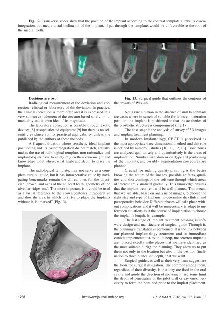
Fig. 12. Transverse slices show that the position of the implant according to the contrast template allows its osseointegration,<br />
but media-distal inclination of the implant, if put through the template, would be unfavorable to the root of<br />
the medial tooth.<br />
Decisions are two:<br />
Radiological measurement of the deviation and correction<br />
- clinical or laboratory of this deviation. In practice,<br />
the clinical correction is more often and it is expressed in a<br />
very subjective judgment of the operator based solely on its<br />
manuality and its own idea of its magnitude.<br />
The laboratory correction is possible through exotic<br />
devices [8] or sophisticated equipment [9] but there is no scientific<br />
evidence for its practical applicability, unless the<br />
published by the authors of these methods.<br />
A frequent situation where prosthetic ideal implant<br />
positioning and its osseointegration do not match, actually<br />
makes the use of radiological template, non rationalize and<br />
implantologists have to solely rely on their own insight and<br />
knowledge about where, what angle and depth to place the<br />
implant.<br />
The radiological template, may not serve as a complete<br />
surgical guide, but it has intraoperative value by navigating<br />
benchmarks remain the clinical ones for the physician<br />
(crowns and axes of the adjacent teeth, geometry of the<br />
alveolar ridges etc.). The more important is it could be used<br />
as a visual reference to the crown contours intraoperative<br />
and thus the area, in which to strive to place the implants<br />
without it, is “marked” (Fig.13).<br />
Fig. 13. Surgical guide that outlines the contours of<br />
the crowns of Wax-up<br />
Not a rare situation in the absence of such benchmark<br />
are cases where in search of suitable for its osseointegration<br />
position, the implant is positioned so that the aesthetics of<br />
the prosthetic structure is compromised (Fig.1).<br />
The next stage is the analysis of survey of 3D images<br />
and implant treatment planning.<br />
In modern implantology, CBCT is perceived as<br />
the most appropriate three-dimensional method, and this role<br />
is defined by numerous studies [10, 11, 12, 13]. Bone zones<br />
are analyzed qualitatively and quantitatively in the areas of<br />
implantation. Number, size, dimension, type and positioning<br />
of the implants, and possibly augmentation procedures are<br />
planned.<br />
Crucial for making quality planning is the better<br />
knowing the nature of the images, possible artifacts, qualities<br />
and shortcomings of the software through which areas<br />
of interest are visualized gradually. This knowledge ensures<br />
that the implant treatment will be well planned. This means<br />
that we are able, based on analysis of images, to choose the<br />
right size and type of implants, to determine the clinical and<br />
postoperative behavior. Different phases will take place without<br />
complications and it will be unnecessary to adapt to unforeseen<br />
situations as in the course of implantation to choose<br />
the implant’s length, for example.<br />
The last stage of implant treatment planning is software<br />
design and manufacture of surgical guide. Through it,<br />
the planning’s translation is performed. It is the link between<br />
our planned implantology treatment and its immediate<br />
clinical implementation. With its help, the selected implants<br />
are placed exactly in the places that we have identified as<br />
the most suitable during the planning. They allow us to put<br />
them not only in the location but also in the position (inclination<br />
to three planes and depth) that we want.<br />
Surgical guides, as well as their very name suggest are<br />
the tools for surgical navigation. The common among them,<br />
regardless of their diversity, is that they are fixed in the oral<br />
cavity and guide the direction of movement, and some limit<br />
the depth of penetration of the pilot drill or any ones, necessary<br />
to form the bone bed prior to the implant placement.<br />
1288 http://www.journal-imab-bg.org / J of IMAB. <strong>2016</strong>, vol. 22, issue 3/