- Page 1 and 2:
e-ISSN: 1312-773X Journal of IMAB -
- Page 3 and 4:
Journal of IMAB ISSN: 1312-773X htt
- Page 5 and 6:
After 1998 the world of oncologic s
- Page 7 and 8:
4. Mammographs Each year more than
- Page 9 and 10:
patients having brain tumors [30, 3
- Page 11 and 12:
CyberKnife measurements. Appl Radia
- Page 13 and 14:
Fig. 1. Risk of emergency situation
- Page 15 and 16:
REFERENCES: 1. Chakurova R, Mihailo
- Page 17 and 18:
In addition, it was reported that l
- Page 19 and 20:
observed between trismus and partia
- Page 21 and 22:
of the neurosensory deficit of an L
- Page 23 and 24:
of the patient (70%) over the right
- Page 25 and 26:
Patel M. Iatrogenic mandibular frac
- Page 27 and 28:
The surgery was held under local an
- Page 29 and 30:
DISCUSSION: Excision of oral lesion
- Page 31 and 32:
Journal of IMAB ISSN: 1312-773X htt
- Page 33 and 34:
from its germ. Missing germs of sec
- Page 35 and 36:
Journal of IMAB ISSN: 1312-773X htt
- Page 37 and 38:
The results of the comparison of ag
- Page 39 and 40:
Lichen simplex chronicus with a cut
- Page 41 and 42:
of a simple lever (Hebelkonstruktio
- Page 43 and 44:
Fig. 7. Gnathodynamometer for anima
- Page 45 and 46:
RESULTS The findings revealed an un
- Page 47 and 48:
Nowadays the quite common fast chew
- Page 49 and 50:
Journal of IMAB ISSN: 1312-773X htt
- Page 51 and 52:
[CrossRef] 4. Rodriguez AL. Atheros
- Page 53 and 54:
le blind, placebo controlled trials
- Page 55 and 56:
et al. Efficacy and safety of choli
- Page 57 and 58:
Fig. 1a b. In the period September
- Page 59 and 60:
A significant correlation (P=0.001)
- Page 61 and 62:
CONCLUSION Manual functional analys
- Page 63 and 64:
Fig. 1. Quadrantectomy with lymph n
- Page 65 and 66:
RESULTS: The performed study covere
- Page 67 and 68:
Journal of IMAB ISSN: 1312-773X htt
- Page 69 and 70:
demic nature of the diseases and re
- Page 71 and 72:
Journal of IMAB ISSN: 1312-773X htt
- Page 73 and 74: Fig. 5. Reduction of sites with PD>
- Page 75 and 76: Journal of IMAB ISSN: 1312-773X htt
- Page 77 and 78: The third condition is related to t
- Page 79 and 80: Journal of IMAB ISSN: 1312-773X htt
- Page 81 and 82: the unilateral location of the defe
- Page 83 and 84: Journal of IMAB ISSN: 1312-773X htt
- Page 85 and 86: Five days after treatment clinical
- Page 87 and 88: ite into the maxillary sinus. Br De
- Page 89 and 90: A 68-year-old woman came to the dep
- Page 91 and 92: in the skull, ethmoid sinuses, mand
- Page 93 and 94: Journal of IMAB ISSN: 1312-773X htt
- Page 95 and 96: Fig. 4. CBCT of the silicone impres
- Page 97 and 98: Fig. 11 a. STL model of the jaw in
- Page 99 and 100: Journal of IMAB ISSN: 1312-773X htt
- Page 101 and 102: Fig. 6. Vacuum-forming foil covered
- Page 103 and 104: Depending on the fixation method, t
- Page 105 and 106: Fig. 23. Stereolithografic printed
- Page 107 and 108: Fig. 28 a, b. - a software model of
- Page 109 and 110: 21. Arisan V, Karabuda CZ, Ozdemir
- Page 111 and 112: PURPOSE: The purpose of this in viv
- Page 113 and 114: Fig. 9. Frequency distribution of t
- Page 115 and 116: Journal of IMAB ISSN: 1312-773X htt
- Page 117 and 118: Fig. 3. Surgical portals are planne
- Page 119 and 120: Fig. 8. Immediate postoperative pho
- Page 121 and 122: Dupuytren’s contracture. N Engl J
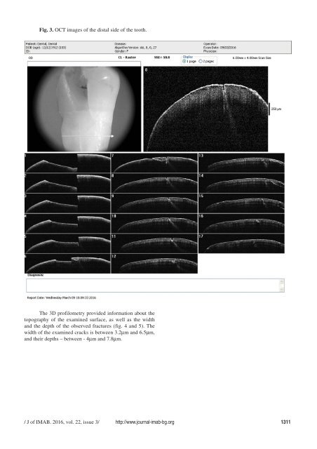
- Page 123: Technologies, Italy). After that, t
- Page 127 and 128: makes this technique a promising no
- Page 129 and 130: The discrepancy in the literature c
- Page 131 and 132: (A) Gleason score 7 Legend: (A) Pea
- Page 133 and 134: of prostate cancer. J Natl Cancer I
- Page 135 and 136: Fig. 2. Scanning process; Several s
- Page 137 and 138: 15. van der Meer WJ, Andriessen FS,
- Page 139 and 140: MATERIALS AND METHODS We use data f
- Page 141 and 142: in particular the need to pay for a
- Page 143: APPLICATION OF 3D DIGITAL SCANNING