JofIMAB-2016-vol22-issue3

You also want an ePaper? Increase the reach of your titles
YUMPU automatically turns print PDFs into web optimized ePapers that Google loves.
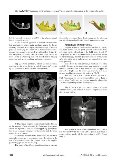
Fig. 1 a, b. CBCT image and its virtual analogue; c, d. Virtual surgical guide created in the images of a and b.<br />
but the second one is not a CBCT of the plastic model,<br />
but an intraoral scanner.<br />
2. The classical approach is difficult or impossible<br />
for application where metal artifacts mask the X-ray<br />
anatomy in which to be repositioned the image of the impression<br />
model (Fig.2). The reason is that anatomical markers<br />
are lost, according to which are adjusted to fit the virtual<br />
images of the model, and the X-ray image of the patient.<br />
There is a very big risk both models not to be fully<br />
compliant and hence to obtain an implant variation.<br />
Fig. 2. Crown contours, which are the anatomic<br />
markers, are invisible due to so called “scattering” caused<br />
by the presence of metal-ceramic constructions.<br />
attempt to overcome these shortcomings in the planning<br />
and use of surgical guides for dental implant treatment.<br />
MATERIALS AND METHODS:<br />
Implant treatment has been conducted on a 52-yearold<br />
male, nonsmoker, in good general health with distal<br />
unlimited partial edentulism in the field from 46 and 47.<br />
The patient had no contraindications to treatment (radioor<br />
chemotherapy, chronic systemic diseases or bruxism).<br />
After the initial view and discuss, we proceeded to treatment<br />
planning.<br />
Since from the clinical view, it has been found that<br />
medially located to the edentulous area teeth have metalceramic<br />
constructions, it has been decided to apply the<br />
method of triple scan to overcome artifacts that these<br />
crowns would cause scan of the patient in CBCT.<br />
The first scan is CBCT of the patient (ProMax 3D<br />
Mid, Planmeca). The special feature of this scan is that it is<br />
made with C-silicone impression material (Zetaplus,<br />
Zhermack®) in the patient’s mouth (Fig. 3).<br />
Fig. 3. CBCT of patient. Despite artifacts of metalceramic<br />
crowns, the outlines of silicone impression material<br />
are seen clearly.<br />
3. The printed surgical guide is hard (rigid). Because<br />
of the absence of any plasticity, its fixing part is arranged<br />
above the equatorial area (in tooth-supporting guides) and<br />
this leads to micro movement of the guide, and deviation<br />
respectively in its use.<br />
We believe that the above three issues are the main<br />
reason for registered by researchers many deviations in the<br />
use of surgical guides to the planned ones in the implant<br />
positioning [9, 10, 11, 12, 13, 14].<br />
The study offers if not a decision, then at least an<br />
The second scan is on the impression itself, and it<br />
has been made with the same CBCT system. It is used to<br />
make a virtual model of the impression in a STL format<br />
(Fig. 4)<br />
1280 http://www.journal-imab-bg.org / J of IMAB. <strong>2016</strong>, vol. 22, issue 3/