JofIMAB-2016-vol22-issue3

You also want an ePaper? Increase the reach of your titles
YUMPU automatically turns print PDFs into web optimized ePapers that Google loves.
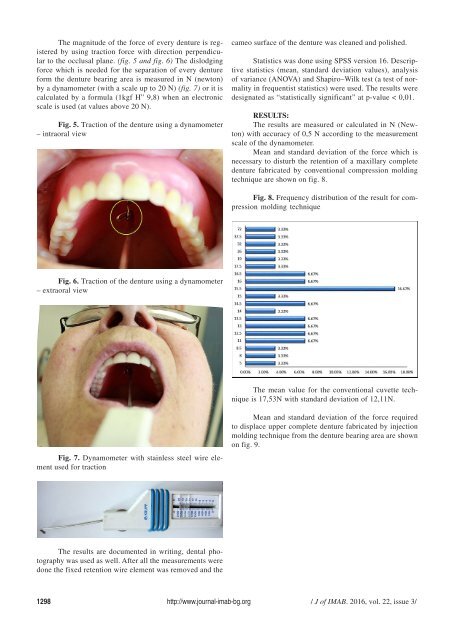
The magnitude of the force of every denture is registered<br />
by using traction force with direction perpendicular<br />
to the occlusal plane. (fig. 5 and fig. 6) The dislodging<br />
force which is needed for the separation of every denture<br />
form the denture bearing area is measured in N (newton)<br />
by a dynamometer (with a scale up to 20 N) (fig. 7) or it is<br />
calculated by a formula (1kgf H” 9,8) when an electronic<br />
scale is used (at values above 20 N).<br />
Fig. 5. Traction of the denture using a dynamometer<br />
– intraoral view<br />
cameo surface of the denture was cleaned and polished.<br />
Statistics was done using SPSS version 16. Descriptive<br />
statistics (mean, standard deviation values), analysis<br />
of variance (ANOVA) and Shapiro–Wilk test (a test of normality<br />
in frequentist statistics) were used. The results were<br />
designated as “statistically significant” at p-value < 0,01.<br />
RESULTS:<br />
The results are measured or calculated in N (Newton)<br />
with accuracy of 0,5 N according to the measurement<br />
scale of the dynamometer.<br />
Mean and standard deviation of the force which is<br />
necessary to disturb the retention of a maxillary complete<br />
denture fabricated by conventional compression molding<br />
technique are shown on fig. 8.<br />
Fig. 8. Frequency distribution of the result for compression<br />
molding technique<br />
Fig. 6. Traction of the denture using a dynamometer<br />
– extraoral view<br />
The mean value for the conventional cuvette technique<br />
is 17,53N with standard deviation of 12,11N.<br />
Fig. 7. Dynamometer with stainless steel wire element<br />
used for traction<br />
Mean and standard deviation of the force required<br />
to displace upper complete denture fabricated by injection<br />
molding technique from the denture bearing area are shown<br />
on fig. 9.<br />
The results are documented in writing, dental photography<br />
was used as well. After all the measurements were<br />
done the fixed retention wire element was removed and the<br />
1298 http://www.journal-imab-bg.org / J of IMAB. <strong>2016</strong>, vol. 22, issue 3/