JofIMAB-2016-vol22-issue3

You also want an ePaper? Increase the reach of your titles
YUMPU automatically turns print PDFs into web optimized ePapers that Google loves.
A 68-year-old woman came to the department of Oral<br />
and Maxillofacial Surgery of Faculty of Dental medicine,<br />
Medical University- Sofia for evaluation of a swelling in the<br />
left side of mandible which she found by chance a couple<br />
of months prior to examination and had grown slowly the<br />
last few months. A mild facial asymmetry was observed.<br />
(figure 1). Clinical examination revealed a non painful firm<br />
well-circumscribed palpable mass in the buccal vestibule<br />
aspect of the left side of the mandible near to the lower<br />
margin of the angle. The lesion was covered by normal skin.<br />
The patient had no paresthesia. The patient was in good<br />
health generally and with negative history of trauma in this<br />
region prior to the onset.<br />
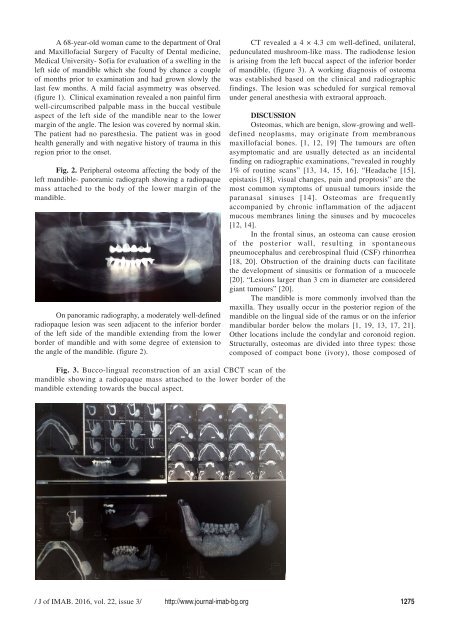
Fig. 2. Peripheral osteoma affecting the body of the<br />
left mandible- panoramic radiograph showing a radiopaque<br />
mass attached to the body of the lower margin of the<br />
mandible.<br />
On panoramic radiography, a moderately well-defined<br />
radiopaque lesion was seen adjacent to the inferior border<br />
of the left side of the mandible extending from the lower<br />
border of mandible and with some degree of extension to<br />
the angle of the mandible. (figure 2).<br />
CT revealed a 4 × 4.3 cm well-defined, unilateral,<br />
pedunculated mushroom-like mass. The radiodense lesion<br />
is arising from the left buccal aspect of the inferior border<br />
of mandible, (figure 3). A working diagnosis of osteoma<br />
was established based on the clinical and radiographic<br />
findings. The lesion was scheduled for surgical removal<br />
under general anesthesia with extraoral approach.<br />
DISCUSSION<br />
Osteomas, which are benign, slow-growing and welldefined<br />
neoplasms, may originate from membranous<br />
maxillofacial bones. [1, 12, 19] The tumours are often<br />
asymptomatic and are usually detected as an incidental<br />
finding on radiographic examinations, “revealed in roughly<br />
1% of routine scans” [13, 14, 15, 16]. “Headache [15],<br />
epistaxis [18], visual changes, pain and proptosis” are the<br />
most common symptoms of unusual tumours inside the<br />
paranasal sinuses [14]. Osteomas are frequently<br />
accompanied by chronic inflammation of the adjacent<br />
mucous membranes lining the sinuses and by mucoceles<br />
[12, 14].<br />
In the frontal sinus, an osteoma can cause erosion<br />
of the posterior wall, resulting in spontaneous<br />
pneumocephalus and cerebrospinal fluid (CSF) rhinorrhea<br />
[18, 20]. Obstruction of the draining ducts can facilitate<br />
the development of sinusitis or formation of a mucocele<br />
[20]. “Lesions larger than 3 cm in diameter are considered<br />
giant tumours” [20].<br />
The mandible is more commonly involved than the<br />
maxilla. They usually occur in the posterior region of the<br />
mandible on the lingual side of the ramus or on the inferior<br />
mandibular border below the molars [1, 19, 13, 17, 21].<br />
Other locations include the condylar and coronoid region.<br />
Structurally, osteomas are divided into three types: those<br />
composed of compact bone (ivory), those composed of<br />
Fig. 3. Bucco-lingual reconstruction of an axial CBCT scan of the<br />
mandible showing a radiopaque mass attached to the lower border of the<br />
mandible extending towards the buccal aspect.<br />
/ J of IMAB. <strong>2016</strong>, vol. 22, issue 3/ http://www.journal-imab-bg.org 1275