ESC Textbook of Cardiovascular Imaging - sample
Discover the ESC Textbook of Cardiovascular Imaging 2nd edition
Discover the ESC Textbook of Cardiovascular Imaging 2nd edition

You also want an ePaper? Increase the reach of your titles
YUMPU automatically turns print PDFs into web optimized ePapers that Google loves.
6<br />
chapter 1 conventional echocardiography—basic principles<br />
Fig. 1.3 Starting with the correct holding <strong>of</strong> the transducer for displaying the parasternal long-axis view (a) the transducer is exactly rotated 90° clockwise (b),<br />
after this movement the pulp <strong>of</strong> the thumb is at the broad side <strong>of</strong> the transducer at the top and the third finger is at the broad side <strong>of</strong> the transducer at the<br />
bottom (b), while the fourth and fifth finger are retracted (c), but they have still contact to the skin. This holding is linked with all parasternal short-axis views (d).<br />
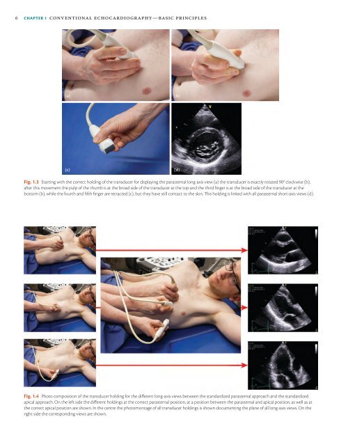
Fig. 1.4 Photo composition <strong>of</strong> the transducer holding for the different long-axis views between the standardized parasternal approach and the standardized<br />
apical approach. On the left side the different holdings at the correct parasternal position, at a position between the parasternal and apical position, as well as at<br />
the correct apical position are shown. In the centre the photomontage <strong>of</strong> all transducer holdings is shown documenting the plane <strong>of</strong> all long-axis views. On the<br />
right side the corresponding views are shown.