ESC Textbook of Cardiovascular Imaging - sample
Discover the ESC Textbook of Cardiovascular Imaging 2nd edition
Discover the ESC Textbook of Cardiovascular Imaging 2nd edition

You also want an ePaper? Increase the reach of your titles
YUMPU automatically turns print PDFs into web optimized ePapers that Google loves.
18<br />
chapter 1 conventional echocardiography—basic principles<br />
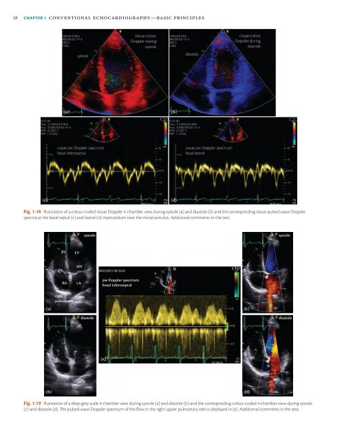
Fig. 1.18 Illustration <strong>of</strong> a colour-coded tissue Doppler 4-chamber view during systole (a) and diastole (b) and the corresponding tissue pulsed wave Doppler<br />
spectra at the basal septal (c) and lateral (d) myocardium near the mitral annulus. Additional comments in the text.<br />
Fig. 1.19 Illustration <strong>of</strong> a deep grey scale 4-chamber view during systole (a) and diastole (b) and the corresponding colour-coded 4-chamber view during systole<br />
(c) and diastole (d). The pulsed wave Doppler spectrum <strong>of</strong> the flow in the right upper pulmonary vein is displayed in (e). Additional comments in the text.