
Create successful ePaper yourself
Turn your PDF publications into a flip-book with our unique Google optimized e-Paper software.
Focus on<br />
Eye Research<br />
Thyroid eye disease, epiphora,<br />
and ptosis after ocular surgery<br />
CHUNDURY RV ET AL. ORBITAL RADIATION<br />
THERAPY IN THYROID EYE DISEASE<br />
Ophthal Plast Reconstr Surg <strong>2016</strong>; 32:83-89<br />
As the role of supportive therapies such as selenium<br />
and biologic agents in the treatment of thyroid eye<br />
disease (TED) continue to evolve, orbital radiation<br />
therapy (ORT) is a longstanding, but contentious<br />
mode of medical treatment of TED. This review<br />
considered a wealth of existing data, and its<br />
shortcomings, in an effort to understand the role of<br />
ORT in TED.<br />
A dose of 20Gy delivered to each eye in 10 fractions<br />
over 10-12 days is common, but different sources of<br />
radiation, classification schemes and compounding<br />
effects of adjunctive glucocorticoid use means<br />
that comparisons between studies is challenging.<br />
This paper considers all the available data as a<br />
whole and concludes that between 50-90% of mild<br />
moderate TED patients treated with ORT will have<br />
some improvement in motility, proptosis and clinical<br />
activity when it is given early in the disease. ORT may<br />
also help prevent vision-threatening complications<br />
such as compressive optic neuropathy. The beneficial<br />
effect of ORT can be enhanced with concomitant use<br />
of glucocorticoids. Re-irradiation has been used with<br />
some success, however there is little data regarding<br />
safety and efficacy.<br />
Much data exists to demonstrate ORT is safe in<br />
patients without predisposition to retinopathy.<br />
Patients with retinopathy risk factors, particularly<br />
severe hypertension and diabetes may be at higher<br />
risk. The risk of cataract development appears very<br />
low.<br />
The theoretical increased risk of brain and bone<br />
malignancy has not been observed in large<br />
retrospective studies with long-term follow up,<br />
although some still limit the use of ORT to patients<br />
greater than 30-35 years because of this concern.<br />
The authors concluded that although ORT is rarely<br />
used as a first-line agent, it is a safe option in the<br />
armamentarium available for management of active<br />
TED.<br />
MANSUR C ET AL. EVALUATION AND<br />
MANAGEMENT OF CHEMOTHERAPY-INDUCED<br />
EPIPHORA, PUNCTAL AND CANALICULAR<br />
STENOSIS, AND NASOLACRIMAL OBSTRUCTION.<br />
OPHTHAL PLAST RECONSTUCT SURG <strong>2016</strong>; JULY<br />
(EPUB AHEAD OF PRINT).<br />
Epiphora is a recognised adverse effect of chemo<br />
therapeutic agents whose extent is probably<br />
under appreciated by both ophthalmologists and<br />
oncologists.<br />
This elegant literature review identifies various<br />
agents known to cause epiphora, the mechanisms<br />
and appropriate management paradigms.<br />
Most commonly associated are 5-Fluorouracil<br />
(5-FU) and Docetaxel which cause dose dependent<br />
epiphora in up to 50 and 64% of patients<br />
respectively; this is reversible with prompt<br />
treatment. The agents are secreted in tears, and<br />
appear to cause canalicular, lacrimal sac and<br />
nasolacrimal duct fibrosis.<br />
A trial of corticosteroid with probe and syringe<br />
appears effective in cases of low dose or short-term<br />
treatment, but patients receiving high frequent<br />
dose 5-FU or docetaxel, silicone stenting at the<br />
first sign of recurrent or progressive canalicular<br />
stenosis can prevent irreversible canalicular scarring.<br />
Unfortunately, prophylactic topical corticosteroid<br />
have been proven to be ineffective.<br />
Other agents less commonly found to cause<br />
epiphora secondary to nasolacrimal obstruction<br />
included radioactive iodine (I-131), which at high<br />
doses is preferentially taken up by nasal tissue.<br />
Females and those over 45 years old were more<br />
likely to develop epiphora.<br />
F-1, an oral drug composed of tegafur (pro-drug of<br />
5-FU) and oteracil, as well as mitomycin C (MMC),<br />
also cause stenosis of the lacrimal drainage pathway<br />
at the punctum, canaliculus or distal nasolacrial<br />
duct. Onset of effect is within three months.<br />
Interestingly MMC is often used to prevent fibrosis<br />
DR JAMES SLATTERY*<br />
of the lacrimal drainage system after surgery.<br />
Epiphora is the second most common side effect<br />
of Imatinib. Unlike other agents, the mechanism<br />
is via hypersecretion and mechanical blockage of<br />
puncta by conjunctivochalasis coupled with pump<br />
dysfunction secondary to periorbital oedema.<br />
Practitioners must be aware of stenosis or<br />
obstruction of the lacrimal drainage system as<br />
a possible adverse effect of these agents. Early<br />
recognition and appropriate treatment may obviate<br />
the need for more invasive and complicated<br />
surgical treatment and so evaluation of drug<br />
type, and thus likely cause of epiphora, with<br />
prompt appropriate referral is critical.<br />
GODFREY K. BLEPHAROPTOSIS FOLLOWING<br />
OCULAR SURGERY: IDENTIFYING RISK FACTORS<br />
Curr Opin Ophthalmol <strong>2016</strong>;27:31-7<br />
Ptosis is common following ocular surgical<br />
procedures. Nearly one third of acquired ptosis is<br />
postsurgical. This literature review attempts to<br />
identify the incidence of this poorly understood<br />
complication post ocular surgery, and shed light on<br />
its likely contributing factors.<br />
Cataract surgery is the most commonly performed<br />
ocular surgery and randomised prospective studies<br />
document the incidence of post cataract ptosis<br />
to be somewhere around 6-12%. Use of a bridal<br />
suture in addition to an eyelid speculum nearly<br />
doubles the risk. The incidence is similarly around<br />
10% post refractive surgery, such as laser in situ<br />
keratomileusis as well as trabeculectomy, as<br />
documented in the collaborative initial glaucoma<br />
treatment study (CIGTS). Adjuvant use of<br />
mitomycin C (MMC) increased the risk. There<br />
are documented cases of ptosis following serial<br />
intravitreal injections of anti-vascular endothelial<br />
growth factor and steroid, but appropriately<br />
powered studies are lacking.<br />
Ptosis can be caused by interruption to any of<br />
the delicate structural and functional relationships<br />
of the eyelids although levator dehiscence is likely<br />
the common denominator. Several studies recognise<br />
individual patient anatomy to confer highest risk of<br />
postoperative ptosis and it is probable that patients<br />
with tenuous levator attachment and subclinical<br />
ptosis are at highest risk. Operative time may play a<br />
role but there is no consensus in the literature.<br />
Theories such as a tight speculum reducing blood<br />
flow to the levator muscle, horizontal eyelid stretch<br />
and contraction of orbicularis against speculum<br />
causing dehiscence have been postulated, or<br />
that compression of myoneural or myovascular<br />
structures contributes. Myotoxicity from local<br />
anaesthetics has also been suggested, but has not<br />
been validated in the literature.<br />
Risk can be minimised with use of topical<br />
anaesthetics and avoidance of bridal sutures and<br />
specula where possible. If a speculum is required,<br />
using the shortest possible horizontal arm and<br />
reduced vertical eyelid displacement should be<br />
considered. Studies have shown disposable specula<br />
are significantly less stiff than reusable ones, which<br />
may reduce the risk.<br />
Even with optimum technique, ptosis can still occur<br />
and this should be discussed during informed<br />
consent. Repair is possible with high rates of<br />
success but requires an additional surgical<br />
procedure. ▀<br />
ABOUT THE AUTHOR:<br />
* Dr James Slattery holds<br />
an MBBS, PhD, B Sci<br />
(Biomed), and is an<br />
oculoplastics fellow<br />
at the University of<br />
Auckland. He trained<br />
in Adelaide, South<br />
Australia.<br />
Labour of love with touch<br />
of madness<br />
BY SIMON ESKOW<br />
What would inspire someone who<br />
doesn’t know French to create the first<br />
translation of a 450-page book written<br />
in that language?<br />
“I’ve got no bloody idea,” says Dr Philip<br />
Polkinghorne, who accomplished that very task,<br />
laughing. “It’s the stupidest thing I ever did.”<br />
Polkinghorne, who describes his grasp of French<br />
as “absolutely useless”, finished the impressive<br />
task of translating Le Decollement de La rétine:<br />
pathogénie, traitement – or Detachment of the<br />
retina: pathogenesis, treatment – in mid-2015. The<br />
400-page tome was authored by the pioneering<br />
Swiss ophthalmologist, Jules Gonin (1870-<br />
1935), who discovered through detailed clinical<br />
observation that the retinal tear was the cause,<br />
and not the result, of retinal detachment, contrary<br />
to prevailing theories of the time.<br />
“People had done all sorts of hocus pocus<br />
before that time,” Polkinghorne says. “But Gonin’s<br />
was the first, if you like, thought-out, scientific<br />
approach to the problem.”<br />
Polkinghorne, a surgeon with Auckland Eye<br />
and an associate professor at the University of<br />
Auckland, embarked on his translation after<br />
spotting a copy on the University of Auckland’s<br />
head of ophthalmology Professor Charles<br />
McGhee’s bookshelf. Polkinghorne has been a<br />
member of the international Club Jules Gonin<br />
for 20 years and was surprised to see a copy<br />
of the original French version as relatively few<br />
are in circulation. Despite the linguistic barrier,<br />
Polkinghorne borrowed the copy, scanned the<br />
400 pages to a PDF file and converted it to Word,<br />
and began the page-by-page, word-by-word<br />
translation, using “a whole lot of search engines”.<br />
In the first section, Gonin details the history of<br />
ophthalmological theories of retinal detachments,<br />
with critique and a presentation of his own theory.<br />
“People did some crazy things for retinal<br />
detachment,” Polkinghorne says. “For example<br />
they cut holes in the iris and injected all sorts of<br />
things into the eye like mercury and cyanide, and<br />
under the conjunctiva.”<br />
The latter section of the book describes<br />
treatment for retinal detachment and Gonin’s<br />
approach using thermocauterisation.<br />
The whole project took Polkinghorne more than<br />
a year.<br />
“I think it took me about 10 hours per page. Once I<br />
got into it, I couldn’t stop. It became an obsession.”<br />
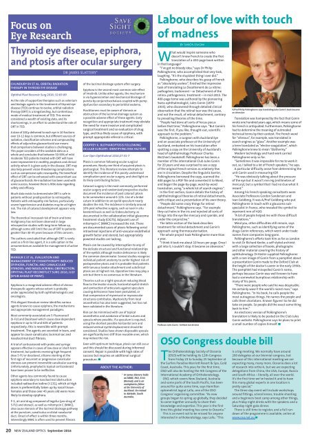
Professor Jules Gonin - brilliant but divisive<br />
A/Prof Philip Polkinghorne says translating Jules Gonin’s book became<br />
an obsession<br />
Translation was hampered by the fact that Gonin<br />
wrote one hundred years ago, which means some of<br />
his French is antiquated. Furthermore, Polkinghorne<br />
had to determine the meaning of outmoded<br />
technical terms by their context. The French word<br />
for “vitreous”, for example, was translated in<br />
search engines as “glass”. Another example was<br />
a term translated as “electro-coagulation”, which<br />
Polkinghorne knew to mean “diathermy.”<br />
Modern technology and hard work carried<br />
Polkinghorne only so far.<br />
“Sometimes it was impossible for me to work it<br />
out, so, I talked to a lot of French speakers,” he says.<br />
A particular difficulty arose from determining the<br />
unit Gonin used in measuring IOP.<br />
“He was obviously talking about the pressure<br />
of the eye but it wasn’t mm Hg (millimetres of<br />
mercury), but a symbol that I had no clue what it<br />
meant.”<br />
Among his French speaking consultants were<br />
Associate Professors Gordon Sanderson and<br />
Ivan Goldberg. It was A/Prof Goldberg who put<br />
Polkinghorne in touch with a glaucoma subspecialist<br />
in France, who revealed the symbol to<br />
represent mm of H2O.<br />
“A lot of people helped me with those difficult<br />
translations.”<br />
Mind you, other difficulties still remain, says<br />
Polkinghorne, such as identifying some of the<br />
drugs Gonin references, which went under trade<br />
names from companies long-gone.<br />
Polkinghorne’s efforts took him as far as London<br />
to visit Dr Richard Keeler, a self-styled archivist<br />
with a large collection of books, photographs<br />
and other material covering the history of<br />
ophthalmology. Dr Keeler supplied Polkinghorne<br />
with a rare image of Gonin from a pamphlet about<br />
a presentation Gonin made to the Oxford Club at<br />
the height of the doctor’s career in the early 1930s.<br />
The pamphlet had misspelled Gonin’s name,<br />
perhaps because Gonin was well-known to have<br />
had a somewhat bumptious relationship with<br />
many of his peers.<br />
“There were people who said he was despicable.<br />
He certainly wasn’t the world’s nicest man,” says<br />
Polkinghorne. “In his book, he calls people the<br />
most outrageous things. He names the people and<br />
calls them charlatans. Known figures! So he did<br />
take on people. So people did the exact same thing<br />
back to him.”<br />
An electronic version of Polkinghorne’s<br />
translation is likely to be posted on the Club Jules<br />
Gonin website. Polkinghorne says he plans to print<br />
a small number of copies himself. ▀<br />
OSO Congress double bill<br />
The Orthokeratology Society of Oceania<br />
(OSO) will be holding its 12th Congress<br />
from Friday 23 to Sunday 25 <strong>Sep</strong>tember at<br />
the Surfers Paradise Marriott Resort & Spa, Gold<br />
Coast, Australia. This year, for the first time,<br />
OSO will also be hosting the 5th Congress of the<br />
International Academy of Orthokeratology.<br />
OSO, which covers New Zealand, Australia<br />
and some parts of the South Pacific, has been<br />
around for quite some time, says Hamilton<br />
optometrist Jagrut Lallu, one of the OSO<br />
Congress’ organising committee. “As new<br />
groups began to spring up globally, they decided<br />
to come together annually to share their<br />
knowledge and expertise. This year is the first<br />
time this global meeting has come to Oceania.”<br />
This is an event not to be missed for anyone<br />
interested in orthokeratology, says Lallu. “This<br />
is a big meeting. We normally have around<br />
200 delegates at our biennial congress, but<br />
because of this international meeting we are<br />
expecting many, many more. Oceania does a lot<br />
of research into ortho-k, but we are expecting<br />
delegations from China, the USA, Europe, Russia<br />
and South Africa – literally, all over the world.<br />
It’s the first time we’ve hosted it and to have<br />
this many global experts in one location is<br />
pretty special.”<br />
The three-day event will include workshops<br />
around fittings, scleral lenses, trouble shooting<br />
and a beginners boot camp among other things,<br />
plus Friday night drinks with the speakers and a<br />
Saturday night gala dinner.<br />
There is still time to register, and a full rundown<br />
of the programme is available, online at<br />
www.osa.net.au ▀<br />
20 NEW ZEALAND OPTICS <strong>Sep</strong>tember <strong>2016</strong>