

Management of stage Ⅳ rectal cancer - World Journal of ...
Management of stage Ⅳ rectal cancer - World Journal of ...
Management of stage Ⅳ rectal cancer - World Journal of ...

You also want an ePaper? Increase the reach of your titles
YUMPU automatically turns print PDFs into web optimized ePapers that Google loves.
J Gastroenterol 2011; 17(7): 938-945 Available from: URL:<br />
http://www.wjgnet.com/1007-9327/full/v17/i7/938.htm DOI:<br />
http://dx.doi.org/10.3748/wjg.v17.i7.938<br />
INTRODUCTION<br />
Substantial efforts have been made with regard to cell<br />
transplantation as an effective supporting system for hepatic<br />
failure and assisted therapies. However, immunological<br />
rejection has always been an important problem for<br />
cell transplantation. Alginate-poly-l-lysine-alginate (APA)<br />
microcapsules have proven to be effective in protecting<br />
enclosed target cells from immune rejection following<br />
transplantation into experimental animals, thereby eliminating<br />
the problems <strong>of</strong> immunosuppressive therapy [1-3] .<br />
Extensive studies have also been conducted on the<br />
core <strong>of</strong> this therapy, namely the cell sources. The investigated<br />
cells have included liver stem cells, embryonic stem<br />
cells, human umbilical cord blood (UCB) cells and bone<br />
marrow stem cells. Human UCB cells have some advantages<br />
that other cells do not have. The frequencies <strong>of</strong><br />
UCB hematopoietic stem/progenitor cells exceed those<br />
from bone marrow and peripheral blood. In our previous<br />
study, we confirmed the differentiation <strong>of</strong> mononuclear<br />
cells (MNCs) from human UCB into hepatocytes in three<br />
different ways, namely co-culture with injured liver cells,<br />
growth factor-assisted culture, and MNC transplantation<br />
in animal models <strong>of</strong> liver injury [4] . In the present study, we<br />
found that CD34 + cells derived from human UCB could<br />
be converted into hepatic-like cells that generate hepatocyte<br />
lineage cells. Furthermore, we encapsulated the hepatic-like<br />
cells using an alginate method and transplanted<br />
them into acute hepatic failure (AHF) rats to evaluate the<br />
effects <strong>of</strong> encapsulated hepatic-like cell transplantation.<br />
MATERIALS AND METHODS<br />
Isolation and identification <strong>of</strong> CD34 + cells<br />
UCB (more than 80 samples) from full-term deliveries<br />
were obtained from the Obstetrics Department <strong>of</strong> Peking<br />
University Shenzhen Hospital. UCB cells were harvested<br />
after written inform consent was obtained. The study<br />
protocol was approved by the Ethics Committee <strong>of</strong> Peking<br />
University Shenzhen Hospital. MNCs were isolated<br />
from the UCB samples by density-gradient centrifugation<br />
at 2000 r/min for 35 min using Ficoll-Hypaque (Huajing,<br />
Shanghai, China). CD34 + subpopulations were isolated<br />
using a Miltenyi Direct CD34 Progenitor Cell Isolation<br />
Kit (Miltenyi Biotec, Bergisch Gladbach, Germany). The<br />
specific steps were as follows: (1) isolated MNCs were resuspended<br />
in a final volume <strong>of</strong> 300 μL <strong>of</strong> PBS that contained<br />
5 g/L bovine serum albumin (BSA); (2) 100 μL <strong>of</strong><br />
FcR Blocking Reagent and 100 μL <strong>of</strong> CD34 Micro Beads<br />
per 1 × 10 8 total cells were sequentially added, mixed well<br />
and incubated for 30 min in a refrigerator at 4℃; (3) cells<br />
were passed through a magnetic column twice and purified;<br />
and (4) CD34 + cells were collected, resuspended in<br />
WJG|www.wjgnet.com<br />
Zhang FT et al . Transplantation <strong>of</strong> microencapsulated hepatic-like cells<br />
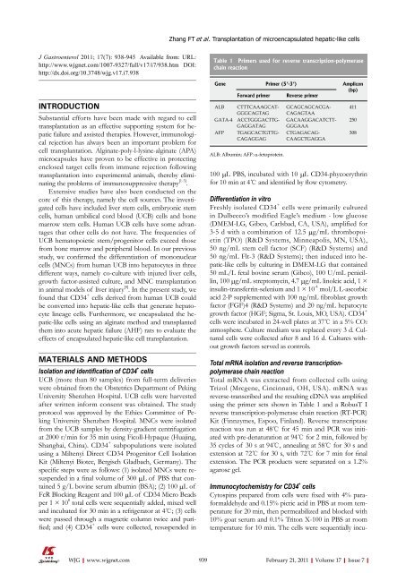
Table 1 Primers used for reverse transcription-polymerase<br />
chain reaction<br />
Gene Primer (5’-3’) Amplicon<br />
(bp)<br />
Forward primer Reverse primer<br />
ALB CTTTCAAAGCAT-<br />
GGGCAGTAG<br />
GATA-4 ACCTGGGACTTG-<br />
GAGGATAG<br />
AFP TGAGCACTGTTG-<br />
CAGAGGAG<br />
ALB: Albumin; AFP: α-fetoprotein.<br />
GCAGCAGCACGA-<br />
CAGAGTAA<br />
GACAAGGACATCTT-<br />
GGGAAA<br />
CTGAGACAG-<br />
CAAGCTGAGGA<br />
100 μL PBS, incubated with 10 μL CD34-phycoerythrin<br />
for 10 min at 4℃ and identified by flow cytometry.<br />
Differentiation in vitro<br />
Freshly isolated CD34 + cells were primarily cultured<br />
in Dulbecco’s modified Eagle’s medium - low glucose<br />
(DMEM-LG, Gibco, Carlsbad, CA, USA), amplified for<br />
3-5 d with a combination <strong>of</strong> 12.5 μg/mL thrombopoietin<br />
(TPO) (R&D Systems, Minneapolis, MN, USA),<br />
50 ng/mL stem cell factor (SCF) (R&D Systems) and<br />
50 ng/mL Flt-3 (R&D Systems); then induced into hepatic-like<br />
cells by culturing in DMEM-LG that contained<br />
50 mL/L fetal bovine serum (Gibco), 100 U/mL penicillin,<br />
100 μg/mL streptomycin, 4.7 μg/mL linoleic acid, 1 ×<br />
insulin-transferrin-selenium and 1 × 10 -4 mol/L L-ascorbic<br />
acid 2-P supplemented with 100 ng/mL fibroblast growth<br />
factor (FGF)4 (R&D Systems) and 20 ng/mL hepatocyte<br />
growth factor (HGF; Sigma, St. Louis, MO, USA). CD34 +<br />
cells were incubated in 24-well plates at 37℃ in a 5% CO2<br />
atmosphere. Culture medium was replaced every 3 d. Cultured<br />
cells were collected after 8 and 16 d. Cultures without<br />
growth factors served as controls.<br />
Total mRNA isolation and reverse transcriptionpolymerase<br />
chain reaction<br />
Total mRNA was extracted from collected cells using<br />
Trizol (Mrcgene, Cincinnati, OH, USA). mRNA was<br />
reverse-transcribed and the resulting cDNA was amplified<br />
using the primer sets shown in Table 1 and a RobusT I<br />
reverse transcription-polymerase chain reaction (RT-PCR)<br />
Kit (Finnzymes, Espoo, Finland). Reverse transcriptase<br />
reaction was run at 48℃ for 45 min and PCR was initiated<br />
with pre-denaturation at 94℃ for 2 min, followed by<br />
35 cycles <strong>of</strong> 30 s at 94℃, annealing at 58℃ for 30 s and<br />
extension at 72℃ for 30 s, with 72℃ for 7 min for final<br />
extension. The PCR products were separated on a 1.2%<br />
agarose gel.<br />
Immunocytochemistry for CD34 + cells<br />
Cytospins prepared from cells were fixed with 4% paraformaldehyde<br />
and 0.15% picric acid in PBS at room temperature<br />
for 20 min, then permeabilized and blocked with<br />
10% goat serum and 0.1% Triton X-100 in PBS at room<br />
temperature for 10 min. The cells were sequentially incu-<br />
939 February 21, 2011|Volume 17|Issue 7|<br />
411<br />
250<br />
308


