

Management of stage Ⅳ rectal cancer - World Journal of ...
Management of stage Ⅳ rectal cancer - World Journal of ...
Management of stage Ⅳ rectal cancer - World Journal of ...

You also want an ePaper? Increase the reach of your titles
YUMPU automatically turns print PDFs into web optimized ePapers that Google loves.
A<br />
C<br />
Liu QY et al . Primary clear cell carcinoma in the liver<br />
the location, number, size and grade <strong>of</strong> tumors were observed<br />
between the two groups [3] . Both tumor types were<br />
prone to occur in patients with hepatitis B, mostly on the<br />
basis <strong>of</strong> liver cirrhosis [3] . PCCCL had a better prognosis<br />
than CHCC, mainly related to capsule formation, vascular<br />
invasion, preoperative liver function and clear cell proportion<br />
[3,4,12] . Surgical resection is an effective treatment for<br />
patients with PCCCL [3,4,7] .<br />
The presence <strong>of</strong> clear cells and fatty changes characterizes<br />
well-differentiated HCC in the early <strong>stage</strong>, and their<br />
ratio is presumed to decrease as the tumor enlarges [15] . In<br />
1999, Monzawa et al [16] analyzed the pathologic and imaging<br />
changes <strong>of</strong> well-differentiated HCC; and found that<br />
some well-differentiated HCCs showed clear cell formation<br />
and/or fatty changes, which presented as high echo<br />
on ultrasound and hyper-intense signals on T1WI. However,<br />
in their study, the proportion <strong>of</strong> clear cells in the recruited<br />
HCC was less than 10%, or only 10%-50%, which<br />
did not meet the diagnosis criteria for PCCCL. In 2008,<br />
Takahashi et al [11] described CT, MR and angiographic<br />
findings <strong>of</strong> PCCCL in a woman with a normal liver. To<br />
our knowledge, no further research on the imaging manifestations<br />
<strong>of</strong> PCCCL has been conducted.<br />
Pseudocapsule formation (consisting mainly <strong>of</strong> peritumoral<br />
hepatic sinusoids and/or fibrosis) is an important<br />
gross pathologic feature <strong>of</strong> HCC. Pseudocapsule indicates<br />
a relatively positive prognosis after tumor resection [17] . Liu<br />
et al [3] found a higher ratio <strong>of</strong> pseudocapsule formation in<br />
PCCCL than in CHCC microscopically (88.4% vs 68.0%,<br />
P < 0.05); and pseudocapsule formation might be related<br />
WJG|www.wjgnet.com<br />
B<br />
D<br />
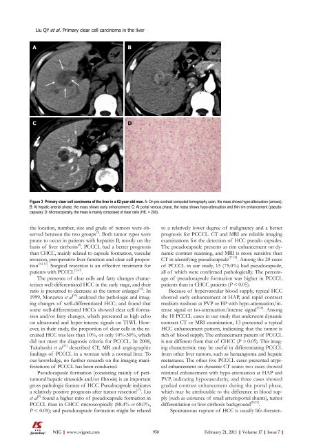
Figure 3 Primary clear cell carcinoma <strong>of</strong> the liver in a 62-year-old man. A: On pre-contrast computed tomography scan, the mass shows hypo-attenuation (arrows);<br />
B: At hepatic arterial phase, the mass shows early enhancement; C: At portal venous phase, the mass shows hypo-attenuation and thin rim enhancement (pseudocapsule);<br />
D: Microscopically, the mass is mainly composed <strong>of</strong> clear cells (HE, × 200).<br />
to a relatively lower degree <strong>of</strong> malignancy and a better<br />
prognosis for PCCCL. CT and MRI are reliable imaging<br />
examinations for the detection <strong>of</strong> HCC pseudo capsules.<br />
The pseudocapsule presents as rim enhancement on dynamic<br />
contrast scanning, and MRI is more sensitive than<br />
CT in identifying pseudocapsule [17-19] . Among the 20 cases<br />
<strong>of</strong> PCCCL in our study, 15 (75.0%) had pseudocapsule,<br />
all <strong>of</strong> which were confirmed pathologically. The percentage<br />
<strong>of</strong> pseudocapsule formation was higher in PCCCL<br />
patients than in CHCC patients (P < 0.05).<br />
Because <strong>of</strong> hypervascular blood supply, typical HCC<br />
showed early enhancement at HAP, and rapid contrast<br />
medium washout at PVP or EP with hypo-attenuation/intense<br />
signal or iso-attenuation/intense signal [9,10] . Among<br />
the 18 PCCCL cases in our study that underwent dynamic<br />
contrast CT or MRI examination, 13 presented a typical<br />
HCC enhancement pattern, indicating that the tumor is<br />
rich <strong>of</strong> blood supply. The enhancement pattern <strong>of</strong> PCCCL<br />
is not different from that <strong>of</strong> CHCC (P > 0.05). This imaging<br />
characteristic may be useful in differentiating PCCCL<br />
from other liver tumors, such as hemangioma and hepatic<br />
metastases. The other five PCCCL cases presented atypical<br />
enhancement on dynamic CT scans: two cases showed<br />
minimal enhancement with hypo-attenuation at HAP and<br />
PVP, indicating hypovascularity, and three cases showed<br />
gradual contrast enhancement during the portal phase,<br />
which may be attributable to the difference in blood supply<br />
(such as existence <strong>of</strong> small arterioportal shunts), tumor<br />
differentiation or liver cirrhosis background [20,21] .<br />
Spontaneous rupture <strong>of</strong> HCC is usually life-threaten-<br />
950 February 21, 2011|Volume 17|Issue 7|


