

Management of stage Ⅳ rectal cancer - World Journal of ...
Management of stage Ⅳ rectal cancer - World Journal of ...
Management of stage Ⅳ rectal cancer - World Journal of ...

Create successful ePaper yourself
Turn your PDF publications into a flip-book with our unique Google optimized e-Paper software.
A<br />
da Silva GGP et al . Neuroprotection in enteric neurons<br />
D<br />
plexus, the area ranged between 89.54 and 426.2 μm 2 ,<br />
between 99.52 and 534.0 μm 2 in group D, and between<br />
72.77 and 435.0 μm 2 in group DT. There was a significant<br />
increase in the mean cell body area in group D (P < 0.05)<br />
when compared to C. The DT group showed no significant<br />
difference in mean cell body area when compared to<br />
group C (Table 3). In the submucous plexus, reduction<br />
in neuronal pr<strong>of</strong>ile area was greater than in the myenteric<br />
plexus; the values in the submucous plexus just below<br />
those <strong>of</strong> the control group.<br />
The distribution <strong>of</strong> the relative frequency <strong>of</strong> areas<br />
<strong>of</strong> cell bodies in the jejunum showed a displacement<br />
curve to the right in the myenteric plexus; thus showing<br />
a higher relative frequency <strong>of</strong> neurons at about 160 μm 2<br />
in both plexuses (Figure 1). There was a similarity in the<br />
curves <strong>of</strong> groups C and DT in both plexuses in the ileum<br />
(Figure 2). Group D showed a displacement to the right<br />
in both plexuses.<br />
DISCUSSION<br />
Streptozotocin (STZ) is widely used in experimental animal<br />
models to induce DM. Its cellular action includes<br />
irreversible changes in genetic material causing lethal<br />
alterations in the metabolism <strong>of</strong> β cells [24] . There is a reduction<br />
in overall myenteric plexus neuron population in<br />
animal models with chronic STZ-diabetes [11,12,25,26] . There<br />
are no studies <strong>of</strong> changes caused by diabetes in the overall<br />
neuronal population <strong>of</strong> the submucous plexus. Our study<br />
showed that the 120-d treatment with purified Ginkgo biloba<br />
extract (EGb 761) has a neuroprotective effect on the<br />
ileum myenteric plexus and on the jejunum submucous<br />
plexus <strong>of</strong> STZ-diabetic rats.<br />
Characteristic diabetic symptoms (polydipsia, polyuria<br />
WJG|www.wjgnet.com<br />
B<br />
20 μm 20 μm<br />
20 μm<br />
20 μm<br />
E<br />
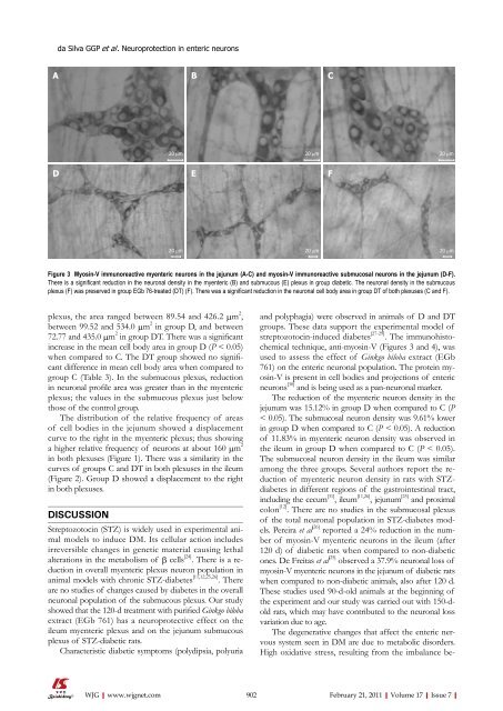
Figure 3 Myosin-V immunoreactive myenteric neurons in the jejunum (A-C) and myosin-V immunoreactive submucosal neurons in the jejunum (D-F).<br />
There is a significant reduction in the neuronal density in the myenteric (B) and submucous (E) plexus in group diabetic. The neuronal density in the submucous<br />
plexus (F) was preserved in group EGb 76-treated (DT) (F). There was a significant reduction in the neuronal cell body area in group DT <strong>of</strong> both plexuses (C and F).<br />
20 μm<br />
C<br />
F<br />
20 μm<br />
and polyphagia) were observed in animals <strong>of</strong> D and DT<br />
groups. These data support the experimental model <strong>of</strong><br />
streptozotocin-induced diabetes [27-29] . The immunohistochemical<br />
technique, anti-myosin-V (Figures 3 and 4), was<br />
used to assess the effect <strong>of</strong> Ginkgo biloba extract (EGb<br />
761) on the enteric neuronal population. The protein myosin-V<br />
is present in cell bodies and projections <strong>of</strong> enteric<br />
neurons [30] and is being used as a pan-neuronal marker.<br />
The reduction <strong>of</strong> the myenteric neuron density in the<br />
jejunum was 15.12% in group D when compared to C (P<br />
< 0.05). The submucosal neuron density was 9.61% lower<br />
in group D when compared to C (P < 0.05). A reduction<br />
<strong>of</strong> 11.83% in myenteric neuron density was observed in<br />
the ileum in group D when compared to C (P < 0.05).<br />
The submucosal neuron density in the ileum was similar<br />
among the three groups. Several authors report the reduction<br />
<strong>of</strong> myenteric neuron density in rats with STZdiabetes<br />
in different regions <strong>of</strong> the gastrointestinal tract,<br />
including the cecum [31] , ileum [11,26] , jejunum [25] and proximal<br />
colon [12] . There are no studies in the submucosal plexus<br />
<strong>of</strong> the total neuronal population in STZ-diabetes models.<br />
Pereira et al [26] reported a 24% reduction in the number<br />
<strong>of</strong> myosin-V myenteric neurons in the ileum (after<br />
120 d) <strong>of</strong> diabetic rats when compared to non-diabetic<br />
ones. De Freitas et al [25] observed a 37.9% neuronal loss <strong>of</strong><br />
myosin-V myenteric neurons in the jejunum <strong>of</strong> diabetic rats<br />
when compared to non-diabetic animals, also after 120 d.<br />
These studies used 90-d-old animals at the beginning <strong>of</strong><br />
the experiment and our study was carried out with 150-dold<br />
rats, which may have contributed to the neuronal loss<br />
variation due to age.<br />
The degenerative changes that affect the enteric nervous<br />
system seen in DM are due to metabolic disorders.<br />
High oxidative stress, resulting from the imbalance be-<br />
902 February 21, 2011|Volume 17|Issue 7|


