11-13 May 2012 Helsingør - Denmark www.networkbio.org
11-13 May 2012 Helsingør - Denmark www.networkbio.org
11-13 May 2012 Helsingør - Denmark www.networkbio.org

Create successful ePaper yourself
Turn your PDF publications into a flip-book with our unique Google optimized e-Paper software.
Network<br />
Medicine<br />
<strong>11</strong>-<strong>13</strong> <strong>May</strong> <strong>2012</strong><br />
<strong>Helsingør</strong> - <strong>Denmark</strong><br />
<strong>www</strong>.<strong>networkbio</strong>.<strong>org</strong>
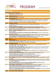
Friday <strong>11</strong>th may <strong>2012</strong><br />
09:00 - 15:00 Registration Open<br />
Session I: KINOME BIOLOGY<br />
Chair: Blagoy Blagoev - University of Southern <strong>Denmark</strong> Session<br />
Programme<br />
15:00 Welcome by Rune Linding<br />
15:10 - 16:00 OPENING LETURE: Eng Lim Goh (SGI, USA) - New Developments in Computing for the Life<br />
Sciences Researcher<br />
16:00 - 16:30 Andrea Califano (Columbia University, New York NY) - Dissecting and interrogating signaling<br />
networks in human malignancies<br />
16:30 - 16:45 Coffee Break and Sponsor Exhibition<br />
16:45 - 17:15 Rune Linding (DTU, Lyngby DK) - Modeling Cancer Kinome Networks<br />
17:15 - 17:30 Gianni Cesareni (Tor Vergata, Rome, Italy) - Mapping the human phosphatome on growth<br />
pathways<br />
17:30 - 17:50 Sponsored Talk (SBV Improver) - Julia Hoeng – Verification of System Biology Research in<br />
the age of Collaborative Competition<br />
17:50 - 18:00 Break<br />
18:00 - 18:15 Sol Efroni (Bar Ilan University, Israel) - Network-based metrics reveals a novel role for<br />
hsa-miR-9 and drug control over the p38 network in glioblastoma multiforme progression<br />
18:15 - 18:45 Garry Nolan (Stanford, San Francisco CA)<br />
19:00 - Poster Session, Drinks and Welcome Dinner at Hotel<br />
Saturday 12th may <strong>2012</strong><br />
Session II: DISEASE NETWORKS<br />
Chair: Ramneek Gutpa - Technical Univeristy of <strong>Denmark</strong> Session<br />
09:10 - 10:00 KEYNOTE LECTURE: Norbert Perrimon (Harvard Medical School, Boston USA) - Building<br />
and validating signaling networks in Drosophila<br />
10:00 - 10:30 Dana Pe’er (Columbia,USA) - On the road to personalized therapy, a systems approach<br />
10:30 - <strong>11</strong>:00 Marc Vidal (DFCI, Boston USA) - Interactome Networks and Human Disease<br />
<strong>11</strong>:00 - <strong>11</strong>:30 Coffe Break and Sponsor Exhibition<br />
<strong>11</strong>:30 - <strong>11</strong>:45 Stephan M. Feller (Oxford, UK) - How are complex signal computations in cells accomplished<br />
by multiprotein complexes assembled on ‘intrinsically disordered’ platform proteins?<br />
The N-terminal folding nucleation (NFN)hypothesis<br />
12:45 -12:00 Theo Knijnenburg (NCI,Amsterdam, NL) - Drug sensitivity of cancer cell lines explained as a<br />
logic combination of mutations<br />
12:00 - 12:30 Søren Brunak (DTU, Lyngby DK) - Interfacing disease phenotypes from electronic patient<br />
records to the underlying network biology<br />
12:30 - <strong>13</strong>:30 Lunch with Sponsored Talk (Thermo Fisher) - Christian Kelstrup (NNF-CPR, Copenhagen,<br />
DK) Optimized Fast and Sensitive Acquisition Methods for Shotgun Proteomics on a Quadru<br />
pole Orbitrap Mass Spectrometer<br />
Programme<br />
Session III: NETWORK DRUGS<br />
Chair: Christopher Workman - Technical University of <strong>Denmark</strong><br />
<strong>13</strong>:30 - 14:20 KEYNOTE LECTURE: Michael Yaffe (MIT, Cambridge USA)<br />
14:20 - 14:50 Matt Onsum (Merrimack Pharmaceuticals, USA) - Using systems biology to accelerate onco<br />
logy drug development<br />
14:50 - 15:15 Coffee Break and Sponsor Exhibition<br />
15:15 - 15:30 Ruth Hüttenhain (ETH Zurich, CH) - A mass spectrometric map for reproducible quantification<br />
of cancer associated proteins in body fluids<br />
15:30 - 16:00 Janine Erler (BRIC, Copenhagen DK) - Molecular networks associated with<br />
cancer progression<br />
16:00 - 16:30 Nevan Krogan (UCFS, San Francisco USA) - Functional Insights from Protein-<br />
Protein and Genetic Interaction Maps<br />
16:30 - 18:30 Poster Session and Sponsor Exhibition<br />
19:00 Transfer to Louisiana Museum for Gala Dinner<br />
20:00 Welcome Speech by SGI<br />
23:00 Buses back to Hotels<br />
Sunday <strong>13</strong>th may <strong>2012</strong><br />
Session IV: INTEGRATIVE NETWORK BIOLOGY<br />
Chair: tba<br />
09:00 -09:50 KEYNOTE LECTURE: Ruedi Aebersold (ETH, Switzerland) - Network Driven Protein Biomar<br />
ker Discovery and Validation<br />
09:50 - 10:20 Anne Claude Gavin (EMBL, Heidelberg DE) - Expanding the interaction space;<br />
protein-metabolite networks<br />
10:20 - 10:45 Coffee Break and Sponsor Exhibition<br />
10:45 - <strong>11</strong>:00 Ulrich Stelzl (MPI Molecular Genetics, Berlin, Germany) A Y2H-seq approach to define the<br />
protein ethyltransferase interactome<br />
<strong>11</strong>:00 - <strong>11</strong>:30 Marian Walhout (UMASS-MED, Worcester MA) - Gene regulatory networks<br />
<strong>11</strong>:30 - 12:00 Ben Lehner (CRG, Barcelona, Spain) - The biology of individuals<br />
12:00 - 12:30 Bernhard Pallson (DTU/UCSD, Lyngby, DK / San Diego, CA) - Systems Biology of<br />
Metabolism<br />
2 / INB <strong>2012</strong> • <strong>11</strong>-<strong>13</strong> <strong>May</strong> <strong>2012</strong> <strong>www</strong>.<strong>networkbio</strong>.<strong>org</strong> / 3
ENG LIM GOh, Ph.D.<br />
SVP & ChIEf TEChNOLOGY OffICER AT SGI<br />
abStract For SPeakerS<br />
Dr. Eng Lim Goh joined SGI in 1989, becoming a chief engineer in 1998 and<br />
then chief technology officer in 2001. He oversees technical computing programs<br />
with the goal to develop the next generation computer architecture for<br />
the new many-core era.<br />
In 2005, InfoWorld named Dr. Goh one of the World’s 25 Most Influential<br />
CTOs. That same year he was also included in the HPCwire list of “15 People<br />
to Watch.” In 2007, he was named “Champions 2.0” of the industry by BioIT<br />
World magazine, and received the HPC Community Recognition Award from<br />
HPCwire. Dr. Goh is a frequent industry speaker and he continues to discuss,<br />
in different forums, innovative technologies and their applications.<br />
Before joining SGI, Dr. Goh worked for Intergraph Systems, Schlumberger Wireline and Shell Research.<br />
A Shell Cambridge University Scholar, Dr. Goh completed his Ph.D. research and dissertation on parallel<br />
architectures and computer graphics, and holds a first-class honors degree in mechanical engineering from<br />
Birmingham University in the U.K.<br />
Dr. Goh has been granted four U.S. patents, two of which as the inventor and the others as co-inventor.<br />
New Developments in Computing for the Life Sciences Researcher<br />
abStract For SPeakerS<br />
ANDREA CALIfANO<br />
COLUMBIA UNIVERSITY, NEW YORK NY<br />
Dissecting and interrogating signaling networks in human malignacies<br />
We will discuss novel experimentally validated computational approaches<br />
to the dissection and interrogation of signal transduction networks in human<br />
malignancies. Specifically, we will address the issue of signaling pathway analysis<br />
from gene expression and phospho-proteomic profile data. We will first<br />
present the identification of KLHL9 deletions as key events in the etiology of<br />
the mesenchymal subtype of high-grade, associated with worst prognosis. We<br />
show that KLHL9, a substrate-specific adapter of a Cul3-based E3 ubiquitinprotein<br />
ligase complex, is responsible for the ubiquitination and proteasomal<br />
degradation of two previously identified master regulators of this glioma<br />
subtype, C/EBPb and C/EBPd. We will also discuss the identification of synergistic synthetic lethality in the<br />
tyrosine kinase signaling network that is dysregulated in non-small cell lung tumors, leading to a potential<br />
application of personalized combination therapy, using available kinase inhibitors.<br />
4 / INB <strong>2012</strong> • <strong>11</strong>-<strong>13</strong> <strong>May</strong> <strong>2012</strong> <strong>www</strong>.<strong>networkbio</strong>.<strong>org</strong> / 5
RUNE LINDING<br />
DTU, LYNGBY - DENMARK<br />
abStract For SPeakerS<br />
Dr Rune Linding is Professor and Research Group Leader for the Cellular Signal<br />
Integration Group (C-SIG) at the Technical University of <strong>Denmark</strong> (DTU),<br />
Center for Biological Sequence Analysis (CBS), Department of Systems Biology,<br />
<strong>Denmark</strong>. He performed his graduate work at the EMBL (Germany), where<br />
he pioneered computational analysis of cell signaling by developing popular<br />
tools like ELM, GlobPlot and DisEMBL for analysing post-translational modifications,<br />
intrinsic protein disorder and modularity of signaling proteins. Dr<br />
Linding was Human Frontiers Science Program Postdoctoral Research Fellow<br />
jointly with Profs Tony Pawson and Mike Yaffe at Samuel Lunenfeld Research<br />
Institute (SLRI, Canada) and Massachusetts Institute of Technology (MIT,<br />
USA), respectively. His postdoctoral work on the cellular phosphorylation networks and development of the<br />
NetworKIN algorithm pioneered Integrative Network Biology and led to the discovery of the quantitative<br />
importance of contextual kinase specificity. He started his own lab (the Cellular & Molecular Logic Team)<br />
at The Institute of Cancer Research (ICR) in London in 2007. At ICR his lab unravelled systems-level models<br />
of JNK and EphR kinase networks, demonstrated a link between specificity and oncogenecity of kinases<br />
and introduced the concept of Network Medicine. Dr Linding leads the NetPhorest community resource<br />
and have pioneered comparative phospho-proteomics and evolutionary studies of signalling networks. Dr<br />
Linding founded the Integrative Network Biology initiative (INBi) which aims to block cancer metastasis by<br />
integration of large-scale, high-dimensional quantitative genomic, proteomic and phenotypic data. His lab<br />
moved to DTU/<strong>Denmark</strong> in 20<strong>11</strong> and the long-term focus of his research group is on studying cellular signal<br />
processing and decision making.<br />
Modeling of Cancer Kinome Networks<br />
Abstract: Biological systems are composed of highly dynamic and interconnected molecular networks that<br />
drive biological decision processes. A goal of integrative network biology is to describe, quantify and predict<br />
the information flow and functional behaviour of living systems in a formal language and with an accuracy<br />
that parallels our characterisation of other physical systems such as Jumbo-jets. Decades of targeted<br />
molecular and biological studies have led to numerous pathway models of developmental and disease<br />
related processes. However, so far no global models have been derived from pathways, capable of predicting<br />
cellular trajectories in time, space or disease. The development of high-throughput methodologies has<br />
further enhanced our ability to obtain quantitative genomic, proteomic and phenotypic readouts for many<br />
genes/proteins simultaneously. Here, I will discuss how it is now possible to derive network models through<br />
computational integration of systematic, large-scale, high-dimensional quantitative data sets. I will review<br />
our latest advances in methods for exploring phosphorylation networks. In particular I will discuss how the<br />
combination of quantitative mass-spectrometry, systems-genetics and computational algorithms (NetworKIN<br />
[1] and NetPhorest [4]) made it possible for us to derive systems-level models of JNK and EphR signalling<br />
networks [2,3]. I shall discuss work we have done in comparative phospho-proteomics and network<br />
evolution[5-7]. Finally, I will discuss our most recent work in analysing genomic sequencing data from NGS<br />
studies and how we have developed new powerful algorithms to predict the impact of disease mutations on<br />
cellular signaling networks and applied them to profiling of ovarian cancer cells.<br />
References:<br />
http://<strong>www</strong>.lindinglab.<strong>org</strong><br />
Linding et al., Cell 2007.<br />
Bakal et al., Science 2008.<br />
Jørgensen et al., Science 2009.<br />
Miller et al., Science Signaling 2008.<br />
Tan et al., Science Signaling 2009.<br />
abStract For SPeakerS<br />
GIANNI CESARENI<br />
Mapping the human phosphatome on growth pathways<br />
fRANCESCA SACCO 1<br />
, PIER fEDERICO GhERARDINI 1<br />
, SERENA PAOLUzI 1<br />
, JULIO SAEz-RODRIGUEz 3<br />
,<br />
MANUELA hELMER-CITTERICh 1<br />
, ANTONELLA RAGNINI-WILSON 1,2<br />
, LUISA CASTAGNOLI 1<br />
AND GIANNI<br />
6 / INB <strong>2012</strong> • <strong>11</strong>-<strong>13</strong> <strong>May</strong> <strong>2012</strong> <strong>www</strong>.<strong>networkbio</strong>.<strong>org</strong> / 7<br />
CESARENI 1<br />
1<br />
DEPARTMENT Of BIOLOGY, UNIVERSITY Of ROME “TOR VERGATA”, ITALY 2hIGh-ThROUGhPUT MICROS-<br />
COPY fACILITY; DEPARTMENT Of TRANSLATIONAL AND CELLULARPhARMACOLOGY, CONSORzIO MARIO<br />
NEGRI SUD, SM. IMBARO, ITALY 3EMBL-EBI, hINxTON, UK AND EMBL-GENOME BIOLOGY UNIT, hEIDEL-<br />
BERG, GERMANY<br />
Large-scale siRNA screenings allow linking the function of poorly characterized genes to phenotypic<br />
readouts. According to this strategy, genes are associated to a function of interest if the alteration of their<br />
expression perturbs the phenotypic readouts. However, given the intricacy of the cell regulatory network, the<br />
mapping procedure is low resolution and the resulting models provide little mechanistic insights. We have<br />
developed a new strategy that combines high-content multiparametric analysis of cell perturbation and logic<br />
modeling to achieve a more detailed functional mapping of human genes onto complex pathways. By this
JULIA hOENG<br />
Verification of Systems Biology Research in the Age of Collaborative-Competition<br />
abStract For SPeakerS<br />
JULIA hOENG1, MARJA TALIKKA1, STéPhANIE BOUé1, PABLO MEYER ROJAS2, RAqUEL NOREL2, JOhN J<br />
RICE2, JöRG SPRENGEL3, MANUEL PEITSCh1, GUSTAVO STOLOVITzKY2<br />
1PhILIP MORRIS INTERNATIONAL R&D, NEUChâTEL, SWITzERLAND, 2IBM COMPUTATIONAL BIOLOGY<br />
CENTER, YORKTOWN hEIGhTS, NY, USA, 3IBM GLOBAL BUSINESS SERVICES, SWITzERLAND<br />
Abstract:<br />
Modern society demands greater scrutiny of the potential health risks and benefits of long-term, and sometimes<br />
lifelong, exposure to drugs, chemicals, and substances found in consumer products and the environment.<br />
Organizations such as companies and academic consortia conduct large multi-year scientific studies that<br />
entail the collection and analysis of thousands of data points. The individual experiments are often conducted<br />
over many physical sites and with internal and outsourced components. To extract maximum value,<br />
the interested parties need to verify the accuracy and reproducibility of automated collection and analysis<br />
workflows in systems biology before the initiation of large multi-year studies.<br />
Traditional verification using the peer-review process has shortcomings, such as lack of scalability, which<br />
renders it insufficient for the assessment of high throughout research. A team of researchers at PMI and<br />
IBM, whose aim is to improve the effectiveness of scientific studies and verification of scientific findings<br />
propose a scheme called IMPROVER, for Industrial Methodology for Process Verification of Research. This<br />
methodology evaluates a research program by dividing its workflow into smaller building blocks, whereby the<br />
verification of each building block can be done internally or externally via challenge-based `crowdsourcing`<br />
to a research community.<br />
Scientific challenges will be broadcast to potential stakeholders in the form of an open call for participation<br />
with the intention of providing the community with the opportunity to test their computational methods on<br />
new data as well as to partake in a collaborative effort whose ultimate goal could contribute to solving a<br />
grand scientific problem.<br />
Considering cancer as the leading cause of death worldwide, we formulate the Diagnostics Signature Challenge<br />
to evaluate novel approaches for the identification of robust and predictive signatures for this disease.<br />
The goal of a Diagnostics Signature Challenge is to verify that transcriptomics data contains enough<br />
information for the determination and prognosis of certain human disease states that could profit from better<br />
diagnostics signatures.<br />
Here we will describe the approach, the necessary operational steps, and how we intend to engage the<br />
wider scientific community to assess the applicability of the IMPROVER approach to molecular diagnostics<br />
(i.e., genomic signatures).<br />
abStract For SPeakerS<br />
ROTEM BEN-hAMO, SOL EfRONI<br />
ThE MINA & EVERARD GOODMAN fACULTY Of LIfE SCIENCES, BAR ILAN UNIVERSITY, RAMAT GAN,<br />
ISRAEL<br />
Network-based metrics reveals a novel role for hsa-miR-9 and drug control over the p38 network in glioblastoma<br />
multiforme progression<br />
Contributors:<br />
Background<br />
Glioblastoma multiforme (GBM) is the most common, aggressive and malignant primary tumor of the brain<br />
and is associated with one of the worst 5-year survival rates among all human cancers. Identification of<br />
molecular interactions that associate with disease progression may be key in finding novel treatments.<br />
Using five independent molecular and clinical datasets with a set of computational algorithms we were able<br />
to identify a gene-gene and gene-microRNA network that significantly stratifies patient prognosis. By combining<br />
gene expression microarray data with microRNA expression levels, copy number alterations, drug<br />
response and clinical data, combined with network knowledge, we were able to identify a single pathway at<br />
the core of glioblastoma.<br />
This network, the p38 network, and an associated microRNA, hsa-miR-9, facilitate prognostic stratification.<br />
The microRNA hsa-miR-9 correlated with network behavior and presents binding affinities with network<br />
members in a manner that suggests control over network behavior. A similar control over network behavior<br />
is possible through a set of drugs. These drugs are part of the treatment regimen for a subpopulation of the<br />
patients that participated in the TCGA study and for which the study provides clinical information. Interestingly,<br />
the patients that were treated with these specific sets of drugs, all of which targeted against p38<br />
network members, demonstrate highly significant stratification of prognosis.<br />
Combined, these results call for attention to p38 network targeted treatment and present the p38 networkhsa-miR-9<br />
control mechanism as critical in GBM progression.<br />
8 / INB <strong>2012</strong> • <strong>11</strong>-<strong>13</strong> <strong>May</strong> <strong>2012</strong> <strong>www</strong>.<strong>networkbio</strong>.<strong>org</strong> / 9
GARRY NOLAN<br />
STANfORD, STANfORD, CA<br />
TBA<br />
abStract For SPeakerS<br />
abStract For SPeakerS<br />
NORBERT PERRIMON<br />
hARVARD MEDICAL SChOOL, BOSTON, MA<br />
Dr. Perrimon is the James Stillman Professor of Developmental Biology at<br />
Harvard medical School and an Investigator of the Howard Hughes Medical<br />
Institute. He has 30 years of experience in the fields of developmental<br />
genetics, signal transduction and genomics. He has made a number of contributions<br />
to the fields of Genetics, Developmental Biology, signal transduction<br />
and functional genomics. His group developed many methods that have<br />
significantly improved the Drosophila toolbox. These include: the FLP-FRT<br />
Dominant Female Sterile technique to generate mosaics in the female germline,<br />
the Gal4-UAS method to control gene expression both spatially and<br />
temporally; the “Positively Marked Labeling Method” for lineage analyses;<br />
and thermosensitive inteins to generate conditional alleles. His contributions<br />
include the characterization of: the maternal effects of zygotic lethal mutations; the logic of head patterning;<br />
the identification of Scribble and the <strong>org</strong>anization of the cell polarity complexes; the discovery of adult<br />
gut stem cells; and the mechanisms of muscle growth and aging. His lab has characterized many signaling<br />
components of receptor tyrosine kinases, Wnt and JAK/STAT pathways, in particular. These include: Raf<br />
kinase and demonstration that it acts downstream of Ras; Corkscrew/SHP2 non receptor tyrosine phosphatase<br />
as a positive transducer of RTK signaling; Spitz as a ligand, and Kekkon as a negative regulator, of<br />
EGFR; Porcupine, Dishevelled and GSK3 as components of Wnt/Wg signaling; Unpaired, Hopscotch/JAK and<br />
Marelle/STAT as members of the JAK/STAT pathway; and Heparan Sulfate Proteglycans in Hedgehog, Wnt<br />
and FGF signaling. Regarding large scale functional genomics, his group established high-throughout genome-wide<br />
RNAi screens to systematically interrogate the entire Drosophila genome in various cell-based<br />
assays, demonstrated that long dsRNAs are associated with off target effects, established a cross-species<br />
method for rescue of RNAi phenotypes, developed RNAi methods in primary embryonic cell cultures, and<br />
generated algorithms for automated image analyses. In 2003 he created the Drosophila RNAi Screening<br />
Center (DRSC) at Harvard Medical School to make this technology available to the community. In addition,<br />
his group developed new shRNA vectors for in vivo RNAi and in 2008 established the Transgenic RNAi Project<br />
(TRiP) at Harvard Medical School to build a genome scale resource of transgenic shRNA flies. Currently,<br />
his laboratory is applying large-scale RNAi and proteomic methods to obtain a global understanding of the<br />
structure of a number of signaling pathways and their cross-talks. In addition, he is studying the roles of<br />
signaling pathways in homeostasis and tissue remodeling in Drosophila muscles and gut stem cells. Dr.<br />
Perrimon has trained more than 80 students and postdoctoral fellows, with most of them currently holding<br />
academic positions.<br />
Building and validating signaling networks in Drosophila<br />
Characterizing the extent and logic of signaling networks is essential to understanding developmental<br />
processes, mechanisms of oncogenesis, and resistance to chemotherapy. A major focus of our lab is to<br />
apply “Omics” approaches to describe the <strong>org</strong>anization of signaling networks. We have spent considerable<br />
effort to implement complementary technologies of genome-wide RNAi HTS, tandem affinity purification/<br />
mass spectrometry (TAP/MS), and transcriptome analyses in Drosophila cell lines, to identify core pathway<br />
components, pathway dynamics, and the extent of cross-talk between pathways. Recently, because studying<br />
networks in the fly is hampered by the paucity of phosphoantibodies available, we have used the Tandem<br />
Mass Tags Mass Spec method to measure quantitative changes in the phosphoproteome. Integration of<br />
various Omic approaches is necessary for network studies because much of the noise associated with each<br />
technology can be filtered by the integration of orthogonal data sets.<br />
We have applied these approaches successfully to date to the study of five pathways in established Drosophila<br />
cell lines: Insulin/PI3K, EGF/MAPK, JAK/STAT, JNK and Hippo and have generated high-confidence<br />
protein-protein interaction (PPI) networks that have been validated to various extents by RNAi and transcriptome<br />
analyses. Our networks are of high quality because they identify most previously known interactions,<br />
and by various means we have validated a number of new components and interactions.<br />
10 / INB <strong>2012</strong> • <strong>11</strong>-<strong>13</strong> <strong>May</strong> <strong>2012</strong> <strong>www</strong>.<strong>networkbio</strong>.<strong>org</strong> / <strong>11</strong>
DANA PE’ER<br />
COLUMBIA UNIVERSITY, NEW YORK, NY<br />
Dana Pe’er is assistant professor at the Department of Biological Sciences at<br />
Columbia and Columbia’s Center for Computational Biology and Bioinformatics<br />
(C2B2). In her Ph.D. (Computer Science) at the Hebrew University of Jerusalem<br />
(with Nir Friedman), Dana pioneered the use of machine learning to uncover<br />
the structure and function of molecular networks from genomics data,<br />
based on Bayesian networks. She subsequently did a postdoc with Ge<strong>org</strong>e<br />
Church at Harvard Medical School and there she began to work towards understanding<br />
of how genetic variation alters the regulatory network between<br />
individuals and subsequently manifests in phenotypic diversity. This is now<br />
the focus of Dana’s lab at Columbia University, where she and her team are<br />
developing methods to infer how variation in sequence modulates signal processing and is manifested in<br />
cellular phenotypes, with applications towards personalized cancer treatment. Dana is recipient of the Burroughs<br />
Wellcome Fund Career Award, NIH Directors New Innovator Award, Stand Up To Cancer Innovative<br />
Research Grant and a Packard Fellow in Science and Engineering.<br />
On the road to personalized therapy, a systems approach<br />
Cancer is an individual disease—unique in how it develops and behaves in every patient. The emergence<br />
of revolutionary technologies has stimulated hope that treatment will improve by becoming more targeted<br />
and individualized in nature. Characterization of cancer genomes has revealed a staggering complexity of<br />
aberrations among individuals, such that the functional importance and physiological impact of most tumor<br />
genetic alterations remains poorly defined. Genomic and proteomic data in tumor samples, using a battery<br />
of using high-throughput, massively parallel technologies is accumulating at a astounding rates. A major<br />
challenge involves the development of analysis methods to integrate this data towards patient-specific tumor<br />
network models. We demonstrate progress on a number of fronts.<br />
• We demonstrate approaches to integrate heterogeneous genomic data types to identify the key<br />
alterations functionally driving the cancer and associate these with their tumorigenic phenotypes (e.g.<br />
proliferation, invasion).<br />
• To understand tumor response to drug and the heterogeneity of this response among patients it is<br />
critical to molecularly interrogate the tumor following drug perturbations. We will demonstrate how such<br />
interrogation of a tumor panel reveals variation in network wiring that connects to drug response.<br />
• Tumors are not only heterogeneous between patients, but there is also pervasive heterogeneity within<br />
a single patient. Mass-cytometry, a novel technology that can measure more than forty signaling mole-<br />
abStract For SPeakerS abStract For SPeakerS<br />
MARC VIDAL<br />
CENTER fOR CANCER SYSTEMS BIOLOGY (CCSB) AND DEPARTMENT Of<br />
CANCER BIOLOGY DANA-fARBER CANCER INSTITUTE & DEPARTMENT Of<br />
GENETICS hARVARD MEDICAL SChOOL BOSTON, MA 02<strong>11</strong>5<br />
Marc Vidal is Professor of Genetics at Harvard Medical School and Director<br />
of the Center for Cancer Systems Biology (CCSB) at the Dana-Farber Cancer<br />
Institute. Dr. Vidal received his PhD in 1991 from Gembloux University (Belgium)<br />
for work performed at Northwestern University (Evanston, IL, USA)<br />
where he identified the yeast genes RPD3 and SIN3, and demonstrated that<br />
they encode global transcriptional regulators. These genes were subsequently<br />
found to encode Histone Deacetylase (HDAC) and its main recruiting<br />
factor, respectively. During postdoctoral training at the Massachusetts General Hospital Cancer Center, he<br />
developed the reverse two-hybrid system, a widely applicable method used to genetically characterize protein-protein<br />
interactions. Having developed interdisciplinary strategies together with collaborators from<br />
the fields of physics, computer science, mathematics, genomics and human genetics, he and his team have<br />
been charting protein-protein and other interactome networks for 15 years and are developing ways to integrate<br />
interactome maps with other large-scale functional genomic and proteomic maps, with the ultimate<br />
objective to discover novel network properties from a systems point-of-view. Dr. Vidal was elected Associate<br />
Member of the Royal Academy for Science and the Arts of Belgium and has received several awards,<br />
including a Chair from the Francqui Foundation (Belgium) and an Abbott Bioresearch Award.<br />
Interactome Networks and Human Disease<br />
For over half a century it has been conjectured that macromolecules form complex networks of functionally<br />
interacting components, and that the molecular mechanisms underlying most biological processes correspond<br />
to particular steady states adopted by such cellular networks. However, until a decade ago, systemslevel<br />
theoretical conjectures remained largely unappreciated, mainly because of lack of supporting experimental<br />
data.<br />
To generate the information necessary to eventually address how complex cellular networks relate to biology,<br />
we initiated, at the scale of the whole proteome, an integrated approach for modeling protein-protein<br />
interaction or “interactome” networks. Our main questions are: How are interactome networks <strong>org</strong>anized at<br />
the scale of the whole cell? How can we uncover local and global features underlying this <strong>org</strong>anization, and<br />
how are interactome networks modified in human disease, such as cancer?<br />
12 / INB <strong>2012</strong> • <strong>11</strong>-<strong>13</strong> <strong>May</strong> <strong>2012</strong> <strong>www</strong>.<strong>networkbio</strong>.<strong>org</strong> / <strong>13</strong>
STEPhAN M. fELLER, WIMM,<br />
OxfORD UNIVERSITY, OxfORD, UK<br />
abStract For SPeakerS<br />
How are complex signal computations in cells accomplished by multi-protein complexes assembled on<br />
‘intrinsically disordered’ platform proteins? The N terminal folding nucleation (NFN) hypothesis<br />
Proteins with little recognisable secondary structure -according to current prediction programs -comprise<br />
one third of the human proteome. Some of these are large and serve as docking platforms for many proteins<br />
involved in the processing of cell signals, thereby creating large signalosomes in which signal computations<br />
are performed. It was always difficult to conceptualise how an intrinsically disordered, i.e. ‘chaotic’ signal<br />
computation platform protein should be able to mediate effective pathway crosstalk in cells. However, we<br />
have now first evidence that the cancer-relevant Gab family proteins display a previously unrecognised<br />
<strong>org</strong>anisation (Simister et al. 20<strong>11</strong>, PLoS Biol; Simister & Feller <strong>2012</strong>, Mol BioSystems). Gab proteins contain<br />
N terminal PH domains followed by long ‘tail regions’ supposedly lacking any structure. Our data point to<br />
several specific interactions of the Gab1 tail with is structured N-terminus. These generate, around the PH<br />
domain, tail loop structures, in which docking sites for pathway-specific signaling proteins are clustered.<br />
Functionally dedicated, distinct sub-complexes can assembly in these loops. If this model is correct, it is<br />
easy to see how effective signaling pathway crosstalk occurs: simply by interactions of distinct sub-complexes<br />
bound to specialised loops, which are held in place by their interactions with the PH domain. We believe<br />
that proving this hypothesis, which is also relevant to multiple similarly structured signaling protein families<br />
(p<strong>13</strong>0Cas, IRS/Dok, Frs etc.), will have a profound impact on the understanding of molecular signal computing<br />
in cells and we will report on our first results.<br />
abStract For SPeakerS<br />
ThEO KNIJNENBURG<br />
NEThERLANDS CANCER INSTITUTE, DIVISION Of MOLECULAR CARCINOGENESIS<br />
PLESMANLAAN 121, 1066Cx AMSTERDAM, ThE NEThERLANDS<br />
MAThEW J. GARNETT<br />
CANCER GENOME PROJECT , WELLCOME TRUST SANGER INSTITUTE<br />
hINxTON CAMBRIDGE CB10 1SA, UNITED KINGDOM<br />
GUNNAR W. KLAU<br />
LIfE SCIENCES GROUP, CENTRUM WISKUNDE & INfORMATICA<br />
SCIENCE PARK 123, 1098 xG AMSTERDAM, ThE NEThERLANDS<br />
fRANCESCO IORIO<br />
EUROPEAN BIOINfORMATICS INSTITUTE, WELLCOME TRUST GENOME CAMPUS<br />
hINxTON CAMBRIDGE CB10 1SD, UNITED KINGDOM<br />
JULIO SAEz-RODRIGUEz<br />
EUROPEAN BIOINfORMATICS INSTITUTE, WELLCOME TRUST GENOME CAMPUS<br />
hINxTON CAMBRIDGE CB10 1SD, UNITED KINGDOM<br />
ULTAN MCDERMOTT<br />
CANCER GENOME PROJECT , WELLCOME TRUST SANGER INSTITUTE<br />
hINxTON CAMBRIDGE CB10 1SA, UNITED KINGDOM<br />
LODEWYK WESSELS<br />
NEThERLANDS CANCER INSTITUTE, DIVISION Of MOLECULAR CARCINOGENESIS<br />
PLESMANLAAN 121, 1066Cx AMSTERDAM, ThE NEThERLANDS<br />
Drug sensitivity of cancer cell lines explained as a logic combination of mutations<br />
Cancer arises as a result of the acquisition of DNA mutations. It is still unclear which and how combinations<br />
of mutations are involved in tumor initiation and development. Two major challenges need to be addressed in<br />
order to systematically unravel these genetic interactions.<br />
First, large amounts of high quality data are necessary in order to ensure the statistical power required<br />
to uncover these genotype-to-phenotype relationships. Current advances in high-throughput biology are<br />
enabling the generation of very large datasets that should facilitate the detection of higher order genetic<br />
interactions. Here, we report on the analysis of a panel of 1000 cancer cell lines. The mutation status of 66<br />
known cancer genes has been characterized for each cell line in this panel. Additionally, these cell lines<br />
have been screened to model drug response with a large number (250+) of anti-cancer therapeutics. This<br />
dataset is part of the Cancer Genome Project at the Wellcome Trust Sanger Institute.<br />
The second challenge is to design a computational framework that employs a mathematical formalism rich<br />
enough to capture the underlying biological complexity, while limiting the computational complexity that<br />
would otherwise prohibit finding optimal (or even good) solutions in the vast combinatorial search space.<br />
We contribute to addressing this second challenge by proposing a novel computational approach based on<br />
integer programming that infers logic combinations of mutations that predict the observed drug response.<br />
The use of a logic formalism enables the formulation of intelligible models, from which the cancer biologists<br />
can generate a deeper understanding based on domain knowledge and easily formulate novel hypotheses<br />
and experiments.<br />
Our models show that for most drugs, combinations of mutations explain the drug response better than<br />
single mutations. For example, of the 8 BRAF inhibitors in the panel, the drug sensitivity to 3 of them is better<br />
explained using a logic combination of a BRAF mutation and one or more mutations in other genes, such<br />
as TET2. These results immediately suggest putative drug combination therapies.<br />
Additionally, cancer signaling pathways as annotated in e.g. the Pathway Interaction Database can be<br />
employed to dramatically reduce the search space and offer a mechanistic explanation of the uncovered<br />
gene combinations.<br />
14 / INB <strong>2012</strong> • <strong>11</strong>-<strong>13</strong> <strong>May</strong> <strong>2012</strong> <strong>www</strong>.<strong>networkbio</strong>.<strong>org</strong> / 15
abStract For SPeakerS<br />
SøREN BRUNAK,<br />
TEChNICAL UNIVERSITY Of DENMARK & UNIVERSITY Of COPENhAGEN<br />
Søren Brunak, Ph.D., is professor of Bioinformatics at the Technical University<br />
of <strong>Denmark</strong> and professor of Disease Systems Biology at the University of Copenhagen.<br />
Prof. Brunak is the founding Director of the enter for Biological Sequence<br />
Analysis, which was formed in 1993 as a multi-disciplinary research<br />
group of molecular biologists, biochemists, medical doctors, physicists, and<br />
computer scientists. Søren Brunak has been highly active within data integration,<br />
where machine learning techniques often have been used to integrate<br />
predicted or experimentally established functional genome and proteome annotation.<br />
His current research does combine molecular level systems biology<br />
and healthcare sector data such as electronic patient records and biobank<br />
questionnaires. The aim is to group and stratify patients not only from their genotype, but also phenotypically<br />
based on the clinical descriptions in the medical records.<br />
Interfacing disease phenotypes from electronic patient records to the underlying network biology<br />
Interfacing sequencing and network biology data to personal healthcare sector information World-wide the<br />
healthcare sector is confronted with the availability of database information which describe the individual<br />
in great detail. These data range all the way from the molecular level, where they for example reveal the<br />
genetic makup of the patient, to the fine-grained descriptions of disease phenotypes as they are found in<br />
electronic patient records at hospitals. Linking these data is a huge undertaking which soon will represent a<br />
major challenge given that it already has become feasible to sequence the DNA of entire populations at low<br />
cost. Combining molecular level data with clinical information and data on the chemical environment may<br />
add complementary types of knowledge which - together with genotype and metagenomic information from<br />
the individual - can reveal disease mechanisms in novel ways. Electronic patient records remain a rather<br />
unexplored, but potentially rich data source for example for discovering correlations between diseases. We<br />
describe a general approach for gathering phenotypic descriptions of patients from medical records in a systematic<br />
and non-cohort dependent manner. By extracting phenotype information from the free-text in such<br />
records we demonstrate that we can extend the information contained in the structured record data, and use<br />
it for producing fine-grained patient stratification and disease co-occurrence statistics. The approach uses<br />
a dictionary based on the WHO International classification of Disease ontology and is therefore in principle<br />
language independent.<br />
References<br />
Using electronic patient records to discover disease correlations and stratify patient cohorts. Roque FS,<br />
Jensen PB, Schmock H, Dalgaard M, Andreatta M, Hansen T, Søeby K, Bredkjær S, Juul A, Werge T, Jensen<br />
LJ, Brunak S. PLoS Comput Biol. 20<strong>11</strong> Aug;7(8):e1002141.<br />
Knowledge engineering for health: A new discipline required to bridge the “ICT gap” between research and<br />
healthcare. Beck T, Gollapudi S, Brunak S, Graf N, Lemke HU, Dash D, Buchan I, Díaz C, Sanz F, Brookes<br />
AJ. Hum Mutat. <strong>2012</strong> Mar 5. doi: 10.1002/humu.22066<br />
Mining electronic health records: towards better research applications and clinical care Jensen PB, Jensen<br />
LJ, and Brunak S, Nature Reviews Genetics, June <strong>2012</strong>, to appear.<br />
abStract For SPeakerS<br />
ChRISTIAN KELSTRUP<br />
NNf-CPR, COPENhAGEN, DENMARK<br />
Optimized Fast and Sensitive Acquisition Methods for Shotgun Proteomics on a Quadrupole Orbitrap Mass<br />
Spectrometer<br />
16 / INB <strong>2012</strong> • <strong>11</strong>-<strong>13</strong> <strong>May</strong> <strong>2012</strong> <strong>www</strong>.<strong>networkbio</strong>.<strong>org</strong> / 17
SPonSorPage<br />
abStract For SPeakerS<br />
MIChAEL YAffE<br />
MIT, CAMBRIDGE, MA<br />
18 / INB <strong>2012</strong> • <strong>11</strong>-<strong>13</strong> <strong>May</strong> <strong>2012</strong> <strong>www</strong>.<strong>networkbio</strong>.<strong>org</strong> / 19
abStract For SPeakerS<br />
MATThEW ONSUM<br />
Ph.D., MERRIMACK PhARMACEUTICALS, CAMBRIDGE, MA<br />
Dr. Onsum is an Associate Director of Translational Research at Merrimack<br />
Pharmaceuticals. He received his B.S., M.S., and Ph.D. degrees in Mechanical<br />
Engineering from the University of California, Berkeley. His doctoral work,<br />
under the supervision of Adam Arkin and Kameshwar Poolla, used both computational<br />
and wet biology to study how immune cells track and capture invading<br />
microbes. Additionally, he was a member of the Alliance for Cellular<br />
Signaling where he developed mathematical models of GPCR mediated calcium<br />
signaling and model validation software. He spent two years at Astra-<br />
Zeneca R&D Boston, where he used model simulations to help identify new<br />
drug targets. He is currently at Merrimack Pharmaceuticals where he is leading<br />
the translational research program for MM-<strong>11</strong>1, a bi-specific antibody against ErbB3 that uses an ErbB2<br />
targeting arm to enhance avidity and inhibitor potency.<br />
Using systems biology to accelerate oncology drug development<br />
This session will discuss how Merrimack uses mathematical models of cancer signaling pathways to design<br />
novel therapeutics, identify predictive biomarkers, and guide clinical development plans. By combining the<br />
knowledge gained from our biochemical model together with biomarker measurements from a large panel<br />
of archived tumors and clinical data from the literature, we simulated the effect of our lead oncology drug in<br />
a variety of cancer indications and used these simulations to help prioritize our clinical development plans.<br />
We will also discuss how we use our mathematical models to assess other targeted oncology drugs and<br />
determine which of these drugs should be combined with our therapies.<br />
abStract For SPeakerS<br />
RUTh hüTTENhAIN 1<br />
, MARTIN SOSTE 2<br />
, NAThALIE SELEVSEK 1<br />
, hANNES RöST 1<br />
, ATUL SEThI 1<br />
, ChRISTINE<br />
CARAPITO 3<br />
, TERRY fARRAh 4<br />
, ERIC W. DEUTSCh 4<br />
, ULRIKE KUSEBAUCh 4<br />
, ROBERT L. MORITz 4<br />
, EMMA<br />
NIMèUS-MALMSTRöM 5<br />
, OLIVER RINNER 6<br />
AND RUEDI AEBERSOLD 1,7<br />
20 / INB <strong>2012</strong> • <strong>11</strong>-<strong>13</strong> <strong>May</strong> <strong>2012</strong> <strong>www</strong>.<strong>networkbio</strong>.<strong>org</strong> / 21<br />
1<br />
DEPARTMENT Of BIOLOGY, INSTITUTE Of MOLECULAR SYSTEMS BIOLOGY, ETh zURICh, zURICh, SWITzERLAND<br />
2<br />
DEPARTMENT Of BIOLOGY, INSTITUTE Of BIOChEMISTRY, ETh zURICh, zURICh, SWITzER-<br />
LAND 3<br />
LABORATOIRE DE SPECTROMéTRIE DE MASSE BIO-ORGANIqUE, INSTITUT PLURIDISCIPLINAIRE<br />
hUBERT CURIEN, UMR7178 CNRS-UNIVERSITé DE STRASBOURG, STRASBOURG, fRANCE 4<br />
INSTITUTE<br />
fOR SYSTEMS BIOLOGY, SEATTLE, WA, USA 5<br />
DEPARTMENT Of ONCOLOGY, LUND UNIVERSITY, LUND, SWE-<br />
DEN 6<br />
BIOGNOSYS AG, zURICh, SWITzERLAND 7<br />
fACULTY Of SCIENCE, UNIVERSITY Of zURICh, zURICh,<br />
SWITzERLAND<br />
A mass spectrometric map for reproducible quantification of cancer associated proteins in body fluids<br />
A major bottleneck in applying targeted proteomic approaches to protein biomarker research is the limited<br />
availability of accurate, reproducible and sensitive assays for testing hypotheses on cohorts of patient<br />
samples. Therefore, we aimed to generate a high-quality resource of selected reaction monitoring (SRM)<br />
assays for cancer associated proteins (CAPs), from an evidence-based list of <strong>11</strong>72 proteins, that have been<br />
previously documented to be differentially expressed in various types of cancer1. Using a protein functional<br />
network, we demonstrated that these proteins are enriched among the interaction partners of genes mutated<br />
in cancer.<br />
Following the development of the SRM assays we examined their detectability in two types of samples which<br />
are highly relevant for biomarker studies, plasma and urine. The concentrations of the detected CAPs in<br />
plasma span six orders of magnitude demonstrating the high sensitivity of these assays for protein quantification.<br />
The developed SRM assays and detectability information are publicly available via PeptideAtlas<br />
SRM Experiment Library (PASSEL)2 to enable researchers to test hypotheses related to these CAPs in any<br />
sample of interest.<br />
We demonstrated the utility of this resource for biomarker research by measuring CAPs from the FDA-approved<br />
OVA1 biomarker panel across plasma samples collected from women diagnosed with a pelvic mass.<br />
The measurements were able to recapitulate the expected capability of this panel to stratify ovarian cancer<br />
(OC) patients and patients with benign ovarian tumors (BOT). Moreover, using this resource, we explored<br />
the prospect of discovering novel biomarker candidates by in silico prediction. Using a functional protein network,<br />
we derived a set of 21 CAPs, that interact with genes mutated in OC, and are measurable in plasma.<br />
12 of these network derived proteins showed a significant difference in abundance between patients with OC<br />
and patients with BOT.<br />
In sum, by developing a publicly accessible resource of SRM assays and testing their detectability in body<br />
fluids, this study will facilitate applying targeted proteomics to protein biomarker research. Furthermore, we<br />
explored the promise of combining genomic data and protein network analysis for predicting novel biomarker<br />
candidates. We demonstrated that this resource can be used to rapidly test these hypotheses in body fluids.<br />
Polanski, M. & Anderson, N.L. A list of candidate cancer biomarkers for targeted proteomics. Biomarker<br />
Insights 1, 1-48 (2007).<br />
Farrah, T. et al. PASSEL: The PeptideAtlas SRM Experiment Library. Proteomics, 10.1002/pmic.20<strong>11</strong>00515<br />
(<strong>2012</strong>).
DR JANINE T ERLER,<br />
BRIC, UNIVERSITY Of COPENhAGEN<br />
COPENhAGEN, DENMARK<br />
abStract For SPeakerS<br />
Elucidating the molecular networks associated with metastasis<br />
Metastasis is responsible for over 90% of cancer patient deaths due to a<br />
lack of effective therapies against metastatic disease. We require a better<br />
understanding of the underlying molecular processes in order to identify and<br />
develop novel effective therapeutic strategies. We aim to identify the signaling<br />
networks associated with metastasis through several different approaches.<br />
Our goal is to identify key nodes in the network and target these to prevent<br />
disease progression, and translate these findings into the clinic.<br />
One approach we are taking is to identify and compare the signaling networks<br />
and associated genetic mutations in metastatic versus non-metastatic samples derived from patients. In<br />
our model, we have used non-metastatic and metastatic matched human cancer cell lines isolated from the<br />
same patient at different stages. We have performed quantitative mass spectrometry to compare the global<br />
proteome and phosphoproteome, and next generation sequencing to both identify and compare mutations,<br />
and investigate their impact on the signaling networks. We are deploying the NetPhorest and NetworKIN<br />
algorithms to predict phospho-binding modules likely to interact with the identified sites. Additionally, computational<br />
integration of the molecular data with quantitative phenotypic data acquired from high throughput<br />
invasion assays allows us to build predictive models of cancer progression. We are using RNAi to perturb<br />
these networks and refine our models to identify the core network predicted to drive metastasis. We will additionally<br />
integrate data collected from fresh frozen human patient samples. Finally, we will test our predictions<br />
using an in vivo model of metastasis.<br />
The results of this study will aid the development of a network-based therapeutic strategy for the treatment<br />
and prevention of metastatic disease.<br />
abStract For SPeakerS<br />
NEVAN KROGAN<br />
CELLULAR AND MOLECULAR PhARMACOLOGY/CALIfORNIA INSTITUTE fOR<br />
qUANTITATIVE BIOMEDICAL SCIENCES, UNIVERSITY Of CALIfORNIA, SAN<br />
fRANCISCO, CA, 94158<br />
Dr. Krogan is an Associate Professor in the Department of Cellular and Molecular<br />
Pharmacology at the University of California-San Francisco and is<br />
an expert in the fields of functional genomics and systems biology. He was<br />
born and raised in Regina, Saskatchewan, Canada and obtained his undergraduate<br />
degree from the University of Regina. As a graduate student at the<br />
University of Toronto, Dr. Krogan led a project that systematically identified<br />
protein complexes in the model <strong>org</strong>anism, Saccharomyces cerevisiae, through<br />
an affinity tagging-purification/mass spectrometry strategy. This work led to<br />
the characterization of 547 complexes, comprising over 4000 proteins, and represents the most comprehensive<br />
protein-protein interaction map to date in any <strong>org</strong>anism. To complement this physical interaction<br />
data, Dr. Krogan developed an approach, termed E-MAP (or epistatic miniarray profile), which allows for<br />
high-throughput generation and quantitative analysis of genetic interaction data. Dr. Krogan’s lab at UCSF<br />
focuses on applying these global proteomic and genomic approaches to formulate hypotheses about various<br />
biological processes, including transcriptional regulation, DNA repair/ replication and RNA processing.<br />
He is now developing and applying methodologies to create genetic and physical interactions between<br />
pathogenic <strong>org</strong>anisms, including HIV and TB, and their hosts, which is providing insight into the human<br />
pathways and complexes that are being hijacked during the course of infection.<br />
Functional Insights from Protein-Protein and Genetic Interaction Maps<br />
Pathways and complexes can be considered fundamental units of cell biology, but their relationship to each<br />
other is difficult to define. Comprehensive tagging and purification experiments have generated networks<br />
of interactions that represent most stable protein complexes. We describe this work in various <strong>org</strong>anisms,<br />
including budding yeast and in infectious <strong>org</strong>anisms like HIV and TB, and show how the analysis of pairwise<br />
epistatic relationships between genes complements the physical interaction data, and furthermore can be<br />
used to classify gene products into parallel and linear pathways.<br />
22 / INB <strong>2012</strong> • <strong>11</strong>-<strong>13</strong> <strong>May</strong> <strong>2012</strong> <strong>www</strong>.<strong>networkbio</strong>.<strong>org</strong> / 23
abStract For SPeakerS<br />
RUEDI AEBERSOLD<br />
ETh zURICh AND UNIVERSITY Of zURICh, SWITzERLAND<br />
Dr. Ruedi Aebersold is a Professor in the field of proteomics and systems biology<br />
with joint appointments at the ETH (Swiss Federal Institute of Technology)<br />
Zurich, Switzerland and the University of Zurich. He has served on<br />
the faculties of the Universities of Washington and British Columbia. He cofounded<br />
the Seattle Institute for Systems Biology, and participates as a member<br />
of Scientific Advisory Boards for a number of academic and private sector<br />
research <strong>org</strong>anizations. Dr. Aebersold, one of the pioneers in the field of<br />
proteomics and systems biology, is known for developing a series of methods<br />
and technologies for quantitative proteomics that can be applied to enhance<br />
our understanding of the structure, function, and control of complex biological<br />
systems. His group was instrumental in the landmark development of methods and reagents for stable<br />
isotopic labeling of protein samples enabling a quantitative dimension to biological mass spectrometry and<br />
the development of software tools for the statistically supported analysis of proteomics data. Recently, the<br />
group has pioneered the use of targeted mass spectrometry for the generation of consistent quantitative<br />
proteomic datasets on differentially perturbed systems. Dr. Aebersold has published more than 500 peer<br />
reviewed papers that have generated > 50.000 citations. He has reached an h-factor of 107 and is the recipient<br />
of numerous awards for his contribution to the field of protein sciences and proteomics including the<br />
MCP-HUPO lectureship (20<strong>11</strong>), the ASBMB Herbert Sober award (2009) the Otto Naegeli Prize (2009), the<br />
ABRF Award (2008), the FEBS Buchner Medal (2006), the HUPO Award (2005), the ASMS Biemann medal<br />
(2002) the Widmer award (2002), and the 2003 World Technology award. His group is currently focused on<br />
establishing novel label-free methods, leveraging new instrumentation and knowledge of representative<br />
“proteotypic” peptides, to rapidly and quantitatively profile global proteomes for discovery of new diagnostic<br />
markers for disease, and to facilitate a more complete understanding of the biochemical processes that<br />
control and constitute cell physiology.<br />
Network Driven Protein Biomarker Discovery and Validation<br />
A key barrier to the realization of personalized medicine for cancer is the identification of biomarkers. Of all<br />
types of biomarkers, plasma protein biomarkers are particularly attractive because they can be measured<br />
in easily accessible samples. Unfortunately, the search for plasma protein biomarkers has been highly<br />
challenging and met with surprisingly low level of success. Specifically, the comparison of plasma sample<br />
proteomes of control and disease affected individuals has to date not uncovered any new markers.<br />
On the backdrop of the emerging personal genome information and large scale cancer genome projects we<br />
have developed and applied a biomarker strategy that is driven by cancer genetic and genomic information.<br />
In a fist stage we use comparative genomic data to computationally predict which signaling systems might<br />
be perturbed in a particular type of cancer. We use targeted proteomic measurements on human tissue<br />
samples or tissue samples from suitable mouse models to experimentally validate these predictions, i.e. to<br />
determine which proteins are disregulated in the specific disease. We then use then the such validated perturbed<br />
molecular networks to select proteins that are likely to be secreted or otherwise released into plasma<br />
and quantify these proteins in sets of plasma samples by selected reaction monitoring, a highly sensitive<br />
targeted mass spectrometry technique.<br />
In this presentation we will discuss this novel biomarker strategy, its present status and expected directions.<br />
A case study on PTEN dependent prostate cancer will illustrate the concept.<br />
abStract For SPeakerS<br />
ANNE-CLAUDE GAVIN<br />
EMBL, hEIDELBERG, GERMANY<br />
Expanding the interaction space; protein-metabolite networks<br />
Biological function emerges from the concerted action of numerous interacting<br />
biomolecules. Deciphering the molecular mechanisms behind cellular<br />
processes requires the systematic charting of the multitude of interactions<br />
between all cellular components. Since the sequencing of the first eukaryotic<br />
genome, Saccharomyces cerevisiae, more than 10 years ago, explosion of<br />
new analytical tools in the fields of transcriptomics, proteomics and metabolomics<br />
contributes ever-growing molecular repertoires of the building blocks<br />
that make up a cell. Biology does not rely on biomolecules acting in isolation.<br />
Biological function depends on the concerted action of molecules acting<br />
in protein complexes, pathways or networks. Biomolecular interactions are<br />
central to all biological functions. In human, for example, impaired or deregulated protein–protein or protein–<br />
metabolite interaction often leads to disease. Recent strategies have been designed that allow the study of<br />
interactions more globally at the level of entire biological systems. We will discuss the use of these biochemical<br />
approaches to genome-wide screen in model <strong>org</strong>anisms.<br />
While protein–protein and protein– DNA networks have been the subject of many systematic surveys, others<br />
critically important cellular components, such as lipids, have to date rarely been studied in large-scale interaction<br />
screens. The importance of protein–lipid interactions is evident from the variety of protein domains<br />
that have evolved to bind particular lipids and from the large list of disorders, such as cancer and bipolar<br />
disorder, arising from altered protein–lipid interactions. The importance of lipids in biological processes and<br />
their under-representation in current biological networks suggest the need for systematic, unbiased biochemical<br />
screens. Here, we report a screen to catalog protein–lipid interactions in yeast using a lipid arrays. To<br />
illustrate the data set’s biological value, we studied further several novel interactions with sphingolipids, a<br />
class of conserved bioactive lipids with an elusive mode of action. Integration of live-cell imaging suggests<br />
new cellular targets for these molecules, including several with pleckstrin homology (PH) domains. The<br />
dataset presented here represents an excellent resource to enhance the understanding of lipids function in<br />
eukaryotic systems.<br />
24 / INB <strong>2012</strong> • <strong>11</strong>-<strong>13</strong> <strong>May</strong> <strong>2012</strong> <strong>www</strong>.<strong>networkbio</strong>.<strong>org</strong> / 25
abStract For SPeakerS<br />
MAREIKE WEIMANN*, JONAThAN WOODSMITh*, ARNDT GROSSMANN, zIYA …zKAN, PETRA BIRTh,<br />
DAVID MEIERhOfER, SASChA SAUER, ULRICh STELzL OTTO-WARBURG LABORATORY, MAx-PLANCK<br />
INSTITUTE fOR MOLECULAR GENETICS (MPIMG)<br />
A Y2H-seq approach to define the protein methyltransferase interactome<br />
Protein methylation, in particular on arginine and lysine residues, is an important, widespread postranslational<br />
modification. The large number of human methyltransferases, potential demethylases and Merecognition<br />
domain containing proteins, which are not only expressed in a large variety of tissues but also at<br />
different subcellular localizations, indicate roles in many cellular processes other than epigenetic regulation.<br />
However, the transient nature of substrate enzyme recognition, the lack of affinity reagents and appropriate<br />
tools to detect methyltransferase substrate pairs largely hampered progress in defining the global role of<br />
non-histone protein methylation.<br />
Here we present a novel proteome wide Y2H protein interaction screening approach involving a second<br />
generation sequencing readout. The method has significantly improved sensitivity in comparison to our state<br />
of the art Y2H matrix screening protocol. Importantly, 2nd generation sequencing provides a quantitative<br />
readout that correlates very well with the retest success rate indicative of the quality of the PPI information.<br />
Y2H-seq will thus accelerate large scale interactome mapping efforts.<br />
We applied the Y2H-seq method to comprehensively screen proteins involved in methylation and demethylation,<br />
i.e. protein methyl transferases (PMTs) and JMJ-domain containing putative demethylases<br />
(PDeMs) such as LSM1, for interacting proteins. We found more than 500 interactions involving 22 PMTs<br />
and PDeMs and 324 potential methylation substrates. Exemplarily, 7 candidate proteins are characterized<br />
with respect to novel R and K methylation sites using a mass spectrometry approach. The final network is<br />
comprehensively annotated, validated in co-IP experiments and will serve as a major informational resource<br />
to define cellular roles of non-histone protein methylation.<br />
abStract For SPeakerS<br />
A.J. MARIAN WALhOUT<br />
UNIVERSITY Of MASSAChUSETTS MEDICAL SChOOL<br />
WORCESTER, MA<br />
A.J. Marian Walhout obtained her BS and PhD degrees in Biochemistry/Medicine<br />
from Utrecht University in the Netherlands. After a post-doc at Harvard<br />
Medical School, she became a Faculty member at the University of Massachusetts<br />
Medical School, Worcester, USA in 2003. She is currently a Professor<br />
in Molecular Medicine and a Co-Director of the Program in Systems Biology.<br />
Gene regulatory networks<br />
Transcriptional regulation of gene expression is pivotal to all biological processes.<br />
Each of our ~20,000 genes must be expressed at the right place, time<br />
and level, and under the right conditions. As a consequence, improper gene expression is associated with<br />
a myriad of human diseases, including congenital disorders, cancer and obesity. The basic mechanisms<br />
of RNA polymerase II transcription have been studied in great detail for decades. However, little is known<br />
about the gene regulatory networks (GRNs) that are composed of physical and regulatory interactions<br />
between transcription factors and their target genes, and that orchestrate spatiotemporal gene expression<br />
during development or upon physiological stresses and pathological insults. Our long-term goal is to comprehensively<br />
characterize the structure, function and evolution of complex metazoan GRNs. As a model we<br />
use the nematode C. elegans, which is amenable to a variety of genetic and genomic approaches. We have<br />
developed a variety of gene-centered methods for the elucidation of GRNs. Progress will be discussed.<br />
26 / INB <strong>2012</strong> • <strong>11</strong>-<strong>13</strong> <strong>May</strong> <strong>2012</strong> <strong>www</strong>.<strong>networkbio</strong>.<strong>org</strong> / 27
abStract For SPeakerS<br />
BEN LEhNER<br />
EMBL-CRG SYSTEMS BIOLOGY RESEARCh UNIT AND ICREA, CENTRE fOR<br />
GENOMIC REGULATION, UPf, BARCELONA, SPAIN<br />
Ben Lehner is an ICREA Research Professor at the EMBL-CRG Systems Biology<br />
Program in Barcelona. He has a degree (Natural Sciences) and PhD (protein interaction<br />
networks, antisense transcription) from the University of Cambridge<br />
and was a post-doctoral fellow in the Fraser lab at the Wellcome Trust Sanger<br />
Institute (genetic interactions, large-scale integrated networks). Since Dec<br />
2006 he has been at the CRG, funded by the ERC, EMBO YIP program, ERASys-<br />
Bio+, AGAUR, Plan Nacional, and the CRG. The main aim of the lab is to answer<br />
basic questions in genetics, using highly quantitative or systematic experimental<br />
and computational approaches in different model systems, as necessary.<br />
The biology of individuals<br />
To what extent is it possible to predict the phenotypic differences among individuals from their completely<br />
sequenced genomes? We use model <strong>org</strong>anisms (yeast, worms) to understand when you can, and why you<br />
cannot, predict the biology of an individual from their genome sequence.<br />
abStract For SPeakerS<br />
onships are utilized.<br />
BERNhARD PALSSON<br />
UCSD / DTU - SAN DIEGO, CA / LYNGBY<br />
Systems Biology of Metabolism<br />
The full genome sequences that began to appear some 15 years ago enabled<br />
the bottom-up reconstruction of biochemical reaction networks that operate<br />
in a particular target <strong>org</strong>anism. Such reconstructions can be converted into a<br />
mathematical format that represents mechanistic genotype-phenotype relationship.<br />
This relationship is fundamental in biology, and it has a very different<br />
characteristic that the basic physical laws elucidated about a century ago. In<br />
this talk we; 1) put the field of molecular systems biology into a historical<br />
context, 2) review the workflows and procedures that have been developed<br />
over the past decade for network reconstruction, and 3) go through a series<br />
of examples that show how mechanistic metabolic genotype-phenotype relati-<br />
28 / INB <strong>2012</strong> • <strong>11</strong>-<strong>13</strong> <strong>May</strong> <strong>2012</strong> <strong>www</strong>.<strong>networkbio</strong>.<strong>org</strong> / 29
You are<br />
ready<br />
Building on the best of two industry-leading capabilities, our new LC/MS systems identify,<br />
quantitate and confi rm compounds in the most demanding samples. Whether unraveling<br />
protein structures, developing new drugs, or analyzing trace pesticides, we provide new<br />
possibilities to help move your science and productivity forward. And as you drive discovery<br />
and face new unknowns, our advanced systems and software provide unprecedented<br />
confi dence in the fi nal result. Whatever challenges arise in the future, you’ll be ready.<br />
for the toughest<br />
analytical challenges<br />
• see our new line up at <strong>www</strong>.thermoscientifi c.com/ready<br />
© 20<strong>11</strong> Thermo Fisher Scientifi c Inc. All rights reserved.<br />
SPonSorPage<br />
Orbitrap Elite Hybrid LC-MS<br />
Gold standard for accurate mass<br />
and now to 240,000 resolution<br />
Q Exactive Benchtop Orbitrap<br />
High resolution, accurate mass and<br />
simultaneous qual/quan<br />
Velos Pro Ion Trap<br />
Fastest, most sensitive ion trap<br />
with HCD fragmentation<br />
TSQ Quantum XLS Ultra<br />
GC-MS/MS<br />
High sensitivity and selectivity in<br />
GC and LC triple quadrupole MS<br />
abStractS For PoSterS<br />
ThOMAS R COx1 , ERWIN SChOOf2, SARA zANIVAN3, RUNE LINDING2 AND JANINE T ERLER1<br />
1 BIOTECh RESEARCh AND INNOVATION CENTRE, UNIVERSITY Of COPENhAGEN, DENMARK 2 CENTER<br />
fOR BIOLOGICAL SEqUENCE ANALYSIS, TEChNICAL UNIVERSITY Of DENMARK, DENMARK 3 BEATSON<br />
INSTITUTE fOR CANCER RESEARCh, GLASGOW, UK<br />
Remodelling of the ECM as a critical mediator of tumour metastasis<br />
Tumour metastasis is a highly complex, dynamic and inefficient process involving multiple steps, yet accounts<br />
for over 90% of cancer patient deaths. The tumour microenvironment and in particular the extracellular<br />
matrix is a key component in driving this process at multiple stages. Both the biochemical and biomechanical<br />
properties or tumour extracellular matrix (ECM) contribute to progression. Metastatic tumours show<br />
elevated ECM remodelling and increased stiffness in comparison to their non-metastatic counterparts and<br />
these changes in stiffness are known to drive metastatic cell behaviour although the underlying molecular<br />
mechanisms remain elusive. The aim of this project is to utilise multiple molecular approaches and evaluate<br />
both the molecular and behavioural changes occurring in tumour cells in response to ECM remodelling and<br />
in particular changes in ECM stiffness. By computationally integrating molecular and phenotypic data, we<br />
aim to derive a molecular network associated with stiffness and identify key enablers of metastatic progression.<br />
Using breast and colorectal cancer models, we have found that metastatic tumours are stiffer than matched<br />
non-metastatic tumours. We have shown that increasing ECM stiffness can drive the invasive behaviour of<br />
the non-metastatic cancer cells. We observe associated cell signalling events and gene expression changes,<br />
and are further investigating the molecular networks associated with enhanced metastasis in response<br />
to increased stiffness.<br />
The goal of the study is to predict and test novel therapeutic strategies for the treatment and prevention of<br />
metastasis that could then be translated into the clinic.<br />
30 / INB <strong>2012</strong> • <strong>11</strong>-<strong>13</strong> <strong>May</strong> <strong>2012</strong> <strong>www</strong>.<strong>networkbio</strong>.<strong>org</strong> / 31
PETER hUSEN1, KIRILL TARASOV2, ALBERT CASANOVAS1, KIM EKROOS2, ChRISTER S. EJSING1<br />
1DEPARTMENT Of BIOChEMISTRY AND MOLECULAR BIOLOGY, UNIVERSITY Of SOUThERN DENMARK<br />
2zORA BIOSCIENCES OY, ESPOO, fINLAND<br />
A software routine for charting the composition and dynamicsof lipid metabolic networks<br />
Lipids constitute a large part of the metabolism of eukaryotic <strong>org</strong>anisms where they play an important role as<br />
constituents of the membranes separating various cellular compartments from each other and the cell itself<br />
from its surroundings. The specific lipid composition of membranes is believed to play an important functional<br />
role in controlling membrane shape and to affect both affinity and function of membrane-embedded<br />
proteins. The importance of the lipidome and its entanglement in the overall cellular metabolic network call<br />
for efficient and reliable methods to quantify lipids as part of the general efforts in mapping the metabolome.<br />
We have established a software tool and framework for streamlined quantification of cellular lipidomes. The<br />
approach is based on high resolution mass spectrometry using direct infusion and application of internal<br />
standards to allow absolute quantification. The software platform is based on targeted extraction of spectral<br />
data using target lists taylored to the individual experiments from a lipid database. The spectral peak<br />
extraction supports offline calibration of the spectra using lock masses to improve mass accuracy and thus<br />
maintain high specificity even in crowded spectra.<br />
The efficient and automated lipidome-wide quantification facilitates studies of the kinematics and complex<br />
regulation of lipid metabolic pathways, e.g. by flux analysis. Particularly, we have employed this framework<br />
to chart the dynamics of the yeast metabolic network under various growth conditions. A total of ~240 lipid<br />
species were quantified simultaneously in this experiment at various time points for each growth condition.<br />
Using the software toolbox, the evolution of both individual lipid species abundances and overall lipidome<br />
features are now efficiently tracked, which provides a huge amount of information on the lipid metabolic<br />
network of this <strong>org</strong>anism.<br />
abStractS For PoSterS abStractS For PoSterS<br />
JOAN ChANG,ANDREAS hADJIPROCOPIS, MARIE-CLAUDE DJIDJA, JOhN SINCLAIR, CLAUS JORGENSEN,<br />
RUNE LINDING, JANINE ERLER<br />
MoleCular Networks Associated with Hypoxia-regulated Metastasis<br />
Hypoxia has been shown to increase metastasis, however the underlying molecular mechanisms are still<br />
unclear. As of yet, there have been no systematic approaches to investigate the proteomic networks associated<br />
with hypoxia-driven tumour progression. Here, we investigated the molecular networks associated with<br />
hypoxia in both in vitro and in vivo samples, using a proteomics approach.<br />
4T1mouse mammary carcinoma cells were subjected to stable isotope labeling by amino acids in cell culture<br />
(SILAC), incubated for 24 hours in air or hypoxia, followed by quantitative mass spectrometry ( MS) using a<br />
ThermoFisher OrbiTrap-Velos. 4T1 cells were also implanted into mice to form in vivo tumours.<br />
Samples were isolated from regions of normoxia and hypoxia in the tumours using laser capture microdissection<br />
(Leica), and proteins were identified through MS (OrbiTrapVelos). Finally, sections of tumours<br />
were subjected to MALDI-MSI using an ABSciex 5800 to determine the distribution of various proteins in the<br />
tumour.<br />
We identified hypoxia-regulated proteins in cancer cells in vitro, and verified our findings with protein identification<br />
in in vivo samples. Furthermore, MALDI-MSI allowed us to visualize the distribution of proteins in<br />
the tumour sections, and confirm hypoxic localization. We developed a computer programme that analysed<br />
all proteins quantitatively detected in the in vitro SILAC system, and visualized them using Cytoscape. This<br />
enabled the build up of an interaction network of all proteins detected, including those that were not significantly<br />
up-or down-regulated, providing a snapshot of the global state of the protein networks within the cells.<br />
This is the first comprehensive study investigating hypoxia-regulated proteins both in vitro and in vivo. This<br />
presents an exciting prospect that with further optimization, these techniques will provide a powerful tool to<br />
identify specific proteins associated with the microenvironment that drive cancer progression.<br />
32 / INB <strong>2012</strong> • <strong>11</strong>-<strong>13</strong> <strong>May</strong> <strong>2012</strong> <strong>www</strong>.<strong>networkbio</strong>.<strong>org</strong> / 33
KRzYSzTOf fUJAREWICz<br />
SILESIAN UNIVERSITY Of TEChNOLOGY, POLAND<br />
Optimal sampling for parameter estimation of cell signaling pathway models based on multivariate measurements<br />
Mathematical modeling of cell signaling pathways became a very important and challenging problem<br />
in recent years. The importance comes from possible application of obtained models. It may help us to<br />
understand phenomena appearing in single cells and in cell populations. Furthermore, it may help us with<br />
discovering new drug therapies.<br />
Mathematical models of cell signaling pathways take different forms. The most popular way of mathematical<br />
modeling is to use a set of nonlinear ordinary differential equations (ODEs).<br />
Usually there are many hypotheses about the structure of the model (set of variables and set of phenomena).<br />
It leads to several rival models and one of them should be chosen among others. This choice may be<br />
supported by so called T-optimal experiment design.<br />
The next step, estimation of the model’s parameters, is also very complicated because of the nature of<br />
measurements. The blotting technique usually gives only semi-quantitative observations which are very<br />
noisy and they are collected only at limited number of time moments. Once more, the accuracy of parameter<br />
estimation may be significantly improved by proper experiment planning.<br />
We present two-stage experiment plan optimization algorithm. The first step is gradient-based D-optimization<br />
procedure to find all stationary points. The second step is pair-wise replacement for finding optimal<br />
numbers of replicates of measurements. The algorithm is applied to one of cell signaling pathway model,<br />
known from the literature.<br />
For multivariate measurements the presented approach gives, in general, different optimal time points for<br />
different variables. In practice it is better to take measurements at the same time points. We show how this<br />
constraint influences the final result of optimization.<br />
This work has been supported by Polish Ministry of Education and Science under grant N N514 4<strong>11</strong>936.<br />
abStractS For PoSterS abStractS For PoSterS<br />
IA VAN DIJK, MJ OUDhOff, J VAN DE AMEIDE, JGM BOLSChER, ECI VEERMAN.<br />
DEPARTMENT Of ORAL BIOChEMISTRY, ACADEMIC CENTRE fOR DENTISTRY AMSTERDAM (ACTA), UNI-<br />
VERSITY Of AMSTERDAM AND VU UNIVERSITY AMSTERDAM, ThE NEThERLANDS.<br />
Histatin from saliva promotes wound healing: Salivary peptide histatin induces GPCR- and ERK-dependent<br />
cell migration<br />
Wounds in the oral cavity heal faster than at other sites in the human body, e.g. the skin. Several factors<br />
have been implicated in this phenomenon, including the presence of saliva, which in rodents is a reservoir of<br />
many growth factors, such as epidermal growth factor (EGF) and nerve growth factor (NGF). In humans the<br />
identity of the involved compounds has remained elusive, since the saliva concentration of growth factors is<br />
1,000 to 100,000 times lower than in rodent saliva. The present study was aimed at the identification of the<br />
factor(s) responsible for the alleged wound healing power of saliva, and a first characterization of the cellular<br />
processes.<br />
Using an in vitro scratch assay we found that human saliva is able to induce epithelial cell migration, without<br />
involvement of EGF. By testing protein fractions of human saliva obtained by HPLC, we identified salivary<br />
histatins as the main migration-inducing factors in saliva. The ᴅ-enantiomer of histatin is not active, pointing<br />
towards the involvement of a stereospecific receptor. N-to-C terminal cyclization of histatin potentiates the<br />
molar activity approximately 1,000-fold, suggesting that the activation of the putative receptor requires a<br />
specific spatial conformation of the peptide. Histatin activity was abolished both by inhibition of mitogen activated<br />
protein kinases ERK1/2 and MEK, and by pertussis toxin, an inhibitor of G protein coupled receptors,<br />
suggesting the involvement of a GPCR-dependent ERK1/2 signaling pathway. Using a functional phosphokinase<br />
assay, we found, in addition, activation as well as inhibition of a large number of other phosphokinases<br />
upon treating epithelial cells with histatin.<br />
Conclusion: Our results emphasize the importance of histatin in human saliva for tissue protection and recovery,<br />
and establish the experimental basis for the development of synthetic histatins as novel wound-healing<br />
agents.<br />
34 / INB <strong>2012</strong> • <strong>11</strong>-<strong>13</strong> <strong>May</strong> <strong>2012</strong> <strong>www</strong>.<strong>networkbio</strong>.<strong>org</strong> / 35
VEIT SChWÄMMLE AND OLE NøRREGAARD JENSEN DEPARTMENT Of BIOChEMISTRY AND MOLECULAR<br />
BIOLOGY, UNIVERSITY Of SOUThERN DENMARK, CAMPUSVEJ 55, DK-5230 ODENSE M, DENMARK<br />
A simple model of histone mark propagation reproduces dynamic features of chromatin structure and transcriptional<br />
regulation<br />
Heterochromatin, the transcriptionally inactive part of the genome, is densely packed and contains histone<br />
H3 that is methylated at Lys 9 (H3K9me). The propagation of H3K9me in nucleosomes along the DNA in<br />
chromatin is antagonized by methylation of H3 Lysine 4 (H3K4me), which is related to euchromatin and<br />
active genes. Both modification marks are assumed to be initiated within distinct nucleation sites in the<br />
DNA and to propagate bi-directionally. We propose a simple computer model that simulates the distribution<br />
of heterochromatin in human chromosomes. Our model explains how heterochromatin is prevented from<br />
occupying regions of active gene expression through continuous competition between the two marks. The<br />
computa¬tional simulations of heterochromatin distribution in chromosomes are in agreement with previously<br />
reported experimental observations. An extended model considers multiple co-existing histone marks<br />
and reproduces important features of chromatin, including switch-like behavior for activation/inactivation of<br />
chromatin domains, and temporal and spatial regulation of genes/chromatin by histone modification domain<br />
rearrangements. The simulations reproduce not only complex spatial conformations but also temporal<br />
features, such as oscillatory behavior found in cell cycle processes and circadian rhythms. The propagation<br />
rates of co-existing histone marks play a crucial role for the transition between distinct chromatin and gene<br />
regulatory states of the system. Our model demonstrates that the interplay of multiple co-existing histone<br />
modifications can explain different chromatin states, thereby controlling for instance cell growth, diff erentiation<br />
and metabolism in a spatio-temporal manner.<br />
abStractS For PoSterS abStractS For PoSterS<br />
ANNA-MARIA LAhESMAA-KORPINENA, SARI JALKANENB, JUKKA VAKKILAB, KIMMO PORKKAB, SATU<br />
MUSTJOKIB, SAMPSA hAUTANIEMIA<br />
A RESEARCh PROGRAMS UNIT, GENOME-SCALE BIOLOGY AND INSTITUTE Of BIOMEDICINE, BIOChEMIS-<br />
TRY AND DEVELOPMENTAL BIOLOGY, UNIVERSITY Of hELSINKI, fINLAND; B hEMATOLOGY RESEARCh<br />
UNIT, BIOMEDICUM, DIVISION Of MEDICINE, hELSINKI UNIVERSITY CENTRAL hOSPITAL AND UNIVER-<br />
SITY Of hELSINKI, fINLAND<br />
Single cell analysis of phosphoprotein network response to tyrosine kinase inhibitor therapy in chronic<br />
myeloid leukemia patients<br />
Tyrosine kinase inhibitors (TKI) are currently the therapy choice for treatment of chronic myeloid leukemia<br />
(CML) patients. TKIs have been shown to be immunosuppressive in vitro, while on the other hand some<br />
patients experience immunoactivation during dasatinib-therapy. Our previous results show an effect of dasatinib<br />
treatment on the basal activation status of the STAT3 signaling pathway, and the monocyte populations<br />
of CML patients at the diagnosis phase were found to respond poorly to cellular cytokine stimulation, which<br />
was restored to normal levels during TKI therapy. Previous studies, however, focused on protein expression<br />
medians of the cell populations studied, and were not able to utilize the single cell information from flow<br />
cytometry (FCM) measurements.<br />
To study the phenomenon in more detail, we used a computational framework designed for large scale FCM<br />
data analysis to automatically gate the lymphocyte populations and interesting sub-populations for individual<br />
patients and various cytokine stimulations. We studied the individual differences in phosphoprotein networks<br />
of CML patients after TKI therapy of patients at diagnosis (n=10), patients after dasatinib (n=10) or imatinib<br />
(n=10) treatment and control subjects (n=7). We measured the expression of phosphorylated ERK1/2,<br />
STAT1, STAT3, STAT5 and STAT6, and cell surface markers with FCM after various cytokine treatments. We<br />
constructed comprehensive signaling networks for the phosphoproteins using known databases for proteinprotein<br />
interactions and pathway information. Using each distinct cell population, we clustered the phosphoprotein<br />
profiles for the various patient groups and looked for clusters of differential signaling networks within<br />
patients and cell populations. We identified various cell population shifts showing differential phosphoprotein<br />
expression affecting immunological signaling pathways.<br />
36 / INB <strong>2012</strong> • <strong>11</strong>-<strong>13</strong> <strong>May</strong> <strong>2012</strong> <strong>www</strong>.<strong>networkbio</strong>.<strong>org</strong> / 37
BLAKE BORGESON(1), CUIhONG WAN(2), ANDREW EMILI(2), EDWARD MARCOTTE(1)<br />
1: ThE UNIVERSITY Of TExAS AT AUSTIN, CENTER fOR SYSTEMS AND SYNThETIC BIOLOGY<br />
2: ThE UNIVERSITY Of TORONTO, BANTING AND BEST DEPARTMENT Of MEDICAL RESEARCh<br />
Conservation and Divergence of Protein Complexes Across Evolution<br />
Despite agreement that the vast majority of life’s processes at a cellular level are carried out by complexes<br />
of multiple proteins, knowledge of all the complexes formed in a cell and their members is a distant goal. By<br />
using a new approach developed by collaborators Havugimana and Hart, et al, consisting of 1) subjecting<br />
biological samples to many levels of many types of fractionations, 2) using LC-MS/MS to quantify protein<br />
levels in each fraction, and 3) processing the data through a machine learning pipeline, we identify putative<br />
soluble (non¬membrane) complexes using a high-throughput all-by-all approach. By incorporating additional<br />
functional genomic information into our learning process, we are able to reconstruct maps of complexes that<br />
so far seem to rival in quality those generated using previous, more labor-intensive methods. Here, we apply<br />
the approach to biological samples from many <strong>org</strong>anisms, including human, sea urchin, fly, worm, and multicellular<br />
amoebas, in order to rapidly learn soluble complexes in many species. From such maps we identify<br />
interesting conservation and divergence of complexes not previously well-understood or studied.<br />
abStractS For PoSterS abStractS For PoSterS<br />
ChRISTOPhER A. BARNES, ALESSIO MAIOLICA, RUEDI AEBERSOLD<br />
INSTITUTE Of MOLECULAR SYSTEMS BIOLOGY, ETh züRICh, SWITzERLAND<br />
A Global Kinase-Substrate Network in Yeast Mapped Using In Vitro Phosphorylation and Phosphoproteomics<br />
Kinase inhibitor drugs have been a major focus in the pharmaceuticals industry over the past decade.<br />
Despite extensive development of these inhibitors, their effectiveness has been limited. It is thought that<br />
the difficulties associated with kinase inhibitor drug effectiveness relates to network compensation events<br />
in the cell that allow other kinases to replace the lost function of the inhibited enzyme. To date, it has been<br />
difficult to link kinases to substrates at the amino acid level with methodologies suitable to creating a global<br />
kinase-substrate wiring diagram. Identifying direct kinase-substrate relationships for each kinase could help<br />
researchers to design pharmaceutical solutions that take into account network compensations. The aim<br />
of this project is to identify kinase-substrate relationships in yeast for all of the kinases in S. cerevisiae. In<br />
previous studies in the lab1, experiments were performed where kinases were sequentially deleted, but the<br />
identification of direct kinase-substrate relationships with these deletion studies proved difficult. Here, we<br />
have taken a large library of the most regulated phosphosites from these deletion experiments and synthesized<br />
unphosphorylated peptides suitable for in vitro phosphorylation followed by LC-MS/MS identification of<br />
the phosphorylated product peptides. We use endogenously expressed immunopurified kinases followed by<br />
a phosphopeptide enrichment step prior to mass spectrometry analysis. Our phosphorylation assays have<br />
shown good specificity in vitro as evidenced by the identification of specific known consensus motifs and<br />
we have begun to identify subfamily redundancies in the substrate networks as this yeast phosphorylation<br />
wiring diagram is beginning to emerge.<br />
References:<br />
1. Bodenmiller et al. Sci. Signal. 3, rs4 (2010).<br />
38 / INB <strong>2012</strong> • <strong>11</strong>-<strong>13</strong> <strong>May</strong> <strong>2012</strong> <strong>www</strong>.<strong>networkbio</strong>.<strong>org</strong> / 39
PAU CREIxELL1, DORIEN WIJTE1, ERWIN SChOOf1, AGATA WESOLOWSKA2, ThOMAS NORDAhL PETERS-<br />
EN2, RAMNEEK GUPTA2, hIROAKI ITAMOChI3, JANINE ERLER4 & RUNE LINDING1<br />
1 CELLULAR SIGNAL INTEGRATION GROUP (C-SIG), CENTER fOR BIOLOGICAL SEqUENCE ANALYSIS (CBS),<br />
DEPARTMENT Of SYSTEMS BIOLOGY, TEChNICAL UNIVERSITY Of DENMARK (DTU), DK-2800LYNGBY,<br />
DENMARK.2 fUNCTIONAL hUMAN VARIATION, CENTER fOR BIOLOGICAL SEqUENCE ANALYSIS (CBS),<br />
DEPARTMENTOf SYSTEMS BIOLOGY, TEChNICAL UNIVERSITY Of DENMARK (DTU), DK-2800 LYNGBY,<br />
DENMARK.3 DEPARTMENT Of OBSTETRICS AND GYNECOLOGY, TOTTORI UNIVERSITY SChOOL Of MEDI-<br />
CINE, 36-1NIShIChO, YONAGO 683-8504, JAPAN.4 BIOTECh RESEARCh & INNOVATION CENTRE (BRIC),<br />
COPENhAGEN UNIVERSITY (KU), DK-2200COPENhAGEN, DENMARK.<br />
Kinomewide Discovery of Functional Cancer Mutations.<br />
Systematic sequencing and targeted studies of tumors are revealing protein kinases as key drivers of cancer.<br />
More than a thousand distinct cancer mutations (equal to around 3fold enrichment) have been identified<br />
in protein kinases alone. However, pinpointing whichof these mutations are functional and how they dysregulate<br />
signaling networks in cells remains a challenge. We have developed an algorithm, ReKINect, that<br />
identifies functionalmutations and predicts how these loss-and gain-of-function mutations lead to changes<br />
insignaling network dynamics and architecture. By combining genome-wide sequencing datawith proteomics<br />
data we want to show how functional mutations can result in kinaseactivation, kinase inactivation, activation<br />
rewiring or substrate rewiring. We define a superSILAC-type strategy to validate our in silico predictions,<br />
which suggests that theeffect of these different types of mutations can be observed at the (phospho)proteomelevel.<br />
• Creixell et al. Navigating Cancer Network Attractors for Tumor-specific Therapy.Submitted.<br />
• Creixell et al. Kinome-wide Discovery of Functional Cancer Mutations. In preparation.<br />
• Creixell et al. Mutational Properties of Amino Acid Residues - Implications for Evolvabilityof Phosphorylatable<br />
Residues. Accepted in Philosophical Transactions of the RoyalSociety B.<br />
abStractS For PoSterS abStractS For PoSterS<br />
SIMONE DAMINELLI†, V. JOAChIM hAUPT†, MATThIAS REIMANN AND MIChAEL SChROEDER<br />
BIOTEC, TU DRESDEN, GERMANY.<br />
Drug Repositioning through Incomplete Bi-cliques in an Integrated Drug-Target-Disease Network<br />
Recently, there has been much interest in gene-disease networks and polypharmacology as a basis for drug<br />
repositioning. Here, we integrate data from structural and chemical databases to create a drug-target-disease<br />
network for 147 promiscuous drugs, their 553 protein targets, and 44 disease indications. Visualizing<br />
and analyzing such complex networks is still an open problem. We approach it by mining the network for<br />
network motifs of bi-cliques. In our case, a bi-clique is a subnetwork in which every drug is linked to every<br />
target and disease. Since the data is incomplete, we identify incomplete bi-cliques, whose completion introduces<br />
novel, predicted links from drugs to targets and diseases. We demonstrate the power of this approach<br />
by repositioning cardiovascular drugs to parasitic diseases, by predicting the cancer-related kinase PIK3CG<br />
as novel target of resveratrol, and by identifying for five drugs a shared binding site in four serine proteases<br />
and novel links to cancer, cardiovascular, and parasitic diseases.<br />
† These authors contributed equally to this work.<br />
40 / INB <strong>2012</strong> • <strong>11</strong>-<strong>13</strong> <strong>May</strong> <strong>2012</strong> <strong>www</strong>.<strong>networkbio</strong>.<strong>org</strong> / 41
ELENA NIKONOVA DIRK fEY† MIKhAIL TSYGANOV‡ BORIS KhOLODENKO<br />
Temporal dynamics of two layer GTPase cascade with GDI binding<br />
GTPases control intracellular signaling and are responsible for a variety of vital cellular mechanisms such<br />
as cytoskeleton formation, motility and vesicle transport. Functioning of GTPases occurs due to monomeric<br />
G proteins cycling between inactive GDP-bound and active GTP-bound states. The reaction is catalyzed<br />
by guanine nucleotide exchange factors (GEFs) for the transformation from GDP to GTP and by GTPase<br />
activating proteins (GAPs) for the reverse transformation. Malfunction of the GTPases often occurs due<br />
to the deregulated expression and activities of GAP and GEF, which was found to be one of the causes of<br />
tumorogenesis [2]. Guanine nucleotide dissociation inhibitors (GDIs) were also found to regulate the GTPase<br />
cycles by binding to the inactive GDB-bound form and transporting the formed complex away from the membrane<br />
to the cytosol. Previous characterization of spatiotemporal dynamics showed that models comprised<br />
of two GTPases without GDI binding can exhibit 3 distinct regimes: sustained oscillations, bistable switches<br />
and excitable behavior [1]. The current work explores the change in dynamics by allowing GDI to bind to<br />
the active and inactive sites of both GTPases. In particular, our results show that as the dissociation and<br />
association rates of GDI binding are varied, the two layer GTPase model can exhibit transformations within<br />
previously outlined regimes.<br />
References<br />
[1] M.A.Tsyganov, W. Kolch and B.N. Kholodenko Molecular BioSystems, <strong>2012</strong>.<br />
[2] D. Vigil, J. Cherfils, K.L. Rossman and C.J. Der Nat. Rev. Cancer, 10(12), 842-857, 2010.<br />
*Systems Biology Ireland, University College Dublin, Belfield, Dublin 4<br />
†Systems Biology Ireland, University College Dublin, Belfield, Dublin 4<br />
‡Institute of Theoretical and Experimental Biophysics, Pushchino, Moscow Region, Russia §Systems Biology<br />
Ireland, University College Dublin, Belfield, Dublin 4<br />
abStractS For PoSterS abStractS For PoSterS<br />
ALExEY GOLTSOV (A), DANA fARATIAN(B), SIMON LANGDON(B), DAVID hARRISON(B), JAMES BOWN(A)<br />
(A) CENTRE fOR RESEARCh IN INfORMATICS AND SYSTEMS PAThOLOGY, UNIVERSITY Of ABERTAY<br />
DUNDEE, DUNDEE, UK (B) EDINBURGh BREAKThROUGh RESEARCh UNIT AND DIVISION Of PAThOLOGY,<br />
WESTERN GENERAL hOSPITAL, UNIVERSITY Of EDINBURGh, EDINBURGh, UK<br />
Systems biology of drug sensitivity-resistance transition in PI3K/AKT signalling in cancer<br />
Systems biology offers a useful approach to study dependence of drug efficacy on oncogene-driven<br />
transformations in drug target pathways relevant to cancer therapy. Systems approach was developed to<br />
elucidate mechanisms underlying the changes of the efficacy of monoclonal antibody therapy (trastuzumab,<br />
pertuzumab) targeting HER2 receptor at cancer genome transformation in the PI3K/AKT signalling network<br />
(SN) [Goltsov et al Cell. Signalling 20<strong>11</strong>]. In silico experiments showed that HER2 inhibition sensitises the<br />
SN both to external signals and to kinetic characteristics of the proteins and their expression levels. We suggested<br />
that a drug-induced increase in SN sensitivity to internal perturbations, and specifically mutations,<br />
causes SN fragility. In particular, the SN is vulnerable to mutations that compensate for drug action and this<br />
may result in a drug sensitivity-to-resistance transition in SN [Goltsov et al Cell. Signalling <strong>2012</strong>]. Modelling<br />
showed the increase of SN sensitivity to typical aberrations in cancer causing drug resistance: loss of<br />
PTEN activity, PI3K and AKT mutations, HER2 overexpression, and overproduction of GSK3ᴅ controlling<br />
PTEN activity. In particular, the SN is vulnerable to mutations that compensate for drug action. PTEN loss or<br />
PIK3CA mutation was shown to cause resistance to HER2 inhibition and leads to the restoration of maximal<br />
pAKT signal with a consequent decrease in SN sensitivity. The drug-induced sensitivity of SN was tested in<br />
experiments on ovarian cancer cells which demonstrated that HER2 inhibition increased SN sensitivity to<br />
the second inhibitor targeting downstream pathway, in particular PI3K inhibition. The developed method is<br />
proposed to be used in the pointed development of combined treatment of cancer which provides both synergetic<br />
inhibition of SN activated in cancer and prevention of the SN from acquired drug resistance caused<br />
by oncogenic mutations.<br />
42 / INB <strong>2012</strong> • <strong>11</strong>-<strong>13</strong> <strong>May</strong> <strong>2012</strong> <strong>www</strong>.<strong>networkbio</strong>.<strong>org</strong> / 43
TRI hIEU NIM1, LE LUO2, JACOB K. WhITE1,3, MARIE-VéRONIqUE CLéMENT2, LISA<br />
TUCKER-KELLOGG4<br />
1 COMPUTATIONAL SYSTEMS BIOLOGY PROGRAMME, SINGAPORE-MIT ALLIANCE 2 DEPARTMENT Of<br />
BIOChEMISTRY, YONG LOO LIN SChOOL Of MEDICINE, NATIONAL UNIVERSITY Of SINGAPORE 3 DEPART-<br />
MENT Of ELECTRICAL ENGINEERING AND COMPUTER SCIENCE, M.I.T. 4 MEChANOBIOLOGY INSTITUTE,<br />
NATIONAL UNIVERSITY Of SINGAPORE<br />
Mathematical modeling of Akt phosphorylation dynamics in serum stimulated fibroblasts<br />
Pathological over-activation of Akt is responsible for cell survival advantages in cancer, and may arise from<br />
mechanisms present in normal cells. In normal cells, peaks of high Akt phosphorylation (activation) occur<br />
briefly after serum stimulation, before reaching a moderate steady-state. It is not known whether canonical<br />
mechanisms of Akt regulation are sufficient to explain the observed dynamics of Akt phosphorylation.<br />
Methods: Possible mechanisms for the brief extreme of Akt phosphorylation were represented by a null<br />
hypothesis and four alternative hypotheses. Ordinary differential equation models of the hypotheses were<br />
constructed using our previously published measurements of phosphatidylinositol(3,4,5)-trisphosphate<br />
(PIP3), total-Akt, and Akt-phosphoThr308 (Aktp308) in mouse embryonic fibroblasts. Simulations revealed<br />
qualitative differences in the dynamics of membrane-cytosol translocation. Additional immuno-blots<br />
were performed to measure the dynamics of membrane localization. Results: Two of five hypotheses were<br />
incompatible with the observed dynamics of PIP3 and PDK1. Measurements of membrane Aktp308 and<br />
membrane total-Akt peaked at 5 and 30 minutes, while measurements of PIP3 peaked at 2 minutes; this<br />
could be explained by a hypothetical non-PIP3 mechanism that increases Akt membrane recruitment. Two<br />
remaining hypotheses (Aktp308 sequestration at membrane, and inhibited Akt dephosphorylation) were able<br />
to reproduce many but not all observed trends. Conclusion: We conclude that a non-canonical enhancement<br />
of the Akt pathway occurs downstream of PDK1. According to computational simulations analyzing the<br />
implied dynamics of different hypotheses, three mechanisms are possible, but the data are most consistent<br />
with a non-PIP3 mechanism to increase recruitment of Akt to the membrane.<br />
abStractS For PoSterS abStractS For PoSterS<br />
DASSI E, MALOSSINI A, RE A, MAzzA T, TEBALDI T, CAPUTI L, qUATTRONE A.<br />
LABORATORY Of TRANSLATIONAL GENOMICS - CENTRE fOR INTEGRATIVE BIOLOGY, UNIVERSITY Of<br />
TRENTO, VIA DELLE REGOLE, 101, 38123 MATTARELLO (TN), ITALY.<br />
AURA: Atlas of UTR Regulatory Activity.<br />
The Atlas of UTR Regulatory Activity (AURA) is a manually compiled and comprehensive catalog of human<br />
mRNA untranslated regions (UTRs) and UTR regulatory annotations. Through its intuitive web interface,<br />
it provides full access to a wealth of information on UTRs that integrates phylogenetic conservation, RNA<br />
sequence and structure data, single nucleotide variation, gene expression and gene functional descriptions<br />
from literature and specialized databases.<br />
Reference<br />
http://aura.science.unitn.it<br />
44 / INB <strong>2012</strong> • <strong>11</strong>-<strong>13</strong> <strong>May</strong> <strong>2012</strong> <strong>www</strong>.<strong>networkbio</strong>.<strong>org</strong> / 45
ROBERT J WEAThERITT1, NORMAN E DAVEY1, TOBY J GIBSON1<br />
1 STRUCTURAL AND COMPUTATIONAL BIOLOGY UNIT, EUROPEAN MOLECULAR BIOLOGY LABORATORY,<br />
MEYERhOfSTRASSE 1, 69<strong>11</strong>7 hEIDELBERG, GERMANY<br />
Linear Motifs confer functional diversity onto splice variants<br />
The pre-translational modification of mRNAs by alternative promoter usage and alternative splicing is an<br />
important source of pleiotropy. Despite intensive efforts, our understanding of the functional implications<br />
of this dynamically created diversity is still incomplete. Using the recent expansion in our knowledge of the<br />
interaction modules within intrinsically disordered regions, we analysed the occurrences of protein modular<br />
architecture within alternative exons. We find that regions affected by pre-translational variation are enriched<br />
in linear motifs and phosphorylation sites suggesting that the modulating of exons containing these interaction<br />
modules is an important regulatory mechanism. In particular, we observe PDZ, PTB and SH2 binding<br />
motifs are particularly prone to be altered between splice variants. We also determine that regions affected<br />
by alternative promoter usage are enriched in IDRs suggesting that protein isoform diversity is tightly coupled<br />
to the modulation of IDRs. This study therefore demonstrates that short linear motifs, and to a lesser<br />
extent phosphorylation sites, are key components for establishing protein diversity between splice variants.<br />
abStractS For PoSterS abStractS For PoSterS<br />
MISS SOPhIE KERShAW, DR. JAMES OSBORNE, PROf. hELEN BYRNE, PROf. DAVID GAVAGhAN.<br />
DEPARTMENT Of COMPUTER SCIENCE, UNIVERSITY Of OxfORD; COLLABORATION WITh ThE WEAThER-<br />
ALL INSTITUTE fOR MOLECULAR MEDICINE, UNIVERSITY Of OxfORD.<br />
Proliferation and Cell Fate in the Colorectal Epithelium<br />
Endemic across the developed world, the prevalence of colorectal cancer (CRC) has not been matched by<br />
equivalent success in pharmaceutical development. Drug failure in clinical trial patients after early-stage<br />
success in laboratory tests motivates a more detailed consideration of the<br />
fundamental differences between in vitro and in vivo cell behaviour. To what extent does tissue geometry<br />
influence expression of the subcellular biochemistry in CRC?<br />
We therefore present a novel framework for in silico translation experiments, enabling experimental hypotheses<br />
to be explored through simulations of colorectal tissue. Developed in collaboration with biochemists<br />
at the Weatherall Institute of Molecular Medicine, we provide a representation of colorectal tissue that incorporates<br />
mathematical equations to govern the biochemical behavior of its constituent cells. Our focus rests<br />
on models for two key pathways implicated in early-stage CRC, namely Notch (involved in cell fate specification)<br />
and Wnt (a governor of proliferation).<br />
We present our model for Notch and Wnt signalling, based upon existing models in the literature, and incorporate<br />
crosstalk between the two pathways. We will discuss an embedding of our model within cells of both<br />
monolayer and colorectal geometries, discussing the spatial constraints imposed in these two scenarios and<br />
demonstrating the resultant cell patterning and proliferative behaviour.<br />
This work presents a new approach to CRC modelling on several counts. Firstly, via the inclusion of crosstalk<br />
between Notch and Wnt pathways to demonstrate coupled control over proliferation and lineage decisions.<br />
This research also represents a novel development in modelling the<br />
Notch-Wnt interaction in situ, in its application of multiscale frameworks to examine the impact of tissue<br />
configuration.<br />
46 / INB <strong>2012</strong> • <strong>11</strong>-<strong>13</strong> <strong>May</strong> <strong>2012</strong> <strong>www</strong>.<strong>networkbio</strong>.<strong>org</strong> / 47
RIKU LOUhIMO, TATIANA LEPIKhOVA, OUTI MONNI, SAMPSA hAUTANIEMI<br />
RESEARCh PROGRAMS UNIT, fACULTY Of MEDICINE, UNIVERSITY Of hELSINKI, fINLAND<br />
Comparative Analysis of Algorithms for Integration of Copy Number and Expression Data<br />
Genomic instability is a key enabling characteristic of cancer, and genes that display differential expression<br />
in regions with notable copy-number alteration are likely to be drivers ofcancer. Identifyingsuchdrivergenesfromhigh-throughput<br />
and sequencingdata requires computational tools that are capable of integrating data<br />
from several sources. Hence, several algorithms that integrate copy-number and expression data have been<br />
developed. Their performance, however, has not so far been assessed relative to one another.<br />
We have compared ten algorithms that integrate high-throughput copy number and transcriptomicsdata<br />
using simulated,head and neck squamous cell carcinoma(HNSCC) cellline andlung squamous cell<br />
carcinoma(LUSC) primary tumordata. The simulated data enabled us to assess algorithms’ sensitivity and<br />
specificity. For the comparison with the HNSCC and LUSC data we selected 30 genes, whose expression<br />
has been shown to be altered due to underlying copy-number aberrations in squamous cell carcinomas.<br />
Algorithms exhibit clear differences in sensitivity and specificity, and their performance decreases with small<br />
sample sets.<br />
abStractS For PoSterS abStractS For PoSterS<br />
APURV GOEL, MARC WILKINS<br />
SYSTEMS BIOLOGY INITIATIVE, SChOOL Of BIOTEChNOLOGY AND BIOMOLECULAR SCIENCES, UNIVER-<br />
SITY Of NEW SOUTh WALES<br />
Visualising the Dynamics of the Interactome<br />
An important part of network biology is the visualisation of networks. Visualisation can provide many benefits<br />
not only to communicate findings but as part of experiments themselves. Some network elements such<br />
as hubs, just-in-time assembly and network motifs were discovered with the help of networks. Protein-protein<br />
interaction networks are typically built with interactions collated from many experiments. These networks<br />
are thus composite and show all possible interactions that might occur in a cell. However, these are static<br />
and ignore the dynamics of protein-protein interactions.<br />
We have adapted a Java-based, open source software package known as GEOMI to visualise protein interaction<br />
data in 3 dimensions. In addition we have developed the capability for the software to co-visualise<br />
3-dimensional networks with non-traditional forms of interaction data (‘integrated networks’). In particular<br />
the integration of abundance-based information, specifically time course gene expression data, permits the<br />
construction of 4-dimensional visualisations. This allows the ‘dynamic’ range of protein abundance to be<br />
visualised with experimental time, in the context of the protein interactions. The software and demonstration<br />
videos are available at <strong>www</strong>.systemsbiology.<strong>org</strong>.au/downloads_geomi.html.<br />
We will demonstrate the utility of the software in visualising post-translational modifications, transcriptional<br />
regulation and synthetic lethality data in association with pairwise protein interaction data. We will also<br />
present results on other experiments we have conducted. These will include experiments that investigate the<br />
how the cell regulates the interactions of singlish hub proteins (one or two interaction interfaces, but many<br />
interaction partners). This is done in the context of the yeast cell cycle.<br />
48 / INB <strong>2012</strong> • <strong>11</strong>-<strong>13</strong> <strong>May</strong> <strong>2012</strong> <strong>www</strong>.<strong>networkbio</strong>.<strong>org</strong> / 49
MAREIKE WEIMANN*, JONAThAN WOODSMITh*, ARNDT GROSSMANN, zIYA özKAN, PETRA BIRTh,<br />
DAVID MEIERhOfER, SASChA SAUER, ULRICh STELzL<br />
OTTO-WARBURG LABORATORY, MAx-PLANCK INSTITUTE fOR MOLECULAR GENETICS (MPIMG), BERLIN<br />
A Y2H-seq approach to define the protein methyltransferase interactome<br />
Protein methylation, in particular on arginine and lysine residues, is a physiologically important post translational<br />
modification (PTM). Whilst a large number of human methyltransferases (PMTs), potential demethylases<br />
(DeMs) and methyl-recognition domain containing genes are annotated in the human genome,<br />
systematic characterisation of this PTM remains poorly developed. Canonically studied with regards to its<br />
role epigenetic regulation, methylation components or substrates have been shown to have diverse subcellular<br />
localisations and molecular functions, indicating a wider role in cellular physiology. However, the lack<br />
of affinity reagents and appropriate tools to detect methyltransferase substrate pairs has largely hampered<br />
progress in defining the global role of non-histone protein methylation.<br />
Here we present a novel proteome wide Y2H protein interaction screening approach involving a 2nd generation<br />
sequencing readout that has a significantly improved sensitivity in comparison to our state of the art<br />
Y2H matrix screening protocol. Furthermore, the sequencing readout provides a quantitation that correlates<br />
very well with the retest success rate, indicative of the quality of the PPI information. Importantly, the workflow<br />
presented here allows for rapid scalability using advances in sequencing technology to enable Y2H-seq<br />
to accelerate large scale interactome mapping efforts.<br />
We applied the Y2H-seq method to comprehensively screen proteins involved in either methylation or<br />
demethylation. We present a network of >500 interactions involving 22 PMTs or putative DeMs and 324<br />
potential methylation substrates. The network is validated using co-IP experiments and will serve as a major<br />
informational resource to define cellular roles of protein methylation Furthermore, 7 candidate proteins are<br />
characterized with respect to novel R and K methylation sites using a mass spectrometry approach, highlighting<br />
the utility of the network to identify enzyme-substrate pairs.<br />
abStractS For PoSterS abStractS For PoSterS<br />
MOhAMMAD MOBAShIR AND DR. TILO BEYER<br />
INSTITUTE Of MOLECULAR AND CLINICAL IMMUNOLOGY OTTO-VON-GUERICKE UNIVERSITY LEIPzIGER-<br />
STRASSE 44 39120 MAGDEBURG GERMANY CONTACT: MOhAMMAD.MOBAShIR@MED.OVGU.DE AND<br />
TILO.BEYER@MED.OVGU.DE<br />
Unraveling of Network Motifs in Signal Transduction Networks Using Evolutionary Approach<br />
Living <strong>org</strong>anisms control their behavior and cellular function by propagating signals across multiple levels.<br />
In T-cell signaling, the receptor, after receiving the signal activates downstream molecules that transmit the<br />
signal to the nucleus, in order to control cellular functions such as proliferation, differentiation, and apoptosis.<br />
During this process the change in the protein-levels, post-translational modification of the proteins, and<br />
interaction strengths affect the final response of the cell. To investigate possible design priciples we developed<br />
an in-silico model using evolutionary algorithm and ordinary differential equations. From our current<br />
simulations, we found that (i) kinetics of the final response of signaling pathway is predominantly controlled<br />
by the expression levels of proteins, (ii) the variations in protein-levels within the cell plays critical roles in<br />
controlling the kinetic behavior of the cell and consequently the cellular functions, and (iii) an increase in<br />
the interaction strengths up to a certain level leads to strong activation patterns, i.e. in the cells become<br />
sensitive to stimuli. Thus, it appears that the kinetic properties of the signaling network mainly determine<br />
the response threshold of a cell while the quantitative behavior is controlled by the expression levels of the<br />
proteins.<br />
50 / INB <strong>2012</strong> • <strong>11</strong>-<strong>13</strong> <strong>May</strong> <strong>2012</strong> <strong>www</strong>.<strong>networkbio</strong>.<strong>org</strong> / 51
GRzEGORz SLODKOWICz1, ChRISTOPhER T. WORKMAN1, OLGA RIGINA1, hANS-hENRIK STæRfELDT1,<br />
KRISTOffER RAPACKI1, SøREN BRUNAK1, KASPER LAGE hANSEN1, 2<br />
1 CENTER fOR BIOLOGICAL SEqUENCE ANALYSIS, TEChNICAL UNIVERSITY Of DENMARK, 2800 KGS.<br />
LYNGBY, DENMARK<br />
2 PROGRAM IN MEDICAL AND POPULATION GENETICS, BROAD INSTITUTE, CAMBRIDGE, USA<br />
Prioritization of causal disease genes with InWeb, the inferred human interactome<br />
Protein-protein interactions are an important tool for understanding cellular processes, in particular inferring<br />
protein function, disease-gene finding and predicting protein complex co-membership. Several genomewide<br />
studies provide direct evidence for physical interactions in human but far more data is available for<br />
other species, in particular S. cerevisiae and D. melanogaster. In the IntAct database, for instance, out of<br />
292,000 reported interactions, only 77,000 (26%) are measured directly in human.<br />
We present InWeb, the human inferred interactome, a database aggregating interactions from BIND,<br />
ConsensusPathDB, DIP, DOMINO, GRID, HPRD, I2D, InnateDB, IntAct, MINT and others. Interactions<br />
from other model <strong>org</strong>anisms are transferred to human by orthology. The aggregated interactions are then<br />
rigorously filtered, scored and validated against a gold standard of high confidence interactions. In total,<br />
InWeb integrates over 10 million redundant interactions from 21 databases.<br />
Earlier versions of InWeb have been previously used in several studies, including an integrative analysis of<br />
protein complexes implicated in genetic disorders (Lage, Karlberg et al., Nature 2007), a disease chemical<br />
biology database (Taboureau, Nielsen et al., Nucleic Acids Research 20<strong>11</strong>), a novel approach to SNP mapping<br />
(Rossin, Lage et al, PLoS Genetics 20<strong>11</strong>) and others. Here we present InWeb version 5 which provides<br />
a state-of-the-art view of the human interactome.<br />
abStractS For PoSterS abStractS For PoSterS<br />
KATRIN SAMEITh1, MARIAN GROOT KOERKAMP1, MARIEL BROK1, DIK VAN LEENEN1, NAThALIE BRA-<br />
BERS1, ChEUK KO1, TINEKE LENSTRA1, JORIS BENSChOP1, SANDER VAN hOOff1, BEREND SNEL2,<br />
PATRICK KEMMEREN1, fRANK hOLSTEGE1<br />
1MOLECULAR CANCER RESEARCh, UNIVERSITY MEDICAL CENTRE UTREChT, UNIVERSITEITSWEG 100,<br />
3584 CG UTREChT, ThE NEThERLANDS 2DEPARTMENT Of BIOLOGY, UTREChT UNIVERSITY, PADUALAAN<br />
8, 3584 Ch UTREChT, ThE NEThERLANDS.<br />
An atlas of genetic interactions between gene specific transcription factors in<br />
Saccharomyces cerevisiae<br />
Transcription plays a key role in cellular processes and its regulation is paramount for a cell’s homeostasis.<br />
Environmental cues, transmitted through signaling pathways, cause activation or repression of gene specific<br />
transcription factors (GSTFs). How GSTFs regulate each other and how they combine to regulate transcription<br />
across the genome are important research questions. To this end, we systematically measure the effect<br />
of deleting each individual GSTF on gene expression genome-wide. So far, a set of 183 gene expression<br />
profiles has been generated. These profiles show that around half of all GSTF deletions do not result in any<br />
transcriptional changes. This can partly be explained by redundancy relationships between GSTFs, whereby<br />
the loss of one GSTFs is compensated by the presence of another GSTF. To study GSTF redundancy<br />
relationships, we selected 98 GSTF pairs. The results demonstrate that gene expression profiling of double<br />
deletion mutants allow for detailed understanding of combinatorial control through GSTF pairs. Regulatory<br />
circuitries and redundancy relationships between GSTFs are investigated through the determination of genes<br />
that are directly affected in transcription upon the deletion of one GSTF, or upon the combined deletion<br />
of two GSTFs. To support novel relationships found between pairs of GSTFs, we are currently examining<br />
how DNA binding of one GSTF is altered in the deletion of its partner GSTF.<br />
52 / INB <strong>2012</strong> • <strong>11</strong>-<strong>13</strong> <strong>May</strong> <strong>2012</strong> <strong>www</strong>.<strong>networkbio</strong>.<strong>org</strong> / 53
ChRISTOf WINTER1, GLEN KRISTIANSEN2, STEPhAN KERSTING3, JANINE ROY1, DANIELA AUST4,<br />
ThOMAS KNOSEL5, PETRA RüMMELE6, BEATRIx JAhNKE3, VERA hENTRICh3, fELIx RüCKERT3, MARCO<br />
NIEDERGEThMANN7, WILKO WEIChERT8, MARCUS BAhRA9, hANS J. SChLITT10, UTz SETTMAChER<strong>11</strong>,<br />
hELMUT fRIESS12, MARKUS B¨UChLER<strong>13</strong>, hANS-DETLEV SAEGER3, MIChAEL SChROEDER1, ChRISTIAN<br />
PILARSKY3, ROBERT GRüTzMANN3<br />
1 DEPARTMENT Of BIOINfORMATICS, BIOTEChNOLOGY CENTER, TEChNISChE UNIVERSIT ¨ AT DRESDEN,<br />
GERMANY, 2 INSTITUTE Of PAThOLOGY, UNIVERSIT ¨ ATSSPITAL z¨ URICh, zURICh, SWITzERLAND, 3<br />
DEPARTMENT Of VISCERAL, ThORACIC, AND VASCULAR SURGERY, UNIVERSITY hOSPITAL CARL GUSTAV<br />
CARUS, TEChNISChE UNIVERSIT ¨ AT DRESDEN, GERMANY, 4 INSTITUTE Of PAThOLOGY, UNIVERSITY<br />
hOSPITAL CARL GUSTAV CARUS, TEChNISChE UNIVERSIT ¨ AT DRESDEN, GERMANY, 5 INSTITUTE Of PA-<br />
ThOLOGY, UNIVERSITY Of JENA, GERMANY, 6 INSTITUTE Of PAThOLOGY, UNIVERSITY Of REGENSBURG,<br />
GERMANY, 7 DEPARTMENT Of SURGERY, UNIVERSITY hOSPITAL MANNhEIM, GERMANY, 8 DEPARTMENT<br />
Of PAThOLOGY, ChARIT ´ E, BERLIN, GERMANY, 9 DEPARTMENT Of SURGERY, ChARIT ´ E, BERLIN,<br />
GERMANY, 10 DEPARTMENT Of SURGERY, UNIVERSITY hOSPITAL REGENSBURG, GERMANY, <strong>11</strong> DEPART-<br />
MENT Of SURGERY, UNIVERSITY hOSPITAL JENA, GERMANY, 12 DEPARTMENT Of SURGERY, TEChNIS-<br />
ChE UNIVERSIT ¨ AT MUNChEN, GERMANY, <strong>13</strong> DEPARTMENT Of SURGERY, UNIVERSITY Of hEIDELBERG,<br />
GERMANY<br />
Google goes cancer: Improving outcome prediction for cancer patients by network-based ranking of marker<br />
genes<br />
Predicting the clinical outcome of cancer patients based on the expression of marker genes in their tumors<br />
has received increasing interest in the past decade. Accurate predictors of out¬come and response to<br />
therapy could be used to personalize and thereby improve therapy. However, state of the art methods used<br />
so far often found marker genes with limited predic¬tion accuracy, limited reproducibility, and unclear biological<br />
relevance. To address this problem, we developed a novel computational approach to identify genes<br />
prognostic for outcome that couples gene expression measurements from primary tumor samples with a<br />
network of known relationships between the genes. Our approach ranks genes according to their prognostic<br />
rel¬evance using both expression and network information in a manner similar to Google’s Page-Rank. We<br />
applied this method to gene expression profiles which we obtained from 30 patients with pancreatic cancer,<br />
and identified seven candidate marker genes prognostic for outcome. Compared to genes found with state<br />
of the art methods, such as Pearson correlation of gene expression with survival time, we improve the<br />
prediction accuracy by up to 7%. Accuracies were assessed using support vector machine classifiers and<br />
Monte Carlo cross-validation. We then validated the prognostic value of our seven candidate markers using<br />
immunohistochemistry on an independent set of 412 pancreatic cancer samples. Notably, signatures derived<br />
from our candidate markers were independently predictive of outcome and superior to established clin¬ical<br />
prognostic factors such as grade, tumor size, and nodal status. As the amount of genomic data of individual<br />
tumors grows rapidly, our algorithm meets the need for powerful computa¬tional approaches that are key to<br />
exploit these data for personalized cancer therapies in clinical practice.<br />
abStractS For PoSterS abStractS For PoSterS<br />
MARKO LAAKSO, RIKU LOUhIMO, SAMPSA hAUTANIEMI<br />
COMPUTATIONAL SYSTEMS BIOLOGY LABORATORY, RESEARCh PROGRAMS UNIT, GENOME-SCALE BIOL-<br />
OGY, AND INSTITUTE Of BIOMEDICINE, BIOChEMISTRY AND DEVELOPMENTAL BIOLOGY, UNIVERSITY Of<br />
hELSINKI<br />
Predicting drug targets for individual tumour sample<br />
The identifcation of therapeutic targets is a major challenge in cancer as the pattern of mu¬tations varies<br />
between individual tumours and during their progression. We have addressed this question by focusing on<br />
the known hallmarks of cancer and by tracing them back to the driving genes independently for each patient<br />
sample. We have analysed over 580 breast cancer samples from The Cancer Genome Atlas. The gene expression<br />
profle of each sample was frst converted to a vector of diferentially expressed genes (DEG), which<br />
in turn was transformed to a vector of afected biological processes representing the hallmarks. The DEGs<br />
that were now mapped to the afected processes were next estimated for their therapeutic relevance. We<br />
checked if drugs were already available for them and how likely the restoration of the gene function would<br />
restore the target process.<br />
The computational framework that has been used for the analysis is freely available at http: //csbi.ltdk.<br />
helsinki.fi/moksiskaan/. We believe an automated in silica pipeline, which takes in gene expression data<br />
is an efcient way of predicting therapeutic candidates. Our method considers many-to-many relationships<br />
between drugs, their targets and the processes driven by these targets and is readily applicable for the<br />
combinations of drugs.<br />
54 / INB <strong>2012</strong> • <strong>11</strong>-<strong>13</strong> <strong>May</strong> <strong>2012</strong> <strong>www</strong>.<strong>networkbio</strong>.<strong>org</strong> / 55
JAKOBSEN JS, WAAGE JE, PORSE BT<br />
1BRIC, UNIVERSITY Of COPENhAGEN AND fINSEN LABORATORY, RIGShOSPITALET, OLE MAALøES VEJ 5,<br />
DK¬2200 COPENhAGEN, DENMARK<br />
A C/EBP binary switch: Transcriptional networks behind the dynamic balancing of cell proliferation and<br />
metabolism in liver regeneration<br />
Understanding liver regeneration at a fundamental level may open up new opportunities in regenerative medicine<br />
for this crucial <strong>org</strong>an, for which donation falls dramatically short. During the regenerative process, a<br />
complex network of transcriptional regulators ensures the dynamic balancing between compensatory growth<br />
and metabolic functions. Central among the regulators are the bZIP transcription factors (TFs) C/EBPᴅ and<br />
C/EBPᴅ. C/ebpᴅ is essential for hepatocyte differentiation and correct expression of key metabolic enzymes,<br />
while C/ebpᴅ is required for a full mitogenic response to liver injury. However, the precise and global<br />
role of these factors in liver regeneration has not been comprehensively mapped on a genomic scale so far.<br />
Here, we employ a time course of chromatin-immunoprecipitation followed by deep sequencing (ChIP-seq)<br />
to map the binding of C/EBPᴅ, C/EBPᴅ and RNA-POL2 in the mouse model. Noticeably, the protein level<br />
ratios of C/EBPᴅ and C/EBPᴅ are precisely reflected in global binding patterns. Two major ‘timed’ binding<br />
patterns are clearly associated with expression of distinct sets of genes, related to either metabolic function<br />
or cell cycle. Using available databases (Uniprobe, Transfac and Jaspar) we generated an extensive list of<br />
TFs potentially involved in either the metabolic or the proliferative phase of liver regeneration. We identify<br />
distinct panels of feed-forward loops associated with C/EBP patterns involving TFs such as circadian clock<br />
regulators, E2fs, Klf-factors, Lxr/Fxrs, Fox-members. A follow-up ChIP-seq examination of EGR1¬binding<br />
surprisingly showed overlap with the C/EBP pattern under¬represented in EGR1¬binding sequence.<br />
Using mice lacking C/ebpᴅ, we demonstrate two binding modes of EGR1 in vivo; direct to its cognate DNA<br />
sequence or assisted/indirect via C/EBPᴅ. This suggests a global scale model for differential regulation of<br />
EGR1¬target genes: with or without dependency on C/EBPs for metabolic or cell cycle regulated genes,<br />
respectively.<br />
In summary, our study shows how ChIP-seq and genomics analyses can be used on a mammalian in vivo<br />
system to elucidate dynamic transcriptional networks required for appropriate physiological gene regulation,<br />
exemplified by the fine-tuned balancing of cell proliferation and homeostasis in liver regeneration.<br />
abStractS For PoSterS abStractS For PoSterS<br />
ANDREI zINOVYEV123, INNA KUPERSTEIN123, DAVID COhEN123, SIMON fOURqUET123, LAURENCE<br />
CALzONE123, STUART POOK4, PAOLA VERA-LICONA123, ERIC BONNET123, DANIEL ROVERA123, EM-<br />
MANUEL BARILLOT123<br />
(1) INSTITUT CURIE, 26 RUE D’ULM, f-75248 PARIS fRANCE, (2) INSERM, U900, PARIS, f-75248<br />
fRANCE, (3) MINES PARISTECh, fONTAINEBLEAU, f-77300 fRANCE, (4) SYSRA, f-75248 PARIS fRANCE<br />
Towards of Atlas of Cancer Signaling Networks<br />
Basis for the Institut Curie systems biology platform for data analysis and interpretation<br />
Cancer is a complex disorder that can be seen as a systems biology disease. There are numerous cell<br />
signaling mechanisms that are dysregulated in cancer. To understand involvement of different mechanisms<br />
in the disease, systematic representation and analysis of the processes are needed. To achieve the goal,<br />
we are currently in the process of creating the Atlas of Cancer Signaling Networks (ACSN), where signaling<br />
mechanisms are represented as comprehensive maps amenable for computational and mathematical analysis<br />
(http://acsn.curie.fr). Currently ACSN consists of four maps: cell-cycle regulation by RB-E2F, DNA repair,<br />
Cell Cycle and checkpoints, Apoptosis and energy metabolism and Cell Survival signaling networks. We<br />
will include in ACSN additional maps for Epithelia-Mesenchimal Transition (EMT), Telomeres maintenance,<br />
Centrosome maintenance, DNA replication and Inflammatory processes. We have developed a Google<br />
Map-based tool NaviCell (http://navicell.curie.fr) for exploring large signaling networks. The tool is characterized<br />
by the unique combination of three essential features: (1) map navigation based on Google Maps<br />
engine, (2) semantic zooming for viewing different levels of details of the map and (3) integrated web-based<br />
blog for collecting the community curation feedbacks. NaviCell facilitates curation of molecular interactions<br />
maps by the community helping to update and maintain maps in an interactive and user-friendly fashion. We<br />
have developed a series of tools for network analysis (BiNoM, OCSANA, etc), which enable structural analysis,<br />
target identification and integration and analysis of high-throughput data using ACSN maps.<br />
56 / INB <strong>2012</strong> • <strong>11</strong>-<strong>13</strong> <strong>May</strong> <strong>2012</strong> <strong>www</strong>.<strong>networkbio</strong>.<strong>org</strong> / 57
fLORIAN MARTIN1; TY M. ThOMSON2; ALAIN SEWER1; DAVID A. DRUBIN2; CAROLE MAThIS1; DExTER<br />
PRATT2; JULIA hOENG1; MANUEL C. PEITSCh1<br />
1PhILIP MORRIS INTERNATIONAL R&D, PhILIP MORRIS PRODUCTS S.A., NEUChâTEL, SWITzERLAND<br />
2SELVENTA, ONE ALEWIfE CENTER, CAMBRIDGE, MA, USA<br />
Assessment of Network Perturbation Amplitude by Applying High-Throughput Data to Causal Biological<br />
Networks<br />
BACKGROUND: High-throughput technologies have the potential to elucidate the biological impact of<br />
disease, drug treatment, and environmental agents on humans. An ongoing challenge has been the analysis<br />
of the generated data to more accurately characterize the perturbed biological processes at the mechanistic<br />
level. Here, a new approach was taken, which uses prior knowledge of cause and effect relationships<br />
structured into biological network models describing specific processes, such as inflammatory signaling or<br />
cell cycle progression.<br />
RESULTS: Four complementary methods and their companion statistics were devised to quantify treatmentinduced<br />
activity changes in processes described by network models. This approach is called Network<br />
Perturbation Amplitude (NPA) scoring because the amplitudes of treatment-induced perturbations are computed<br />
for biological network models. The NPA methods were tested on two transcriptomic data sets: normal<br />
human bronchial epithelial (NHBE) cells treated with the pro-inflammatory signaling mediator TNFᴅ, and<br />
HCT<strong>11</strong>6 colon cancer cells treated with the CDK cell cycle inhibitor R547. Each data set was scored against<br />
network models representing different aspects of inflammatory signaling and cell cycle progression. The<br />
NPA scores successfully quantified the amplitude of the TNFᴅ-induced perturbations in treated NHBE cells,<br />
as confirmed by NF-ᴅB nuclear localization measurements. In HCT<strong>11</strong>6 cells, the degree and specificity to<br />
which CDK-inhibition affected cell cycle and inflammatory signaling were also meaningfully determined by<br />
the NPA results.<br />
CONCLUSION: The NPA scoring method leverages high-throughput measurements and a priori knowledge<br />
in the form of causal network models to characterize the activity change for a broad collection of biological<br />
processes. Applications of this framework include quantitative assessment of the biological impact caused<br />
by drug treatments, environmental factors, or toxic substances.<br />
abStractS For PoSterS abStractS For PoSterS<br />
ALESSANDRO ORI1, AMPARO ANDRES-PONS1, NICCOLò BANTERLE1, MURAT ISKAR1, CLAUDIA ES-<br />
ChER2, OLIVER RINNER2, PEER BORK1, EDWARD A. LEMKE1 AND MARTIN BECK1<br />
1 STRUCTURAL AND COMPUTATIONAL BIOLOGY UNIT, EUROPEAN MOLECULAR BIOLOGY LABORATORY,<br />
hEIDELBERG, GERMANY. 2 BIOGNOSYS AG, SChLIEREN, SWITzERLAND.<br />
Compositional remodelling of the nuclear pore complex<br />
Structure determination of nuclear pores complexes (NPCs) turned out to be challenging due to their positioning<br />
in a membranous environment, their sheer size and their intricate composition. In order to generate<br />
structural models of the human NPC, data obtained by several different structure determination techniques<br />
have to be integrated into a common framework. To achieve this goal, the knowledge of human nucleoporin<br />
stoichiometry is critical. We have absolutely quantified human nucleoporins using targeted proteomics in<br />
combination with a heavy labeled reference peptide strategy. We used selected reaction monitoring (SRM)<br />
to establish multiple independent measurements per protein across several biological replicates and different<br />
biological states which allowed us to accurately define the absolute composition of fully assembled<br />
NPCs. Our data demonstrate that nucleoporin abundance is structured in discreet steps reflecting the<br />
geometrical <strong>org</strong>anization of subcomplexes into NPC scaffold structure. The integration of these data with<br />
complementary techniques including fluorescence and electron microscopy enable deciphering the compositional<br />
inventory of the human NPC and thus provide crucial information for the generation of structural<br />
models at atomic resolution. We have furthermore absolutely quantified NPC composition in different human<br />
cell lines and identified several nucleoporins that are differentially incorporated into the NPC. Our data imply<br />
that the human nuclear pore is remodeled in a cell-type specific manner.<br />
58 / INB <strong>2012</strong> • <strong>11</strong>-<strong>13</strong> <strong>May</strong> <strong>2012</strong> <strong>www</strong>.<strong>networkbio</strong>.<strong>org</strong> / 59
ALI ALTINTAS1, ChRISTOPhER WORKMAN1<br />
1TEChNICAL UNIVERSITY Of DENMARK (DTU), DEPARTMENT Of SYSTEMS BIOLOGY, CENTER fOR BIO-<br />
LOGICAL SEqUENCE ANALYSIS (CBS)<br />
Growth Physiology and Time Dependent Metabolic Remodelling of Saccharomyces cerevisiae under Oxidative<br />
Stress<br />
Oxidative stress caused by an excess of reactive oxygen species (ROS), is known to damage cellular<br />
components and triggers a number of signalling pathways that contribute to a remodelling of transcription,<br />
translation and metabolism. ROS are produced during normal aerobic physiological processes or when<br />
cells are exposed to oxidizing agents such as hydrogen peroxide (HP). Cells have to sense ROS to trigger<br />
a response against its harmful effects. Protein kinases (PK) and protein phosphatases (PP) regulate this<br />
sense-response switch to create adaptation to oxidative stress.<br />
The objective of the project was to investigate the time-dependent growth effects of oxidizing environments<br />
on PK and PP deletion mutants of budding yeast. To do this, 38 different PK and PP mutants and two<br />
wild-type strains of S. cerevisiae were investigated for oxidative stress responses. These 38 mutants were<br />
selected for their known activities in both general/oxidative stress response and DNA damage response. HP<br />
was used to create an oxidizing environment at a number of different concentrations (0.25, 0.50, 1.0 and 2.0<br />
mM) ranging from mild to moderate stress. Growth physiology and the time dynamics of the stress response<br />
were characterized in a microfermentation system (m2p BioLector) for all deletion strains.<br />
The microfermentation platform allowed us to measure biomass with high time resolution (3 minute sample<br />
rate). Important metabolites of carbon metabolism were measured by HPLC before, during and after the<br />
diauxic shift (DS) of stressed and un¬stressed S. cerevisiae. The metabolic delay of DS after stress provided<br />
a quantitative measure of strain sensitivity. Measured DS delays, due to growth arrest and/or a decrease<br />
in the metabolic activity with increasing HP, allow for the detection of phenotypic variation in mild stress<br />
conditions.<br />
The most important growth physiology findings relate to the opposing mechanism of PK and PP, the central<br />
activity of OCA1 gene among responses of oxidative stress, and also the investigation of slow growing mutants:<br />
bud32∆, vps15∆, ptc1∆ and oca1∆.<br />
abStractS For PoSterS abStractS For PoSterS<br />
AGNIESzKA SzWAJDA1, LEENA KARhINEN1, SAWAN KUMAR JhA1, TEA PEMOVSKA1, BhAGWAN YA-<br />
DAV1, KRISTER WENNERBERG1, TERO AITTOKALLIO1<br />
1 INSTITUTE fOR MOLECULAR MEDICINE fINLAND, fIMM, UNIVERSITY Of hELSINKI, hELSINKI, fINLAND<br />
Identification of molecular drivers in breast cancer using kinome-wide drug-target network and drug<br />
sensitivity screening<br />
To systematically study how kinase inhibitors and their cellular targets function together as networks, we<br />
integrated three recent large-scale studies of kinase inhibitor specificities1-3, along with drug binding<br />
information from the ChEMBL database and individual experiments, into a quantitative data matrix for a<br />
total of 1679 chemical compounds and 445 kinase targets. When using relatively stringent thresholds for<br />
biochemical drug inhibition levels (drug dissociation constant and IC50 34% kinase activity<br />
inhibition at 0.5µM), the resulting drug-target network (26416 interactions between 1464 drugs and 444<br />
kinases) exhibited approximately a scale-free property with a degree distribution ( ) . Drugs with highly<br />
correlated target profiles (Spearman correlation > 0.5) were then linked to construct a drug-drug network.<br />
The interconnected drugs turned out to have similar chemical structure and mode of action, demonstrating<br />
that such global drug networks could be used to reveal mechanisms of drug action or suggest effective drug<br />
combinations.<br />
In a specific case study, we elucidated the signaling pathways driving 10 breast cancer cell lines by correlating<br />
cellular viability inhibition measurements (EC50 values) of 35 kinase inhibitors with the biochemical drug<br />
inhibition information from the drug-target network. The analysis was based on the assumption that kinase<br />
inhibitors targeting the essential kinases should effectively decrease cell numbers. As a result, we obtained<br />
a ranking of kinases according to their likelihood of being the key molecular drivers in each cell line. The<br />
ERBB2/HER2 receptor tyrosine kinase, a well-known oncogenic driver in a subset of breast cancers, served<br />
as a positive control and was found among the top hits in cell lines known to be driven by this kinase, but not<br />
in others. Other breast cancer- related kinases, such as PTK6/BRK or members of the PI3K pathway, were<br />
also highly ranked in relevant cell lines. The significance of the predicted driver kinases for cancer progression<br />
will be experimentally tested with siRNA-mediated knockdowns and other kinase inhibitors or their<br />
combinations.<br />
References:<br />
1. Davis et al., “Comprehensive analysis of kinase inhibitor selectivity”, Nat Biotechnol. 20<strong>11</strong>; 29:1046-1051.<br />
2. Metz et al., “Navigating the kinome”, Nat Chem Biol. 20<strong>11</strong>; 7:200-202.<br />
3. Anastassiadis et al., “Comprehensive assay of kinase catalytic activity reveals features of kinase inhibitor<br />
selectivity”, Nat Biotechnol. 20<strong>11</strong>; 29:1039-1045.<br />
60 / INB <strong>2012</strong> • <strong>11</strong>-<strong>13</strong> <strong>May</strong> <strong>2012</strong> <strong>www</strong>.<strong>networkbio</strong>.<strong>org</strong> / 61
NAThANIEL STANLEY, GIANNI DE fABRITIIS<br />
UNIVERSITAT POMPEU fABRA (UPf), RESEARCh UNIT ON BIOMEDICAL INfORMATICS (GRIB), PARC DE<br />
RECERCA BIOMèDICA DE BARCELONA (PRBB), DR. AIGUADER 88, 08003 BARCELONA, SPAIN<br />
Kinetic, Thermodynamic, and Conformational Characterization of Biological Network Components Using<br />
Molecular Dynamics: Towards better understanding and better drugs<br />
The full description of the kinetics and thermodynamics of interacting components in a biological system are<br />
critically important for us to accurately represent and understand them. With recent advancements in both<br />
methodology and hardware, molecular dynamics simulations are at a point where they can now be used<br />
to accurately calculate such data for those components where traditional experimental techniques prove<br />
inadequate or prohibitive. In addition, conformational changes in the structure of interacting molecules can<br />
be assessed to high degree of precision. Finally, target conformations can be subjected to high throughput<br />
fragment docking in order to build novel inhibitors and allosteric modulators. We present our work in this vein<br />
done thus far on multiple components of the endocannabinoid system.<br />
abStractS For PoSterS abStractS For PoSterS<br />
LUISIER R, LEMPIÄINEN h, MUELLER A, hAhNE f, UNTERBERGER E, BOLOGNANI f, BALWIERz P, BRAE-<br />
UNING A, DUBOST V, GOODMAN J, ChIBOUT SD, ThEIL D, hEARD D, MOULIN P ,GRENET O, SChWARz<br />
M, MOGGS J, VAN NIMWEGEN E, TERRANOVA R<br />
Prediction of early regulatory networks underlying liver non-genotoxic carcinogenesis upon xenobiotic<br />
exposure.<br />
Non-genotoxic carcinogenesis (NGC) is a common drug-induced toxicity that occurs in rodent models, for<br />
which no well-validated short-term assays exist. Here we have used genome-wide transcriptome profiling<br />
and computational modeling to investigate the temporal sequence of events and identify potential gene regulatory<br />
networks involved in NGC using a well-characterized rodent model for liver tumor promotion.<br />
Singular value decomposition was used to identify and quantify early dynamics of gene expression changes<br />
in mouse liver upon exposure to the non-genotoxic carcinogen phenobarbital (PB). Applying Motif Activity<br />
Response Analysis (MARA), which models gene expression dynamics in terms of predicted cis-regulatory<br />
sites, led to the identification of transcription factors perturbed upon PB treatment and potentially responsible<br />
for specific kinetic responses to treatment. Genetic analyses revealed the relation to cancer-relevant<br />
pathways of candidate NGC biomarkers and enabled the disentangling of tumor promotion pathways from<br />
the xenobiotic response.<br />
Altogether our results indicate that in vivo gene expression changes upon PB exposure are not linearly<br />
dependent on time, consistent with possible changes in tissue composition over time. Gene regulatory<br />
networks underlying tumor promotion upon PB are not fully “active” at early stages of tumor promotion,<br />
“inactive” in tumor-resistant mouse models and “active” in promoted end-tumors.<br />
Our data highlight novel potential early mechanisms and pathways for liver tumor promotion, providing new<br />
opportunities for assessing the carcinogenic potential of environmental cues including novel therapeutics.<br />
62 / INB <strong>2012</strong> • <strong>11</strong>-<strong>13</strong> <strong>May</strong> <strong>2012</strong> <strong>www</strong>.<strong>networkbio</strong>.<strong>org</strong> / 63
ROSSUKON ThONGWIChIAN 1*, hONOR ROSE 1*, fRANCOIS-xAVIER ThEILLET 1, JONAS KOSTEN1, AL-<br />
ExANDER DOSE 2, UWE BENARY 3, DIRK SChWARzER 2, JANA WOLf 3, PhILIPP SELENKO 1.<br />
1DEPARTMENT Of NMR-ASSISTED STRUCTURAL BIOLOGY IN-CELL NMR GROUP AND 2DEPARTMENT Of<br />
ChEMICAL BIOLOGY, LEIBNIz INSTITUTE fOR MOLECULAR PhARMACOLOGY (fMP), BERLIN, GERMANY.<br />
3MAThEMATICAL MODELING Of CELLULAR PROCESS, MAx DELBRUECK CENTER fOR MOLECULAR MEDI-<br />
CINE (MDC), BERLIN, GERMANY. ROSE@fMP-BERLIN.DE * EqUAL CONTRIBUTION.<br />
Systems level profiling of cellular kinase activities by multiplexed NMR spectroscopy.<br />
Complex kinase activities lie at the heart of most eukaryotic signaling networks. Here we introduce a novel<br />
concept to directly monitor cellular kinase activities by high-resolution NMR spectroscopy. Using isotopelabeled<br />
Kinase Activity Reporters (KARs) and multiplexed NMR readouts, we follow the activities of up to 15<br />
different, cellular kinases in parallel, and within a single set of time-resolved NMR experiments. Quantitative<br />
in situ monitoring of reversible kinase and phosphatase activities-, in connection with multiplexed kinase<br />
inhibitor screening-, enables high-accuracy mathematical modeling of cellular signal response behaviors,<br />
which, in turn, can be directly applied to network medicine approaches.<br />
abStractS For PoSterS abStractS For PoSterS<br />
Negative Feedback is a Subordinated Control Structure in Signal Transduction<br />
Protein signaling and metabolic systems often contain multiple feedback interactions which are a prominent<br />
motif of regulation. We hypothesized that feedback interactions influence concentration range, timing, signal<br />
transmission strength and the appearance of transients in a predictable and computable manner. In steadystate,<br />
feedback interactions are resolved, i.e. cyclic structures can be eliminated and subsumed by pure<br />
input-output relations. We use this fact in order to systematically analyze the effect of a multiple negative<br />
feedback structure (figure). We employ a tool for steady-state analysis that operates over input ranges and<br />
calculates systemic delays ([1]).<br />
We find that negative feedback universally shortens delays and limits concentration ranges. Furthermore,<br />
it linearizes signal transmission, increases the appearance of transients, but also shortens their decay<br />
times. In other words, the directed path of signal transduction from receptor to cytosolic and nuclear targets<br />
is adorned by negative feedback interactions, such that signal transduction becomes better controlled in<br />
timing, concentration and by reduction of nonlinearity. An additional property, the increased appearance of<br />
transients, shows that below the time resolution of steady-state analysis, feedback has an additional role<br />
in providing for fast interactions. However, in steady-state analysis, negative feedback has a subordinated<br />
control function compared to a primary role of signal transduction along pathways.<br />
[1] Scheler, G.: Transfer Functions in Signal Transduction: Applying a Protein Signaling Function (PSF) to a<br />
Model of Striatal Neural Plasticity. Plos Comp Biol, submitted.<br />
64 / INB <strong>2012</strong> • <strong>11</strong>-<strong>13</strong> <strong>May</strong> <strong>2012</strong> <strong>www</strong>.<strong>networkbio</strong>.<strong>org</strong> / 65
SEBASTIAN STUDENT, MAGDALENA SKONIECzNA, JOANNA RzESzOWSKA<br />
BIOSYSTEMS GROUP, INSTITUTE Of AUTOMATIC CONTROL, SILESIAN UNIVERSITY Of TEChNOLOGY, 44-<br />
100 GLIWICE, POLAND<br />
Osteopontin expression in human colon cancer cells<br />
Osteopontin is a phosphorylated glycoprotein which plays a crucial role in many normal physiological as<br />
well as pathological cellular processes. It was originally identified as a important factor in bone remodeling,<br />
cell adhesion, migration and cell survival, activation, chemotaxis and apoptosis. Osteopontin is an important<br />
factor in the progression of several cancer types and in various aspect of metastasis. Regulation of the<br />
osteopontin gene is only incompletely understood, and cell types may differ in the regulatory mechanisms of<br />
the gene and the roles of osteopontin.<br />
In this study we concentrated on identifying the relationships between osteopontin and the apoptosis signaling<br />
pathway in a model of colon cancer. We used different oligonucleotide microarray datasets to study the<br />
potential role of osteopontin, mainly in the p53-dependent apoptosis signaling pathway. Experiments were<br />
conducted with the Affymetrix platform on HCT <strong>11</strong>6 wild-type and p53-mutated colon cancer cell lines. The<br />
cells were irradiated with 4 Gy of ionizing radiation, and non-irradiated cells were used as a control group.<br />
Ionizing radiation induces the production of reactive oxygen species which play an important role in apoptotic<br />
cell death, and by inducing DNA damage activates the p53-dependent apoptosis signaling pathway which<br />
can cause cell cycle arrest and apoptosis. Not all interactions between apoptosis-related genes that are<br />
transcriptionally regulated by p53 have been identified.<br />
The computational analysis was carried out using the PLS (partial least squares)-based gene selection<br />
method, which enables assignment of biological meanings for the genes with the highest weights in the PLS<br />
model. The PLS method, in contrast to the PCA (principal component analysis) criterion based on maximization<br />
of the variance of a linear combination of genes, extracts components by maximizing the sample<br />
covariance between the class variable and a linear combination of genes. The information for genes included<br />
in components described by PLS can be directly related to the biological meaning by this analysis. The<br />
relationship between the expression level of the osteopontin gene and p53 was confirmed by real time PCR<br />
experiments on the same cell lines. The list of genes involved in apoptosis signaling pathways was prepared<br />
based on KEGG, Biocarta and other published apoptosis signal interaction models.<br />
Our results show that the osteopontin expression level depends strongly on the p53 status of colon cancer<br />
cells, and is lower in the HCT<strong>11</strong>6 p53-mutated cell line. We also observed an inverse correlation with expression<br />
of the PTEN gene, which has a growth suppressive effect through its inhibition of the phosphatidylinositol<br />
3-kinase (PI3K) pathway. It is known that this pathway is involved in promoting cell growth, survival<br />
and tumorigenesis when overstimulated. Our results suggest that osteopontin plays an important role in<br />
PI3K regulation, and in this way can affect the apoptosis pathway. This analysis is the first step in the investigation<br />
of osteopontin’s role in the apoptosis pathway, which will require more detailed investigation.<br />
This work was supported by grants No. NN5144<strong>11</strong>936 and N N518497639 from MNiSW and ZIS ¬BK/ 274<br />
/20<strong>11</strong> t.<strong>13</strong> from Silesian University of Technology in Gliwice.<br />
abStractS For PoSterS abStractS For PoSterS<br />
ANNA hEGELE 1,*, ATANAS KAMBUROV 1,*, ARNDT GROSSMANN 1, ChRYSOVALANTIS SOURLIS 1, SYL-<br />
VIA WOWRO 1, MAREIKE WEIMANN 1, CINDY L. WILL 2, VLAD PENA 2, REINhARD LühRMANN 2, ULRICh<br />
STELzL 1,#<br />
1 MAx-PLANCK INSTITUTE fOR MOLECULAR GENETICS (MPI-MG), OTTO-WARBURG LABORATORY, BER-<br />
LIN, GERMANY. AND 2 MAx-PLANCK INSTITUTE Of BIOPhYSICAL ChEMISTRY (MPI-BPC), DEPARTMENT<br />
Of CELLULAR BIOChEMISTRY, GöTTINGEN, GERMANY.<br />
Dynamic protein-protein interaction wiring of the human spliceosome<br />
Pre-mRNA splicing is catalyzed by the spliceosome, a highly complex, dynamic and protein rich ribonucleoprotein<br />
complex (RNP) that assembles de novo on each intron to be spliced. During spliceosome assembly,<br />
activation, catalysis and disassembly, defined large RNP complexes are formed in an ordered, stepwise<br />
manner. More than 200 proteins copurify at one or more stages with human spliceosomes assembled on<br />
prototype pre-mRNAs in cellular extracts. To better understand protein -protein interactions governing splicing,<br />
we systematically investigated interactions between human spliceosomal proteins. A comprehensive<br />
Y2H interaction matrix screen generated a protein interaction map comprising 632 interactions between 196<br />
proteins. 242 interactions were found between spliceosomal core proteins, and largely validated by co-immunoprecipitation.<br />
To reveal dynamic changes in protein interactions, we integrated spliceosomal complex<br />
purification information with our interaction data and performed link clustering. These data, together with<br />
interaction competition experiments, suggest that during step 1 of splicing, hPRP8 interactions with SF3b<br />
proteins are replaced by hSLU7, positioning this second step factor close to the active site, and that the<br />
DEAH-box helicases hPRP2 and hPRP16 cooperate through ordered interactions with GPKOW. Our data<br />
provide extensive information about the spliceosomal protein interaction network and its dynamics.<br />
Reference:<br />
Hegele et al., Mol Cell. <strong>2012</strong> Feb 24;45(4):567-80.<br />
66 / INB <strong>2012</strong> • <strong>11</strong>-<strong>13</strong> <strong>May</strong> <strong>2012</strong> <strong>www</strong>.<strong>networkbio</strong>.<strong>org</strong> / 67
CAMILLE TERfVE & JULIO SAEz-RODRIGUEz, EMBL-EBI, WELLCOME TRUST GENOME CAMPUS, CAM-<br />
BRIDGE, CB10 1SD, U.K.<br />
Logic modeling of signalling networks with high-content phosphoproteomic data<br />
Cells are continuously exposed to signals, which need to be received, interpreted and propagated in an integrated<br />
manner to produce the appropriate response. This information processing is performed through the<br />
use of highly dynamic and context specific networks whose deregulation is often involved in the development<br />
of diseases. Mathematical modeling is a very useful tool to make sense of this complexity. Many different<br />
formalisms can be applied depending on the type of data available and the question to be answered.<br />
Logic models are a flexible yet computationally simple way to approach this problem. Cause-effect relationships<br />
in biological pathways can often be found in the literature, but rarely include specific gates, nor cell-type<br />
specific information. An approach to provide predictive power and context-specificity models was introduced<br />
in [1], by using perturbation data to train a Boolean model from a generic prior knowledge network. Here we<br />
present CellNOptR, and R/Bioconductor package that implements this method and extends it including a<br />
palette of logic formalisms that handle both quantitative and time-dependent aspects with various levels of<br />
complexity.<br />
We also using this as a framework to investigate the challenges and opportunities [2] associated with using<br />
mass spectrometry proteomics as a primary data collection technology to build models of signalling networks.<br />
References<br />
[1] J. Saez-Rodriguez, L. G. Alexopoulos, J. Epperlein, R. Samaga, D. A. Lauffenburger,<br />
S. Klamt, and P. K. S<strong>org</strong>er. Discrete logic modelling as a means to link protein signalling networks with<br />
functional analysis of mammalian signal transduction. Molecular Systems Biology, 5:331, 2009. PMID:<br />
19953085.<br />
[2] C. Terfve and J. Saez-Rodriguez. Modeling signaling networks using high-throughput phospho-proteomics.<br />
Advances in Experimental Medicine and Biology, 736:19–57, <strong>2012</strong>. PMID: 2216<strong>13</strong>21.<br />
abStractS For PoSterS abStractS For PoSterS<br />
ThOMAS SChLITT1, NIKOLAOS BARKAS1, ChRISTOPhER TEBBE2, fRAUKE SPRENGEL2, VOLKER AhL-<br />
ERS2, BENJAMIN LEhNE1<br />
1DEPARTMENT Of MEDICAL AND MOLECULAR GENETICS, SChOOL Of MEDICINE, KING’S COLLEGE LON-<br />
DON, GUY’S hOSPITAL, LONDON SE1 9RT, UNITED KINGDOM<br />
2DEPARTMENT Of COMPUTER SCIENCE, fACULTY IV, UNIVERSITY Of APPLIED SCIENCES AND ARTS hAN-<br />
NOVER, P.O. BOx 920251, 30441 hANNOVER, GERMANY<br />
De-novo pathway discovery for genes linked to complex diseases<br />
Genome-wide association studies (GWAS) proved successful in the identification of sequence variants<br />
associated with complex diseases. However, the necessity to apply very stringent thresholds to ensure genome-wide<br />
significance suggests many disease associated genes might be missed. We developed a novel<br />
approach to support the discovery of genes and pathways underlying complex diseases that is independent<br />
of previous pathway annotation. Based on data from GWAS we derive gene-specific p-values by mapping<br />
SNPs to genes controlling for confounding effects such as the number of SNPs per gene. We use additional<br />
biological information in the form of protein or gene networks to identify additional genes that might be associated<br />
with disease. Our hypothesis is that genes truly linked to the disease under study (true-positives) are<br />
localised in proximity in gene networks while genes not linked to the disease (false-positives) will be scattered<br />
randomly over the network. Our novel algorithm -Region Growing Analysis (RGA) -maps gene-specific<br />
p-values to molecular gene networks and identifies regions enriched for genes with association signal.<br />
These regions could span several densely connected subnetworks (modules or cliques) and do not have to<br />
be identical to known pathways. We demonstrate the utility of our RGA algorithm by applying it to proteinprotein<br />
interaction networks to identify regions associated with Crohn’s disease and Type-1¬diabetes. We<br />
are able to identify several candidate disease genes in addition to the genes identified in the GWAS studies<br />
we used to obtain the gene-specific p-values; many of these candidate genes have been confirmed by larger<br />
meta-analysis and functional studies, others we are currently validating experimentally.<br />
68 / INB <strong>2012</strong> • <strong>11</strong>-<strong>13</strong> <strong>May</strong> <strong>2012</strong> <strong>www</strong>.<strong>networkbio</strong>.<strong>org</strong> / 69
abStractS For PoSterS<br />
ANDREI zINOVYEV1,2,3*, INNA KUPERSTEIN1,2,3, EMMANUEL BARILLOT1,2,3 , AND WOLf-DIETRICh<br />
hEYER4<br />
1INSTITUT CURIE, 26 RUE D’ULM, f-75248 PARIS fRANCE<br />
2INSERM, U900, PARIS, f-75248 fRANCE<br />
3MINES PARISTECh, fONTAINEBLEAU, f-77300 fRANCE<br />
4DEPARTMENTS Of MICROBIOLOGY AND Of MOLECULAR AND CELLULAR BIOLOGY, UNIVERSITY Of CALIfORNIA,<br />
DAVIS, ONE ShIELDS AVENUE, DAVIS, CA 95616-8665<br />
Synthetic lethality within one pathway and cancer treatment<br />
A synthetic lethal interaction is usually stated when defects in two non-essential genes cause cell death.<br />
Synthetic lethality and synthetic dosage lethality studies in model <strong>org</strong>anisms and human cells give hope<br />
to develop cancer drugs that would kill cancer cells very selectively: if a cancer cell has a characteristic<br />
deletion or amplification of a gene, then inhibiting or overexpressing another nonessential gene forming a<br />
synthetic interaction pair, will lead to specific lethality of cancer cells. For example, some breast cancers are<br />
characterized by loss-of-function mutation in BRCA1 gene involved in DNA repair. The PARP1 gene forms a<br />
synthetic lethal pair with BRCA1 in cellular models and, therefore, inhibitors of PARP1 for treating BRCA1deficient<br />
breast cancer were developed and went to clinical trials.<br />
The genes from a synthetic interaction pair are generally assumed functioning in two parallel and mutually<br />
compensatory pathways (multi-pathway Synthetic Lethality). However, several examples of synthetic lethal<br />
relationships involving genes implicated in the homologous recombination DNA repair pathway extend this<br />
paradigm. In this situation defects in two genes which function in the same pathway lead to cell death (single-pathway<br />
Synthetic Lethality). We explored the inherent system properties of such a genetic relationship<br />
using mathematical modeling. We found that three circumstances are pre-requisites for the single-pathway<br />
Synthetic Lethality scenario: reversibility of pathway steps, presence of a compensatory pathway and toxicity<br />
of at least one pathway intermediate. Further modeling revealed the potential contribution of synthetic dosage<br />
lethal interactions in such a genetic system.<br />
We discuss implications of single-pathway synthetic lethality on cancer treatment modalities acting through<br />
DNA damage.<br />
SPonSorS<br />
PLATINUM LEVEL<br />
PREMIUM LEVEL<br />
PARTNER LEVEL<br />
SUPPORTER LEVEL<br />
PROMOTIONAL LEVEL<br />
70 / INB <strong>2012</strong> • <strong>11</strong>-<strong>13</strong> <strong>May</strong> <strong>2012</strong> <strong>www</strong>.<strong>networkbio</strong>.<strong>org</strong> / 71
Ruedi Aebersold<br />
aebersold@imsb.biol.ethz.ch<br />
ETH Zürich<br />
Zürich<br />
CH<br />
Hassan Ahmed<br />
hahmed@eisbm.<strong>org</strong><br />
EISBM<br />
Massy, France<br />
Ali Altintas<br />
ali@cbs.dtu.dk<br />
Center for Biological Sequence Analysis<br />
(CBS)<br />
Department of Systems Biology<br />
Technical University of <strong>Denmark</strong><br />
(DTU)<br />
Lyngby, <strong>Denmark</strong><br />
Emmanuel BARILLOT<br />
Emmanuel.Barillot@curie.fr<br />
Institut Curie<br />
PARIS, FRANCE<br />
Christopher Barnes<br />
barnes@imsb.biol.ethz.ch<br />
ETH Zürich<br />
Copenhagen, Switzerland<br />
Tilo Beyer<br />
tilo.beyer@med.ovgu.de<br />
Otto-von-Guericke University, Institute<br />
of Molecular and Clinical Immunology<br />
Magdeburg, Germany<br />
Willy Björklund<br />
willy.bjorklund@thermofisher.com<br />
Thermo Fisher Scientific<br />
Kungens Kurva, Sverige<br />
Blagoy Blagoev<br />
bab@bmb.sdu.dk<br />
SDU<br />
Odense, <strong>Denmark</strong><br />
Nikolaj Blom<br />
blom@cbs.dtu.dk<br />
DTU - Technical University of <strong>Denmark</strong><br />
Kongens Lyngby, <strong>Denmark</strong><br />
Blake B<strong>org</strong>eson<br />
b<strong>org</strong>eson@utexas.edu<br />
University of Texas at Austin<br />
Austin, US<br />
Susanne Brix<br />
sbp@cbs.dtu.dk<br />
DTU Systems Biology<br />
Kgs. Lyngby, <strong>Denmark</strong><br />
Søren Brunak<br />
brunak@cbs.dtu.dk<br />
Center for Biological Sequence<br />
Analysis<br />
Kgs. Lyngby, <strong>Denmark</strong><br />
Andrea Califano<br />
califano@c2b2.columbia.edu<br />
Columbia University<br />
New York, MA, USA<br />
Gianni Cesareni<br />
cesareni@uniroma2.it<br />
University of Rome Tor Vergata<br />
Roma, Italy<br />
Joan Chang<br />
joan.chang@icr.ac.uk<br />
Institute of Cancer Research<br />
London, UK<br />
Sara Holm Christiansen<br />
sara@intomics.com<br />
Intomics<br />
Lyngby DK, <strong>Denmark</strong><br />
Morten Colding-Jørgensen<br />
mcj@novonordisk.com<br />
Novo Nordisk A/S<br />
Søb<strong>org</strong> DK, <strong>Denmark</strong><br />
Thomas Cox<br />
coxthomasr@gmail.com<br />
Biotech Research & Innovation Centre<br />
(BRIC)<br />
Copenhagen, <strong>Denmark</strong><br />
Pau Creixell<br />
creixell@cbs.dtu.dk<br />
Cellular Signal Integration Group (C-<br />
SIG) - Center for Biological Sequence<br />
Analysis (CBS) - Department of Systems<br />
Biology - Technical University<br />
of <strong>Denmark</strong> (DTU)<br />
Lyngby, <strong>Denmark</strong><br />
Simone Daminelli<br />
simone.daminelli@biotec.tu-dresden.<br />
de, Biotechnology Center, Technische<br />
Universitet Dresden<br />
Dresden, Germany<br />
Federico De Masi<br />
fdemasi@cbs.dtu.dk<br />
CBS - DTU<br />
Kgs. Lyngby, <strong>Denmark</strong><br />
ParticiPantS ParticiPantS<br />
Sol Efroni<br />
sol.efroni@biu.ac.il<br />
Bar Ilan University<br />
Ramat Gan, Israel<br />
Janine Erler<br />
Janine.erler@bric.ku.dk<br />
BRIC<br />
Copenhagen, <strong>Denmark</strong><br />
Stephan Feller<br />
stephan.feller@imm.ox.ac.uk<br />
WIMM, Oxford University<br />
Oxford, United Kingdom<br />
Mogens Fenger<br />
mogens.fenger@hvh.regionh.dk<br />
Hvidovre Univeristy Hospital<br />
Hvidovre, <strong>Denmark</strong><br />
Robin Friedman<br />
robin.friedman@gmail.com<br />
Institut Pasteur<br />
Paris, France<br />
Krzysztof Fujarewicz<br />
krzysztof.fujarewicz@polsl.pl<br />
Silesian University of Technology<br />
Gliwice, Poland<br />
Adam Galuszka<br />
adam.galuszka@polsl.pl<br />
Silesian University of Technology<br />
Gliwice, Poland<br />
Christian Garde<br />
garde@cbs.dtu.dk<br />
Center for Biologcal Sequence Analysis<br />
(CBS), Department of Systems<br />
Biology, Technical University of<br />
<strong>Denmark</strong> (DTU)<br />
Lyngby, <strong>Denmark</strong><br />
Anne Claude Gavin<br />
gavin@embl.de<br />
EMBL-Heidelberg<br />
Heidelberg, Germany<br />
Pier Federico Gherardini<br />
federico.gherardini@gmail.com<br />
University of Rome Tor Vergata<br />
Rome, Italy<br />
Apurv Goel<br />
a.goel@student.unsw.edu.au<br />
University of New South Wales<br />
Sydney, Australia<br />
Eng Lim Goh<br />
englim@sgi.com<br />
SGI<br />
USA<br />
Alexey Golstov<br />
a.goltsov@abertay.ac.uk<br />
Abertay University<br />
Dundee DD1 1HG, SCOTLAND<br />
Valb<strong>org</strong> Gudmundsdottir<br />
valb<strong>org</strong>g@gmail.com<br />
CBS<br />
Kemitorvet, Building 208<br />
Lyngby, <strong>Denmark</strong><br />
Alejandro Guiliani<br />
guiliani@gmail.com<br />
BRIC<br />
Copenhagen, <strong>Denmark</strong><br />
Ramneek Gupta<br />
ramneek@cbs.dtu.dk<br />
Center for Biological Sequence<br />
Analysis<br />
Lyngby, <strong>Denmark</strong><br />
Frederik Gwinner<br />
frederik.gwinner@pasteur.fr<br />
Institut Pasteur<br />
Paris,France<br />
René Normann Hansen<br />
rnha@novonordisk.com<br />
Novo Nordisk A/S<br />
Søb<strong>org</strong>, <strong>Denmark</strong><br />
V. Joachim Haupt<br />
joachim.haupt@biotec.tu-dresden.de<br />
Biotechnology Center, Technische<br />
Universitet Dresden<br />
Dresden, Germany<br />
Peter Henriksen<br />
peterhe@cpr.ku.dk<br />
Copenhagen University, CPR<br />
København, <strong>Denmark</strong><br />
Markus Herrgard<br />
herrgard@biosustain.dtu.dk<br />
DTU Biosustainability<br />
Hørsholm, <strong>Denmark</strong><br />
Sanna Herrgard<br />
herrgard@cbs.dtu.dk<br />
Danish Technical University<br />
Kongens Lyngby, <strong>Denmark</strong><br />
Carl-Magnus Høgerkorp<br />
cmgh@novonordisk.com<br />
Novo Nordisk<br />
Måløv, <strong>Denmark</strong><br />
Heiko Horn<br />
heiko.horn@cpr.ku.dk<br />
NNF Center for Protein Research,<br />
Faculty of Health Sciences, University<br />
of Copenhagen<br />
København, Danmark<br />
Martin Hornshaw<br />
martin.hornshaw@thermofisher.com<br />
Thermo<br />
Hemel Hempstead, UK<br />
Luisa Hugerth<br />
luisa.hugerth@scilifelab.se<br />
Science for Life Laboratory<br />
Solna, Sweden<br />
Peter Husen<br />
phusen@bmb.sdu.dk<br />
Department of Biochemistry and Molecular<br />
Biology, University of Southern<br />
<strong>Denmark</strong><br />
Odense M, <strong>Denmark</strong><br />
Ruth Hüttenhain<br />
huettenhain@imsb.biol.ethz.ch<br />
Institute for Molecular Systems Biology,<br />
ETH Zurich<br />
Zurich, Switzerland<br />
Tae Hyun Hwang<br />
hwang071@umn.edu<br />
University of Minnesota<br />
Minneapolis, USA<br />
Janus Jakobsen<br />
janus.jakobsen@bric.ku.dk<br />
University of Copenhagen<br />
Copenhagen N, <strong>Denmark</strong><br />
Janette Jones<br />
janette.jones@unilever.com<br />
Unilever<br />
Bebington, Wirral, UK<br />
Haja Kadarmideen<br />
hajak@life.ku.dk<br />
Faculty of Health and Medical<br />
Sciences<br />
Frederiksberg C, Danmark<br />
Kumaran Kandasamy<br />
kkandasamy@cemm.oeaw.ac.at<br />
Center for Molecular Medicine<br />
Vienna, Austria<br />
Sophie Kershaw<br />
sophie.kershaw@keble.ox.ac.uk<br />
University of Oxford<br />
Oxford, United Kingdom<br />
Theo Knijnenburg<br />
t.knijnenburg@nki.nl<br />
Netherlands Cancer Institute<br />
Amsterdam 1066 CX, Netherlands<br />
Lisette Kogelman<br />
lkog@life.ku.dk<br />
Copenhagen University<br />
Frederiksberg C, <strong>Denmark</strong><br />
Alexey Kopylov<br />
kopylov.alex@gmail.com<br />
Chemistry Department of Moscow<br />
State University, Apto-pharm<br />
Moscow, Russian Federation<br />
Nevan Krogan<br />
Nevan.Krogan@ucsf.edu<br />
UCSF<br />
San Francisco, CA, USA<br />
Inna Kuperstein<br />
inna.kuperstein@curie.fr<br />
Institut Curie<br />
Paris, France<br />
Marie Kveib<strong>org</strong><br />
marie.kveib<strong>org</strong>@bric.ku.dk<br />
Copenhagen University<br />
Copenhagen, <strong>Denmark</strong><br />
Anna-Maria Lahesmaa-Korpinen<br />
anna-maria.lahesmaa@helsinki.fi<br />
University of Helsinki<br />
Finland<br />
Janne Marie Laursen<br />
jml@cbs.dtu.dk<br />
Technical University of <strong>Denmark</strong><br />
Kgs. Lyngby, <strong>Denmark</strong><br />
Ben Lehner<br />
lehner.ben@gmail.com<br />
EMBL-CRG<br />
Barcelona, Spain<br />
Rune Linding<br />
linding@cbs.dtu.dk<br />
C-SIG, CBS, DTU<br />
Lyngby, DENMARK<br />
Riku Louhimo<br />
Riku.Louhimo@Helsinki.FI<br />
University of Helsinki<br />
Finland<br />
Raphaëlle Luisier<br />
raphaelle.luisier@unibas.ch<br />
Basel University, Biozenturm<br />
Basel, Switzerland<br />
72 / INB <strong>2012</strong> • <strong>11</strong>-<strong>13</strong> <strong>May</strong> <strong>2012</strong> <strong>www</strong>.<strong>networkbio</strong>.<strong>org</strong> / 73
Kenji Maeda<br />
maeda@embl.de<br />
EMBL-Heidelberg<br />
Heidelberg, Germany<br />
Mohammad Mobashir<br />
mohammad.mobashir@med.ovgu.de<br />
Otto-von-Guericke University<br />
Magdeburg, Germany<br />
Gian Luca Negri<br />
gian.negri@ucd.ie<br />
University College of Dublin<br />
Dublin, Ireland<br />
Elena Nikonova<br />
elena.nikonova@ucdconnect.ie<br />
University College Dublin<br />
Dublin, Ireland<br />
Tri Hieu Nim<br />
nimtrihieu<strong>11</strong><strong>11</strong>@gmail.com<br />
Singapore-MIT Alliance<br />
Singapore, Singapore<br />
Garry Nolan<br />
gnolan@stanford.edu<br />
Stanford University<br />
San Francisco, CA, USA<br />
Matt Onsum<br />
monsum@merrimackpharma.com<br />
Merrimack Pharmaceuticals<br />
Cambridge, MA, USA<br />
Alessandro Ori<br />
alessandro.ori@embl.de<br />
EMBL<br />
Heidelberg, Germany<br />
Bernhard Palsson<br />
bpalsson@ucsd.edu<br />
UCSD/DTU<br />
San Diego/Lyngby, USA/<strong>Denmark</strong><br />
Galina Pavlova<br />
lkorochkin@mail.ru<br />
Institute of Gene Biology, Apto-pharm<br />
Moscow, Russian Federation<br />
Dana Pe’er<br />
dpeer@biology.columbia.edu<br />
Columbia University<br />
New York, MA, USA<br />
Helle Krogh Pedersen<br />
hellekp@cbs.dtu.dk<br />
DTU<br />
Copenhagen, <strong>Denmark</strong><br />
Stine Falsig Pedersen<br />
sfpedersen@bio.ku.dk<br />
University of Copenhagen<br />
Copenhagen 2100, <strong>Denmark</strong><br />
Lars Hagsholm Pedersen<br />
lap@bioneer.dk<br />
Bioneer<br />
Hørsholm, Danmark<br />
Norbert Perrimon<br />
perrimon@receptor.med.harvard.edu<br />
Harvard Medical School<br />
Boston, MA, USA<br />
Ian Prior<br />
iprior@liv.ac.uk<br />
University of Liverpool<br />
Liverpool, UK<br />
Christian Hove Rasmussen<br />
chvr@novonordisk.com<br />
Novo Nordisk A/S<br />
Søb<strong>org</strong>, Søb<strong>org</strong><br />
Angela Re<br />
re@science.unitn.it<br />
Trento University<br />
Trento, Italy<br />
Xavier Robin<br />
Xavier.Robin@unige.ch<br />
University of Geneva<br />
Genève, Switzerland<br />
Honor <strong>May</strong> Rose<br />
rose@fmp-berlin.de<br />
Leibniz Institute of Molecular Pharmacology<br />
(FMP Berlin)<br />
Berlin, Germany<br />
Janine Roy<br />
janine.roy@biotec.tu-dresden.de<br />
Biotechnology Center, Technische<br />
Universitet Dresden<br />
Dresden, Germany<br />
Francesca Sacco<br />
francesca.sacco@uniroma2.it<br />
University of Rome Tor Vergata<br />
Roma, Italia<br />
Katrin Sameith<br />
k.sameith@umcutrecht.nl<br />
Molecular Cancer Research, UMC<br />
Utrecht<br />
Utrecht, The Netherlands<br />
ParticiPantS ParticiPantS<br />
Gabriele Scheler<br />
gscheler@gmail.com<br />
Carl-Correns Foundation for Mathematical<br />
Biology<br />
Mountain View, USA<br />
Thomas Schlitt<br />
thomas.schlitt@kcl.ac.uk<br />
King’s College London<br />
London, UK<br />
Erwin Schoof<br />
schoofe@cbs.dtu.dk<br />
Center for Biological Sequence<br />
Analysis<br />
Lyngby, <strong>Denmark</strong><br />
Veit Schwemmle<br />
veitveit@gmail.com<br />
SDU<br />
Odense, <strong>Denmark</strong><br />
Benno Schwikowski<br />
benno@pasteur.fr<br />
Institut Pasteur<br />
Paris, France<br />
Alain Sewer<br />
Alain.Sewer@pmi.com<br />
PMP SA<br />
Neuchatel, Switzerland<br />
Greg Slodkowicz<br />
greg@cbs.dtu.dk<br />
DTU<br />
Kgs. Lyngby, <strong>Denmark</strong><br />
Nathaniel Stanley<br />
nathaniel.stanley@gmail.com<br />
Universitat Pompeu Fabra<br />
Barcelona, Spain<br />
Ulrich Stelzl<br />
stelzl@molgen.mpg.de<br />
MPI-MG<br />
Berlin, Germany<br />
Sebastian Student<br />
sebastian.student@polsl.pl<br />
Silesian University of Technology<br />
Gliwice, Poland<br />
Anthony Sullivan<br />
anthony.sullivan@absciex.com<br />
AB SCIEX<br />
Warrington WA1 1RX, UK<br />
Damian Szklarczyk<br />
damian.szk@gmail.com<br />
NNF Center for Protein Research<br />
Copenhagen, <strong>Denmark</strong><br />
Agnieszka Szwajda<br />
agnieszka.szwajda@helsinki.fi<br />
Institute for Molecular Medicine Finland<br />
(FIMM)<br />
Helsinki FI-00014, Finland<br />
Camille Terfve<br />
terfve@ebi.ac.uk<br />
EMBL-EBI<br />
Hinxton, United Kingdom<br />
Rossukon Thongwichian<br />
thongwichian@fmp-berlin.de<br />
Leibniz Institute of Molecular Pharmacology<br />
(FMP Berlin)<br />
Berlin, Germany<br />
Ala Trusina<br />
trusina@nbi.dk<br />
Niels Bohr Institute<br />
Copenhagen<br />
<strong>Denmark</strong><br />
Kalliopi Tsafou<br />
ptsafou@gmail.com<br />
NNF Center for Protein Research,<br />
Faculty of Health Sciences, University<br />
of Copenhagen<br />
København, Danmark<br />
Bora Uyar<br />
bora.uyar@embl.de<br />
EMBL<br />
Heidelberg, Germany<br />
Anatoly Uzdensky<br />
auzd@yandex.ru<br />
Southern Federal University<br />
Rostov-on-Don, Russia<br />
Irene van Dijk<br />
I.v.Dijk@acta.nl<br />
Academic Centre Dentistry Research<br />
(ACTA)<br />
Amsterdam, Netherlands<br />
Hanne Varmark<br />
hvarmark@gmail.com<br />
Dept. of systems biology<br />
Lyngby, <strong>Denmark</strong><br />
Jose Velazquez<br />
jvelazqu@mail.nih.gov<br />
National Institute on Aging<br />
Bethesda, USA<br />
Marc Vidal<br />
marc_vidal@dcfi.edu<br />
DCFI, HMS<br />
Boston, USA<br />
Marian Walhout<br />
marian.walhout@cbs.dtu.dk<br />
UMASS Medical School<br />
Worcester, USA<br />
Robert Weatheritt<br />
robert.weatheritt@embl.de<br />
EMBL<br />
Heidelberg , Germany<br />
Rasmus Wernersson<br />
raz@intomics.com<br />
Intomics<br />
Lyngby, <strong>Denmark</strong><br />
Lodewyk Wessels<br />
l.wessels@nki.nl<br />
Netherlands Cancer Institute<br />
Amsterdam, Netherlands<br />
Dorien Wijte<br />
dorien.wijte@gmail.com<br />
DTU<br />
Lyngby, <strong>Denmark</strong><br />
Christof Winter<br />
christof.winter@med.lu.se<br />
Lund University<br />
Lund, Sweden<br />
Jonathan Woodsmith<br />
woodsmit@molgen.mpg.de<br />
MPIMG<br />
Berlin, Germany<br />
Christopher Workman<br />
workman@cbs.dtu.dk<br />
DTU<br />
Kgs. Lyngby, <strong>Denmark</strong><br />
Michael Yaffe<br />
myaffe@mit.edu<br />
MIT<br />
Cambridge, MA, USA<br />
Andrei Zinovyev<br />
andrei.zinovyev@curie.fr<br />
Institut Curie<br />
Paris, France<br />
74 / INB <strong>2012</strong> • <strong>11</strong>-<strong>13</strong> <strong>May</strong> <strong>2012</strong> <strong>www</strong>.<strong>networkbio</strong>.<strong>org</strong> / 75
conFerence venue<br />
Comwell Borupgaard<br />
Nørrevej 80<br />
3070 Snekkersten<br />
<strong>Denmark</strong><br />
+45 4838 0399<br />
WiFi<br />
For wireless internet choose TDC Hotspot and log in with<br />
Password: comwell<br />
Username: comwell<br />
inFo<br />
Production<br />
COVER AND POSTER DESIGN<br />
Studio Grafico L'Asterisco<br />
Rome, Italy<br />
<strong>www</strong>.asterisco.eu<br />
LAYOUT<br />
JS - CEC - GP<br />
PRINTED AT<br />
<strong>www</strong>.GP-Reklame.dk<br />
76 / INB <strong>2012</strong> • <strong>11</strong>-<strong>13</strong> <strong>May</strong> <strong>2012</strong> <strong>www</strong>.<strong>networkbio</strong>.<strong>org</strong> / 77
Network<br />
Medicine