

UNIVERSITE DE BOURGOGNE U.F.R de MEDECINE Anne ... - TEL
UNIVERSITE DE BOURGOGNE U.F.R de MEDECINE Anne ... - TEL
UNIVERSITE DE BOURGOGNE U.F.R de MEDECINE Anne ... - TEL

Create successful ePaper yourself
Turn your PDF publications into a flip-book with our unique Google optimized e-Paper software.
<strong>UNIVERSITE</strong> <strong>DE</strong> <strong>BOURGOGNE</strong><br />
U.F.R <strong>de</strong> ME<strong>DE</strong>CINE<br />
Année 2006 N° attribué par la bibliothèque :<br />
THESE<br />
Présentée pour l’obtention du titre <strong>de</strong><br />
DOCTEUR <strong>DE</strong> L’<strong>UNIVERSITE</strong> <strong>DE</strong> <strong>BOURGOGNE</strong><br />
Mention : Biochimie, Biologie Cellulaire et Moléculaire<br />
Présentée et soutenue publiquement le 1 er décembre 2006<br />
par<br />
<strong>Anne</strong> VEJUX<br />
Caractérisation <strong>de</strong>s figures myéliniques associées à l'accumulation <strong>de</strong> lipi<strong>de</strong>s polaires<br />
induites par différents oxystérols cytotoxiques i<strong>de</strong>ntifiés dans les lésions<br />
athéromateuses: étu<strong>de</strong> <strong>de</strong>s relations entre apoptose et métabolisme <strong>de</strong>s lipi<strong>de</strong>s<br />
Membres du jury :<br />
Directeur <strong>de</strong> thèse : Gérard LIZARD<br />
Pr Bernard Mignotte (Professeur et Directeur d’étu<strong>de</strong>s EPHE, Versailles) Rapporteur<br />
Dr Xavier Ronot (Directeur d’étu<strong>de</strong>s EPHE, La Tronche) Rapporteur<br />
Dr Pierre Bischoff (Chargé <strong>de</strong> recherche INSERM, Strasbourg) Examinateur<br />
Dr Edmond Kahn (Directeur <strong>de</strong> recherche INSERM, Paris) Examinateur<br />
Pr Eric Solary (Professeur <strong>de</strong>s Universités/Praticien Hospitalier, Dijon) Prési<strong>de</strong>nt du Jury<br />
Dr Gérard Lizard (Chargé <strong>de</strong> recherche INSERM, Dijon) Directeur <strong>de</strong> thèse<br />
- 1 -
Qu’il me soit permis <strong>de</strong> remercier,<br />
Monsieur le Professeur Eric Solary, qui me fait l’honneur <strong>de</strong> prési<strong>de</strong>r mon jury <strong>de</strong> thèse,<br />
Messieurs le Professeur Bernard Mignotte et le Docteur Xavier Ronot qui ont accepté <strong>de</strong><br />
bien vouloir relire et critiquer ce travail. Qu’ils soient assurés <strong>de</strong> ma reconnaissance et <strong>de</strong> ma<br />
profon<strong>de</strong> considération,<br />
Monsieur le Docteur Pierre Bischoff d’avoir accepté d’examiner ce travail,<br />
Monsieur le Docteur Edmond Kahn pour sa collaboration lors <strong>de</strong> mon travail <strong>de</strong> thèse, et<br />
pour avoir accepté d’examiner ce travail,<br />
Monsieur le Professeur Philippe GAMBERT, <strong>de</strong> m’avoir accueillie au sein du laboratoire<br />
INSERM U498,<br />
Monsieur le Docteur Gérard LIZARD, mon directeur <strong>de</strong> thèse, pour m’avoir fait découvrir<br />
les méandres <strong>de</strong> la Science,
J’aimerais également remercier toute l’unité INSERM U498 :<br />
Arnaud Berthier pour sa gentillesse, sa disponibilité et pour m’avoir tout <strong>de</strong> suite<br />
aidée lors <strong>de</strong> mon arrivée au labo<br />
Thomas Montange, pour sa gentillesse, sa disponibilité, pour nos discussions<br />
variées, pour tout le reste, j’espère que tu pourras trouver le poste que tu souhaites<br />
après ton EPHE, si un jour tu as le cafard pense à Bouddha !!<br />
Marie-Charlotte Royer, pour sa bonne humeur, nos fous rires, les pauses café,<br />
« The Eye of the Tiger », DTC, pour toutes ses petites expressions bien marrantes,<br />
pour être restée toi-même. Je te souhaite le meilleur.<br />
<strong>Anne</strong> Athias, Ginette Bessè<strong>de</strong> (merci pour m’avoir aidée lors <strong>de</strong> mon <strong>DE</strong>A), Aurélia<br />
Boualam, Laure Dumont, Thomas Gautier, Alexis Klein (médaille d’or <strong>de</strong>s paris coupe du<br />
mon<strong>de</strong> <strong>de</strong> football 2006, à moi l’argent !!), Stéphanie Lemaire-Ewing, Laure Malvitte (merci<br />
pour les gouttes contre l’allergie et l’uvéite), Franck Ménétrier (pour les petites pauses café,<br />
les restos chinois !), Serge Monier (comment tu vas faire maintenant pour télécharger les<br />
mises à jour <strong>de</strong> Baldur ?), Naig Le Guern, Dominique Néel, Nicolas Ogier, Jean-Paul Pais De<br />
Barros (Ah la tarte au citron !), Céline Prunet, Martina Schnei<strong>de</strong>r, Zoulika Zak…<br />
Nathalie Decologne (pour les sorties cinés et restos, les pauses café et les DVDs) et mon petit<br />
stagiaire préféré Christophe Maiza,<br />
L’équipe BT : Daniela Lakomy, Cédric Rébé, Magalie Raveneau ( la championne <strong>de</strong> Zuma,<br />
merci pour les petites causettes autour <strong>de</strong> la bouilloire, les restos …) et Arnold.<br />
Toute l’équipe du LBMC (nouvelle équipe BMN du CRI !!) : Norbert Latruffe, Patrick<br />
Dutartre, Mustapha Cherkaoui Malki, Brigitte Jannin, Valerie Nicolas, Stéphane Savary,<br />
Pierre Andreoletti, Dominique Delmas, Marie-Clau<strong>de</strong> Clémencet, Catherine Gondcaille,<br />
David Oaxaca-Castillo, Alan Lançon (tout ce qui ne me d…), Didier Colin, Fabien Gueugnon,<br />
Sabrina Leclercq, Aurore Vluggens, Ségolène, Marco and Co …
Ainsi que toute l’équipe <strong>de</strong> l’unité FLAVIC, département Œil et Nutrition, INRA pour<br />
m’avoir accueillie : Corinne Joffre, Lionel Bretillon, Niazi Acar, Olivier Ber<strong>de</strong>aux, Bénédicte<br />
Buteau, Stéphanie Cabaret, Stéphane Grégoire, Pierre Juanéda, Laurent Leclere, Marie-<br />
Annick Maire, Chantal Vicente ainsi que les doctorantes : Agnès André, Véronique Fournier,<br />
Coralie Schnebelen, Sabrina Viau et Omega 21 : Valérie Lasseigne et Florent Joffre. Et<br />
surtout Lucy Martine pour m’avoir fait partager ses connaissances en chromatographie.
A mes parents,<br />
Maman sans toi je n’en serais pas là,<br />
A mes grands-parents,<br />
A Marie-France et Daniel,<br />
A Jean-Marie et Danièle,<br />
A Maryline et Romain.
A Alex,<br />
A Audrey,<br />
A Marie,
SOMMAIRE<br />
Liste <strong>de</strong>s figures…………………………………………………………………………….…….- 6 -<br />
Liste <strong>de</strong>s tableaux………………………………………………………………………………...- 8 -<br />
Liste <strong>de</strong>s abréviations……………………………………………………………………………- 9 -<br />
CONTEXTE BIBLIOGRAPHIQUE<br />
I. AVANT PROPOS.........................................................................................................................- 14 -<br />
II. L’ATHEROSCLEROSE .............................................................................................................- 16 -<br />
1. Généralités................................................................................................................................................ - 16 -<br />
2. Structure <strong>de</strong> la paroi vasculaire................................................................................................................. - 17 -<br />
3. Anatomopathologie <strong>de</strong> l’athérosclérose.................................................................................................... - 18 -<br />
4. Athérogenèse ............................................................................................................................................ - 24 -<br />
a. Les différentes théories........................................................................................................................ - 24 -<br />
b. Evolution <strong>de</strong> la plaque d’athérosclérose .............................................................................................. - 25 -<br />
Le rôle <strong>de</strong>s lipoprotéines <strong>de</strong> basse <strong>de</strong>nsité dans l’athérosclérose ............................................... - 26 -<br />
Le recrutement <strong>de</strong>s monocytes ................................................................................................... - 28 -<br />
Formation <strong>de</strong>s cellules spumeuses et progression <strong>de</strong> l’athérosclérose ....................................... - 31 -<br />
5. Facteurs <strong>de</strong> risque ..................................................................................................................................... - 33 -<br />
6. Athérosclérose et mort cellulaire .............................................................................................................. - 35 -<br />
III. LES OXYSTEROLS................................................................................................................- 38 -<br />
1. Origine alimentaire ................................................................................................................................... - 39 -<br />
2. Origine endogène...................................................................................................................................... - 40 -<br />
a. Synthèse enzymatique.......................................................................................................................... - 40 -<br />
b. Synthèse par auto-oxydation du cholestérol ........................................................................................ - 43 -<br />
3. Nature <strong>de</strong>s oxystérols i<strong>de</strong>ntifiés in vivo .................................................................................................... - 44 -<br />
4. Métabolisme <strong>de</strong>s oxystérols...................................................................................................................... - 46 -<br />
a. Elimination et transformation <strong>de</strong>s oxystérols....................................................................................... - 46 -<br />
b. Transport <strong>de</strong>s oxystérols ...................................................................................................................... - 46 -<br />
c. Estérification <strong>de</strong>s oxystérols ................................................................................................................ - 47 -<br />
5. Récepteurs intracellulaires aux oxystérols................................................................................................ - 48 -<br />
a. Récepteurs cytosoliques....................................................................................................................... - 48 -<br />
Les OSBPs : ............................................................................................................................... - 48 -<br />
Les ORPs.................................................................................................................................... - 49 -<br />
L’ AEBS..................................................................................................................................... - 51 -<br />
Les récepteurs arylhydrocarbones .............................................................................................. - 51 -<br />
b. Récepteurs nucléaires........................................................................................................................... - 51 -<br />
Les SREBPs ............................................................................................................................... - 51 -<br />
Les LXRs.................................................................................................................................... - 51 -<br />
Le SF-1....................................................................................................................................... - 52 -<br />
Autres récepteurs nucléaires....................................................................................................... - 53 -<br />
6. Effets biologiques <strong>de</strong>s oxystérols ............................................................................................................. - 53 -<br />
a. Oxystérols et homéostasie du cholestérol ............................................................................................ - 53 -<br />
Le cholestérol ............................................................................................................................. - 53 -<br />
Oxystérols et synthèse du cholestérol......................................................................................... - 57 -<br />
Oxystérols et estérification du cholestérol.................................................................................. - 58 -<br />
- 2 -
Oxystérols et absorption digestive du cholestérol ...................................................................... - 59 -<br />
Oxystérols et transport reverse du cholestérol............................................................................ - 59 -<br />
b. Oxystérols et récepteur aux LDLs ....................................................................................................... - 60 -<br />
c. Oxystérols et métabolisme <strong>de</strong>s aci<strong>de</strong>s gras .......................................................................................... - 61 -<br />
d. Oxystérols et membrane cellulaire....................................................................................................... - 62 -<br />
e. Oxystérols, prolifération et différenciation.......................................................................................... - 63 -<br />
f. Oxystérols et immunité ........................................................................................................................ - 64 -<br />
g. Oxystérols et inflammation.................................................................................................................. - 65 -<br />
7. Toxicité <strong>de</strong>s oxystérols ............................................................................................................................. - 66 -<br />
a. Mort cellulaire induite par les oxystérols sur les cellules <strong>de</strong> la paroi vasculaire.................................. - 66 -<br />
L’apoptose.................................................................................................................................. - 66 -<br />
• Voie mitochondriale ................................................................................................................... - 67 -<br />
• Voie <strong>de</strong>s récepteurs <strong>de</strong> mort ....................................................................................................... - 68 -<br />
L’autophagie............................................................................................................................... - 69 -<br />
Le stress du reticulum................................................................................................................. - 69 -<br />
b. Propriétés cytotoxiques et mutagènes <strong>de</strong>s oxystérols sur d’autres types cellulaires ............................ - 70 -<br />
Propriétés cytotoxiques .............................................................................................................. - 70 -<br />
Propriétés mutagènes.................................................................................................................. - 71 -<br />
c. Résistance <strong>de</strong>s cellules aux oxystérols................................................................................................. - 71 -<br />
8. Mélanges d’oxystérols.............................................................................................................................. - 71 -<br />
a. Effets <strong>de</strong>s mélanges d’oxystérols sur l’expression <strong>de</strong> cytokines.......................................................... - 72 -<br />
b. Mélanges d’oxystérols et mort cellulaire ............................................................................................. - 72 -<br />
IV. MORT CELLULAIRE............................................................................................................- 74 -<br />
1. Oncose ...................................................................................................................................................... - 75 -<br />
2. Autophagie ............................................................................................................................................... - 76 -<br />
3. Apoptose................................................................................................................................................... - 77 -<br />
a. Les aspects morphologiques <strong>de</strong> l’apoptose .......................................................................................... - 77 -<br />
b. Différentes voies apoptotiques............................................................................................................. - 78 -<br />
La voie <strong>de</strong>s récepteurs à domaine <strong>de</strong> mort ................................................................................. - 78 -<br />
La voie intrinsèque mitochondriale ............................................................................................ - 80 -<br />
La voie <strong>de</strong> la p53 ........................................................................................................................ - 82 -<br />
La voie du Granzyme B.............................................................................................................. - 83 -<br />
c. Les enzymes exécutrices <strong>de</strong> la mort par apoptose : les caspases ......................................................... - 84 -<br />
Nomenclature <strong>de</strong>s caspases: ....................................................................................................... - 84 -<br />
Classification, structure et localisation <strong>de</strong>s caspases:................................................................. - 84 -<br />
Activation <strong>de</strong>s caspases .............................................................................................................. - 86 -<br />
Substrats <strong>de</strong>s caspases ................................................................................................................ - 86 -<br />
Régulation <strong>de</strong>s caspases ............................................................................................................. - 88 -<br />
La caspase-2 ............................................................................................................................... - 92 -<br />
d. Les protéines <strong>de</strong> la famille Bcl-2 ......................................................................................................... - 93 -<br />
e. La lipoapoptose.................................................................................................................................... - 95 -<br />
4. Le stress du réticulum............................................................................................................................... - 98 -<br />
a. La signalisation UPR médiée par IRE1 et ATF6 ............................................................................... - 100 -<br />
b. La signalisation UPR médiée par PERK............................................................................................ - 101 -<br />
c. Rôles adaptatifs et cytotoxiques du système UPR dans la fonction cellulaire et la survie cellulaire. - 102 -<br />
Le Système ERAD ................................................................................................................... - 102 -<br />
Homéostasie du calcium........................................................................................................... - 102 -<br />
Biosynthèse <strong>de</strong>s lipi<strong>de</strong>s ............................................................................................................ - 103 -<br />
Mort cellulaire programmée associée au stress du reticulum ................................................... - 103 -<br />
- 3 -
V. LA VOIE <strong>DE</strong> SIGNALISATION PI3-K/c-Akt........................................................................- 105 -<br />
1. Les PI3-kinases....................................................................................................................................... - 105 -<br />
a. PI3-K <strong>de</strong> classe I ................................................................................................................................ - 106 -<br />
PI3-K <strong>de</strong> classe IA.................................................................................................................... - 107 -<br />
PI3-K <strong>de</strong> classe IB.................................................................................................................... - 107 -<br />
Régulation négative <strong>de</strong> la signalisation PI3-K.......................................................................... - 109 -<br />
Fonctions biologiques <strong>de</strong>s PI3-K <strong>de</strong> classe I ............................................................................ - 110 -<br />
Applications médicales et industrielles possibles..................................................................... - 110 -<br />
b. PI3-K <strong>de</strong> classe II............................................................................................................................... - 111 -<br />
c. PI3-K <strong>de</strong> classe III ............................................................................................................................. - 111 -<br />
2. c-Akt....................................................................................................................................................... - 112 -<br />
a. Structure............................................................................................................................................. - 112 -<br />
b. Activation .......................................................................................................................................... - 112 -<br />
c. Substrats et fonctions ......................................................................................................................... - 115 -<br />
d. Régulation négative ........................................................................................................................... - 116 -<br />
3. PDK-1…………………………………………………………………………………………………....-117 -<br />
a. Structure............................................................................................................................................. - 117 -<br />
b. Activation .......................................................................................................................................... - 117 -<br />
c. Substrats............................................................................................................................................. - 118 -<br />
d. Régulation négative ........................................................................................................................... - 118 -<br />
TRAVAUX <strong>DE</strong> RECHERCHE<br />
I. CARACTERISATION <strong>DE</strong>S FIGURES MYELINIQUES .....................................................- 120 -<br />
1. Figures myéliniques et céroï<strong>de</strong>s.............................................................................................................. - 120 -<br />
2. Relations entre figures myéliniques et autophagie ................................................................................. - 120 -<br />
a. Coloration à la monodansylcadavérine .............................................................................................. - 120 -<br />
b. Effets <strong>de</strong> différents inhibiteurs d’autophagie sur la formation <strong>de</strong>s figures myéliniques .................... - 126 -<br />
c. Expression <strong>de</strong> marqueurs d’autophagie : la protéine LC3 ................................................................. - 128 -<br />
d. Determination <strong>de</strong> la composition lipidique <strong>de</strong>s figures myéliniques avec le Nile Red associé à la<br />
technique <strong>de</strong> FRET et par chromatographie en phase gazeuse associée à la spectrométrie <strong>de</strong> masse......... - 129 -<br />
II. RELATIONS ENTRE FIGURES MYELINIQUES, ACCUMULATION <strong>DE</strong> LIPI<strong>DE</strong>S ET<br />
ACTIVATION <strong>DE</strong>S CASPASES.......................................................................................................- 136 -<br />
III. EFFETS <strong>DE</strong> LA VITAMINE-E SUR LA PHOSPHOLIPIDOSE ET LA VOIE <strong>DE</strong><br />
SIGNALISATION PI3-K/PDK-1/Akt AU COURS <strong>DE</strong> LA MORT CELLULAIRE INDUITE PAR<br />
LE 7-CETOCHOLESTEROL ...........................................................................................................- 141 -<br />
DISCUSSION<br />
I. Marqueurs ultrastructuraux <strong>de</strong> la cytotoxicité <strong>de</strong>s oxystérols...............................................- 145 -<br />
II. Relations figures myéliniques - céroï<strong>de</strong>s...................................................................................- 145 -<br />
III. Relations figures myéliniques et autophagie........................................................................- 146 -<br />
IV. Rôles possibles <strong>de</strong>s figures myéliniques................................................................................- 147 -<br />
1. Réponse adaptative à l’accumulation <strong>de</strong> cholestérol .............................................................................. - 147 -<br />
2. Oxystérols et lipoapoptose...................................................................................................................... - 148 -<br />
3. Oxystérols et phospholipidose ................................................................................................................ - 149 -<br />
V. Origines possibles <strong>de</strong>s figures myéliniques...............................................................................- 150 -<br />
- 4 -
VI. Relations entre la formation <strong>de</strong>s figures myéliniques, l’accumulation <strong>de</strong> lipi<strong>de</strong>s polaires et<br />
l’activité caspase..................................................................................................................................- 151 -<br />
VII. Effets <strong>de</strong> la vitamine-E sur la formation <strong>de</strong>s figures myéliniques et la voie PI3-K/PDK-1/Akt<br />
……………………………………………………………………………………………….- 153 -<br />
CONCLUSIONS ET PERSPECTIVES……………………………………………………..- 155 -<br />
BIBLIOGRAPHIE……………………………………………………………………………- 158 -<br />
Liste <strong>de</strong>s publications et communications…………………………………………………...- 207 -<br />
- 5 -
LISTE <strong>DE</strong>S FIGURES<br />
Figure 1: Coupe d’Artère 17<br />
Figure 2: Lésion <strong>de</strong> type II. Artère humaine avec strie lipidique 19<br />
Figure 3: Lésion <strong>de</strong> type Va: Plaque fibro-lipidique précoce 21<br />
Figure 4: Lésion <strong>de</strong> type Vb 21<br />
Figure 5: Lésion <strong>de</strong> type Vb – plaque d’athérosclérose aortique avec<br />
une calcification étendue 22<br />
Figure 6: Lésion <strong>de</strong> type VI - Plaque athérosclérotique aortique en rupture 22<br />
Figure 7: Lésion <strong>de</strong> type VIb 23<br />
Figure 8: Lésion <strong>de</strong> type VIc 23<br />
Figure 9: Structure d’une LDL (Low Density Lipoprotein) 26<br />
Figure 10: Schéma général <strong>de</strong> la peroxydation lipidique 27<br />
Figure 11: Initiation du développement d’une lésion 31<br />
Figure 12: Progression <strong>de</strong> la lésion 31<br />
Figure 13: Rupture <strong>de</strong> plaque et Thrombose 32<br />
Figure 14: Structures chimiques du cholestérol et <strong>de</strong> ses dérivés oxydés<br />
majoritairement i<strong>de</strong>ntifiés in vivo 38<br />
Figure 15: Synthèse enzymatique du 27-hydroxycholestérol 41<br />
Figure 16: Synthèse enzymatique du 25-hydroxycholestérol 42<br />
Figure 17: Synthèse enzymatique du 24(S)-hydroxycholestérol 42<br />
Figure 18: Auto-oxydation du cholestérol 43<br />
Figure 19: Présentation schématique <strong>de</strong> la structure <strong>de</strong> la protéine OSBP 48<br />
avec les membranes <strong>de</strong> l’appareil <strong>de</strong> Golgi<br />
Figure 20: Les différentes protéines ORPs (OSBP-Related Protein) humaines 49<br />
Figure 21: Clivage régulé par les stérols <strong>de</strong> SREBP 51<br />
Figure 22 : Voie <strong>de</strong> signalisation LXR dans les macrophages: implication 52<br />
sur le métabolisme <strong>de</strong>s lipi<strong>de</strong>s, l’efflux <strong>de</strong> cholestérol et l’inflammation<br />
Figure 23 : Synthèse du cholestérol 57<br />
Figure 24 : Transformation du cholestérol en aci<strong>de</strong>s biliaires 58<br />
Figure 25 : Rôles <strong>de</strong>s gènes cibles <strong>de</strong> LXR dans l’homéostasie du cholestérol 59<br />
Figure 26 : Différentes voies menant à la mort cellulaire 74<br />
Figure 27 : Modèle schématique <strong>de</strong> l’autophagie 76<br />
Figure 28 : Différentes voies apoptotiques: la voie Fas et la voie mitochondriale 79<br />
- 6 -
Figure 29: Deux modèles non-exclusifs <strong>de</strong> perméabilisation <strong>de</strong> la membrane<br />
externe mitochondriale 80<br />
Figure 30 : Molécules relarguées par la mitochondrie au cours<br />
<strong>de</strong> la mort cellulaire par apoptose 81<br />
Figure 31 : Formation <strong>de</strong> l’apoptosome 82<br />
Figure 32 : Analyse phylogénétique <strong>de</strong> la famille <strong>de</strong>s caspases 84<br />
Figure 33: Organisation <strong>de</strong>s procaspases 85<br />
Figure 34: Schéma d’activation <strong>de</strong>s caspases 86<br />
Figure 35: Spécificité <strong>de</strong> substrats <strong>de</strong>s caspases 87<br />
Figure 36 : Les trois sous-familles <strong>de</strong>s protéines <strong>de</strong> la famille Bcl-2 93<br />
Figure 37 : La voie <strong>de</strong> la synthèse <strong>de</strong> novo <strong>de</strong>s cérami<strong>de</strong>s connue pour<br />
être la route majeure <strong>de</strong> la lipoapoptose dans les îlots <strong>de</strong> rats ZDF 95<br />
Figure 38: Les trois détecteurs UPR 99<br />
Figure 39 : Signalisation UPR médiée par IRE1 et ATF6 100<br />
Figure 40 : Signalisation UPR médiée par PERK 101<br />
Figure 41: Structure chimique du phosphatidylinositol 105<br />
Figure 42: Caractéristiques structurales <strong>de</strong>s membres <strong>de</strong> la famille PI3 kinase 106<br />
Figure 43: Recrutement et activation <strong>de</strong>s PI3K <strong>de</strong> classe IA 108<br />
Figure 44: Recrutement et Activation <strong>de</strong>s PI3K <strong>de</strong> classe IB 109<br />
Figure 45 : Liste <strong>de</strong>s différents stimuli activant la protéine kinase B (PKB/Akt ) 112<br />
Figure 46: Modèle d’activation <strong>de</strong> c-Akt 113<br />
Figure 47 : Formule chimique <strong>de</strong> la monodansylcadavérine 121<br />
Figure 48 : Coloration par la monodansylcavérine (MDC) <strong>de</strong> cellules U937 traitées ou<br />
non par le 7-cétocholestérol 121<br />
Figure 49 : Fractionnement sub-cellulaire sur gradient <strong>de</strong> sucrose (10%-60%). 121<br />
Figure 50 : Caractérisation <strong>de</strong>s lipi<strong>de</strong>s neutres (cholestérol, 7-cétocholestérol)<br />
et polaires (phosphatidylcholine, sphyngomyéline) dans les échantillons<br />
issus du fractionnement sub-cellulaire 122<br />
Figure 51 : Principe <strong>de</strong> la microscopie confocale 123<br />
Figure 52 : Modèle mathématique <strong>de</strong> la FAMIS 125<br />
Figure 53 : Effets <strong>de</strong> différents inhibiteurs d’autophagie sur le pourcentage <strong>de</strong> cellules U937<br />
traitées ou non par le 7-cétocholestérol et colorées par la monodansylcadavérine (MDC) 127<br />
Figure 54 : Analyse <strong>de</strong> la présence la protéine LC3-II par immunofluorescence indirecte 128<br />
Figure 55 : Caractéristiques chimique et spectrale du Nile Red 129<br />
Figure 56 : Conditions requises pour le FRET 130<br />
- 7 -
Figure 57 : Formule chimique <strong>de</strong> la Filipine 133<br />
Figure 58 : Analyse du cholestérol cellulaire en chromatographie en phase gazeuse<br />
après extraction selon la métho<strong>de</strong> <strong>de</strong> Folch 133<br />
Figure 59 : Séquence <strong>de</strong>s changements morphologiques associés au traitement<br />
par le 7-cétocholestérol. 135<br />
Figure 60 : Analyse <strong>de</strong> la caspase-2 140<br />
Figure 61 : Voies <strong>de</strong> signalisation induites par le 7-cétocholestérol 157<br />
LISTE <strong>DE</strong>S TABLEAUX<br />
Tableau 1: Principales causes estimées <strong>de</strong> décès dans le mon<strong>de</strong> en 2002 16<br />
Tableau 2: Définition <strong>de</strong>s lésions d’athérosclérose aux différents sta<strong>de</strong>s selon<br />
la classification <strong>de</strong> l’American Heart Association 18<br />
Tableau 3: Principaux facteurs <strong>de</strong> risque cardio-vasculaire 33<br />
Tableau 4: Nomenclature <strong>de</strong> certains oxystérols 38<br />
Tableau 5: Enzymes impliquées dans la formation d’oxystérols in vivo 40<br />
Tableau 6: Famille gène/protéine en relation avec les OSBPs 49<br />
Tableau 7: Les différentes classes <strong>de</strong> PI3K et leurs caractéristiques:<br />
sous-unités, régulation, substrats. 107<br />
Tableau 8 : Liste <strong>de</strong>s substrats connus d’Akt 114<br />
- 8 -
LISTE <strong>DE</strong>S ABREVIATIONS<br />
ABC : ATP Binding Cassette<br />
ACAT : Acyl CoA:cholesterol AcylTransferase<br />
ADN : Aci<strong>de</strong> DesoxyriboNucléique<br />
AEBS : AntiEstrogene Binding Site<br />
AhR : récepteur arylhydrocarbone<br />
AIF : Apoptosis Inducing Factor<br />
ANT : A<strong>de</strong>nine Nucleoti<strong>de</strong> Translocator<br />
AP-1 : Activator Protein-1<br />
Apaf-1 : Apoptotic Protease Activating Factor-1<br />
Arf1p : A<strong>de</strong>nosinediphosphate Ribosylation Factor<br />
ASK1 : Apoptosis-Signaling Kinase 1<br />
ATF6 : Activating Transcription Factor 6<br />
ATP : a<strong>de</strong>nosine triphosphate<br />
ATPase : A<strong>de</strong>nosine TriPhosphate phosphatase<br />
Bad : Bcl-2 Antagonist of cell Death<br />
BAR : Bifunctional Apoptosis Regulator<br />
Bcl-2 : B-Cell Leukemia/Lymphoma 2<br />
BH : Bcl-2 Homology Domain<br />
BH : Breakpoint-cluster-region Homology<br />
Bim : Bcl-2 Interacting Mediator of cell <strong>de</strong>ath<br />
BIR : Baculovirus IAP Repeat<br />
CaMKK : Calcium/calmodulin <strong>de</strong>pen<strong>de</strong>nt Kinase Kinase<br />
CARD : Caspase Recrutment Domain<br />
Caspase : Cystein Aspartic Acid Protease<br />
CD36 : Cluster Differentiation 36<br />
CE : cholestérol estérase<br />
CETP : Cholesterol Ester Transfer Protein<br />
CHOP/GADD153 : C/EBP Homologous Protein-10/Growth Arrest- and DNA Damage-inducible<br />
genes<br />
CLSM : Confocal Laser Scanning Microscopy<br />
CMH : complexe majeur d’histocompatibilité<br />
CML : cellules musculaires lisses<br />
CNBP : Cellular Nucleic acid Binding Protein<br />
COX2 : cyclooxygenase 2<br />
CPG/SM : chromatographie en phase gazeuse couplée à la spectrométrie <strong>de</strong> masse<br />
CrmA : Cytokine Response Modifier A<br />
CRP : protéine reactive C<br />
CYP : cytochrome P450<br />
DD : Death Domain<br />
<strong>DE</strong>D : Death Effector Domain<br />
DISC : Death-Inducing Signalling Complex<br />
DMLA : Dégénérescence Maculaire Liée à l’Age<br />
DR : Death Receptor<br />
ENTH : Epsin N-Terminal Homology<br />
ERAD : ER-Associated protein Degradation<br />
- 9 -
ERK : Extracellular signal-Regulated Kinase<br />
ERSE : Endoplasmic Reticulum Stress Elements<br />
FABPpm : plasma membrane Fatty Acid-Binding Protein<br />
FADD : Fas-Associated Death Domain<br />
FAK : Focal Adhesion Kinase<br />
FAMIS : analyse factorielle <strong>de</strong> séquences d’images<br />
FLIM : Fluorescence Lifetime Imaging Microscopy<br />
FLIP : protéine inhibitrice <strong>de</strong> Flice<br />
FRET : Fluorescence Resonance Energy Transfer<br />
GLUT4 : GLUcose Transporter 4<br />
GRO-α α : : Growth Regulated Oncogene<br />
GSK-3 : Glycogen Synthase Kinase 3<br />
GTP : Guanosine TriPhosphate<br />
HDL : High Density Lipoproteins<br />
HEK : Human Epithelial Kidney<br />
Hg : mercure<br />
HMG-CoA : 3-hydroxy-3-méthylglutaryl coenzyme A<br />
HMG-CoA reductase : β-hydroxy-β-méthyl-glutaryl-coenzyme A réductase<br />
HSP : Heat Shock Protein<br />
HtrA2 : High-temperature-requirement protein A2<br />
H2O2 : peroxy<strong>de</strong> d’hydrogène<br />
HO • : radical hydroxyl<br />
IAP : Protéines Inhibitrices <strong>de</strong> l’Apoptose<br />
ICAD : Inhibitor Caspase-Activated Deoxyribonuclease<br />
ICAM : endothelialI InterCellular Adhesion Molecule 1<br />
IDL : Intermediate Density Lipoproteins<br />
IFN-γ : interferon γ<br />
IKK : IKappaB Kinase<br />
IL-1β : interleukin-1β<br />
ILK : Integrin-Linked Kinase<br />
Ire1p/Ern 1p : Inositol requiring 1/ER to nucleus signaling 1<br />
IRF-1 : Interferon Regulatory Factor-1<br />
iSH2 : inter-SH2<br />
JAK : Janus Activated Kinase<br />
Jam : Junctional adhesion molecule<br />
JIK : Jun N-terminal Inhibitory Kinase<br />
JNK : c-Jun N-terminal Kinase<br />
laser : light amplification by stimulated emission of radiation<br />
LCAT : Lecithin-Cholesterol AcylTransferase<br />
LC3 : microtubule-associated protein 1 light chain 3<br />
LDL : Low Density Lipoproteins<br />
LDLox : LDL oxydées<br />
LDL-receptor : Recepteur aux LDLs<br />
LXR : Liver X Receptor<br />
LFA-1 : Leucocyte Function Associated-1<br />
Lp(a) : lipoprotéine a<br />
- 10 -
MAC : Mitochondria Apoptosis-induced Channel<br />
MAP kinases : Mitogen-Activated Protein<br />
MAPKAPK2 : MAPK-Activated Protein Kinase 2<br />
MCP-1 : Macrophage Chemotactic Protein 1<br />
M-CSF : Monocyte-Colony Stimulating Factor<br />
MDC : monodansylcadavérine<br />
Mdm-2 : Mouse Double-Minute 2 homologue<br />
MEK-1 : Mitogen-activated protein kinase Kinase<br />
MIP-1β β : : Macrophage Inflammatory Protein 1 beta<br />
MMAC1 : Mutated in Multiple Advanced Cancers<br />
MTP : Microsomal Triglyceri<strong>de</strong> Transfer Protein<br />
NADPH : Nicotinami<strong>de</strong> Adénine Dinucleoti<strong>de</strong><br />
NAIP : Neuronal apoptosis inhibitory protein<br />
NEK6 : NIMA-related protein Kinase-6<br />
NF-κB : Nuclear Factor-kappa B<br />
NGFR :Nerve Growth Factor Receptor<br />
NO : monoxy<strong>de</strong> d’azote<br />
Nox : NADPH oxidase<br />
NK : Natural Killer<br />
Nrf2 : NF-E2-related factor-2<br />
O3 : ozone<br />
ORP : OSBP-Related Protein<br />
OSBP : OxySterol Binding Proteins<br />
OSBPL : OxySterol Binding Like-Protein<br />
PAF-AH : Platelet Activating Factor-Acetylhydrolase ou lipoprotein-associated phospholipase A2<br />
PARP : Poly(ADP-ribose)polymerase<br />
PDGF : Platelet-Derived Growth Factor<br />
PDK-1 : 3’Phosphoinositi<strong>de</strong>-regulated Kinase<br />
PECAM-1 : Platelet/Endothelial Cell Adhesion Molecule-1<br />
PERK : double-strand RNA-activated Protein kinase-like ER Kinase<br />
PH : Pleckstrin Homology<br />
PI(3)P : phosphatidylinositol-3-phosphate<br />
PI(4)P : phosphatidylinositol-4-phosphate<br />
PI(4,5)P2 : phosphatidylinositol-4,5-bisphosphate<br />
PI(3,4,5)P3 : phosphatidylinositol-3,4,5-triphosphate<br />
PIF : PDK-1-Interacting Fragment<br />
PIK : PI-Kinase region<br />
PKB : Protein Kinase B<br />
PLA2 :Phospholipase A2<br />
PLAD : Pre Ligand Assembly Domain<br />
PLTP : protéine <strong>de</strong> transfert <strong>de</strong>s phospholipi<strong>de</strong>s<br />
PMT : photomultiplicateur<br />
PP2A : Protein Phosphatase 2A<br />
PP1c : protéine phosphatase 1c<br />
PRK2 : Protein kinase C-Related protein Kinase-2<br />
PSGL-1 : P-Selectin Glycoprotein-1<br />
PTEN : Phosphatase and TENsin homologue <strong>de</strong>leted from chromosome 10<br />
PTPC : Permeability Transition Pore Complex<br />
PXR : Pregnana X Receptor<br />
- 11 -
RAC-PKα : protéine α en relation avec les protéines kinases A et C<br />
RAIDD : RIP-associated ICH1/CED-3-homologous protein with <strong>de</strong>ath domain<br />
RANTES : Regulated on Activation Normal T cell Expressed and Secreted<br />
RING : Really Interesting New Gene<br />
RIP : Receptor Interactig Protein<br />
RSK : Ribosomal protein S6 Kinase<br />
RXR : Retinoid X Receptor<br />
S1P/S2P : Site-1/2 Protease<br />
SAM : Sterile Alpha Motif<br />
SCAP : SREBP Cleavage Activating Protein<br />
SDF-1α : Stroma cell-Derived Factor 1<br />
SERCA : Sarco-Endoplasmic Reticulum Calcium ATPase<br />
SF-1 : Steroidogenic Factor-1<br />
SH2 : Src Homology 2<br />
SH3 : Src Homology 3<br />
SHIP : 145-kDa SH2-containing Inositol (poly)phosphate 5-Phosphatase<br />
SIMP : Soluble Intermembrane Mitochondrial Proteins<br />
Smac/DIABLO : Second Mitochondria-<strong>de</strong>rived Activator of Caspase / Direct IAP-Binding protein<br />
with Low pI<br />
SP1 : Site Protease 1<br />
SR-B1 : Scavenger Receptor class B1<br />
SRE : Sterol Responsive Element<br />
SREBP : Sterol Response Element Binding Protein<br />
SRA-A : récepteur scavenger <strong>de</strong> classe A<br />
STAT : Signal Transducer and Activator of Transcription<br />
SXR : Steroid X Receptor<br />
TEP1 : TGFβ-regulated and Epithelial cell-enriched Phosphatase<br />
TGF-β1 : Transforming Growth Factor β1<br />
Th1/Th2 : T helper 1/2<br />
TNF-α : Tumor Necrosis Factor α<br />
Tor : Target of rapamycine<br />
TRADD : Tumor Necrosis Factor Receptor 1 Associated protein with Death Domain).<br />
TRAIL : TNF-Related Apoptosis-Inducing Ligand<br />
TRAF2 : Tumor necrosis factor Receptor-Associated Factor 2<br />
UPR : Unfol<strong>de</strong>d Protein Response<br />
UV : Ultra-Violet<br />
VCAM : Vascular Cell Adhesion Molecule 1<br />
VDAC : Voltage Depen<strong>de</strong>nt Anion Channel<br />
VE-Cadherin : Vascular Endothelial Cadherin<br />
VLA-4 : Very Late Antigen-4<br />
VLDL : Very Low Density Lipoproteins<br />
Vps34p : Vacuolar protein sorting mutant<br />
XBP1 : X-box DNA-Binding Protein<br />
XIAP : X-linked Inhibitor of Apoptosis<br />
- 12 -
Contexte Bibliographique<br />
- 13 -
CONTEXTE BIBLIOGRAPHIQUE Avant Propos<br />
I. AVANT PROPOS<br />
L’athérosclérose est un processus dégénératif multifactoriel lent qui fait intervenir <strong>de</strong>s<br />
phénomènes inflammatoires, oxydatifs et cytotoxiques. Dans les pathologies associées à<br />
l’athérosclérose (infarctus du myocar<strong>de</strong>, acci<strong>de</strong>nts vasculaires cérébraux, artérites), l’intervention<br />
<strong>de</strong>s dérivés oxydés du cholestérol (appelés aussi oxystérols), et en particulier du 7-cétocholestérol et<br />
du 7β-hydroxycholestérol, est fortement suspectée en raison <strong>de</strong> la présence <strong>de</strong> ces composés en<br />
quantités importantes dans le plasma et les plaques d’athérome <strong>de</strong> sujets athéromateux. Ceux-ci<br />
interviendraient également dans certaines pathologies comme la maladie d’Alzheimer 1 et la<br />
dégénérescence maculaire liée à l’âge (DMLA) 2 (Koldamova et al., 2003 ; Malvitte et al., 2006). En<br />
effet, <strong>de</strong> récentes étu<strong>de</strong>s ont montré que certains oxystérols, via leurs actions sur LXR (Liver X<br />
Receptor), diminuent la secretion d’amyloï<strong>de</strong> β dans <strong>de</strong>s cellules du cerveau, ceci pourrait ainsi<br />
constituer une nouvelles stratégie thérapeutique (Koldamova et al., 2003).<br />
Les processus cytotoxiques intervenant au cours <strong>de</strong> l’athérosclérose affectent essentiellement<br />
les cellules musculaires lisses et les cellules d’origine monocytaire (en particulier les cellules<br />
spumeuses 3 ). Les dérivés oxydés du cholestérol en position 7 (7-cétocholestérol et 7β-<br />
hydroxycholestérol) entraînent in vitro la mort par apoptose <strong>de</strong> ces cellules (Lizard et al, 1997 ;<br />
Miguet et al, 2001), et dans certains cas la sécrétion d'interleukines (MCP-1, MIP-1β, IL-8, et TNF-<br />
α) et l'expression sur l'endothélium vasculaire <strong>de</strong> molécules d'adhérence (Lemaire et al, 1998 ;<br />
Prunet et al., 2006).<br />
Il a été démontré au laboratoire que ces <strong>de</strong>ux oxystérols induisaient la mort <strong>de</strong>s cellules<br />
promonocytaires U937 en empruntant la voie mitochondriale <strong>de</strong> l’apoptose, c'est-à-dire en<br />
entraînant une chute du potentiel transmembranaire mitochondrial, une fuite dans le cytosol du<br />
cytochrome c, d’Apoptosis Inducing Factor (AIF) et <strong>de</strong> l’endonucléase G. Il s’en suit l’activation<br />
<strong>de</strong>s caspases-9 et -3, celle-ci entraînant à son tour l’activation <strong>de</strong>s caspases-9, -7 et -8.<br />
1<br />
Maladie d’Alzheimer : démence sénile, caractérisée par <strong>de</strong>s dépots extracellulaires <strong>de</strong> pepti<strong>de</strong>s β amyloï<strong>de</strong>s.<br />
2<br />
La DMLA est une maladie dégénérative rétinienne chronique, évolutive et invalidante, aboutissant à la cécité, qui<br />
débute après l’âge <strong>de</strong> 50 ans.<br />
3<br />
Les cellules spumeuses sont <strong>de</strong>s cellules présentant une accumulation <strong>de</strong> lipi<strong>de</strong>s dans leur cytoplasme.<br />
- 14 -
CONTEXTE BIBLIOGRAPHIQUE Avant Propos<br />
Des structures myéliniques 4 ont été observées, localisées dans le cytoplasme <strong>de</strong> ces cellules<br />
(Kahn et al., 2004 ; Vejux et al., 2005). De telles structures sont induites par du cholestérol non<br />
estérifié ainsi que par <strong>de</strong>s oxystérols inducteurs <strong>de</strong> mort cellulaire (7-cétocholestérol, 7β-<br />
hydroxycholestérol). Leur morphologie, leur constitution biochimique, leurs caractéristiques<br />
antigéniques et leurs rôles physiologiques (protecteurs ou non dans le processus <strong>de</strong> mort) ainsi que<br />
les voies métaboliques impliquées dans leur genèse sont actuellement pratiquement inconnus. Nous<br />
nous sommes donc interessés à la nature <strong>de</strong> ces structures, dans <strong>de</strong>s cellules monocytaires, traitées<br />
par différents oxystérols (7-cétocholestérol, 7β-hydroxycholestérol, 25-hydroxycholestérol, 5,6α-<br />
époxycholestérol, 5,6β-époxycholestérol).<br />
Ceci nous a aussi mené à étudier les changements potentiels dans la composition en lipi<strong>de</strong>s<br />
<strong>de</strong>s cellules traitées par ces oxystérols. En effet, <strong>de</strong>s macrophages traités par du cholestérol non<br />
estérifié présentent <strong>de</strong>s modifications <strong>de</strong> leur contenu en lipi<strong>de</strong>s notamment à travers la régulation<br />
<strong>de</strong> l’enzyme CTP :phosphocholine cytidylyltransférase (Shiratori et al., 1995). Compte-tenu du rôle<br />
<strong>de</strong>s caspases dans <strong>de</strong> nombreux processus physiologiques, nous nous sommes interessés à<br />
l’implication possible <strong>de</strong> celles-ci dans les changements du contenu en lipi<strong>de</strong>s <strong>de</strong>s cellules.<br />
En fonction <strong>de</strong> ces différentes observations, le travail <strong>de</strong> recherche réalisé a consisté :<br />
-à caractériser les structures myéliniques au cours <strong>de</strong> la mort cellulaire induite par le 7-<br />
cétocholestérol<br />
-à évaluer les relations entre l’accumulation <strong>de</strong> lipi<strong>de</strong>s induite par le 7-cétocholestérol et<br />
l’activité caspase<br />
-à étudier la voie PI3-kinase / c-Akt (PKB, Protein Kinase B) dont l’inhibition peut déclencher<br />
la mort par apoptose.<br />
4 Les structures myéliniques sont <strong>de</strong>s structures multilamellaires présentes dans le cytoplasme <strong>de</strong> cellules traitées par les<br />
oxystérols.<br />
- 15 -
Rang<br />
1<br />
2<br />
3<br />
4<br />
5<br />
6<br />
7<br />
8<br />
9<br />
10<br />
11<br />
12<br />
13<br />
14<br />
15<br />
16<br />
17<br />
18<br />
19<br />
20<br />
Cause <strong>de</strong>s décès<br />
Cardiopathies ischémiques<br />
Maladies cérébrovasculaires<br />
Infections <strong>de</strong>s voies respiratoires inférieures<br />
VIH/SIDA<br />
Bronchopneumopathie obstructive chronique<br />
Affections périnatales<br />
Tuberculose<br />
Paludisme<br />
Cancers Trachée/Bronches/Poumons<br />
Acci<strong>de</strong>nts <strong>de</strong> la route<br />
Affections neuropsychiatriques<br />
Diabète sucré<br />
Traumatismes auto-infligés<br />
Cirrhose du foie<br />
Néphrite/Néphrose<br />
Cancers colorectaux<br />
Cancers du foie<br />
Rougeole<br />
Violence<br />
Affections maternelles<br />
Nombre annuel<br />
<strong>de</strong> décès (x10 3 )<br />
Tableau 1: Principales causes estimées <strong>de</strong> décès dans le mon<strong>de</strong> en 2002 (Rapport sur la santé dans le mon<strong>de</strong> 2004,<br />
OMS)<br />
7 208<br />
5 509<br />
3 884<br />
2 777<br />
2 748<br />
2 462<br />
1 566<br />
1 272<br />
1 243<br />
1 192<br />
1 112<br />
988<br />
873<br />
786<br />
677<br />
622<br />
618<br />
611<br />
559<br />
493
CONTEXTE BIBLIOGRAPHIQUE Athérosclérose<br />
II. L’ATHEROSCLEROSE<br />
1. Généralités<br />
En 1958, l’Organisation Mondiale <strong>de</strong> la Santé (OMS) donne une définition, <strong>de</strong>scriptive,<br />
mais toujours d’actualité: « L’athérosclérose est une association variable <strong>de</strong> remaniement <strong>de</strong><br />
l’intima <strong>de</strong>s artères <strong>de</strong> gros et moyen calibre. Elle consiste en une accumulation focale <strong>de</strong> lipi<strong>de</strong>s, <strong>de</strong><br />
gluci<strong>de</strong>s complexes, <strong>de</strong> sang et <strong>de</strong> produits sanguins, <strong>de</strong> tissus fibreux et <strong>de</strong> dépôts calcaires. Le tout<br />
est accompagné <strong>de</strong> modifications <strong>de</strong> la média. ».<br />
Le mot « athérosclérose », inventé par Félix Marchand en 1904, vient du grec athéré qui<br />
veut dire bouillie et <strong>de</strong> skléros qui veut dire dur.<br />
L’athérosclérose n’est pas en elle-même une maladie, mais un processus focal qui est aussi<br />
et surtout le principal facteur <strong>de</strong> survenue <strong>de</strong>s infarctus du myocar<strong>de</strong>, <strong>de</strong>s acci<strong>de</strong>nts artériels<br />
cérébraux et <strong>de</strong> l’artériopathie oblitérante <strong>de</strong>s membres inférieurs.<br />
En 2002, les complications <strong>de</strong> l’athérosclérose sont responsables <strong>de</strong>s <strong>de</strong>ux premières causes<br />
<strong>de</strong> mortalité dans le mon<strong>de</strong> (Tableau 1). Les cardiopathies ischémiques sont responsables d’environ<br />
7 millions <strong>de</strong> décès par an et les acci<strong>de</strong>nts vasculaires cérébraux <strong>de</strong> plus <strong>de</strong> 5 millions. La mortalité<br />
coronarienne est donc à la première place <strong>de</strong>s causes <strong>de</strong> mortalité au niveau mondiale.<br />
L’athérosclérose est une pathologie très complexe, elle fait intervenir aussi bien <strong>de</strong>s facteurs<br />
métaboliques, environnementaux, vasculaires que génétiques. Les différents facteurs <strong>de</strong> risque <strong>de</strong><br />
l’athérosclérose participent à la modulation <strong>de</strong> l’âge <strong>de</strong> survenue <strong>de</strong>s lésions, la précocité <strong>de</strong>s<br />
lésions et la rapidité <strong>de</strong> leur développement augmentent la fréquence <strong>de</strong> survenue d’une pathologie<br />
liée à l’athérosclérose.<br />
A partir <strong>de</strong> différentes étu<strong>de</strong>s anatomo-cliniques, d’imagerie vasculaire-clinique ainsi<br />
qu’épidémiologiques, la prévalence, l’extension et la distribution <strong>de</strong>s lésions selon les territoires<br />
artériels, l’âge, le sexe et le type <strong>de</strong> lésion ont été déterminés. Celles-ci ont également démontré que<br />
la lésion d’athérosclérose est une lésion évolutive, passant par différents sta<strong>de</strong>s dont chacun est<br />
l’évolution du précé<strong>de</strong>nt. Ces lésions surviennent très tôt dans la vie, chez <strong>de</strong>s jeunes enfants voir<br />
chez <strong>de</strong>s nourrissons (Emmerich et Bruneval, 2000).<br />
- 16 -
Cellules musculaires<br />
lisses (média)<br />
Tunique externe (adventice)<br />
Limitante élastique externe<br />
Limitante élastique interne<br />
Tissu conjonctif<br />
Endothélium (intima)<br />
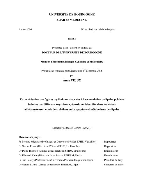
Figure 1: Coupe d’Artère<br />
Les artères répon<strong>de</strong>nt toutes à un modèle commun d’organisation. Leur paroi est constituée <strong>de</strong> trois tuniques qui, <strong>de</strong><br />
l’intérieur vers l’extérieur, sont: l’intima, la média et l’adventice. La tunique interne ou intima possè<strong>de</strong> trois<br />
couches: 1°) l’endothélium reposant sur une lame basale mince, 2°) l'espace subendothelial contenant <strong>de</strong>s fibres<br />
élastiques et <strong>de</strong> collagènes minces ainsi que <strong>de</strong>s protéoglycanes, 3°) <strong>de</strong>s cellules musculaires lisses orientées <strong>de</strong><br />
manière longitudinale. La limitante élastique interne, relativement fine, possè<strong>de</strong> une ban<strong>de</strong> bien délimitée <strong>de</strong> fibres<br />
élastiques. La média est composée <strong>de</strong> cellules musculaires lisses. L’adventice est composée <strong>de</strong> tissu conjonctif<br />
contenant <strong>de</strong>s quantités variable <strong>de</strong> fibres <strong>de</strong> collagène et élastiques.
CONTEXTE BIBLIOGRAPHIQUE Athérosclérose<br />
2. Structure <strong>de</strong> la paroi vasculaire<br />
La paroi artérielle est composée <strong>de</strong> trois couches cellulaires concentriques (Figure 1):<br />
l’intima, la média et l’adventice.<br />
L’intima est la tunique la plus interne et la plus fine. C’est à son niveau que débute le<br />
développement <strong>de</strong> l’athérosclérose. Elle est constituée d’une couche unique <strong>de</strong> cellules<br />
endothéliales, imbriquées les unes dans les autres et formant une couverture étanche. Cet<br />
endothélium exerce différentes fonctions régulatrices telles que la synthèse <strong>de</strong> molécules<br />
vasoactives (NO (monoxy<strong>de</strong> d’azote), endothéline), la thromborésistance (synthèse <strong>de</strong><br />
prostaglandine I2, antithrombine III, activateur tissulaire du plasminogène) ou la régulation <strong>de</strong>s<br />
phénomènes immunitaires. Le sous-endothélium est formé <strong>de</strong> tissu conjonctif fibro-élastique. La<br />
couche sous-endothéliale est le site préférentiel <strong>de</strong> développement <strong>de</strong>s lésions d’athérosclérose et<br />
sert <strong>de</strong> zone <strong>de</strong> stockage <strong>de</strong>s lipoprotéines et <strong>de</strong>s monocytes/macrophages provenant du sang. La<br />
limitante élastique interne (lame <strong>de</strong> fibres élastiques constituées d’élastine) sépare l’intima <strong>de</strong> la<br />
média.<br />
La média est la tunique moyenne, le constituant principal <strong>de</strong> l’artère, elle est la plus épaisse.<br />
Elle est essentiellement constituée par <strong>de</strong>s cellules musculaires lisses, empilées <strong>de</strong> façon<br />
concentrique en couches appelées unités lamellaires. Le nombre <strong>de</strong> ces couches varie suivant le<br />
type d’artère: d’une couche pour les artérioles, à plusieurs couches, pour les artères élastiques.<br />
Chaque unité lamellaire est composée <strong>de</strong> cellules musculaires lisses entourées d’une matrice<br />
extracellulaire constituée <strong>de</strong> protéines fibreuses et élastiques (collagène et élastine) et <strong>de</strong><br />
mucopolysacchari<strong>de</strong>s. Dans sa partie externe, la média reçoit l’irrigation <strong>de</strong>s vasa vasorum 5 <strong>de</strong><br />
l’adventice. La limitante élastique externe (lame d’élastine) sépare la média <strong>de</strong> l’adventice.<br />
L’adventice est la tunique externe, elle se situe entre média et graisse péri-artérielle, elle est<br />
constituée par un tissu conjonctif peu organisé, riche en collagène et en fibres élastiques, et<br />
contenant <strong>de</strong>s fibroblastes et <strong>de</strong>s adipocytes. Elle assure l’ancrage <strong>de</strong> l’artère aux structures<br />
avoisinantes. Un réseau <strong>de</strong> nerfs vasomoteurs non myélinisés rejoint les fibres musculaires lisses <strong>de</strong><br />
la media. Elle est irriguée par <strong>de</strong>s vasa vasorum qui se prolongent dans la partie externe <strong>de</strong> la<br />
media.<br />
5 Vasa vasorum : capillaires dont la paroi n’est constituée que d’une seule cellule endothéliale.<br />
- 17 -
Type I, Lésion initiale<br />
Type II, Strie Lipidique<br />
Type III, Pré-athérome<br />
Type IV, Athérome<br />
Nomenclature<br />
Type V, Fibro-athérome ou plaque simple<br />
-Va: cœur lipidique et chape fibreuse<br />
-Vb: en plus, calcifications<br />
-Vc: cœur lipidique minuscule ou absent<br />
Type VI, Fibro-athérome compliqué<br />
- VIa: ulcération<br />
- VIb: hématome ou hémorragie intraplaque<br />
- VIc: thrombose<br />
Type VII, Lésion calcifiée<br />
Type VIII, Lésion fibreuse<br />
Principales caractéristiques<br />
Dépôt <strong>de</strong> lipi<strong>de</strong>s dans un épaississement <strong>de</strong> l’intima<br />
Cellules spumeuses macrophagiques, esters <strong>de</strong> cholestérol<br />
Accumulation <strong>de</strong> cellules spumeuses dans l’intima<br />
Esters <strong>de</strong> cholestérol<br />
Apparition <strong>de</strong> lipi<strong>de</strong>s extracellulaires<br />
Cellules spumeuses, lipi<strong>de</strong>s extracellulaires<br />
Formation du cœur lipidique sans fibrose (cellules<br />
spumeuses, cristaux <strong>de</strong> cholestérol)<br />
Individualisation <strong>de</strong> la chape fibreuse (collagène fibrillaire I<br />
et III, glycoprotéines, cellules musculaires lisses)<br />
Cœur lipidique (cellules musculaires lisses, lipi<strong>de</strong>s: esters <strong>de</strong><br />
cholestérol, cholestérol libre, cholestérols oxydés,<br />
phospholipi<strong>de</strong>s)<br />
Plaque instable par rupture, pouvant former un thrombus<br />
mural et/ou un hématome à l’intérieur <strong>de</strong> la plaque<br />
Plaque avancée, calcifiée<br />
Plaque avancée, principalement formée <strong>de</strong> collagène<br />
Tableau 2: Définition <strong>de</strong>s lésions d’athérosclérose aux différents sta<strong>de</strong>s selon la classification <strong>de</strong> l’American Heart<br />
Association. (Stary, 2000)
CONTEXTE BIBLIOGRAPHIQUE Athérosclérose<br />
3. Anatomopathologie <strong>de</strong> l’athérosclérose<br />
Les lésions ont été classées en fonction du caractère évolutif dynamique <strong>de</strong> l’athérosclérose<br />
mis en évi<strong>de</strong>nce par <strong>de</strong>s étu<strong>de</strong>s anatomopathologiques et épidémiologiques.<br />
La classification histogénétique proposée sous l’égi<strong>de</strong> <strong>de</strong> l’American Heart Association<br />
(AHA) (Tableau 2) dans le cadre du Committee on Vascular Lesions of the Council on<br />
Atherosclerosis tient compte <strong>de</strong> ce caractère évolutif et reconnaît six principaux types lésionnels<br />
successifs (Stary, 2000).<br />
Un épaississement fibro-musculaire <strong>de</strong> l’intima, présent dès la vie fœtale, se charge durant<br />
l’enfance et l’adolescence <strong>de</strong> cellules spumeuses (type I) pour <strong>de</strong>venir une strie lipidique 6 (type II).<br />
Les lésions <strong>de</strong> type I et II ne désorganisent pas la structure normale <strong>de</strong> l’intima, ne déforment pas<br />
l’artère et ne se manifestent pas cliniquement. Les lésions <strong>de</strong> type I et II sont incluses dans l’histoire<br />
naturelle <strong>de</strong> l’athérosclérose car la séquence <strong>de</strong>s changements histologiques indique que, chez les<br />
individus symptomatiques, les lésions cliniques se développent à partir <strong>de</strong> ces changements au<br />
départ inoffensif. Chez le jeune adulte, la lésion <strong>de</strong> type III, lien histologique entre le type II et<br />
l’athérome (type IV), précè<strong>de</strong> la plaque « simple » fibrolipidique (type IV et V). Ensuite les<br />
acci<strong>de</strong>nts <strong>de</strong> rupture avec thrombose et infiltration hémorragique surviennent (type VI). Aux âges<br />
avancés <strong>de</strong> la vie, on observe <strong>de</strong>s plaques fortement calcifiées (type VII) ou exclusivement<br />
scléreuses (type VIII).<br />
Type I<br />
Première lésion initiale, elle correspond à l’accumulation <strong>de</strong> cellules spumeuses, macrophages<br />
gorgés <strong>de</strong> lipi<strong>de</strong>s modifiés (cholestérol et ses dérivés oxydés appelés oxystérols), dans le sous-<br />
endothélium, plus précisément au niveau <strong>de</strong>s coussinets intimaux. Ces coussinets sont formés<br />
naturellement par la paroi artérielle dans certains territoires, comme les zones d’écoulement<br />
turbulent, sous l’effet <strong>de</strong> forces <strong>de</strong> cisaillement. Il existe également à ce sta<strong>de</strong> un début <strong>de</strong><br />
prolifération <strong>de</strong> cellules musculaires lisses. Il n’y a aucune modification <strong>de</strong> la morphologie <strong>de</strong> la<br />
lumière vasculaire. Cette lésion ne se développera qu’à la suite d’une exposition à divers facteurs <strong>de</strong><br />
risque, elle évoluera vers les stries lipidiques (Type II).<br />
6 Strie lipidique : cellules chargées <strong>de</strong> lipi<strong>de</strong>s organisées en petits amas dans la couche superficielle sous-endothéliale <strong>de</strong><br />
l’intima et formant <strong>de</strong>s lésions visibles macroscopiquement.<br />
- 18 -
CONTEXTE BIBLIOGRAPHIQUE Athérosclérose<br />
Type II (Figure 2)<br />
Il est caractérisé par l’accumulation d’un plus grand nombre <strong>de</strong> cellules spumeuses dans<br />
l’intima <strong>de</strong>s artères. Ces cellules sont organisées en petits amas dans la couche sous-endothéliale <strong>de</strong><br />
l’intima. Ces lésions sont l’évolution du type I. Elles peuvent soit régresser, soit évoluer vers <strong>de</strong>s<br />
lésions plus avancées (type III).<br />
A B<br />
Figure 2: Lésion <strong>de</strong> type II. Artère humaine avec strie lipidique.<br />
A: De nombreuses cellules sont présentes dans l’espace subendothélial: <strong>de</strong>s macrophages spumeux<br />
(flèches) et <strong>de</strong>s lymphocytes (têtes <strong>de</strong> flèches) sous un endothélium intact (E). (Hématoxyline<br />
Eosine, x 200)<br />
B: Une strie lipidique (SL) développée sous un endothélium intact structurellement (flèches). Bien<br />
que la majorité <strong>de</strong>s cellules dans l’espace subendothélial soit <strong>de</strong>s macrophages spumeux, plusieurs<br />
lymphocytes sont aussi présents. La média (M) est intacte. (Hématoxyline Eosine, x 80).<br />
(www.images.md)<br />
Type III<br />
Ce type <strong>de</strong> lésion se caractérise par l’accumulation <strong>de</strong> lipi<strong>de</strong>s extracellulaires sous les cellules<br />
spumeuses.<br />
Type IV<br />
Les lésions <strong>de</strong> type IV représentent les premières lésions avancées à cause d’une sévère<br />
désorganisation intimale, provoquée par le cœur lipidique (Stary et al., 1995). Une accumulation<br />
<strong>de</strong>nse <strong>de</strong> lipi<strong>de</strong>s extracellulaires occupe une région étendue mais bien définie <strong>de</strong> l’intima. Ce type<br />
d’accumulation <strong>de</strong> lipi<strong>de</strong>s extracellulaires est appelé « cœur lipidique » (Stary et al., 1995). La<br />
lésion <strong>de</strong> type IV est aussi connue sous le nom d’athérome. Le cœur lipidique semble se développer<br />
à partir d'une augmentation <strong>de</strong> pools isolés <strong>de</strong> lipi<strong>de</strong>s extracellulaires qui caractérise la lésion <strong>de</strong><br />
type III. Elles peuvent soit <strong>de</strong>venir <strong>de</strong>s lésions compliquées thrombotiques (type VIc), soit évoluer<br />
en plaques fibreuses (type V).<br />
- 19 -<br />
SL
CONTEXTE BIBLIOGRAPHIQUE Athérosclérose<br />
Type V<br />
Ce type <strong>de</strong> lésion correspond à la définition <strong>de</strong> l’OMS citée précé<strong>de</strong>mment. Les lésions <strong>de</strong> type<br />
V sont définies comme <strong>de</strong>s lésions dans lesquelles un tissu conjonctif fibreux s’est formé (Stary et<br />
al., 1995). Quand le nouveau tissu fait partie d’une lésion avec un cœur lipidique, cette lésion peut<br />
être référencée comme un fibroathérome ou une lésion <strong>de</strong> type Va (Figure 3). Une lésion <strong>de</strong> type V,<br />
avec un cœur lipidique ou d’autres parties <strong>de</strong> la lésion, calcifiés, peut être référencée comme une<br />
lésion <strong>de</strong> type Vb (Figures 4-5). Une lésion <strong>de</strong> type V avec un cœur lipidique absent et <strong>de</strong>s lipi<strong>de</strong>s<br />
présents en quantités minimales peut être définie comme une lésion <strong>de</strong> type Vc (Stary et al., 1995).<br />
Dans le cas <strong>de</strong> ces lésions, les artères ont une lumière rétrécie plus que dans le type IV. Comme<br />
avec le type IV, les lésions <strong>de</strong> type V peuvent développer <strong>de</strong>s fissures, <strong>de</strong>s hématomes et/ou une<br />
thrombose pour <strong>de</strong>venir une lésion <strong>de</strong> type VI (Stary et al., 1995). Pour cette raison, ces lésions sont<br />
cliniquement importantes.<br />
Le tissu conjonctif se forme dans et autour <strong>de</strong>s régions <strong>de</strong> l’intima au niveau <strong>de</strong>squelles les<br />
accumulations importantes <strong>de</strong> lipi<strong>de</strong>s extracellulaires (cœur lipidique) désorganisent ou oblitèrent la<br />
structure normale <strong>de</strong> la cellule et <strong>de</strong> la matrice extracellulaire. Le nouveau tissu fibreux est composé<br />
<strong>de</strong> collagène et <strong>de</strong> cellules musculaires lisses riches en réticulum endoplasmique rugueux. Les<br />
capillaires, présents aux limites du cœur lipidique, peuvent être plus grands et plus nombreux que<br />
dans les lésions <strong>de</strong> type IV, ils peuvent être aussi présents dans le tissu nouvellement formé (Stary<br />
et al., 1995).<br />
Les lésions <strong>de</strong> type Va peuvent se présenter sous forme multicouches: plusieurs cœurs<br />
lipidiques, séparés par <strong>de</strong>s couches épaisses <strong>de</strong> tissu fibreux conjonctif, sont empilés irrégulièrement<br />
les uns au <strong>de</strong>ssus <strong>de</strong> autres (Stary et al., 1995). Le terme « fibroathérome multicouches » peut être<br />
utilisé pour décrire cette morphologie.<br />
Les lésions, contenant une gran<strong>de</strong> quantité <strong>de</strong> calcium, ont généralement aussi un tissu fibreux<br />
conjonctif plus important. Les lésions, dans lesquelles la minéralisation est la caractéristique<br />
dominante, sont appelées lésions <strong>de</strong> type Vb (calcifiées) (Figures 4-5) (Stary et al., 1995). Les<br />
lésions calcifiées ont été dénommées lésions <strong>de</strong> type VII.<br />
Dans les lésions <strong>de</strong> type Vc, souvent observées dans les artères <strong>de</strong>s extrémités inférieures,<br />
l’intima normal est remplacée et épaissie avec du tissu fibreux conjonctif, alors que la composante<br />
lipidique est minimale voire absente (Stary et al., 1995). Les lésions fibrotiques ont été dénommées<br />
lésions <strong>de</strong> type VIII.<br />
- 20 -
CONTEXTE BIBLIOGRAPHIQUE Athérosclérose<br />
Les cellules musculaires lisses <strong>de</strong> la média adjacente, <strong>de</strong> l’intima transformée dans les lésions <strong>de</strong><br />
type V, peuvent être désorganisées et leur quantité diminuée. La média et l’adventice adjacente<br />
peuvent contenir <strong>de</strong>s accumulations <strong>de</strong> lymphocytes, macrophages et cellules macrophagiques<br />
spumeuses (Stary et al., 1995).<br />
CL<br />
CF<br />
A B<br />
Figure 3: Lésion <strong>de</strong> type Va: Plaque fibro-lipidique précoce.<br />
A : La média sous le cœur lipidique (CL) apparaît fine avec une fibrose évi<strong>de</strong>nte (F). Les <strong>de</strong>ux<br />
plaques présentent une chape fibreuse (CF) et un cœur lipidique d’importance modérée (CL). Lors<br />
<strong>de</strong> la progression <strong>de</strong>s lésions intermédiaires, une chape fibreuse (CF) d'épaisseur variable se forme<br />
au-<strong>de</strong>ssus <strong>de</strong> la lésion. B :La chape fibreuse est habituellement plus fine à l’endroit où s’accumulent<br />
les cellules spumeuses et les cellules musculaires lisses (CML). Le dépôt <strong>de</strong> tissu fibreux au niveau<br />
d’une plaque contribue significativement à la progression <strong>de</strong> la lésion qui apparaît être régulée par<br />
<strong>de</strong>s facteurs <strong>de</strong> croissance agissant sur les fibroblastes et les cellules musculaires lisses. (Trichrome<br />
<strong>de</strong> Masson, x 80). A: adventice E: endothélium M: média.<br />
(www.images.md)<br />
FC FC<br />
PM<br />
PM<br />
A B<br />
Figure 4: Lésion <strong>de</strong> type Vb.<br />
La plaque fibro-lipidique calcifiée occupe les <strong>de</strong>ux tiers <strong>de</strong> la lumière vasculaire. Un tissu fibromusculaire<br />
s’étend entre la chape fibreuse (CF) <strong>de</strong> la plaque originale et la média (*) à travers une<br />
fissure (F) <strong>de</strong> la chape fibreuse au niveau <strong>de</strong> la région charnière. En plus d’être le site <strong>de</strong> croissance<br />
(recrutement <strong>de</strong> monocytes), les régions charnières <strong>de</strong> la plaque montrent une néovascularisation<br />
(angiogenèse). Des sites <strong>de</strong> calcification (C) sont présents à côté du site <strong>de</strong> prolifération<br />
myofibroblastique (PM). A: adventice ; E: endothélium ; LEI: limitante élastique interne<br />
(Hématoxyline Eosine, A x 40, B x 80)<br />
(www.images.md)<br />
- 21 -<br />
CF<br />
FC<br />
F<br />
CL<br />
PM<br />
CML<br />
PM<br />
LEI
CONTEXTE BIBLIOGRAPHIQUE Athérosclérose<br />
Figure 5: Lésion <strong>de</strong> type Vb – plaque d’athérosclérose aortique avec une calcification étendue.<br />
Bien que la plaque possè<strong>de</strong> un coeur lipidique réduit (CL), la chape fibreuse entière (*) est calcifiée<br />
(flèches). (Hématoxyline Eosine, x 80). A: adventice ; M: média ; (www.images.md)<br />
Type VI<br />
CL<br />
La morbidité et la mortalité <strong>de</strong> l’athérosclérose sont largement dues aux lésions <strong>de</strong> type IV et V<br />
dans lesquelles les ruptures <strong>de</strong> la surface <strong>de</strong> la lésion, les hématomes ou les hémorragies et les<br />
dépôts thrombotiques se sont développés (Stary et al., 1995). Les lésions <strong>de</strong> type IV et V avec une<br />
ou plusieurs <strong>de</strong> ces caractéristiques additionnelles sont classifiées comme <strong>de</strong>s lésions <strong>de</strong> types VI et<br />
appelées aussi lésions compliquées (Stary et al., 1995). Ainsi, la rupture <strong>de</strong> la plaque est définie<br />
comme le type VIa (Figure 6), l’hématome ou l’hémorragie le type VIb (Figure 7) et la thrombose<br />
type VIc (Figure 8) (Stary et al., 1995).<br />
PH<br />
Figure 6: Lésion <strong>de</strong> type VI - Plaque athérosclérotique aortique en rupture.<br />
Les extrémités rompues (flèches) <strong>de</strong> la chape fibreuse (CF) sont considérablement plus épaisses que<br />
le reste <strong>de</strong> l’intima riche en collagène. (Trichrome <strong>de</strong> Masson, x 380) A: adventice ; M: média ; PH:<br />
plaque hémorragique, CL: cœur lipidique. (www.images.md)<br />
- 22 -<br />
CL<br />
CF
CONTEXTE BIBLIOGRAPHIQUE Athérosclérose<br />
DM<br />
Figure 7: Lésion <strong>de</strong> type VIb.<br />
La chape fibreuse (CF) s’amincit au niveau <strong>de</strong> la charnière droite <strong>de</strong> la plaque. Le cœur lipidique<br />
(CL) présente <strong>de</strong>s centres hémorragiques (PH, plaques hemorragiques), bien qu’il n’y ait aucune<br />
communication apparente entre l’amas <strong>de</strong> lipi<strong>de</strong>s et la lumière vasculaire. La rupture <strong>de</strong>s capillaires<br />
à côté du cœur lipidique peut être la source <strong>de</strong> ces hémorragies. Les couches inférieures <strong>de</strong> la média<br />
(M), à côté du cœur lipidique, sont focalement détruites. (Trichrome <strong>de</strong> Masson, x 20) DM:<br />
<strong>de</strong>struction <strong>de</strong> la média. (www.images.md)<br />
TM<br />
CL<br />
Figure 8: Lésion <strong>de</strong> type VIc.<br />
Il y a une rupture <strong>de</strong> la fine chape fibreuse (flèches), avec exposition du noyau nécrotique (NN) à la<br />
circulation générale et une thrombose murale (TM). (Trichrome <strong>de</strong> Masson, x 45) A: adventice ; M:<br />
média ; C: collagène. (www.images.md)<br />
- 23 -<br />
CF<br />
NN<br />
PH
CONTEXTE BIBLIOGRAPHIQUE Athérosclérose<br />
4. Athérogenèse<br />
a. Les différentes théories<br />
Différentes théories concernant l’athérogenèse ont été avancées.<br />
La théorie d'incrustation, émise en 1852 par Karl Von ROKITANSKY, proposait que la<br />
plaque résulte d'un dépôt excessif <strong>de</strong> produits dérivés du sang, principalement <strong>de</strong> la fibrine. On a<br />
retenu <strong>de</strong> cette théorie l'aspect thrombotique <strong>de</strong> l'athérogenèse.<br />
La théorie inflammatoire: basée sur l'observation <strong>de</strong>s modifications que subissait l'intima<br />
(prolifération <strong>de</strong> tissu conjonctif, calcification et dégénérescence graisseuse), cette théorie présentée<br />
par Rudolf VIRCHOW (1821-1902) dans les années 1860 avait été mise à l'écart pendant un peu<br />
plus d'un siècle.<br />
La théorie <strong>de</strong> l'imbibition, dont dérive l'actuelle théorie lipidique, est née en 1908 à la suite<br />
d'expérimentations <strong>de</strong> IGNATOWSKI qui montra la relation, chez le lapin, entre un régime carné et<br />
la survenue <strong>de</strong> l'athérosclérose. IGNATOWSKI cherchait alors à prouver le lien entre régime<br />
alimentaire et apparition <strong>de</strong> la plaque. Et c'est en 1913 qu' ANITSCHOW et CHALATOW ont<br />
montré la responsabilité du cholestérol dans la survenue <strong>de</strong> l'athérosclérose, émettant l'hypothèse<br />
que la formation <strong>de</strong> l'athérome est le résultat d'une imbibition lipidique <strong>de</strong> la paroi artérielle.<br />
En 1973, ROSS et GLOMSET proposent l’hypothèse que l’athérosclérose est une « réponse à<br />
l’effraction endothéliale » (response to injury). Cette hypothèse a rapi<strong>de</strong>ment été révisée et<br />
modifiée. Elle peut être résumée ainsi. 1) Beaucoup <strong>de</strong> facteurs (hyperlipidémie, diabète,<br />
hypertension, tabac, hyperhomocystéinémie …) peuvent endommager et activer l’endothélium. 2)<br />
La dysfonction endothéliale mène à une augmentation <strong>de</strong> la perméabilité, les lipi<strong>de</strong>s et les cellules<br />
circulantes (particulièrement les lymphocytes et les monocytes qui <strong>de</strong>viennent <strong>de</strong>s macrophages<br />
dans la paroi vasculaire) entrent dans l’espace sub-endothélial et forment la lésion initiale <strong>de</strong><br />
l’athérosclérose: la strie lipidique. 3) Comme le nombre <strong>de</strong> cellules augmente en même temps que<br />
l’accumulation <strong>de</strong> lipi<strong>de</strong>s, ceci peut augmenter la rupture <strong>de</strong> l’endothélium menant à <strong>de</strong>s surfaces<br />
thrombogéniques au niveau <strong>de</strong>squelles les plaquettes peuvent adhérer. 4) Les plaquettes, les<br />
macrophages, les cellules endothéliales et probablement les cellules musculaires lisses libèrent <strong>de</strong>s<br />
facteurs <strong>de</strong> croissance provoquant la prolifération <strong>de</strong>s cellules musculaires lisses et <strong>de</strong>s fibroblastes.<br />
5) Ce processus mène à la formation <strong>de</strong> la plaque fibreuse et ultimement aux lésions avancées <strong>de</strong><br />
l’athérosclérose (Schachter, 1997).<br />
- 24 -
CONTEXTE BIBLIOGRAPHIQUE Athérosclérose<br />
Cette hypothèse a été très controversée, car in vivo il n’a jamais été mis en évi<strong>de</strong>nce <strong>de</strong> rupture,<br />
<strong>de</strong> <strong>de</strong>squamation ou <strong>de</strong> dégénérescence <strong>de</strong>s cellules endothéliales au niveau <strong>de</strong>s plaques<br />
d’athérosclérose. L’endothélium conserve en effet son intégrité structurale.<br />
Les étu<strong>de</strong>s expérimentales les plus récentes, ainsi que les observations anatomo-pathologiques<br />
réalisées sur <strong>de</strong>s plaques d’athérome humaines remettent en avant la théorie inflammatoire, avec<br />
la mise en évi<strong>de</strong>nce <strong>de</strong> la présence <strong>de</strong> cellules inflammatoires (macrophages et lymphocytes T)<br />
(Ross, 1999). Ceci démontre donc la survenue <strong>de</strong> processus inflammatoires dans les différentes<br />
lésions d’athérosclérose. Dès les premiers sta<strong>de</strong>s <strong>de</strong> l’athérosclérose, le processus inflammatoire<br />
intervient en réponse à une agression externe. Ensuite, la réaction inflammatoire mal contrôlée<br />
<strong>de</strong>vient chronique et participerait alors à la progression <strong>de</strong> la lésion d’athérosclérose. Enfin, les<br />
complications thrombotiques associées à l’athérosclérose seraient aussi liées à l’état d’inflammation<br />
généralisée généré par la plaque d’athérosclérose (Tedgui & Mallat, 2001).<br />
In fine, ces différentes théories sont proches dans certains domaines et très complémentaires.<br />
Initialement, la paroi vasculaire réagit à différents facteurs pathologiques, l’endothélium conservant<br />
son intégrité structurale. Ces différentes réactions conduisent au développement <strong>de</strong> l’athérosclérose.<br />
Ces facteurs peuvent être d’origine génétique, liés aux composants chimiques du sang ou aux<br />
influences environnementales.<br />
b. Evolution <strong>de</strong> la plaque d’athérosclérose<br />
Les lésions d’athérosclérose se développent dans certaines zones prédisposées <strong>de</strong>s artères <strong>de</strong><br />
gros et moyen calibre, au niveau <strong>de</strong>s zones <strong>de</strong> bifurcations artérielles où les forces <strong>de</strong> cisaillements<br />
« shear stress », forces biomécaniques déterminées par le flux sanguin, la géométrie <strong>de</strong>s vaisseaux<br />
et la viscosité du flui<strong>de</strong>, sont faibles. Il s’agit <strong>de</strong>s bifurcations carotidiennes, <strong>de</strong>s artères<br />
coronariennes, <strong>de</strong> l’aorte abdominale et <strong>de</strong>s artères <strong>de</strong>s membres inférieures. Dans ces zones, où il<br />
existe une stase <strong>de</strong>s particules circulantes, l’exposition locale <strong>de</strong> l’endothélium et <strong>de</strong> la paroi<br />
artérielle aux agents athérogènes est accrue. Ceci explique le développement préférentiel <strong>de</strong>s lésions<br />
d’athérosclérose à ces niveaux.<br />
- 25 -
A<br />
Figure 9: Structure d’une LDL (Low Density Lipoprotein) (Segrest et al., 1994)<br />
A: La LDL est la particule majeure du transport du cholestérol chez l’être humain et dans d’autres espèces, avec un cœur<br />
constitué principalement d’esters <strong>de</strong> cholestérol et d’une petite quantité <strong>de</strong> triglycéri<strong>de</strong>s et une surface composée <strong>de</strong><br />
cholestérol non-estérifié et <strong>de</strong> phosphatidylcholine.<br />
B: L’apolipoprotéine B-100, pratiquement l’unique apoprotéine au niveau <strong>de</strong> la surface, sert <strong>de</strong> ligand pour le récepteur<br />
aux LDL qui favorise la consommation <strong>de</strong>s LDL par les cellules. Elle est constituée <strong>de</strong> 3 hélices α et <strong>de</strong> 2 feuillets β.<br />
B
CONTEXTE BIBLIOGRAPHIQUE Athérosclérose<br />
On peut ainsi diviser l'évolution <strong>de</strong> la plaque d’athérosclérose en différentes étapes:<br />
1. Pénétration <strong>de</strong>s LDLs (low <strong>de</strong>nsity lipoprotein) au niveau <strong>de</strong> l'intima.<br />
2. Oxydation <strong>de</strong> ces LDLs.<br />
3. Activation <strong>de</strong>s cellules endothéliales ; adhésion <strong>de</strong>s monocytes à l'endothélium et<br />
pénétration <strong>de</strong> ces monocytes au niveau <strong>de</strong> l'intima.<br />
4. Formation <strong>de</strong>s cellules spumeuses à partir <strong>de</strong>s macrophages et <strong>de</strong>s cellules musculaires lisses<br />
(CMLs).<br />
5. Prolifération <strong>de</strong>s cellules musculaires lisses (CMLs) et migration <strong>de</strong> ces CMLs <strong>de</strong> la media<br />
vers l'intima.<br />
6. Sécrétion <strong>de</strong> collagène, <strong>de</strong> fibres élastiques et <strong>de</strong> protéoglycanes par les CMLs.<br />
7. Accumulation <strong>de</strong> tissu conjonctif, <strong>de</strong> lipi<strong>de</strong>s, <strong>de</strong> CML et <strong>de</strong> cellules spumeuses.<br />
8. Formation du noyau lipidique à partir <strong>de</strong>s éléments lipidiques accumulés.<br />
9. Ulcération <strong>de</strong> la paroi vasculaire et mise à nu du sous-endothélium.<br />
10. Adhésion et activation plaquettaire provoquant une thrombose.<br />
Ainsi les LDLs (Figure 9), les cellules endothéliales, les monocytes et les cellules musculaires<br />
lisses sont les différents acteurs <strong>de</strong> l’athérosclérose.<br />
Le rôle <strong>de</strong>s lipoprotéines <strong>de</strong> basse <strong>de</strong>nsité dans l’athérosclérose<br />
Les lipoprotéines transportent la majeure partie <strong>de</strong>s lipi<strong>de</strong>s dans l’organisme. Ces<br />
lipoprotéines sont constituées d’un cœur hydrophobe entouré par une couronne <strong>de</strong> phospholipi<strong>de</strong>s,<br />
cholestérol et protéines (Figure 9). Les protéines sont <strong>de</strong>s apoprotéines. Elles solubilisent les lipi<strong>de</strong>s<br />
hydrophobes et jouent un rôle majeur dans le ciblage cellulaire et le métabolisme <strong>de</strong>s lipoprotéines.<br />
On classe les lipoprotéines en fonction <strong>de</strong> leur <strong>de</strong>nsité. Des moins <strong>de</strong>nses au plus <strong>de</strong>nses, on<br />
distingue : les chylomicrons, les lipoprotéines <strong>de</strong> très basse <strong>de</strong>nsité (Very Low Density<br />
Lipoproteins : VLDL), les lipoprotéines <strong>de</strong> <strong>de</strong>nsité intermédiaire (Intermediate Density<br />
Lipoproteins : IDL), les lipoprotéines <strong>de</strong> basse <strong>de</strong>nsité (LDL), et les lipoprotéines <strong>de</strong> haute <strong>de</strong>nsité<br />
(High Density Lipoproteins : HDL). Des transferts <strong>de</strong> lipi<strong>de</strong>s ou d’apoprotéines sont possibles entre<br />
les différentes lipoprotéines.<br />
Des étu<strong>de</strong>s in vitro ont montrées les propriétés athérogènes <strong>de</strong>s LDLs petites et <strong>de</strong>nses. Ces<br />
<strong>de</strong>rnières possè<strong>de</strong>nt en effet une affinité plus faible pour le récepteur aux LDLs et une résistance<br />
plus faible au stress oxydant.<br />
- 26 -
.•<br />
-O - O •<br />
-O - O -H<br />
•<br />
RH<br />
R •<br />
-<br />
H +<br />
Résonance<br />
+ O 2<br />
Fragmentation<br />
Produits secondaires<br />
(Notamment aldéhy<strong>de</strong>s, MDA, HNE)<br />
COOH<br />
Aci<strong>de</strong><br />
arachidonique<br />
COOH<br />
Radical<br />
allylique R• COOH<br />
COOH<br />
Diène<br />
conjugué<br />
Radical<br />
peroxy<strong>de</strong><br />
ROO •<br />
COOH<br />
Hydroperoxy<strong>de</strong><br />
(HPETE)<br />
Figure 10: Schéma général <strong>de</strong> la peroxydation lipidique<br />
En exemple, l’aci<strong>de</strong> arachidonique, composant polyinsaturé <strong>de</strong>s phospholipi<strong>de</strong>s et <strong>de</strong>s esters <strong>de</strong> cholestérol <strong>de</strong>s LDL, est<br />
transformé en hydroperoxy<strong>de</strong> lipidique, l’aci<strong>de</strong> hydroperoxyeicosatétranoïque (HPETE), suite à la perte d’un atome<br />
d’hydrogène lors d’une attaque radicalaire et <strong>de</strong> l’addition <strong>de</strong> l’oxygène moléculaire. La fragmentation <strong>de</strong> cet<br />
hydroperoxy<strong>de</strong> peut ensuite donner lieu à la formation <strong>de</strong> produits secondaires tels que <strong>de</strong>s aldéhy<strong>de</strong>s à courte chaîne<br />
possédant <strong>de</strong>s activités chimiques et biologiques élevées. Le radical R• peut subir un nouveau processus <strong>de</strong><br />
peroxydation lipidique. HNE : HydroxyNonEnal ; MDA: MalonDiAldéhy<strong>de</strong>
CONTEXTE BIBLIOGRAPHIQUE Athérosclérose<br />
La traversée <strong>de</strong> l’endothélium vasculaire par les LDLs initie le processus d’athérogenèse. La<br />
pénétration <strong>de</strong>s LDLs dans l’espace sous-endothélial est inversement proportionnelle à leur taille, ce<br />
qui fait jouer un rôle prépondérant aux LDLs <strong>de</strong>nses et <strong>de</strong> petites tailles dans l’athérosclérose.<br />
L’oxydation <strong>de</strong>s LDLs est une étape prépondérante dans l’athérogenèse. Elle se produit<br />
majoritairement in situ, dans la paroi (Steinberg, 1997). On ne retrouve en effet que <strong>de</strong> très faibles<br />
quantités <strong>de</strong> LDLs oxydées (LDLox) circulantes alors qu’elles sont présentes en abondance dans la<br />
plaque d’athérosclérose (Chisolm & Steinberg, 2000). L’oxydation <strong>de</strong>s composants lipidiques ou<br />
peroxydation lipidique se déroule en plusieurs étapes (Figure 10). Il se produit tout d’abord une<br />
phase d’initiation où les radicaux oxygénés s’attaquent aux lipi<strong>de</strong>s <strong>de</strong>s LDLs. Les lipi<strong>de</strong>s oxydés<br />
sont principalement ceux présentant une double liaison.<br />
Dans une secon<strong>de</strong> phase, il y a une propagation, aux autres lipi<strong>de</strong>s, <strong>de</strong>s modifications chimiques<br />
induites par l’oxydation <strong>de</strong>s lipi<strong>de</strong>s insaturés. Cette <strong>de</strong>uxième phase dépend en partie <strong>de</strong> l’enzyme<br />
PAF-AH (Platelet Activating Factor-Acetylhydrolase ou lipoprotein-associated phospholipase A2)<br />
qui possè<strong>de</strong> une activité phospholipase A2 (Navab et al., 1996). Ceci conduit dans une troisième<br />
phase à la dégradation et à la libération <strong>de</strong> fragments lipidiques. Il y a alors formation <strong>de</strong> peroxy<strong>de</strong>s<br />
et d’aldéhy<strong>de</strong>s <strong>de</strong> lipi<strong>de</strong>s. Les aldéhy<strong>de</strong>s formés peuvent alors se lier à l’apolipoprotéine ApoB100<br />
(Figure 9), ligand clé du récepteur cellulaire <strong>de</strong>s LDLs, via <strong>de</strong>s résidus lysine ce qui modifie son<br />
activité physiologique. Il s’ensuit une modification covalente <strong>de</strong> l’apoB100 avec augmentation <strong>de</strong><br />
sa charge négative, entraînant une perte <strong>de</strong> la reconnaissance par le récepteur <strong>de</strong>s LDLs natives,<br />
mais lui conférant la capacité <strong>de</strong> se lier aux récepteurs « scavenger » (SRA-A et CD 36) (Berliner et<br />
al., 1995).<br />
Les forces <strong>de</strong> cisaillement influencent directement l’intégrité <strong>de</strong> l’endothélium et ainsi sa<br />
perméabilité, ceci est en partie du à la modulation <strong>de</strong>s fonctions contractiles du cytosquelette. Des<br />
forces <strong>de</strong> faible intensité peuvent ainsi augmenter le temps <strong>de</strong> passage <strong>de</strong>s LDLs et ainsi favoriser<br />
leur pénétration dans l’endothélium (Van<strong>de</strong>rLaan et al., 2004). Suite à leur pénétration et à leur<br />
rétention, les LDLox s’accumulent dans l’espace sous-endothélial, déclenchant le recrutement et<br />
l’infiltration <strong>de</strong> monocytes circulants dans l’intima, conduisant à la constitution <strong>de</strong> stries lipidiques<br />
à la surface luminale (Ross, 1999 ; Nor<strong>de</strong>stgaard & Nielson, 1994 ; Boren et al., 2000). Des forces<br />
<strong>de</strong> cisaillements faibles, comme il en existe au niveau <strong>de</strong>s zones <strong>de</strong> prédisposition <strong>de</strong><br />
l’athérosclérose, favorisent donc la consommation <strong>de</strong>s LDLox par les macrophages (Niwa et al.,<br />
2004).<br />
- 27 -
CONTEXTE BIBLIOGRAPHIQUE Athérosclérose<br />
Le recrutement <strong>de</strong>s monocytes<br />
En conditions normales, le recrutement <strong>de</strong> monocytes au sein <strong>de</strong> la paroi artérielle et leur<br />
différenciation en macrophages est un phénomène protecteur qui permet l’élimination <strong>de</strong>s LDLox<br />
cytotoxiques et pro-inflammatoires ainsi que <strong>de</strong>s cellules apoptotiques. Mais l’accumulation <strong>de</strong><br />
macrophages chargés en LDLox peut induire la formation d’une lésion athérosclérotique. Le<br />
recrutement <strong>de</strong>s monocytes aux sites <strong>de</strong> dysfonction endothéliale est régulé par <strong>de</strong>s molécules<br />
d’ahésion exprimées à la surface <strong>de</strong> l’endothélium vasculaire. Les forces <strong>de</strong> cisaillement, faibles,<br />
influencent l’adhésion <strong>de</strong>s leucocytes à l’endothélium activé, en favorisant l’augmentation <strong>de</strong><br />
l’expression <strong>de</strong>s molécules d’adhésion (Cunningham & Gotlieb, 2005). Ces molécules d’ahésion<br />
appartiennent à la famille <strong>de</strong>s sélectines, aux ligands <strong>de</strong>s sélectines, aux intégrines et aux<br />
immunoglobulines (Huo & Ley, 2001). Les LDLox sont également <strong>de</strong>s stimuli majeurs pour<br />
l’expression <strong>de</strong> ces molécules d’adhésion. La régulation transcriptionnelle <strong>de</strong>s molécules<br />
d’adhérence est sous la dépendance du facteur <strong>de</strong> transcription NF-κB (Nuclear Factor-kappa B),<br />
qui peut être activé par les LDLox (Berliner et al., 1995) ou par les cytokines inflammatoires TNF-<br />
α (Tumor Necrosis Factor α) et IL-1β (interleukin-1β) (Tedgui & Mallat, 2001).<br />
*Les sélectines : « le rolling »<br />
Les sélectines sont responsables du phénomène <strong>de</strong> « rolling » <strong>de</strong>s leucocytes à la surface <strong>de</strong><br />
l’endothélium (Giddings, 2005). Ce phénomène correspond à l’établissement puis à la rupture <strong>de</strong>s<br />
liaisons établies entre les sélectines, exprimées par les cellules endothéliales, avec leurs ligands<br />
exprimés, à la surface <strong>de</strong>s leucocytes, et se traduit par un ralentissement <strong>de</strong>s leucocytes qui roulent à<br />
la surface <strong>de</strong> l’endothélium. La E-sélectine et la P-sélectine sont exprimées au niveau <strong>de</strong><br />
l’endothélium activé, alors que la L-sélectine est constitutivement exprimée par l’endothélium.<br />
Le principal ligand <strong>de</strong> ces sélectines est la P-Selectin Glycoprotein-1 (PSGL-1). L’inhibition<br />
du phénomène <strong>de</strong> « rolling » se traduit par une forte diminution <strong>de</strong> l’adhésion <strong>de</strong>s leucocytes au site<br />
inflammatoire. Ainsi, sur un modèle <strong>de</strong> caroti<strong>de</strong> isolée et perfusée <strong>de</strong> souris athérosclérotiques<br />
ApoE -/- , Ramos et al. ont observé une forte diminution du « rolling » et <strong>de</strong> l’attachement <strong>de</strong>s<br />
leucocytes mononuclées en présence d’anticorps anti P-sélectine ou anti PSGL-1 (Ramos et al.,<br />
1999). D’autre part, la taille <strong>de</strong>s plaques d’athérome observées à 4 mois au niveau <strong>de</strong> l’aorte <strong>de</strong>s<br />
souris obtenues par croisement <strong>de</strong> souris P-sélectine -/- et apoE -/- est 3,5 fois plus faible que celle <strong>de</strong><br />
souris apoE -/- (Dong et al., 2000).<br />
- 28 -
CONTEXTE BIBLIOGRAPHIQUE Athérosclérose<br />
Le rôle <strong>de</strong> la E-sélectine dans la formation <strong>de</strong> la plaque d’athérome est controversée. Ainsi<br />
O’Brian a observé en immunohistochimie sur <strong>de</strong>s aortes humaines que la E-sélectine est exprimée<br />
dans 32% <strong>de</strong>s plaques et est absente <strong>de</strong>s aortes saines (O’Brian et al., 1996). En revanche, Iiyama et<br />
al. n’ont pas détecté la présence <strong>de</strong> la protéine ou <strong>de</strong> son ARNm chez la souris et chez le lapin<br />
(Iiyama et al., 1999).<br />
*Les immunoglobulines et VCAM-1 : adhésion et transmigration<br />
L’adhésion <strong>de</strong>s leucocytes à travers l’endothélium nécessite une interaction entre les<br />
immunoglobulines, molécules d’adhésion exprimées à la surface <strong>de</strong> l’endothélium vasculaire, et<br />
leurs ligands, <strong>de</strong>s intégrines exprimées par les leucocytes. Cette liaison permet la transition entre la<br />
phase <strong>de</strong> « rolling » et une adhésion ferme <strong>de</strong>s leucocytes à la surface <strong>de</strong> l’endothélium. Les<br />
molécules <strong>de</strong> la superfamille <strong>de</strong>s immunoglobulines sont <strong>de</strong>s protéines transmembranaires<br />
possédant une série <strong>de</strong> motifs extracellulaires communs à ceux <strong>de</strong>s immunoglobulines G (IgG-like<br />
domains) et un court domaine cytoplasmique. Parmi les molécules d’adhésion appartenant à cette<br />
superfamille, on distingue les molécules d’adhésion intercellulaires 1, 2 et 3 (ICAM-1, 2, 3), la<br />
molécule d’adhésion endothéliale et plaquettaire 1 (PECAM-1 ou CD31) et la molécule <strong>de</strong>s cellules<br />
vasculaires 1 (VCAM-1).<br />
Leurs ligands, les intégrines, sont <strong>de</strong>s hétérodimères composés d’une sous-unité β liée <strong>de</strong><br />
manière non covalente à une sous-unité α. Les intégrines formées avec la sous-unité β1 (CD29,<br />
Cluster Differention) sont aussi appelées antigènes très tardifs (VLA, Very Late Antigen) car elles<br />
sont exprimées tardivement par les lymphocytes à la suite <strong>de</strong> leur activation. L’une d’elles, VLA-4<br />
(α4β1) est le principal ligand <strong>de</strong> VCAM-1. ICAM-1, 2 et 3 (endothelial InterCellular Adhesion<br />
Molecule 1) sont <strong>de</strong>s ligands <strong>de</strong> l’intégrine LFA-1 (αLβ2) (Leucocyte Function Associated-1), et<br />
ICAM-1 et 2 lient également Mac-1 (αMβ2). PECAM-1 (platelet/endothelial cell adhesion<br />
molecule-1) est un ligand <strong>de</strong> l’intégrine αVβ3 mais réalise principalement <strong>de</strong>s liaisons homotypiques<br />
avec <strong>de</strong>s molécules PECAM-1 exprimées à la surface d’autres cellules. Les expressions <strong>de</strong> ICAM-1<br />
et VCAM-1 sont augmentées ou induites lors d’une activation endothéliale. En effet, <strong>de</strong>s molécules,<br />
présentes dans les LDLox comme la lysophosphatidylcholine, peuvent augmenter la transcription<br />
du gène VCAM-1 (Kume et al., 1992). En revanche, PECAM-1 est constitutivement exprimée par<br />
l’endothélium.<br />
- 29 -
CONTEXTE BIBLIOGRAPHIQUE Athérosclérose<br />
Les trois principales molécules d’ahésion dont l’expression est induite au niveau <strong>de</strong><br />
l’endothélium lors <strong>de</strong> la formation <strong>de</strong> la plaque d’athérome sont donc la P-sélectine, VCAM-1 et<br />
ICAM-1 (Huo & Ley, 2001).<br />
La transmigration endothéliale ou diapédèse est un processus rapi<strong>de</strong> durant lequel le<br />
leucocyte émet un pseudopo<strong>de</strong> pour franchir la barrière endothéliale. Il nécessite le désassemblage<br />
du cytosquelette du leucocyte du côté apical <strong>de</strong> l’endothélium puis son réassemblage dans l’espace<br />
sous-endothélial. Deux interactions moléculaires homophiliques jouent un rôle majeur lors <strong>de</strong> la<br />
diapédèse : la liaison PECAM-1/PECAM-1 et la liaison CD99-CD99. Des étu<strong>de</strong>s réalisées in vitro<br />
et in vivo indiquent que la suppression ou le blocage <strong>de</strong> l’une ou l’autre <strong>de</strong> ces molécules stoppe la<br />
diapédèse <strong>de</strong>s leucocytes (Schenkel et al., 2002 ; Bogen et al., 1994). Cependant, lorsque PECAM-<br />
1 est bloquée, les leucocytes sont stoppés en surface <strong>de</strong> l’endothélium alors que lorsque CD99 est<br />
bloqué, ils sont stoppés à l’intérieur <strong>de</strong> la jonction endothéliale, ce qui indique que CD99 est<br />
impliqué lors d’une étape distale <strong>de</strong> celle impliquant PECAM-1. Les molécules d’adhésion Jam-A<br />
(Junctional adhesion molecule), B et C et la Vascular Endothelial Cadherin (VE-Cadherin) sont<br />
également impliquées dans ce processus.<br />
Le signal <strong>de</strong> transmigration nécessite une augmentation transitoire <strong>de</strong> la concentration<br />
calcique à l’intérieur <strong>de</strong>s cellules endothéliales adjacentes au site <strong>de</strong> transmigration (Huang et al.,<br />
1993). Ce signal active la chaîne légère <strong>de</strong> la myosine et induit la rétractation <strong>de</strong>s cellules<br />
endothéliales. Il est induit par <strong>de</strong>s protéines cationiques solubles relarguées par les leucocytes<br />
activés ainsi que par la liaison <strong>de</strong>s leucocytes aux sélectines et à VCAM-1.<br />
*Les cytokines 7<br />
L’IL-1β, le TNF-α, et l’IFN-γ favorisent le recrutement <strong>de</strong>s monocytes, <strong>de</strong>s lymphocytes T<br />
et Natural Killer au site inflammatoire en stimulant la libération <strong>de</strong>s facteurs chimioattractifs :<br />
MCP-1 (Macrophage Chemotactic Protein 1), l’interleukine 8 (IL-8) (se lie aux récepteurs CXC 2<br />
(CXCR-2)) (Glass & Witztum, 2001 ; Libby, 2002) et la fractalkine par les macrophages, les<br />
cellules endothéliales et les cellules musculaires lisses (Van<strong>de</strong>rlaan & Reardon, 2005). Le<br />
recrutement <strong>de</strong>s monocytes est aussi amplifié par les plaquettes activées, via RANTES (Regulated<br />
7 Les cytokines, petites protéines ou glycoprotéines solubles, sont <strong>de</strong>s molécules <strong>de</strong> signalisation intercellulaire qui sont<br />
sécrétées dans le milieu interstitiel par <strong>de</strong> nombreuses cellules activées en réponse à une agression. Elles régulent la<br />
prolifération, la migration, la différenciation ou l’apoptose <strong>de</strong>s cellules.<br />
- 30 -
lumière<br />
paroi<br />
lumière<br />
paroi<br />
monocyte<br />
matrice<br />
extracellulaire<br />
Différenciation<br />
M-CSF<br />
lumière<br />
vasculaire<br />
LDL<br />
Oxydation<br />
<strong>de</strong>s LDL<br />
LDL ox<br />
macrophage<br />
cellules<br />
endothéliales<br />
HDL<br />
cellules<br />
spumeuses<br />
Cellules<br />
musculaires<br />
lisses<br />
Figure 11: Initiation du développement d’une lésion (Glass & Witztum, 2001)<br />
Les LDL sont sujets à <strong>de</strong>s modifications oxydatives dans l’espace sous-endothélial. Les monocytes s’attachent aux<br />
cellules endothéliales qui ont été induites pour exprimer <strong>de</strong>s molécules d’adhésion par les cytokines et les LDL<br />
modifiées <strong>de</strong> manière minime. Les monocytes adhérents migrent dans l’espace sous-endothélial et se différencient en<br />
macrophages. La consommation <strong>de</strong>s LDL oxydées (LDLox) via les récepteurs scavengers mène à la formation <strong>de</strong><br />
cellules spumeuses.<br />
matrice<br />
extracellulaire<br />
IL-4<br />
IL-10<br />
IL-13<br />
Th2<br />
IL-4<br />
IL-10<br />
cellules<br />
endothéliales<br />
cytokines<br />
IFNγ<br />
CD40<br />
Th1<br />
Chape<br />
Fibreuse<br />
Cellules<br />
macrophagiques<br />
spumeuses<br />
Cellules<br />
musculaires<br />
lisses<br />
Figure 12: Progression <strong>de</strong> la lésion (Glass & Witztum, 2001)<br />
Les interactions entre les cellules macrophagiques spumeuses et les cellules Th1 et Th2 établissent un processus<br />
inflammatoire chronique. Les cytokines, secrétées par les lymphocytes et les macrophages, exercent <strong>de</strong>s effets pro- et<br />
anti-athérogèniques sur chaque élément du mur vasculaire. Les cellules musculaires lisses migrent <strong>de</strong> la portion<br />
médiane du mur artériel, prolifèrent et secrètent <strong>de</strong>s protéines <strong>de</strong> matrice extracellulaire qui forment une plaque fibreuse
CONTEXTE BIBLIOGRAPHIQUE Athérosclérose<br />
on Activation Normal T cell Expressed and Secreted) et le facteur plaquettaire 4 (CXCL4), ainsi que<br />
par la fraction active du complément C5a. D’autres chemokines seraient également impliquées<br />
telles que l’éotaxine, GRO-α (Growth Regulated Oncogne), Mig, IP10, I-TAC ou SDF-1α (Stroma<br />
cell-Derived Factor 1) (Raines & Ferri, 2005).<br />
Formation <strong>de</strong>s cellules spumeuses et progression <strong>de</strong> l’athérosclérose<br />
Les monocytes adhérents pénètrent dans l’intima à travers les jonctions inter-endothéliales.<br />
Une fois que les monocytes circulant sont dans l’espace sous-endothélial, ils se différencient en<br />
macrophages sous l’influence du facteur hématopoïétique <strong>de</strong> différenciation et <strong>de</strong> prolifération <strong>de</strong>s<br />
monocytes, le M-CSF (Monocyte-Colony Stimulating Factor) (Figure 11) (Tedgui & Mallat, 2001).<br />
Les macrophages captent par leurs récepteurs « éboueurs » (scavengers) les LDLox. Cette<br />
internalisation <strong>de</strong>s LDLs aboutit à la formation <strong>de</strong> cellules gorgées <strong>de</strong> lipi<strong>de</strong>s, les cellules<br />
spumeuses qui seront à l’origine <strong>de</strong> la strie lipidique, première étape <strong>de</strong> la lésion athéromateuse<br />
(Figure 11).<br />
En réponse à la libération <strong>de</strong> chimiokines et <strong>de</strong> cytokines (PDGF: Platelet-Derived Growth<br />
Factor), M-CSF, IL-1β), par les plaquettes, les cellules endothéliales et les macrophages <strong>de</strong> la<br />
plaque d’athérome (Ross, 1993), les cellules musculaires lisses présentes dans la média se<br />
multiplient et migrent <strong>de</strong> la média vers la zone endommagée (Figure 12). Certaines cellules<br />
musculaires lisses accumulent <strong>de</strong>s lipi<strong>de</strong>s et <strong>de</strong>viennent <strong>de</strong>s cellules spumeuses. Ceci définit le<br />
passage d’une strie lipidique simple à une lésion plus complexe. Ces cellules musculaires lisses<br />
synthétisent une matrice extracellulaire qui mène au développement <strong>de</strong> la chape fibreuse (Figure<br />
12). Cette phase <strong>de</strong> développement lésionnel est influencée par les interactions entre les<br />
monocytes/macrophages et les lymphocytes T provoquant un large spectre <strong>de</strong> réponses cellulaires et<br />
humorales et l’acquisition <strong>de</strong>s caractéristiques d’un état inflammatoire chronique (Glass &<br />
Witztum, 2001). Les différentes réponses immunologiques régulent la progression <strong>de</strong> la plaque. En<br />
effet, elles peuvent exercer <strong>de</strong>s effets pro-athérogènes (IFNγ stimule la production par les<br />
macrophages <strong>de</strong> cytokines pro-inflammatoires et augmente l’expression <strong>de</strong> molécule <strong>de</strong> CMH<br />
(complexe majeur d’histocompatibilité) <strong>de</strong> classe 2) et anti-athérogènes (IFNγ réduit l’expression<br />
du récepteur scavenger à la surface <strong>de</strong>s monocytes) (Glass & Witztum, 2001).<br />
- 31 -
lumière<br />
paroi<br />
Cellules<br />
spumeuses<br />
macrophagiques<br />
plaquettes<br />
Cellules<br />
spumeuses<br />
dérivées<br />
<strong>de</strong>s SMC<br />
Cœur<br />
nécrotique<br />
Cellules<br />
musculaires<br />
lisses<br />
Figure 13: Rupture <strong>de</strong> plaque et Thrombose (Glass et Witztum, 2001)<br />
La nécrose <strong>de</strong>s cellules spumeuses dérivées <strong>de</strong>s cellules musculaires lisses et <strong>de</strong>s macrophages mène à la<br />
formation d’un cœur nécrotique et à l’accumulation <strong>de</strong> cholestérol extracellulaire. La sécrétion par les<br />
macrophages <strong>de</strong> métalloprotéinases <strong>de</strong> matrice et la néovascularisation contribuent à la rupture <strong>de</strong> plaque. La<br />
rupture <strong>de</strong> plaque expose les composants sanguins au facteur tissulaire, à l’initiation <strong>de</strong> la coagulation, au<br />
recrutement <strong>de</strong>s plaquettes et à la formation d’un thrombus.
CONTEXTE BIBLIOGRAPHIQUE Athérosclérose<br />
Les principaux lymphocytes T, présents au niveau <strong>de</strong> la plaque, sont les lymphocytes T<br />
auxiliaires ou helper et plus particulièrement <strong>de</strong>s Th1 (T helper type 1). La différenciation <strong>de</strong>s Th1<br />
requièrt l’intervention <strong>de</strong>s cellules <strong>de</strong>ndritiques elles-même ainsi que différentes molécules comme<br />
l’interleukine-12, et l’interleukine-18. Ces <strong>de</strong>rnières sont exprimées au niveau <strong>de</strong> la plaque et sont<br />
impliquées dans le maintient <strong>de</strong> l’inflammation au niveau <strong>de</strong> la plaque et <strong>de</strong> l’instabilité <strong>de</strong> celle-ci<br />
(Mallat et al., 2005). Les réponses <strong>de</strong>s Th1 seraient ainsi impliqées dans le développement et la<br />
progression <strong>de</strong> l’athérosclérose. Les Th2, moins présents au niveau <strong>de</strong> la plaque, peuvent avoir <strong>de</strong>s<br />
effets anti-athérogènes (en antagonisant les effets <strong>de</strong>s Th1 et en produisant <strong>de</strong>s anticorps anti-<br />
LDLox) et pro-athérogènes selon les différentes étapes <strong>de</strong> développement <strong>de</strong> la plaque (Mallat et<br />
al., 2005). Récemment, un nouveau sous-type <strong>de</strong> cellules T, appelé cellules T régulatrices, a été<br />
montré pour être un inhibiteur <strong>de</strong> l’athérosclérose et pourrait constituer une cible thérapeutique<br />
(Ait-Oufella, 2006).<br />
La présence <strong>de</strong> fibres <strong>de</strong> collagène interstitiel au niveau du tissu conjonctif fibreux, formé<br />
par les cellules musculaires lisses, et les néo-capillaires qui vascularisent l’intima participent à la<br />
stabilisation <strong>de</strong> la plaque. La mort <strong>de</strong>s cellules spumeuses dérivées <strong>de</strong>s macrophages et <strong>de</strong>s cellules<br />
musculaires lisses, provoquée entre autre par les stérols oxydés, participe à la formation d’un cœur<br />
nécrotique et à l’accumulation <strong>de</strong> cholestérol extracellulaire (Figure 13) (Glass & Witztum, 2001).<br />
Les métalloprotéinases, endopeptidases dirigées contre les différents constituants <strong>de</strong> la matrice<br />
extracellulaire, agissent sur la stabilité <strong>de</strong> la plaque en dégradant les protéines <strong>de</strong> la matrice<br />
extracellulaire. La mise à nu <strong>de</strong> l’endothélium, après la fissuration <strong>de</strong> la plaque, entraîne l’adhésion<br />
<strong>de</strong>s plaquettes, puis leur agrégation, autour <strong>de</strong> la lésion (Figure 13). Les plaques à risque en vue <strong>de</strong><br />
rupture, contiennent généralement un cœur lipidique acellulaire <strong>de</strong>veloppé, avec un contenu réduit<br />
en collagène, très peu <strong>de</strong> cellules musculaires lisses synthétisant du collagène, une chape fibreuse<br />
réduite, une inflammation au niveau <strong>de</strong> l’adventice et <strong>de</strong> l’intima, avec <strong>de</strong>struction <strong>de</strong>s membranes<br />
élastiques internes et externes, une néovascularisation augmentée au niveau <strong>de</strong> la plaque et un<br />
remo<strong>de</strong>lage du mur vasculaire (Shah, 2005).<br />
- 32 -
CONTEXTE BIBLIOGRAPHIQUE Athérosclérose<br />
5. Facteurs <strong>de</strong> risque<br />
Les facteurs <strong>de</strong> risque sont <strong>de</strong>s variables indépendantes, qui sont déterminées lors <strong>de</strong> gran<strong>de</strong>s<br />
étu<strong>de</strong>s <strong>de</strong> cohorte et répon<strong>de</strong>nt à un ensemble <strong>de</strong> propriétés: relation linéaire <strong>de</strong> type dose effet avec<br />
le risque vasculaire, cohérence avec <strong>de</strong>s données <strong>de</strong> physiopathologie et expérimentales,<br />
indépendance entre les différents facteurs.<br />
Les facteurs <strong>de</strong> risque <strong>de</strong> l’athérosclérose font intervenir <strong>de</strong>s situations physiologiques (âge,<br />
sexe, hérédité), <strong>de</strong>s habitu<strong>de</strong>s <strong>de</strong> vie (alimentation, tabac) ou pathologiques (hypercholestérolémie,<br />
diabète, hypertension).<br />
non modifiables<br />
- 33 -<br />
modifiables<br />
Sexe masculin Tabac<br />
Age Hypertension artérielle<br />
Ménopause<br />
Antécé<strong>de</strong>nts personnels<br />
Facteurs <strong>de</strong> risque cardio-vasculaire<br />
Dyslipidémies<br />
LDLc<br />
Triglycéri<strong>de</strong>s<br />
HDLc<br />
Lp(a)<br />
Insulino-résistance<br />
Obésité abdominale<br />
Diabète<br />
Antécé<strong>de</strong>nts familiaux Sé<strong>de</strong>ntarité<br />
Tableau 3: Principaux facteurs <strong>de</strong> risque cardio-vasculaire.<br />
Lp(a) : lipoprotéine a<br />
L’âge et le sexe: La prévalence <strong>de</strong> la maladie coronaire et <strong>de</strong> l’artériopathie <strong>de</strong>s membres<br />
inférieurs augmente <strong>de</strong> façon nette à partir <strong>de</strong> 45 ans pour atteindre 6 à 7% à l’âge <strong>de</strong> 60 ans<br />
(Emmerich & Bruneval, 2000). Le sexe masculin est un facteur particulier car les femmes sont<br />
protégées jusqu’à l’âge <strong>de</strong> la ménopause du fait d’un meilleur profil lipidique secondaire à<br />
l’imprégnation oestroprogestative, à 45 ans le risque d’un acci<strong>de</strong>nt coronaire est dix fois supérieur<br />
chez l’homme (Emmerich & Bruneval, 2000). Cette protection particulière <strong>de</strong>s femmes n’est tout <strong>de</strong><br />
fois pas prolongée par les traitements substitutifs hormonaux <strong>de</strong> la ménopause (Hulley et al., 1998).
CONTEXTE BIBLIOGRAPHIQUE Athérosclérose<br />
Le tabac: Des étu<strong>de</strong>s épidémiologiques supporte l’évi<strong>de</strong>nce que la consommation <strong>de</strong><br />
cigarettes que cela soit chez les hommes ou chez les femmes augmente l’inci<strong>de</strong>nce <strong>de</strong> l’infarctus du<br />
myocar<strong>de</strong> et <strong>de</strong>s maladies <strong>de</strong>s artères coronaires (Ambrose & Barua, 2004). Les fumeurs passifs,<br />
avec une exposition à la cigarette d’un centième comparée à celle <strong>de</strong>s fumeurs actifs, ont<br />
approximativement une augmentation <strong>de</strong> 30% du risque <strong>de</strong> maladies coronariennes, comparée avec<br />
une augmentation <strong>de</strong> 80% chez les fumeurs actifs (Glantz & Parmley, 1991). L’arrêt du tabac<br />
entraîne, en cinq ans, une diminution rapi<strong>de</strong> du risque coronaire qui <strong>de</strong>vient <strong>de</strong>ux fois inférieur aux<br />
sujets qui continuent <strong>de</strong> fumer (Emmerich & Bruneval, 2000).<br />
L’hypercholestérolémie: le taux <strong>de</strong> cholestérol plasmatique est un facteur majeur <strong>de</strong> risque<br />
<strong>de</strong> maladies cardiovasculairse. Dès 1913, les travaux d’Anitschkow montrent que le lapin, soumis à<br />
un régime riche en lipi<strong>de</strong>s développe <strong>de</strong>s lésions d’athérosclérose. Ultérieurement, l’étu<strong>de</strong><br />
Framingham montre chez l’homme une corrélation positive entre le taux <strong>de</strong> cholestérol plasmatique<br />
et l’inci<strong>de</strong>nce <strong>de</strong>s maladies coronariennes (Kannel et al., 1971 ; Castelli, 1988).<br />
Le diabète: les diabètes insulino et non insulino-dépendant constituent <strong>de</strong>s facteurs <strong>de</strong><br />
risque majeurs d’athérosclérose. Le rôle du diabète dans le développement <strong>de</strong>s lésions<br />
athérosclérotiques est mal connu. Le diabète modifie le profil lipidique, en diminuant les HDL et en<br />
favorisant les modifications <strong>de</strong> LDLs qui <strong>de</strong>viennent plus <strong>de</strong>nses et plus athérogènes (Emmerich &<br />
Bruneval, 2000). L’étu<strong>de</strong> <strong>de</strong> Framingham indique que le risque relatif <strong>de</strong> mortalité coronarienne<br />
chez les diabétiques, comparé à celui <strong>de</strong>s sujets non diabétiques, est <strong>de</strong> 1,7 chez les hommes et 3,3<br />
chez les femmes, quelque soit le type <strong>de</strong> diabète (Kannel & McGee, 1979). Il existe <strong>de</strong> nombreux<br />
liens entre le diabète et l’athérosclérose. Ainsi, <strong>de</strong> nombreuses molécules pro-inflammatoires sont<br />
produites lors du diabète, tel que le INF-γ libéré par le tissu adipeux ou encore l’IL-6 et la CRP (C<br />
reactive protein). Le diabète est responsable d’un ensemble d’altérations <strong>de</strong> l’endothélium, <strong>de</strong>s<br />
cellules musculaires lisses et <strong>de</strong>s plaquettes, qui favorisent le développement <strong>de</strong> l’athérosclérose.<br />
L’hypertension artérielle: une relation entre la pression artérielle et l’athérosclérose est<br />
retrouvée dans toutes les enquêtes épidémiologiques. Par exemple, une étu<strong>de</strong> parisienne montre que<br />
lorsque la pression artérielle systolique est à 170 mm <strong>de</strong> Hg, le risque <strong>de</strong> maladie coronaire est<br />
multiplié par quatre (Emmerich & Bruneval, 2000). Les effets <strong>de</strong> la pression artérielle sur la paroi<br />
vasculaire sont complexes. L’augmentation <strong>de</strong>s forces <strong>de</strong> cisaillement favorise l’adhésion <strong>de</strong>s<br />
monocytes à l’endothélium. Chez les patients présentant une hypertension, les taux plasmatiques<br />
d’angiotensine II, puissant vasoconstricteur, stimulent la prolifération <strong>de</strong>s cellules musculaires lisses<br />
et favorisent la sécrétion <strong>de</strong> cytokines par les macrophages.<br />
- 34 -
CONTEXTE BIBLIOGRAPHIQUE Athérosclérose<br />
L’homocystéinémie: l’homocystéine, aci<strong>de</strong> aminé soufré dérivé du métabolisme <strong>de</strong> la<br />
méthionine, est retrouvée dans le plasma sous forme oxydée (homocystine et cystéine-homocystéine<br />
disulfite) et en partie fixée aux protéines plasmatiques. L’homocystéinémie est augmentée chez 15 à<br />
40 % <strong>de</strong>s patients présentant <strong>de</strong>s pathologies artérielles coronaires, cérébrales ou périphériques<br />
(Malinow, 1994). In vivo, l’homocystéine est toxique pour l’endothélium (Van Den Berg et al.,<br />
1995). L’homocystéine pourrait ainsi initier la lésion d’athérosclérose en altérant les fonctions <strong>de</strong><br />
l’endothélium vasculaire.<br />
Les infections: différents agents infectieux bactériens et viraux sembleraient être<br />
impliqués dans l’athérosclérose et les infections multiples prédisposeraient plus à l’initiation et au<br />
développement du processus athéromateux. Parmi les bactéries et les virus i<strong>de</strong>ntifiés au niveau <strong>de</strong>s<br />
lésions d’athérosclérose Figurent: Chlamydia pneumoniae (Gram -), Helicobacter pylori (Gram -),<br />
Streptococcus sanguis (Gram +) et Porphyromonas gingivalis (Gram -), les virus herpès simplex <strong>de</strong><br />
type 1 et 2, les coxsakievirus B et le virus <strong>de</strong> l’hépatite A. La migration et la prolifération <strong>de</strong>s<br />
cellules musculaires lisses sont stimulées en présence <strong>de</strong> cytomégalovirus (Zhou et al., 1999), qui<br />
pourrait comme Chlamydia pneumoniae s’opposer à l’induction <strong>de</strong> la mort cellulaire par apoptose<br />
(Tanaka et al., 1999 ; Fan et al., 1998) favorisant <strong>de</strong> ce fait l’épaississement sous-intimal et<br />
conduisant à une diminution <strong>de</strong> la lumière vasculaire. L’expression <strong>de</strong> molécules d’adhérence est<br />
plus élevée à la surface <strong>de</strong> fibroblastes et <strong>de</strong> cellules endothéliales infectées par du cytomégalovirus<br />
(Grundy et al., 1993), ce qui favorise un recrutement plus important <strong>de</strong>s cellules monocytaires et<br />
lymphocytaires.<br />
6. Athérosclérose et mort cellulaire<br />
Lors du développement <strong>de</strong> l’athérosclérose, il existe une forte implication <strong>de</strong> la prolifération<br />
<strong>de</strong>s cellules monocytaires et <strong>de</strong>s cellules musculaires lisses notamment. Il semble que non<br />
seulement la prolifération mais aussi la mort <strong>de</strong>s cellules jouent un rôle important dans<br />
l’athérogenèse. La mort par apoptose <strong>de</strong>s cellules musculaires lisses est relativement faible, elle<br />
affecte moins <strong>de</strong> 5% <strong>de</strong> ces cellules alors que dans certaines zones jusqu’à 50% <strong>de</strong>s macrophages<br />
peuvent être engagés dans un processus <strong>de</strong> mort (Tedgui & Mallat, 1999).<br />
Un certain nombre <strong>de</strong> facteurs est responsable <strong>de</strong> l’induction <strong>de</strong> l’apoptose <strong>de</strong>s cellules<br />
endothéliales parmi lesquels les cytokines (TNF-α en particulier), le stress oxydatif, l’angiotensine<br />
II (Bombeli et al., 1997, Mallat & Tedgui, 2000). L’apoptose <strong>de</strong>s cellules endothéliales conduit à :<br />
- 35 -
CONTEXTE BIBLIOGRAPHIQUE Athérosclérose<br />
1) une augmentation du risque <strong>de</strong> thrombose. Durant le processus apoptotique, les cellules<br />
endothéliales per<strong>de</strong>nt <strong>de</strong>s composants membranaires anticoagulants et <strong>de</strong>viennent procoagulantes<br />
par une exposition augmentée <strong>de</strong>s phosphatidylsérines à leur surface. En présence <strong>de</strong> facteurs <strong>de</strong><br />
coagulation V et VII, la phosphatidylcholine peut promouvoir la production <strong>de</strong> thrombine (Bombeli<br />
et al., 1997).<br />
2) une progression <strong>de</strong> l’athérosclérose. Les vésicules membranaires <strong>de</strong>s cellules endothéliales<br />
apoptotiques contiennent <strong>de</strong>s phospholipi<strong>de</strong>s oxydés biologiquement actifs qui induisent l’adhésion<br />
<strong>de</strong>s monocytes à l’endothélium (Huber et al., 2002).<br />
Les cellules musculaires lisses apoptotiques sont principalement localisées dans la portion<br />
fibrotique <strong>de</strong> l’athérome où elles synthétisent du collagène qui maintient la structure <strong>de</strong> la chape<br />
fibreuse. L’apoptose <strong>de</strong>s cellules musculaires lisses est induite par les mêmes facteurs que ceux<br />
provoquant celle <strong>de</strong>s cellules endothéliales (Lesauskaitè et al., 2003). La voie médiée par Fas est<br />
très importante dans l’apoptose <strong>de</strong>s cellules musculaires lisses. Par cette voie, les macrophages<br />
activés peuvent induire l’apoptose <strong>de</strong>s cellules musculaires lisses et participer au remo<strong>de</strong>lage <strong>de</strong>s<br />
plaques athérosclérotiques (Imanishi et al., 2002).<br />
L’apoptose <strong>de</strong>s cellules musculaires lisses mène à :<br />
1) la disparition <strong>de</strong>s cellules musculaires lisses <strong>de</strong> la chape fibreuse <strong>de</strong> la plaque<br />
athérosclérotique. Les cellules musculaires lisses sont les seules cellules qui peuvent synthétiser du<br />
collagène maintenant ainsi la structure et la résistance <strong>de</strong> la chape fibreuse. Ainsi une augmentation<br />
<strong>de</strong> l’apoptose <strong>de</strong> cellules musculaires lisses à l’intérieur <strong>de</strong> la région vulnérable <strong>de</strong> la plaque peut<br />
participer à l’affaiblissement voir à la rupture <strong>de</strong> cette région (Von <strong>de</strong>r Thusen et al., 2002 ; Kockx<br />
& Herman, 2000 ; Clarke et al., 2006).<br />
2) une augmentation du risque <strong>de</strong> thrombose. L’exposition <strong>de</strong> la phosphatidylsérine sur la<br />
membrane <strong>de</strong>s cellules musculaires lisses apoptotiques peut mener à la production <strong>de</strong> thrombine<br />
(Flynn et al., 1997).<br />
3) une calcification <strong>de</strong> la plaque. Ce qu’il reste <strong>de</strong>s cellules musculaires lisses apoptotiques<br />
peut souvent <strong>de</strong>meurer dans la plaque ce qui peut initier la calcification <strong>de</strong> la plaque (Kockx &<br />
Herman, 1998).<br />
4) une protection <strong>de</strong>s artères vis-à-vis <strong>de</strong> la resténose durant la formation néo-intimale après<br />
une agression artérielle (Matsuda et al., 2002). L’apoptose <strong>de</strong>s cellules musculaires lisses est d’une<br />
importance physiopathologique considérable dans la mesure où la rupture <strong>de</strong> plaque, associée à la<br />
thrombose, est la cause majeure d’angor instable, d’infarctus et <strong>de</strong> mort subite (Tedgui & Mallat,<br />
1999).<br />
- 36 -
CONTEXTE BIBLIOGRAPHIQUE Athérosclérose<br />
Les macrophages apoptotiques sont regroupés principalement à l’intérieur du cœur lipidique<br />
<strong>de</strong> la lésion athérosclérotique (Geng & Libby, 1995). Le stress oxydatif et le chargement <strong>de</strong>s<br />
cellules en cholestérol ont une gran<strong>de</strong> importance dans l’induction <strong>de</strong> l’apoptose <strong>de</strong>s macrophages<br />
(Salvayre et al., 2002 ; Yao & Tabas, 2001). En effet, l’ensemble <strong>de</strong>s évènements (accumulation <strong>de</strong><br />
cholestérol dans le réticulum endoplasmique, altérations <strong>de</strong> la fonction <strong>de</strong>s protéines intégrales <strong>de</strong> la<br />
membrane du réticulum, induction du stress du réticulum) induit par l’accumulation <strong>de</strong> cholestérol<br />
favorise la déstabilisation <strong>de</strong> la plaque dans les lésions athérosclérotiques avancées, qui est<br />
responsable <strong>de</strong> l’occlusion vasculaire athérothrombotique aigue et <strong>de</strong> l’infarctus tissulaire (Tabas,<br />
2004). L’apoptose <strong>de</strong>s macrophages induit <strong>de</strong>s changements dans les plaques athérosclérotiques :<br />
1) elle favorise la formation et l’agrandissement du cœur lipidique (Geng & Libby, 1995)<br />
dont la taille est en corrélation avec l’instabilité <strong>de</strong> la plaque (Libby et al., 1996)<br />
2) un nombre réduit <strong>de</strong> macrophages, à cause <strong>de</strong> leur mort par apoptose, peut conduire à une<br />
baisse <strong>de</strong> l’activité métalloprotéinase et à une stabilisation <strong>de</strong> la plaque à travers la diminution <strong>de</strong> la<br />
<strong>de</strong>struction du collagène (les macrophages étant responsables <strong>de</strong> cette <strong>de</strong>struction), (Kockx &<br />
Herman, 2000). Ceci permettrait une diminution du risque <strong>de</strong> rupture <strong>de</strong> plaque.<br />
3) la diminution du nombre <strong>de</strong> macrophages conduit à un manque dans l’élimination <strong>de</strong>s<br />
corps apoptotiques. Si les cellules musculaires lisses et les macrophages ne sont pas éliminés, un<br />
cœur nécrotique avec une thrombogénicité peut se former (Kockx & Herman, 2000). Dans le cas<br />
<strong>de</strong>s macrophages survivants, une baisse <strong>de</strong> la phagocytose <strong>de</strong>s autres cellules apoptotiques peut être<br />
observée. Plusieurs facteurs présents dans les plaques athérosclérotiques, comme l’accumulation <strong>de</strong><br />
matériel non-digerable dans le cytoplasme <strong>de</strong>s macrophages, le stress oxydatif et la présence <strong>de</strong><br />
LDLox ou <strong>de</strong> cellules sanguines oxydées peuvent contribuer à cette inhibition <strong>de</strong> la phagocytose<br />
(Schrijvers et al., 2005). Les macrophages, chargés <strong>de</strong> lipi<strong>de</strong>s, seraient défectueux en phagocytose à<br />
cause <strong>de</strong>s changements induits par le cholestérol dans la fonction et la structure membranaire ou à<br />
cause <strong>de</strong> changements dans les voies <strong>de</strong> signalisation en relation avec la phagocytose (Tabas, 2004).<br />
L’apoptose est à la fois un processus normal intervenant dans le remo<strong>de</strong>lage <strong>de</strong> la paroi<br />
vasculaire au cours du développement et du vieillissement et pathologique participant à la formation<br />
<strong>de</strong>s lésions athérosclérotiques. L’apoptose peut avoir <strong>de</strong>s effets bénéfiques et néfastes suivant le<br />
sta<strong>de</strong> <strong>de</strong> développement <strong>de</strong> l’athérosclérose. Dans les phases tardives, les effets dommageables<br />
prennent le pas sur les effets favorables, l’apoptose pourrait donc être une cible pharmacologique.<br />
Les composants <strong>de</strong>s LDLs oxydées connus pour provoquer l’apoptose in vitro incluent les<br />
produits <strong>de</strong> l’oxydation du cholestérol ou oxystérols.<br />
- 37 -
Nom trivial<br />
4α-hydroxycholestérol<br />
4β-hydroxycholestérol<br />
6β-hydroxycholestérol<br />
7α-hydroperoxycholestérol<br />
7β-hydroperoxycholestérol<br />
7αααααααα-hydroxycholest<br />
hydroxycholestérol rol<br />
7ββββββββ-hydroxycholest<br />
hydroxycholestérol rol<br />
7-cétocholest tocholestérol rol<br />
Cholestérol-5α,6α-epoxi<strong>de</strong><br />
Cholestérol-5β,6β-epoxi<strong>de</strong><br />
Cholestanetriol<br />
20α-hydroxycholestérol<br />
22-hydroxycholestérol<br />
(24S)-hydroxycholestérol<br />
Cholestérol-(24S),25-epoxi<strong>de</strong><br />
25-hydroxycholestérol<br />
27-hydroxycholest<br />
27 hydroxycholestérol rol<br />
7α,27-dihydroxycholestérol<br />
7α,24-dihydroxycholestérol<br />
Nom Systématique<br />
Cholest-5-en-3β,4α-diol<br />
Cholest-5-en-3β,4β-diol<br />
Cholest-5-en-3β,6β-diol<br />
3β-hydroxycholest-5-en-7α-hydroperoxi<strong>de</strong><br />
3β-hydroxycholest-5-en-7β-hydroperoxi<strong>de</strong><br />
Cholest-5-en-3β,7α-diol<br />
Cholest-5-en-3β,7β-diol<br />
3β-hydroxycholest-5-en-7-one<br />
5,6α-Epoxy-5α-cholestan-3β-ol<br />
5,6β-Epoxy-5β-cholestan-3β-ol<br />
5α-Cholestan-3β,5,6β-triol<br />
Cholest-5-en-3β,20α-diol<br />
Cholest-5-en-3β,22ξ-diol<br />
(24S)-Cholest-5-en-3β,24-diol<br />
(24S),25-Epoxy-5α-cholestan-3β-ol<br />
Cholest-5-en-3β,25-diol<br />
(25R)-Cholest-5-en-3β,26-diol<br />
(25R)-Cholest-5-en-3β,7α,26-triol<br />
(24S)-Cholest-5-en-3β,7α,24-triol<br />
Tableau 4: Nomenclature <strong>de</strong> certains oxystérols<br />
Les oxystérols, 7α-hydroxycholestérol, 7β-hydroxycholestérol, 7-cétocholestérol, cholestanetriol, 27hydroxycholestérol,<br />
sont présents <strong>de</strong> façon prépondérante dans les plaques d’athérosclérose.
CONTEXTE BIBLIOGRAPHIQUE Oxystérols<br />
III. LES OXYSTEROLS<br />
Les oxystérols sont <strong>de</strong>s molécules à 27 atomes <strong>de</strong> carbones issues <strong>de</strong> l’oxydation du<br />
cholestérol (Figure 14). L’oxydation peut avoir lieu sur le noyau stérol, où la double liaison<br />
présente sur le cycle B est l’objet d’attaques radicalaires. Les positions 5, 6 et 7 sont sensibles à<br />
l’oxydation, c’est là où elle est la plus fréquente. Les oxydations peuvent également avoir lieu sur la<br />
chaîne latérale.<br />
HO<br />
2<br />
3<br />
1<br />
A<br />
4<br />
18<br />
10<br />
5<br />
11<br />
9<br />
B<br />
6<br />
19 20<br />
12<br />
14<br />
C<br />
13<br />
D 15<br />
8<br />
7<br />
21 22<br />
17<br />
16<br />
cholestérol<br />
HO OH<br />
HO<br />
23<br />
24<br />
7-hydroxyxholestérol (α ou β)<br />
25-hydroxycholestérol<br />
OH<br />
25<br />
27<br />
26<br />
- 38 -<br />
HO<br />
HO<br />
HO<br />
O<br />
O<br />
7-cétocholestérol<br />
5,6-epoxycholestérol<br />
27-hydroxycholestérol<br />
Figure 14: Structures chimiques du cholestérol et <strong>de</strong> ses dérivés oxydés majoritairement<br />
i<strong>de</strong>ntifiés in vivo.<br />
Dans la littérature, les oxystérols peuvent être désignés sous différents noms. Le Tableau 4<br />
présente la nomenclature <strong>de</strong> certains oxystérols.<br />
OH
CONTEXTE BIBLIOGRAPHIQUE Oxystérols<br />
Les oxystérols, présents in vivo, peuvent être produits <strong>de</strong> manière endogène (synthèse par<br />
auto-oxydation ou par voie enzymatique) ou être apportés par l’alimentation.<br />
1. Origine alimentaire<br />
Les oxystérols proviennent <strong>de</strong> nourritures riches en cholestérol. Parmi les différents produits<br />
alimentaires, on trouve les œufs, la poudre d’œuf séchée souvent utilisée dans un grand nombre <strong>de</strong><br />
produits commerciaux (Boselli et al., 2001), la poudre <strong>de</strong> lait présente dans la composition <strong>de</strong><br />
produits pour enfants (Przygonski et al., 2000), le beurre clarifié (ou ghee), les fromages, les<br />
produits laitiers, la vian<strong>de</strong> rouge (bœuf, porc, veau), le jambon, le foie, les reins, le poisson séché ou<br />
en boites (morue, anchois, harengs). Tous les produits contenant du cholestérol sont sujets à<br />
l’oxydation, spécialement ceux qui sont déshydratés, sujets à <strong>de</strong>s radiations ou à <strong>de</strong>s hautes<br />
températures et ceux qui sont cuisinés en présence d’oxygène (Leonarduzzi et al., 2002 ; Yan,<br />
1999). Sous ces conditions, les aliments sont exposés à <strong>de</strong>s espèces réactives <strong>de</strong> l’oxygène tels que<br />
-<br />
O2 • , H2O2 (peroxy<strong>de</strong> d’hydrogène), HO • (radical hydroxyl), O3 (ozone). Le stockage sur <strong>de</strong> longues<br />
pério<strong>de</strong>s, même sous vi<strong>de</strong>, peut aussi favoriser la production d’oxystérols par les même effets<br />
décrits précé<strong>de</strong>mment (Leonarduzzi et al., 2002).<br />
Les oxystérols, les plus communément détectés, sont oxydés en position 7 tels que les 7-<br />
cétocholestérol, 7α-hydroxycholestérol, 7β-hydroxycholestérol et ceux oxydés en positions 5,6 tels<br />
que les 5α,6α-epoxycholestérol, 5β,6β-epoxycholestérol, cholestan-3β,5α,6β-triol. On trouve<br />
également, en petite quantité, <strong>de</strong>s oxystérols oxydés en position 25, 19 et 20α, ainsi que le 3β-<br />
hydroxy-5α-cholestan-6-one et le 3β,5α-dihydroxycholestan-6-one (Leonarduzzi et al., 2002 ;<br />
Schroepfer, 2000).<br />
Les oxystérols d’origine alimentaire sont principalement absorbés sous forme d’esters au<br />
niveau du tractus intestinal supérieur et transportés ensuite dans le plasma à l’intérieur <strong>de</strong><br />
chylomicrons. Le 7-cétocholestérol est l’oxystérol le moins absorbé alors que le 7β-<br />
hydroxycholestérol est celui qui l’est le plus. Ensuite, ils sont transportés par <strong>de</strong>s lipoprotéines dans<br />
le plasma. Tous les types <strong>de</strong> lipoprotéines peuvent les véhiculer que ce soit <strong>de</strong>s VLDLs, LDLs ou<br />
HDLs. La majeure partie <strong>de</strong>s oxystérols se trouve dans les LDLs, on en détecte un peu moins dans<br />
les LDLs et encore moins dans les VLDLs (Babiker et Diczfalusy, 1998). Différentes étu<strong>de</strong>s ont<br />
démontré que les oxystérols non-estérifiés peuvent également être transportés, dans le plasma, liés à<br />
l’albumine (Babiker et Diczfalusy, 1998). Des étu<strong>de</strong>s in vivo montrent également clairement que<br />
comparés avec le cholestérol, les oxystérols sont absorbés plus rapi<strong>de</strong>ment et plus complètement au<br />
- 39 -
CONTEXTE BIBLIOGRAPHIQUE Oxystérols<br />
niveau <strong>de</strong> l’intestin et possè<strong>de</strong>nt une clairance plasmatique plus rapi<strong>de</strong> (Krut et al., 1997). Ils sont<br />
ensuite rapi<strong>de</strong>ment captés par les tissus et les organes (Krut et al., 1997).<br />
2. Origine endogène<br />
Les oxystérols peuvent être produits à l’intérieur <strong>de</strong>s tissus en faisant intervenir <strong>de</strong>s<br />
oxydations non-enzymatiques du cholestérol, <strong>de</strong> manière générale par <strong>de</strong>s espèces réactives pro-<br />
oxydantes ou par catalyse enzymatique.<br />
a. Synthèse enzymatique<br />
La synthèse <strong>de</strong>s oxystérols a été pour la première fois abordée par Fre<strong>de</strong>rickson et Ono en<br />
1956. Ils incubèrent du cholestérol radiomarqué avec une fraction subcellulaire enrichie en<br />
mitochondries et montrèrent que le 25-hydrocholestérol et le 27-hydrocholestérol étaient formés. La<br />
synthèse enzymatique d’un troisième oxystérol, le 24-hydroxycholestérol, a été décrite beaucoup<br />
plus tard en 1974 par Lin et Smith.<br />
Les 27-hydroxycholestérol, 25-hydroxycholestérol, 24-hydroxycholestérol, le 7α-<br />
hydroxycholestérol et le 4β-hydroxycholestérol sont synthétisés par voie enzymatique,<br />
respectivement par la 27-hydroxylase, la 25-hydroxylase, la 24-hydroxylase, la 7α-hydroxylase et<br />
la CYP3A4 (Tableau 5).<br />
Enzyme<br />
CYP27 ou 27-<br />
hydroxylase<br />
25-hydroxylase<br />
CYP46 ou 24-<br />
hydroxylase<br />
CYP7A ou 7α-<br />
hydroxylase<br />
Expression<br />
tissulaire<br />
Hépatocytes<br />
Macrophages<br />
Cellules endothéliales<br />
Hépatocytes<br />
Autres tissus<br />
Neurones<br />
Hépatocytes<br />
- 40 -<br />
Localisation<br />
subcellulaire<br />
Mitochondries<br />
Réticulum endoplasmique<br />
ou Golgi<br />
Réticulum endoplasmique<br />
Hépatocytes Microsomes<br />
Produit<br />
formé<br />
27-hydroxycholestérol<br />
Aci<strong>de</strong> cholestanoïque<br />
25-hydroxycholestérol<br />
24-hydroxycholestérol<br />
25-hydroxycholestérol<br />
7α-hydroxycholestérol<br />
CYP3A4 Hépatocytes Microsomes 4β-hydroxycholestérol<br />
7α-hydroxycholestérol<br />
déshydrogénase<br />
Hépatocytes Microsomes<br />
7-cétocholestérol<br />
7β-hydroxycholestérol<br />
Tableau 5: Enzymes impliquées dans la formation d’oxystérols in vivo. (Russell, 2000)
CONTEXTE BIBLIOGRAPHIQUE Oxystérols<br />
L’enzyme qui catalyse l’hydroxylation en position 27 du cholestérol, la 27-hydroxylase,<br />
appartient à la famille <strong>de</strong>s enzymes à cytochrome P450, elle est aussi appelée cyp27. Elle est<br />
présente dans les tissus extrahépatiques (en particulier le poumon) (Björkhem et Diczfalusy, 2002).<br />
Cette enzyme catalyse l’addition d’un groupe hydroxyl sur plusieurs substrats stérols, incluant le<br />
cholestérol pour produire le 27-hydroxycholestérol (Figure 15) et <strong>de</strong>s intermédiaires dans la voie <strong>de</strong><br />
synthèse <strong>de</strong>s aci<strong>de</strong>s biliaires comme le 5β-cholestane-3α,7α,12α-triol et le 5β-cholestane-3α,7α-<br />
diol pour produire leurs formes hydroxylées en position 27 (Russell, 2000). La 27-hydroxylase joue<br />
un rôle important dans la synthèse dite aci<strong>de</strong> <strong>de</strong>s aci<strong>de</strong>s biliaires. Elle catalyse les <strong>de</strong>ux premières<br />
réactions en oxydant le cholestérol en 27-hydroxycholestérol. Ce <strong>de</strong>rnier est ensuite transformé en<br />
aci<strong>de</strong> 3β-hydroxy-5-cholesténoïque (Chiang, 1998). Le 27-hydroxycholestérol est aussi un puissant<br />
suppresseur <strong>de</strong> la biosynthèse du cholestérol (Russell, 2000).<br />
HO<br />
27-hydroxylase<br />
Cholestérol 27-hydroxycholestérol<br />
- 41 -<br />
HO<br />
CH 2OH<br />
Figure 15: Synthèse enzymatique du 27-hydroxycholestérol. La 27-hydroxylase appartient à la<br />
famille <strong>de</strong>s cytochromes P450, elle est présente dans la plupart <strong>de</strong>s tissus.<br />
L’enzyme responsable <strong>de</strong> la production du 25-hydroxycholestérol est la 25-hydroxylase<br />
(Figure 16), petite enzyme hydrophobe (Lund et al., 1998), qui appartient à la famille <strong>de</strong>s protéines<br />
non hémiques contenant du fer (Russell, 2000). Elle est située au niveau du réticulum<br />
endoplasmique (Lund et al., 1998). Elle est exprimée dans la plupart <strong>de</strong>s tissus à <strong>de</strong>s niveaux faibles<br />
(Lund et al., 1998). Le 25-hydroxycholestérol est un puissant suppresseur <strong>de</strong> la synthèse <strong>de</strong>s stérols<br />
dans les cellules en culture (Lund et al., 1998). Chacun <strong>de</strong>s gènes qui sont régulés possè<strong>de</strong>nt une<br />
courte séquence d’ADN: un élément <strong>de</strong> réponse aux stérols (SRE, Sterol Responsive Element)<br />
(Edwards et al., 2000). Cette séquence est reconnue par la SREBP (Sterol Response Element<br />
Binding Protein). La SREBP est synthétisée sous forme <strong>de</strong> précurseur et doit subir une série<br />
d’étapes protéolytiques afin d’être dirigée vers le noyau où elle va se lier aux séquences SRE<br />
(Edwards et al., 2000). Le 25-hydroxycholestérol régule les étapes protéolytiques, lorsque ce stérol<br />
est en excès, la protéolyse ne se produit pas et la SREBP reste sous forme <strong>de</strong> précurseur. Cette<br />
régulation <strong>de</strong> la SREBP par les stérols est médiée par la protéine SCAP (SREBP Cleavage
CONTEXTE BIBLIOGRAPHIQUE Oxystérols<br />
Activating Protein) qui est un senseur du taux d’oxystérol. La SCAP possè<strong>de</strong> un domaine qui est<br />
décrit pour lier un oxystérol permettant cette régulation <strong>de</strong> la SREBP par le 25-hydroxycholestérol.<br />
HO<br />
25-hydroxylase<br />
Cholestérol 25-hydroxycholestérol<br />
Figure 16: Synthèse enzymatique du 25-hydroxycholestérol. Elle appartient à la famille <strong>de</strong>s<br />
protéines non hémiques contenant du fer, elle est située au niveau du réticulum endoplasmique et<br />
est exprimée dans la plupart <strong>de</strong>s tissus à <strong>de</strong>s niveaux faibles.<br />
La 24-hydroxylase catalyse la réaction permettant la synthèse du 24-hydroxycholestérol à<br />
partir du cholestérol (Figure 17). Elle appartient à la famille <strong>de</strong>s cytochromes P450 (Lund et al.,<br />
1999). Son nom officiel est CYP46. Elle est située dans le réticulum endoplasmique. Elle est<br />
principalement exprimée dans le cerveau, au niveau <strong>de</strong>s neurones à partir <strong>de</strong> la naissance et tout au<br />
long <strong>de</strong> la vie (Björkhem et Diczfalusy, 2002, Russell, 2000, Lund et al., 1999). La 24-hydroxylase<br />
serait impliquée dans le transport reverse du cholestérol (Russell, 2000). La 24-hydroxylase semble<br />
permettre aussi la synthèse <strong>de</strong> 25-hydroxycholestérol mais à <strong>de</strong>s niveaux beaucoup plus faibles<br />
(Lund et al., 1999). Le 24-hydroxycholestérol et la 24-hydroxylase jouent <strong>de</strong>s rôles importants dans<br />
le métabolisme du cholestérol au niveau du cerveau. L’excès <strong>de</strong> cholestérol serait converti en 24-<br />
hydroxycholestérol par cette enzyme, ce <strong>de</strong>rnier est plus rapi<strong>de</strong>ment secrété, à travers la barrière<br />
sang/cerveau, dans la circulation (Lund et al., 1999).<br />
24-hydroxylase<br />
HO HO<br />
Cholestérol 24(S)-hydroxycholestérol<br />
Figure 17: Synthèse enzymatique du 24(S)-hydroxycholestérol. La 24-hydroxylase appartient à<br />
la famille <strong>de</strong>s cytochromes P450, elle est située dans le réticulum endoplasmique.<br />
- 42 -<br />
HO<br />
OH<br />
OH
HO<br />
HO<br />
OH<br />
OH<br />
C 8H 17<br />
C 8H 17<br />
HO<br />
HO<br />
OOH<br />
épimérisation<br />
C8H17 C8H17 C8H17 épimérisation<br />
HO<br />
HO<br />
C 8H 17 C 8H 17 C8H 17<br />
HO HO HO HO<br />
OO<br />
radical cholestérol radical cholestérol .<br />
cholestérol centré sur le carbone c-7 7-peroxyl<br />
HO<br />
cholestanetriol<br />
O<br />
cholestérol-5,6-époxi<strong>de</strong>s<br />
ROH<br />
ROOH<br />
25-cholestérol<br />
hydroperoxi<strong>de</strong><br />
radical cholestérol<br />
centré sur le carbone C-25<br />
HO<br />
OH OH<br />
7α-hydroxycholestérol 7β-hydroxycholestérol<br />
Figure 18: Auto-oxydation du cholestérol<br />
HO<br />
HO<br />
OOH<br />
O<br />
[OH- ]<br />
O<br />
OH<br />
25-hydroxycholestérol<br />
radical cholestérol<br />
25-peroxyl<br />
7β-cholestérol<br />
hydropéroxy<strong>de</strong><br />
OO.<br />
OOH<br />
C 8<br />
H 17<br />
C 8H 17<br />
Cholesta-3,5-diène-7-one<br />
C 8H 17
CONTEXTE BIBLIOGRAPHIQUE Oxystérols<br />
Une enzyme <strong>de</strong> la famille <strong>de</strong>s cytochromes P450, la 7α-hydroxylase ou CYP7A,<br />
transforme le cholestérol en 7α-hydroxycholestérol lors <strong>de</strong> la synthèse classique <strong>de</strong>s aci<strong>de</strong>s biliaires.<br />
Cette enzyme est l’enzyme limitante dans la conversion du cholestérol en aci<strong>de</strong>s biliaires (Li &<br />
Spencer, 2000). Elle est située dans les microsomes du foie (Ogishima et al., 1987). Elle est régulée<br />
par le taux d’aci<strong>de</strong>s biliaires qui retournent au foie via la circulation entérohépatique (Ogishima et<br />
al., 1987).<br />
Il a également été démontré que le 4β-hydroxycholestérol est formé par l’action du<br />
cytochrome P-450 3A4 (CYP3A4) sur le cholestérol (Björkhem et Diczfalusy, 2002).<br />
Les oxystérols peuvent également être formés par auto-oxydation du cholestérol.<br />
b. Synthèse par auto-oxydation du cholestérol<br />
Les radicaux libres, sécrétés par les différents types cellulaires <strong>de</strong> la paroi artérielle<br />
(macrophages, cellules endothéliales lésées et cellules musculaires lisses) au niveau <strong>de</strong>s plaques<br />
d’athérome, induisent l’oxydation <strong>de</strong>s LDLs ainsi que celle du cholestérol qu’elles contiennent<br />
(Figure 18).<br />
L’auto-oxydation du cholestérol a lieu préférentiellement sur le cycle B, formant les 7-<br />
hydroxycholestérols (α et β), le 7-cétocholestérol, les epoxy<strong>de</strong>s 5,6 (5α,6α et 5β,6β) et le<br />
cholestanétriol (Patel et al., 1996). Le 7-cétocholestérol est le principal oxystérol <strong>de</strong>s LDLs oxydées<br />
et il semble être un bon marqueur <strong>de</strong> leur oxydation (Dyer et al., 1997). L’oxydation in vivo est<br />
provoquée via <strong>de</strong>s formes radicalaires actives par le contact entre les LDLs, certaines cellules<br />
vasculaires (monocytes/macrophages et cellules endothéliales) et <strong>de</strong>s composants sanguins<br />
(myoglobine).<br />
Ces réactions d’oxydation peuvent avoir lieu dans les différents tissus et notamment dans la<br />
paroi artérielle au cours du processus athéromateux.<br />
- 43 -
CONTEXTE BIBLIOGRAPHIQUE Oxystérols<br />
3. Nature <strong>de</strong>s oxystérols i<strong>de</strong>ntifiés in vivo<br />
La détermination <strong>de</strong> petites quantités d’oxystérols dans <strong>de</strong>s échantillons biologiques<br />
contenant <strong>de</strong> gran<strong>de</strong>s quantités <strong>de</strong> cholestérol est difficile. En effet, une oxydation mineure peut se<br />
produire durant l’échantillonnage, le transport, le stockage et l’analyse en elle-même et conduire à<br />
la génération d’oxystérols. Ce biais peut être contourné en utilisant du cholestérol marqué au<br />
<strong>de</strong>utérium ou au tritium qui sert alors <strong>de</strong> standard interne pour évaluer l’autooxydation (Diczfalusy,<br />
2004). Dans les tissus, les oxystérols sont présents sous forme libre et sous forme estérifiée.<br />
Beaucoup <strong>de</strong> procédures analytiques incluent une étape hydrolytique pour transformer les formes<br />
estérifiées en formes libres. Ces procédures peuvent provoquer <strong>de</strong>s artefacts si certaines précautions<br />
ne sont pas prises (Park et al., 1996). Un autre problème peut être <strong>de</strong> trouver un standard interne<br />
aproprié. Ces différents facteurs peuvent expliquer les gran<strong>de</strong>s différences observées dans les<br />
diverses étu<strong>de</strong>s. Il est actuellement admis que les taux d’oxystérols ciculant chez <strong>de</strong>s sujets sains<br />
sont 1000 fois plus faibles que ceux du cholestérol. Ces taux sont que 100 fois plus faibles chez <strong>de</strong>s<br />
sujets athéromateux (Diczfalusy, 2004).<br />
Quand le sang est analysé, le plasma est préféré au sérum, car lors <strong>de</strong> la préparation du<br />
sérum certains produits utilisés peuvent modifier la composition en oxystérols. Un <strong>de</strong>s oxystérols,<br />
quantitativement prédominant dans le plasma humain est le (24S)-hydroxycholestérol qui est le<br />
produit <strong>de</strong> la cholestérol 24-hydroxylase. Ceci est du à un flux continu entre le cerveau et la<br />
circulation (Diczfalusy, 2004). Les concentrations <strong>de</strong> 7α-hydroxycholestérol varient avec une<br />
gran<strong>de</strong> amplitu<strong>de</strong> <strong>de</strong> 10-43 nmol/L à 286 nmol/L selon les analyses (Kudo et al., 1989 ; Breuer &<br />
Björkhem, 1990), ceci parce qu’elles reflètent l’activité <strong>de</strong> la 7α-hydroxylase dans le foie. Le 7β-<br />
hydroxycholestérol et le cholestérol-5,6-epoxi<strong>de</strong>, présents en petites quantités dans le plasma, ne<br />
sont pas sujets à <strong>de</strong> gran<strong>de</strong>s variations. Les différences dans les concentrations plasmatiques <strong>de</strong>s<br />
oxystérols peuvent être dues à l’âge, au sexe, ou à <strong>de</strong>s maladies spécifiques comme la maladie<br />
d’Alzheimer, le syndrome Smith-Lemli-Opitz et leurs traitements associés (Diczfalusy, 2004 ;<br />
Bodin et al., 2001). Les oxystérols, dans la ciculation, sont, en majorité, estérifiés avec <strong>de</strong>s aci<strong>de</strong>s<br />
gras à longue chaîne (Diczfalusy, 2004). Le 4α-hydroxycholestérol, 4β-hydroxycholestérol, 7α-<br />
hydroxycholestérol, 7β-hydroxycholestérol, (24S)-hydroxycholestérol et le 27-hydroxycholestérol<br />
sont présents principalement sous forme estérifiée (> 70%) (Dzeletovic et al., 1995a). Les<br />
oxystérols polaires, comme l’aci<strong>de</strong> 3β-hydroxy-5-cholestenoïque, sont présents principalement dans<br />
la fraction libre <strong>de</strong> lipoprotéines, probablement associés à l’albumine.<br />
- 44 -
CONTEXTE BIBLIOGRAPHIQUE Oxystérols<br />
Dans la fraction <strong>de</strong> lipoprotéines, les oxystérols présents en plus gran<strong>de</strong> quantité sont le 7α-<br />
hydroxy-, 7β-hydroxy-, et le 7-céto-cholestérol ainsi que les 5,6-epoxycholestérols. Ces taux élevés<br />
d’oxystérols peuvent s’expliquer par le fait que lors <strong>de</strong> l’isolement <strong>de</strong>s lipoprotéines une oxydation<br />
du cholestérol peut se produire et générer ces oxystérols (Babiker & Diczfalusy, 1998). Le 27-<br />
hydroxycholestérol, qui est formé seulement par voie enzymatique, est distribué principalement<br />
entre les LDLs et les HDLs. Le (24S)-hydroxycholestérol a également la même distribution<br />
(Babiker & Diczfalusy, 1998). Dans les lipoprotéines oxydées, le 7-cétocholestérol est l’oxystérol le<br />
plus abondant, viennent ensuite le 7α-hydroxycholestérol, le 7β-hydroxycholestérol, et le<br />
cholestérol-5β,6β-epoxi<strong>de</strong> (Patel et al., 1996 ; Brown et al., 1996 ; Dzeletovic et al., 1995b).<br />
Lorsque l’on analyse les oxystérols au niveau tissulaire, leurs taux sont souvent rapportés au<br />
taux <strong>de</strong> cholestérol (nmol ou µg d’oxystérols/ mmol ou 100µg <strong>de</strong> cholestérol) ou au poids du tissu<br />
(pmol ou µg d’oxystérols/ mg <strong>de</strong> tissu). Au niveau <strong>de</strong>s plaques d’athérosclérose, les produits non-<br />
enzymatiques <strong>de</strong> l’oxydation du cholestérol et le 27-hydroxycholestérol, un produit <strong>de</strong> l’enzyme 27-<br />
hydroxylase, sont présents. Le 27-hydroxycholestérol est l’oxystérol dominant dans les plaques<br />
avancées et sa concentration augmente avec l’aggravation <strong>de</strong> la sévérité <strong>de</strong> la lésion (Carpenter et<br />
al., 1995 ; Fumagalli et al., 1971 ; Garcia-Cruset et al., 1999). Elle est plus élevée dans les lésions<br />
avancées riches en macrophages que dans les lésions fibreuses (Carpenter et al., 1995). Le 27-<br />
hydroxycholestérol est plus abondant dans le cœur que dans la chape <strong>de</strong>s lésions athérosclérotiques<br />
avancées (Garcia-Cruset et al., 1999). La concentration <strong>de</strong> 27-hydroxycholestérol dans les plaques<br />
avancées est comprise entre 2,1 et 8,3 mg/g cholestérol (Brown & Jessup, 1999). Ces observations<br />
sont liées au rôle <strong>de</strong> la 27-hydroxylase dont l’activité est stimulée par les macrophages <strong>de</strong>s lésions<br />
avancées en réponse à une accumulation <strong>de</strong> cholestérol, comme mécanisme <strong>de</strong> défense (Carpenter<br />
et al., 1993a). En plus du 27-hydroxycholestérol, le 7-cétocholestérol, le 7β-hydroxycholestérol et<br />
le 7α-hydroxycholestérol sont également abondants dans les plaques avancées avec <strong>de</strong>s<br />
concentrations comprises entre 20 et 50 µg/g <strong>de</strong> tissu (Crisby et al., 1997 ; Björkhem et al., 1994).<br />
Le cerveau est l’organe le plus riche en cholestérol, il n’est donc pas surprenant qu’il<br />
contienne <strong>de</strong>s oxystérols. Un oxystérol isolé <strong>de</strong> cerveau humain est le (24S)-hydroxycholestérol ou<br />
cerebrostérol. La concentration <strong>de</strong> (24S)-hydroxycholestérol, dans le cerveau humain, peut atteindre<br />
15 µg/g <strong>de</strong> cerveau ce qui correspond à 2 mg/g <strong>de</strong> cholestérol (Lütjohann et al., 1996). La<br />
conversion du cholestérol en (24S)-hydroxycholestérol est probablement une réaction importante<br />
pour maintenir l’homéostasie du cholestérol dans le cerveau (Björkhem et al., 1998). Le 4α-<br />
hydroxycholestérol et le 4β-hydroxycholestérol sont présents dans le cerveau, à <strong>de</strong>s concentrations<br />
<strong>de</strong> 88 et 165 µg/g <strong>de</strong> cholestérol, respectivement (Bodin et al., 2001).<br />
- 45 -
CONTEXTE BIBLIOGRAPHIQUE Oxystérols<br />
Quelques travaux rapportent la présence d’oxystérols dans le foie. Les produits d’oxydation<br />
primaires non-enzymatiques du cholestérol, les 7-hydroperoxycholestérols, ont été i<strong>de</strong>ntifiés dans le<br />
foie humain (Adachi et al., 2000). La concentration <strong>de</strong> 7α-hydroxycholestérol a été déterminée dans<br />
le foie humain et a été trouvée pour être <strong>de</strong> 4,1 µg/g <strong>de</strong> foie (Honda et al., 1995). Le 4α-<br />
hydroxycholestérol et le 4β-hydroxycholestérol sont présents en faibles concentrations, 2 et 21 µg/g<br />
cholestérol, dans le foie humain. Le (24S)-hydroxycholestérol et le 27-hydroxycholestérol sont<br />
présents en plus gran<strong>de</strong>s quantités 90 et 390 µg/g cholestérol, respectivement.<br />
4. Métabolisme <strong>de</strong>s oxystérols<br />
a. Elimination et transformation <strong>de</strong>s oxystérols<br />
Il est généralement accepté que le catabolisme <strong>de</strong>s oxystérols est similaire à celui du<br />
cholestérol. Les oxystérols seraient métabolisés en aci<strong>de</strong>s biliaires. La synthèse <strong>de</strong>s aci<strong>de</strong>s biliaires<br />
se produit dans le foie à travers la 7α-hydroxylation du cholestérol. L’oxystérol, 7α-<br />
hydroxycholestérol, ainsi formé, peut être utilisé pour synthétiser l’aci<strong>de</strong> cholique et l’aci<strong>de</strong><br />
chénodéoxycholique. Il n’est pas encore bien défini si les oxystérols et le cholestérol empruntent la<br />
même voie pour la synthèse <strong>de</strong>s aci<strong>de</strong>s biliaires.<br />
Les oxystérols, spécialement ceux avec un groupe hydroxyl sur la chaîne latérale, peuvent<br />
servir <strong>de</strong> substrats pour la synthèse <strong>de</strong>s hormones stéroï<strong>de</strong>s dans les cellules stéroïdogéniques. Dans<br />
du syncytiotrophoblaste en culture, l’ajout <strong>de</strong> 25-hydroxycholestérol à un milieu sans sérum<br />
provoque une augmentation dans le contenu en progestérone cellulaire, suggérant son utilisation<br />
dans la synthèse <strong>de</strong> la progestérone (Tuckey et al., 1994 ; Chaudhary et al., 1992).<br />
b. Transport <strong>de</strong>s oxystérols<br />
A cause <strong>de</strong> la similarité dans la structure entre les oxystérols et le cholestérol, il a été<br />
longtemps admis que leur transport <strong>de</strong>vait être similaire dans le sérum. D’après <strong>de</strong>s étu<strong>de</strong>s réalisées<br />
chez <strong>de</strong>s animaux, les oxystérols s’associent principalement avec les LDLs et les HDLs (Peng et al.,<br />
1985 ; Javitt et al., 1981). En revanche, dans <strong>de</strong>s étu<strong>de</strong>s in vitro, les oxystérols ne s’associent que<br />
partiellement avec les lipoprotéines, le reste <strong>de</strong>s oxystérols se trouve dans la fraction <strong>de</strong> plus haute<br />
<strong>de</strong>nsité du sérum (Streuli et al., 1981). De récentes étu<strong>de</strong>s ont démontré que les oxystérols non-<br />
estérifiés s’associent rapi<strong>de</strong>ment avec l’albumine sérique (Lin & Morel, 1995). L’association avec<br />
l’albumine est <strong>de</strong> faible affinité mais <strong>de</strong> haute capacité. L’association <strong>de</strong>s oxystérols avec<br />
l’albumine ou les lipoprotéines dépend <strong>de</strong> l’étendue <strong>de</strong> l’estérification <strong>de</strong>s oxystérols (Lin & Morel,<br />
- 46 -
CONTEXTE BIBLIOGRAPHIQUE Oxystérols<br />
1996). En effet, lorsque l’estérification est bloquée, les oxystérols se fixent principalement à<br />
l’albumine en revanche lorsque l’estérification <strong>de</strong>s oxystérols se met en place, ces <strong>de</strong>rniers se lient<br />
plus aux lipoprotéines.<br />
Un autre élément régulateur <strong>de</strong> la distribution <strong>de</strong>s oxystérols dans le sérum est la CETP<br />
(Cholesterol Ester Transfer Protein), qui permet le transfert <strong>de</strong>s esters du 25-hydroxycholestérol<br />
associés aux HDLs vers les LDLs (Lin & Morel, 1996).<br />
c. Estérification <strong>de</strong>s oxystérols<br />
Les oxystérols peuvent être estérifiés. Leur estérification peut être inhibée par <strong>de</strong>s agents<br />
connus pour empêcher l’estérification du cholestérol tels que les inhibiteurs <strong>de</strong> la LCAT, <strong>de</strong><br />
l’ACAT (Lin & Morel, 1996 ; Szedlacsek et al., 1995).<br />
Dans le plasma, la Lecithin-Cholesterol Acyltransferase (LCAT) catalyse le transfert d’un<br />
groupe acyl <strong>de</strong> la phosphatidylcholine en position sn-2 du cholestérol formant la<br />
lysophosphatidylcholine et d’un ester <strong>de</strong> cholestérol. Tous les oxystérols détectés dans le plasma<br />
humain sont estérifiés par la LCAT pour former <strong>de</strong>s 3β-acyl esters (Szedlacsek et al., 1995).<br />
Dans les tissus, les oxystérols peuvent également être estérifiés par l’Acyl CoA:Cholesterol<br />
AcylTransferase (ACAT) (Lichtenstein & Brecher, 1983). Deux ACAT sont présentes chez les<br />
mammifères, ACAT-1 et ACAT-2. L’enzyme ACAT-1 est hautement exprimée chez les<br />
macrophages. Bien que les oxystérols aient une fonction oxygène additionnelle, qui peut être<br />
disponible pour l’estérification d’un aci<strong>de</strong> gras, la formation d’un monoester en position 3β du<br />
noyau stérol apparaît pour être prédominante (Lin & Morel, 1996 ; Lichtenstein & Brecher, 1983).<br />
Les formes estérifiées d’oxystérols, détectées in vivo, au niveau <strong>de</strong>s plaques d’athérome<br />
constitueraient un processus protecteur empêchant l’accumulation en excès <strong>de</strong> cholestérol, qui est<br />
toxique (Vaya et al., 2001 ; Tabas, 2002). De plus, il a été montré que les esters d’oxystérols,<br />
contrairement aux formes non estérifiées, ne sont pas pro-apoptotiques in vitro (Monier et al.,<br />
2003). Ils peuvent aussi être métabolisés et transportés par les LDLs en vue d’une utilisation ou<br />
d’une excrétion, ce qui en revanche accroît la production <strong>de</strong> lipoprotéines athérogènes (Brown &<br />
Jessup, 1999). Les esters d’oxystérols peuvent donc avoir <strong>de</strong>s effets pro- et anti-athérogènes suivant<br />
leur <strong>de</strong>venir.<br />
- 47 -
Domaine PH<br />
Domaine <strong>de</strong><br />
liaison du ligand<br />
Figure 19: Présentation schématique <strong>de</strong> la structure <strong>de</strong> la protéine OSBP avec les membranes <strong>de</strong> l’appareil <strong>de</strong><br />
Golgi (Lehto & Olkkonen, 2003)<br />
Après la liaison du 25-hydroxycholestérol au domaine <strong>de</strong> liaison <strong>de</strong> son ligand en position carboxy-terminale, un<br />
domaine PH, au niveau <strong>de</strong> la région amino-terminale se trouve exposé. Il peut donc interagir avec les phosphoinositi<strong>de</strong>s<br />
présents dans la membrane du golgi et cibler la protéine OSBP au golgi.<br />
OSBP : OxySterol Binding Protein ; PH : pleckstrin homology.
CONTEXTE BIBLIOGRAPHIQUE Oxystérols<br />
5. Récepteurs intracellulaires aux oxystérols<br />
Les oxystérols sont <strong>de</strong>s molécules <strong>de</strong> signalisation importantes, capables <strong>de</strong> moduler<br />
l’activité <strong>de</strong> nombreux facteurs <strong>de</strong> transcription afin, entre autres, <strong>de</strong> limiter l’accumulation<br />
excessive <strong>de</strong> cholestérol. En effet, la présence d’un groupe oxygéné en plus du β hydroxyl sur le<br />
carbone en position 3, permet aux oxystérols <strong>de</strong> s’insérer parfaitement dans le double feuillet<br />
lipidique <strong>de</strong> la membrane plasmique, exerçant alors un effet similaire à celui du cholestérol<br />
(Guardiola et al., 1996).<br />
oxystérols.<br />
Deux catégories <strong>de</strong> récepteurs, cytosoliques et nucléaires, sont susceptibles <strong>de</strong> lier les<br />
Les OSBPs :<br />
a. Récepteurs cytosoliques<br />
Les OxySterol Binding Proteins (OSBPs) sont <strong>de</strong>s protéines cytosoliques <strong>de</strong> masse<br />
moléculaire apparente <strong>de</strong> 95-101 kDa, dont la spécificité <strong>de</strong> liaison aux stérols est corrélée avec la<br />
capacité <strong>de</strong> ces composés à supprimer l’activité 3-hydroxy-3-methylglutaryl coenzyme A (HMG-<br />
CoA) qui conduit à la synthèse du mévalonate, précurseur du cholestérol (Taylor et al., 1984). Elles<br />
existent sous <strong>de</strong>ux formes, 1 et 2. La région <strong>de</strong> liaison aux oxystérols se situe dans la région<br />
carboxy-terminale <strong>de</strong> l’OSBP, la région responsable du ciblage à l’appareil <strong>de</strong> Golgi se situe elle<br />
dans la partie amino-terminale. Les OSBPs contiennent un domaine d’homologie à la pleckstrin<br />
(domaine PH) (Letho & Olkkonen, 2003). Un domaine <strong>de</strong> dimérisation peut en plus être localisé<br />
entre le domaine PH et le domaine <strong>de</strong> liaison du ligand (Ridgway et al., 1992).<br />
Les domaines PH contiennent <strong>de</strong>s informations <strong>de</strong> ciblage suffisantes pour une localisation<br />
d’OSBP au niveau du Golgi (Figure 19). In vitro, le domaine PH <strong>de</strong>s OSBPs a été trouvé pour lier<br />
le phosphatidylinositol-4,5-bisphosphate PI(4,5)P2, le phosphatidylinositol-4-phosphate PI(4)P, et le<br />
phosphatidylinositol-3,4,5-triphosphate PI(3,4,5)P3 (Levine & Munro, 1998 ; Rameh et al., 1997).<br />
Ce ciblage, au niveau <strong>de</strong> l’appareil <strong>de</strong> Golgi, est dépendant d’une GTPase Arf1p<br />
(A<strong>de</strong>nosinediphosphate Ribosylation Factor), régulateur important <strong>de</strong>s fonctions <strong>de</strong> transport <strong>de</strong>s<br />
vésicules Golgiennes (Roth, 1999 ; Presley et al., 2002 ; Levine & Munro, 2002).<br />
- 48 -
Gènes humains Anciens noms<br />
Localisation chromosomique<br />
Tableau 6: Famille gène/protéine en relation avec les OSBPs (OxySterol Binding Protein) (Lehto & Olkkonen,<br />
2003).<br />
Figure 20: Les différentes protéines ORPs (OSBP-Related Protein) humaines (Lehto & Olkkonen, 2003).<br />
Les protéines ont été organisées en six sous-familles (indiquées par <strong>de</strong>s chiffres romains) en se basant sur la structure du<br />
gène et l’homologie <strong>de</strong> la séquence en aci<strong>de</strong>s aminés. Les séquences sont alignées en prenant comme séquence<br />
d’alignement la séquence « EQVSHHPP » (colorée en jaune). Le domaine PH est en bleu et le domaine <strong>de</strong> liaison au<br />
ligand en rouge. Les domaines transmembranaires possibles et les «ankyrin-like repeats» sont en vert et orange,<br />
respectivement Le domaine <strong>de</strong> dimérisation pour OSBP est en rose. La région responsable <strong>de</strong> l’interaction d’OSBP avec<br />
les protéines <strong>de</strong>s vésicules <strong>de</strong> transport VAP-A est indiquée avec une barre noire. Les variants longs (L) et courts (S)<br />
ont été i<strong>de</strong>ntifiés pour ORP4, ORP1 et ORP9.<br />
OSBP : OxySterol Binding Protein; PH : Pleckstrin Homology ; VAP-A : vesicle-associated membrane proteinassociated<br />
protein A.
CONTEXTE BIBLIOGRAPHIQUE Oxystérols<br />
Le 25-hydroxycholestérol est le composé présentant la plus haute affinité pour OSBP1 mais<br />
d’autres oxystérols comme le 20-hydroxycholestérol et le 7-cétocholestérol peuvent aussi s’y fixer.<br />
Les OSBPs possè<strong>de</strong>nt <strong>de</strong> nombreux effets régulateurs sur le métabolisme du cholestérol<br />
cellulaire et <strong>de</strong> la sphingomyéline. La surexpression d’OSBP dans <strong>de</strong>s cellules d’ovaires <strong>de</strong><br />
hamster, (CHO)-K1, montre une diminution <strong>de</strong> la biosynthèse <strong>de</strong>s esters du cholestérol et <strong>de</strong><br />
l’activité <strong>de</strong> l’ACAT (Lagace et al., 1997), ainsi qu’une production accrue <strong>de</strong> sphingomyéline<br />
(Lehto & Olkkonen, 2003). Les OSBPs, en modulant les transports du réticulum endoplasmique<br />
aux membranes du Golgi, pourraient modifier le transport <strong>de</strong> SCAP et donc l’activation <strong>de</strong> la<br />
protéase SP1 (Site Protease 1). De plus, OSBPs pourraient contrôler la disponibilité <strong>de</strong>s oxystérols<br />
pour les récepteurs en aval (SREBP et LXR (Liver X Receptor) qui sont plus directement impliqués<br />
dans le contrôle <strong>de</strong> l’homéostasie du cholestérol (Lehto & Olkkonen, 2003). Les OSBPs pourraient<br />
donc relayer les effets <strong>de</strong>s oxystérols sur l’homéostasie du cholestérol.<br />
Les OSBPs peuvent également participer à la régulation <strong>de</strong> voies <strong>de</strong> signalisation comme la<br />
voie <strong>de</strong> signalisation ERK (Extracellular signal-Regulated Kinase). La <strong>de</strong>pletion en cholestérol<br />
membranaire augmente les niveaux <strong>de</strong> ERK dans la cavéole et les fractions cytosoliques <strong>de</strong>s<br />
cellules. Ceci passe par l’inactivation d’une phosphatase. Quand les niveaux <strong>de</strong> cholestérol<br />
cellulaire sont normaux, cette phosphatase travaille en tan<strong>de</strong>m avec la ERK kinase MEK-1<br />
(Mitogen-activated protein kinase Kinase) pour réguler le niveau d’ERK phosphorylée dans la<br />
cellule. Cette phosphatase est un hétéro-oligomère composé <strong>de</strong> <strong>de</strong>ux phosphatases, une appartient à<br />
la famille <strong>de</strong> tyrosine phosphatases PTPPBS et l’autre est PP2A (Protein Phosphatase 2A) (Wang et<br />
al., 2005). Le cholestérol se lie donc aux OSBPs qui subissent un changement conformationnel qui<br />
masque son domaine PH (Wang et al., 2005). Dans cette configuration, OSBP est capable <strong>de</strong> se lier<br />
à ces <strong>de</strong>ux phosphatases qui se retrouvent dans un environnement favorable pour ôter sélectivement<br />
les phosphates d’ERK1/2. L’activité phosphatase conférée par OSBPs est positivement régulée par<br />
le cholestérol et négativement régulée par les oxystérols (Wang et al., 2005).<br />
Les ORPs<br />
La famille ORP (OSBP-Related Protein) chez les humains est composée <strong>de</strong> 12 protéines<br />
subdivisées en 6 sous-familles selon l’organisation du gène et l’homologie en aci<strong>de</strong>s aminés<br />
(Tableau 6, Figure 20) (Lehto et al., 2001). La plupart <strong>de</strong>s protéines ORPs humaines possè<strong>de</strong> <strong>de</strong>ux<br />
caractéristiques structurales hautement homologues: le domaine PH à l’extrémité amino-terminale<br />
et le domaine LB à l’extrémité carboxy-terminale (Figure 20) (Lehto & Olkkonen, 2003). Il peut<br />
exister <strong>de</strong>s variants longs (L) et <strong>de</strong>s variants courts (S) composés seulement d’un domaine <strong>de</strong> liaison<br />
- 49 -
CONTEXTE BIBLIOGRAPHIQUE Oxystérols<br />
du ligand (décrits pour ORP1 et ORP4). ORP4 et ORP8 possè<strong>de</strong>nt <strong>de</strong>s séquences <strong>de</strong> domaine<br />
transmembranaire au niveau <strong>de</strong> l’extrémité carboxy-terminale, indiquant que ces protéines peuvent<br />
entièrement s’ancrées dans les membranes.<br />
ORP2, comme OSBP, apparaît pour se distribuer entre le cytosol et l’appareil <strong>de</strong> Golgi (Xu<br />
et al., 2001 ; Laitinen et al., 2002). ORP1 est présent sous forme d’un variants longs (ORP1L) et<br />
d’un court (ORP1S). Le variant long se localise à la surface <strong>de</strong>s compartiments endosomaux tardifs,<br />
le variant court se distribue entre le cytosol et le noyau. Le variant court d’ORP4 se localise au<br />
niveau <strong>de</strong>s filaments intermédiaires <strong>de</strong> vimentine. Ces filaments influencent le métabolisme et le<br />
trafic <strong>de</strong>s stérols et sphingolipi<strong>de</strong>s cellulaires. Il semblerait qu’il y ait une possible implication <strong>de</strong><br />
l’interaction entre ORP4S et les filaments <strong>de</strong> vimentine dans le transport du cholestérol dérivé <strong>de</strong>s<br />
LDLs au compartiment d’estérification (Wang et al., 2002).<br />
Moreira et al. (2001) suggèrent qu’ORP4, le plus proche homologue d’OSBP, lie le 7-<br />
cétocholestérol et possè<strong>de</strong> une plus faible affinité pour le 25-hydroxycholestérol. Alors que Wang et<br />
al. (2002) montrent qu’ORP4 et OSBP présentent une spécificité similaire pour les oxystérols. Tous<br />
les ORP ne lient pas les oxystérols, ORP1S et ORP2 ne lient pas le 25-hydroxycholestérol. Ces<br />
protéines montrent une affinité pour l’aci<strong>de</strong> phosphatidique et lient faiblement la cardiolipine et le<br />
phosphatidylinositol-3-phosphate PI(3)P (Laitinen et al., 2002).<br />
Les ORPs semblent être impliquées dans divers processus cellulaires tels que le transport<br />
vésiculaire, le métabolisme du cholestérol. ORP1S et ORP2 pourraient intervenir au niveau du<br />
transport vésiculaire intracellulaire. ORP2 peut réguler le transport vésiculaire dans l’appareil <strong>de</strong><br />
Golgi et entre le réticulum endoplasmique et le Golgi, ce qui peut conduire à <strong>de</strong>s altérations dans le<br />
transport intracellulaire <strong>de</strong>s lipi<strong>de</strong>s et <strong>de</strong>s protéines et éventuellement provoquer <strong>de</strong>s changements<br />
dans le métabolisme du cholestérol cellulaire (Lehto & Olkkonen, 2003). Le variant court d’ORP4,<br />
localisé au niveau <strong>de</strong>s filaments intermédiaires <strong>de</strong> vimentine, peut provoquer <strong>de</strong>s changements dans<br />
l’organisation <strong>de</strong> ces éléments du cytosquelette et ainsi modifier le métabolisme du cholestérol. Des<br />
cellules surexprimant cette protéine montrent une réduction <strong>de</strong> 40% dans l’estérification du<br />
cholestérol dérivé <strong>de</strong>s LDLs. Des étu<strong>de</strong>s récentes montrent que ORP1L affecte les fonctions <strong>de</strong><br />
LXR et serait ainsi impliquée dans le contrôle du métabolisme <strong>de</strong>s lipi<strong>de</strong>s cellulaires (Johansson et<br />
al., 2003).<br />
- 50 -
Stérols cellulaires élevés Stérols cellulaires faibles<br />
Réticulum<br />
Endoplasmique<br />
Membrane<br />
nucléaire<br />
NF-Y<br />
NF-Y<br />
SCAP<br />
gène FPP<br />
synthase<br />
WD<br />
N<br />
C<br />
SREBP<br />
SREBP<br />
WD C<br />
S1P<br />
S2P<br />
NF-Y NF-Y<br />
SRE-3 SRE-3<br />
Transcription faible Transcription élevée<br />
Figure 21: Clivage régulé par les stérols <strong>de</strong> SREBP (Sterol Responsive Element Binding Protein) (Edwards &<br />
Ericsson, 1998)<br />
Le domaine WD <strong>de</strong> SCAP s’associe avec SREBP, en réponse à <strong>de</strong>s niveaux en stérols cellulaires réduits il se produit un<br />
clivage <strong>de</strong> SREBP par S1P (Site-1 protease) et S2P (Site-2 protease). L’association <strong>de</strong> SCAP avec SREBP et le clivage <strong>de</strong><br />
SREBP ne se produit pas dans les cellules chargées en stérols. Le promoteur du gène <strong>de</strong> farnésyl diphosphate synthase<br />
contient <strong>de</strong>s sites <strong>de</strong> liaison pour SREBP (SRE-3) et NF-Y . Lorsque SREBP est clivée, la partie N-terminale vient se<br />
lier à SRE-3, et la transcription du gène <strong>de</strong> la farnésyl diphosphate synthase ou d’autres gènes présentant une séquence SRE<br />
est activée grâce à la présence <strong>de</strong> NF-Y sur ses sites <strong>de</strong> liaison.<br />
SCAP : SREBP cleavage activating protein ; SRE : Sterol Regulatory Element ; WD : Tryptophane Aspartate<br />
N
CONTEXTE BIBLIOGRAPHIQUE Oxystérols<br />
L’ AEBS<br />
Certains oxystérols peuvent se fixer sur une protéine intracellulaire liant les anti-<br />
oestrogènes, l’Anti Estrogene Binding Site (AEBS) (Schroepfer, 2000). Les 6- et 7-cholestanols,<br />
ainsi que le 7β-hydroxycholestérol et le 7-cétocholestérol, possè<strong>de</strong>nt la plus forte affinité pour ce<br />
récepteur.<br />
Les récepteurs arylhydrocarbones<br />
Le 7-cétocholestérol peut être un inhibiteur endogène <strong>de</strong> la transactivation du récepteur<br />
arylhydrocarbone (AhR) par liaison compétitive avec ses ligands non physiologiques, comme les<br />
xénobiotiques <strong>de</strong> type dioxine, benzopyrène et polychlorobiphényl (Savouret et al., 2001).<br />
L’association du 7-cétocholestérol avec le récepteur AhR empêcherait la liaison <strong>de</strong> ce <strong>de</strong>rnier avec<br />
l’ADN. En revanche, les 7α- et 7β-hydroxycholestérols, l’epoxycholestérol et le cholestanetriol ne<br />
se lient pas à ce récepteur (Savouret et al., 2001).<br />
Les SREBPs<br />
b. Récepteurs nucléaires<br />
Les SREBPs sont une famille <strong>de</strong> trois facteurs <strong>de</strong> transcription, SREBP1a, SREBP1c et<br />
SREBP2. Les SREBPs contrôlent les niveaux d’enzymes impliqués dans la synthèse du cholestérol.<br />
SREBP est activée via clivage d’un précurseur en une forme active par une protéine du réticulum<br />
endoplasmique SCAP (Figure 21). Cette forme active est transloquée au noyau. Certains oxystérols<br />
se lient aux SREBPs, abaissant leur maturation protéique par la SCAP et donc leur translocation<br />
nucléaire (Schroepfer, 2000). In vitro, sur <strong>de</strong>s cellules d’hépatomes, l’utilisation <strong>de</strong>s 25-<br />
hydroxycholestérol et 20-hydroxycholestérol (à la concentration <strong>de</strong> 0,1 µg/ml) ainsi que du 7-<br />
cétocholestérol (à la concentration <strong>de</strong> 1 µg/ml) a également démontré cette inhibition <strong>de</strong> l’activité<br />
<strong>de</strong> la SREBP (Wang et al., 1994).<br />
Les LXRs<br />
Les LXRs forment <strong>de</strong>s hétérodimères fonctionnels avec RXR (Retinoid X Receptor) et<br />
activent la transcription en réponse à différents ligands. Ce complexe LXR/RXR est appelé un<br />
hétérodimère permissif car il peut être activé par les ligands <strong>de</strong> LXR ou <strong>de</strong> RXR. LXRα possè<strong>de</strong><br />
- 51 -
Stimuli<br />
inflammatoires<br />
Figure 22 : Voie <strong>de</strong> signalisation LXR dans les macrophages: implication sur le métabolisme <strong>de</strong>s lipi<strong>de</strong>s,<br />
l’efflux <strong>de</strong> cholestérol et l’inflammation. (Tontonoz & Mangelsdorf, 2003)<br />
Les LDLox, après internalisation via le récepteur CD36, peuvent subir <strong>de</strong>s modifications aboutissant à la formation<br />
d’oxystérols. Certains oxystérols sont <strong>de</strong>s récepteurs pour LXR.<br />
LXR, en association avec RXR, agit comme un facteur <strong>de</strong> transcription qui permet l’augmentation <strong>de</strong> la<br />
transcription <strong>de</strong> gènes cibles dont ABCA1, celui <strong>de</strong> la PLTP et <strong>de</strong> l’ApoE/CII. La protéine ABCA1 est importante<br />
pour l’efflux du cholestérol cellulaire en excès. L’ApoE est le composant protéique principal <strong>de</strong>s remnants, VLDLs<br />
et IDLs. La reconnaissance <strong>de</strong> l’ApoE par les récepteurs aux LDLs favorise l’internalisation au niveau du foie <strong>de</strong>s<br />
remnants, VLDLs et IDLs. LXR favorise l’expression inductible par les lipi<strong>de</strong>s du gène <strong>de</strong> l’ApoE dans le tissu<br />
adipeux et les macrophages mais pas dans le foie. L’expression au niveau <strong>de</strong>s macrophages <strong>de</strong> l’ApoE exerce <strong>de</strong>s<br />
effets anti-athérogèniques. La PLTP a été i<strong>de</strong>ntifiée comme étant un modulateur du métabolisme <strong>de</strong>s HDLs et est<br />
impliquée dans le transport reverse du cholestérol. Les agonistes <strong>de</strong> LXR contrôlent l’expression <strong>de</strong> la PLTP dans<br />
les macrophages et cette enzyme est exprimée à <strong>de</strong>s taux élevés au niveau <strong>de</strong>s lésions athérosclérotiques.<br />
Des agonistes <strong>de</strong> LXR inhibent l’expression <strong>de</strong> toute une batterie <strong>de</strong> gènes <strong>de</strong> l’inflammation dans les macrophages<br />
activés parmi lesquels les gènes <strong>de</strong> l’iNOS, IL-1β, IL-6, COX2, MMP9. Les médiateurs comme l’IL-1β et l’ IL-6<br />
favorisent le recrutement <strong>de</strong>s monocytes et stimulent la prolifération <strong>de</strong>s cellules musculaires lisses. Les<br />
métalloprotéinases comme MMP9 ont été impliquées dans le remo<strong>de</strong>lage <strong>de</strong>s lésions et la rupture <strong>de</strong> la plaque.<br />
ABCA1 : ATP Binding Cassette A1 ; ApoE : Apolipoprotéine E ; CE : cholestérol estérifié ; COX2 : cyclooxygénase<br />
2 ; IDL : Intermediate Density Lipoprotein ; IL : interleukine ; iNOS : NO Synthase inductible ; LDL :<br />
Low Density Lipoprotein ; LXR : Liver X Receptor ; MMP9 : Matrix MetalloProteinase-9 ; PLTP : Protéine <strong>de</strong><br />
transfert <strong>de</strong>s phospholipi<strong>de</strong>s ; RXR :récepteur <strong>de</strong> l’aci<strong>de</strong> 9-cis-rétinoïque ; VLDL : Very Low Density Lipoprotein.
CONTEXTE BIBLIOGRAPHIQUE Oxystérols<br />
une expression limitée aux organes viscéraux, intestin, foie, reins, rate mais également dans les<br />
macrophages. Contrairement à LXRα, LXRβ est exprimé <strong>de</strong> manière constitutionnelle à <strong>de</strong>s<br />
niveaux faibles dans tous les tissus. Les hétérodimères LXRα/RXRα et LXRβ/RXRα se lient à une<br />
séquence LXRE qui est un variant <strong>de</strong> la séquence idéale AGGTCA N4 AGGTCA.<br />
Les gènes cibles <strong>de</strong>s LXRs co<strong>de</strong>nt <strong>de</strong>s protéines qui ont <strong>de</strong>s rôles critiques en régulant le<br />
catabolisme du cholestérol en aci<strong>de</strong>s biliaires (CYP7A1), la synthèse <strong>de</strong>s aci<strong>de</strong>s gras à partir <strong>de</strong><br />
précurseurs (SREBP1c, aci<strong>de</strong>s gras synthase), le métabolisme <strong>de</strong>s lipoprotéines plasmatiques<br />
(CETP, lipoprotéine lipase), le transport transmembranaire <strong>de</strong>s phospholipi<strong>de</strong>s et/ou <strong>de</strong>s stérols<br />
(ABCA1, ABCG1, ABCG5, ABCG8 (ATP Binding Cassette), la protéine <strong>de</strong> transfert <strong>de</strong>s<br />
phospholipi<strong>de</strong>s (PLTP)) et l’inflammation (NO Synthase inductible, IL-1β, IL-6, COX2<br />
(cyclooxygénase 2), metalloprotéinase-9) (Edwards et al., 2002, Ory, 2004, Tontonoz &<br />
Mangelsdorf, 2003) (Figure 22).<br />
Les oxystérols se lient aux LXRs. Les ligands les plus efficaces <strong>de</strong> l’isoforme α sont ceux<br />
oxydés sur la chaîne latérale, tels que le 22(R)-hydroxycholestérol, le 20(S)-hydroxycholestérol, le<br />
24(S),25-epoxycholestérol, le 24-hydroxycholestérol, le 25-hydroxycholestérol, ainsi que le 27-<br />
hydroxycholestérol (Janowski et al., 1999, Ory, 2004). Le 7-cétocholestérol, le 7α-<br />
hydroxycholestérol et le 7β-hydroxycholestérol sont quant à eux <strong>de</strong>s ligands moins efficaces <strong>de</strong>s<br />
LXRs (Janowski et al., 1999). Les différents oxystérols activent LXR à <strong>de</strong>s concentrations<br />
physiologiques, similaires à celles rencontrées dans les tissus (Lehmann et al., 1997).<br />
Le SF-1<br />
SF-1 (Steroidogenic Factor-1) est un membre <strong>de</strong> la famille <strong>de</strong>s récepteurs nucléaires. SF-1<br />
se lie comme monomère à un élément <strong>de</strong> l’ADN qui contient la séquence « AGGTCA » <strong>de</strong>mi-site<br />
<strong>de</strong>s récepteurs aux estrogènes. SF-1 régule les gènes impliqués dans la stéroïdogenèse et dans le<br />
développement <strong>de</strong>s gona<strong>de</strong>s. Dans les différentes expériences menées sur les fonctions <strong>de</strong> SF-1<br />
décrites précé<strong>de</strong>mment, SF-1 apparaît pour fonctionner indépendamment d’un ligand. Il a ensuite<br />
été suggéré que les oxystérols pouvaient être <strong>de</strong>s ligands pour SF-1. Plusieurs mécanismes<br />
d’activation sont envisagés: les oxystérols peuvent se lier directement à SF-1 ; un métabolite <strong>de</strong>s<br />
oxystérols ou un métabolite induit par les oxystérols peut servir <strong>de</strong> ligand ; les oxystérols peuvent se<br />
lier et recruter d’autres protéines qui peuvent interagir avec SF-1 et l’activer indirectement. Les<br />
oxystérols oxydés en position 25, 26 et 27 sont les activateurs les plus efficaces <strong>de</strong> SF-1 (Lala et al.,<br />
1997). Dans un environnement riche en stérols, les oxystérols pourraient agir comme signaux pour<br />
- 52 -
CONTEXTE BIBLIOGRAPHIQUE Oxystérols<br />
augmenter la biosynthèse <strong>de</strong>s stéroï<strong>de</strong>s. Ceci pourrait passer par SF-1 qui peut augmenter<br />
l’expression d’enzymes impliquées dans le métabolisme <strong>de</strong>s stéroï<strong>de</strong>s (Lala et al., 1997).<br />
Autres récepteurs nucléaires<br />
Les oxystérols peuvent également constituer <strong>de</strong>s ligands <strong>de</strong>s récepteurs SXR (Steroid X<br />
Receptor) et <strong>de</strong> son orthologue chez la souris PXR (Pregnana X Receptor). Le SXR est un récepteur<br />
nucléaire orphelin qui joue un rôle clé dans la réponse aux xénobiotiques en contrôlant l’expression<br />
d’enzymes métabolisant ces drogues et impliquées dans leur clairance (Kawana et al., 2003). Il est<br />
également un senseur biologique <strong>de</strong>s dérivés secondaires <strong>de</strong>s aci<strong>de</strong>s biliaires incluant l’aci<strong>de</strong><br />
lithocholique (Kawana et al., 2003). SXR active l’expression <strong>de</strong> certaines protéines comme<br />
CYP3A4 et le transporteur ABCB1 impliqué dans l’efflux <strong>de</strong> drogues (glycoprotéine P), qui agit en<br />
réduisant la concentration <strong>de</strong> ces xénobiotiques et aci<strong>de</strong>s biliaires toxiques (Kawana et al., 2003).<br />
PXR est exprimé principalement dans le foie et l’intestin, il est défini comme un récepteur aux<br />
xénobiotiques qui permet <strong>de</strong> défendre l’organisme contre <strong>de</strong>s produits étrangers potentiellement<br />
toxiques (Sonoda et al., 2005). Le (24S),25-epoxycholestérol a été décrit comme pouvant activer le<br />
PXR dans les hépatocytes <strong>de</strong> souris (Sonoda et al., 2005).<br />
6. Effets biologiques <strong>de</strong>s oxystérols<br />
Le cholestérol<br />
a. Oxystérols et homéostasie du cholestérol<br />
Le cholestérol, dérivé cyclopentanoperhydrophénantrènique, est présent dans les tissus et<br />
dans les lipoprotéines plasmatiques sous forme <strong>de</strong> cholestérol libre, ou combiné à un aci<strong>de</strong> gras à<br />
longue chaîne sous la forme d’esters <strong>de</strong> cholestérol. Le métabolisme du cholestérol comprend sa<br />
synthèse à partir <strong>de</strong> l’acétyl-CoA, sa transformation en aci<strong>de</strong>s biliaires et les réactions<br />
d’estérification et d’hydrolyse <strong>de</strong>s esters (les esters <strong>de</strong> cholestérol sont la forme <strong>de</strong> stockage et <strong>de</strong><br />
transport du cholestérol).<br />
- 53 -
CONTEXTE BIBLIOGRAPHIQUE Oxystérols<br />
Le cholestérol a un double rôle :<br />
- structural : le cholestérol est au côté <strong>de</strong>s phospholipi<strong>de</strong>s, l’un <strong>de</strong>s constituants<br />
lipidiques (10% du poids sec) <strong>de</strong>s membranes cellulaires chez les animaux, (et non<br />
chez les végétaux, où l’on trouve d’autres stérols, sigmastérol et β-sitostérol) et, dans<br />
une moindre mesure, <strong>de</strong> la membrane <strong>de</strong>s organites. Sa molécule est amphiphile et<br />
s’intercale entre les molécules <strong>de</strong> phospholipi<strong>de</strong>s dans la bicouche lipidique. De part sa<br />
structure tétracyclique rigi<strong>de</strong>, le cholestérol dans une biomembrane en réduit la<br />
fluidité.<br />
- métabolique : le cholestérol est le précurseur 8 :<br />
* <strong>de</strong> la synthèse <strong>de</strong>s aci<strong>de</strong>s biliaires, dans le foie, indispensables à la digestion <strong>de</strong>s lipi<strong>de</strong>s<br />
* <strong>de</strong>s hormones stéroï<strong>de</strong>s, dans les organes stéroïdogènes (corticosurrénales, gona<strong>de</strong>s et<br />
placenta)<br />
* <strong>de</strong> la vitamine D, dans la peau<br />
Les besoins <strong>de</strong> l’organisme en cholestérol (1,2 g / 24h) sont couverts par l’alimentation (0,2<br />
g) et surtout par la synthèse endogène (1 g) qui a lieu dans le foie (les 4/5), dans l’intestin (le 1/5) et<br />
un peu dans la peau.<br />
lipoprotéines.<br />
Le métabolisme du cholestérol a lieu dans l’intestin, le foie, les tissus périphériques et les<br />
- dans l’intestin : le cholestérol alimentaire libre ou liberé <strong>de</strong> ses esters est absorbé par les<br />
entérocytes. Ce cholestérol exogène se mèle au cholestérol endogène synthétisé à partir<br />
<strong>de</strong> l’acétyl-CoA et au cholestérol déversé par la bile dans l’intestin et repris dans un<br />
cycle entérohépatique. Après estérification avec <strong>de</strong>s aci<strong>de</strong>s gras à longue chaîne, le<br />
cholestérol est incorporé dans les chylomicrons. Les remnants, issus du catabolisme<br />
plasmatique <strong>de</strong>s chylomicrons, transportent les esters <strong>de</strong> cholestérol vers le foie.<br />
Un grand nombre <strong>de</strong> protéines cellulaires distinctes peut influencer, <strong>de</strong> manière positive ou<br />
négative, l’absorption intestinale du cholestérol alimentaire comme FABPpm (plasma membrane<br />
8 Des intermédiaires <strong>de</strong> la synthèse du cholestérol sont les précurseurs <strong>de</strong> l’ubiquinone, du dolichol et <strong>de</strong>s groupements<br />
géranyle et farnésyle qui ancrent les protéines dans les membranes.<br />
- 54 -
CONTEXTE BIBLIOGRAPHIQUE Oxystérols<br />
Fatty Acid-Binding Protein), la Caveolin-1, SR-B1 (Scavenger Receptor class B1) (CLA-1),<br />
ABCA1, ABCG5, ABCG8, ou encore MTP (Microsomal Triglyceri<strong>de</strong> Transfer Protein).<br />
- dans le foie, le cholestérol a trois origines :<br />
* le cholestérol libéré <strong>de</strong>s esters <strong>de</strong> cholestérol <strong>de</strong>s lipoprotéines issus du catabolisme<br />
plasmatique <strong>de</strong>s chylomicrons (remnants) et <strong>de</strong>s VLDLs (IDL, LDL).<br />
*le cholestérol liberé <strong>de</strong>s esters <strong>de</strong> cholestérol <strong>de</strong>s HDLs d’origine instestinale et hépatique<br />
qui transportent le cholestérol <strong>de</strong>s tissus périphériques vers le foie. La captation <strong>de</strong>s esters <strong>de</strong><br />
cholestérol <strong>de</strong>s HDLs par le foie impliquerait le récepteur SR-B1 (Acton et al., 1996). Le<br />
recepteur SR-B1 ouvre la principale voie par laquelle les esters <strong>de</strong> cholestérol <strong>de</strong>s HDL sont<br />
délivrés au foie et aux tissus stéroïdogènes (Williams et al., 1999). Après liaison directe <strong>de</strong>s<br />
HDLs au récepteur SR-B1, il se produit la formation d’un canal hydrophobe le long duquel<br />
les molécules d’esters <strong>de</strong> cholestérol diffuseraient <strong>de</strong>s HDLs vers la membrane plasmique le<br />
long d’un gradient <strong>de</strong> concentration (Williams et al., 1999). Ceci se déroulerait au niveau<br />
<strong>de</strong>s cavéoles.<br />
* le cholestérol synthétisé à partir <strong>de</strong> l’acétyl-Co A provenant <strong>de</strong> la β-oxydation <strong>de</strong>s aci<strong>de</strong>s<br />
gras.<br />
Ce cholestérol a trois <strong>de</strong>stinations :<br />
* 25% sont éliminés tels quels par la bile dans l’intestin, où une partie (les 2/3) est réduite<br />
par une enzyme bactérienne en coprostanol éliminé dans les fécès (0,6 g / 24 h), et l’autre<br />
partie subit un cycle entérohépatique.<br />
* 50 % sont catabolisés en aci<strong>de</strong>s biliaires éliminés par la bile dans l’intestin (0,6 g / 24 h).<br />
* 25% sont incorporés dans les VLDLs au sein <strong>de</strong>squels il est estérifié. Les IDLs qui sont<br />
issues du catabolisme plasmatique <strong>de</strong>s VLDLs retournent soit au foie, soit sont transformées<br />
en LDL dont 1/3 retourne au foie, 1/3 est capté par les macrophages, et 1/3 est internalisé<br />
par endocytose grâce aux récepteurs <strong>de</strong>s LDLs <strong>de</strong>s tissus périphériques.<br />
- dans les tissus prériphériques : les esters <strong>de</strong> cholestérol <strong>de</strong>s LDL internalisées sont<br />
hydrolysés. Le cholestérol libéré est soit incorporé dans les membranes cellulaires, soit<br />
utilisé comme précurseur dans les tissus stéroïdogènes, soit réestérifié pour stockage. Le<br />
cholestérol libéré par les cellules est capté par les HDL, qui l’emportent vers le foie.<br />
L’efflux <strong>de</strong> cholestérol cellulaire, étape au cours <strong>de</strong> laquelle le cholestérol est transféré <strong>de</strong>s<br />
cellules périphériques vers <strong>de</strong>s HDLs, constitue la première phase <strong>de</strong> transport reverse du<br />
- 55 -
CONTEXTE BIBLIOGRAPHIQUE Oxystérols<br />
cholestérol. Les mouvements <strong>de</strong> cholestérol peuvent se faire par une diffusion passive à travers la<br />
phase aqueuse, suivie d’une incorporation dans <strong>de</strong>s particules acceptrices ou par l’intervention <strong>de</strong><br />
protéines spécifiques. Par exemple, le récepteur SR-B1 pourrait contribuer à l’efflux du cholestérol<br />
cellulaire (Williams et al., 1999 ; Rothblat et al., 1999 ; Krieger, 2001). Il a aussi été démontré que<br />
la capacité <strong>de</strong>s HDL à extraire le cholestérol <strong>de</strong>s tissus périphériques est largement sous le contrôle<br />
<strong>de</strong> la protéine codée par le gène ABCA1 (Oram & Lawn, 2001). ABCA1 est une protéine intégrale<br />
<strong>de</strong> membrane, qui appartient à la famille <strong>de</strong>s transporteurs ABC. ABCA1 transporte le cholestérol et<br />
les phospholipi<strong>de</strong>s cellulaires, principalement la phosphatidylcholine, aux apolipoprotéines liées à<br />
la surface <strong>de</strong>s cellules (Oram & Lawn, 2001).<br />
- dans les lipoprotéines :<br />
* les chylomicrons et les VLDLs échangent avec les HDLs grâce à la CETP,<br />
<strong>de</strong>s triglycéri<strong>de</strong>s contre <strong>de</strong>s esters <strong>de</strong> cholestérol, qui seront pris en charger par<br />
les remnants (IDLs et VLDLs) pour retourner au foie.<br />
* le cholestérol capté par les HDLs au niveau <strong>de</strong>s tissus périphériques est<br />
estérifié au sein même <strong>de</strong> la lipoprotéine.<br />
Ainsi la circulation du cholestérol entre le foie et les tissus périphériques est à double sens :<br />
les LDLs convoient le cholestérol du foie aux tissus périphériques tandis que les HDLs le<br />
transportent <strong>de</strong>s tissus périphériques au foie.<br />
Les enzymes responsables <strong>de</strong> l’estérification du cholestérol et <strong>de</strong> l’hydrolyse <strong>de</strong>s esters <strong>de</strong><br />
cholestérol sont au nombre <strong>de</strong> quatre :<br />
- La lipase pancréatique non spécifique hydrolyse dans la lumière intestinale les esters <strong>de</strong><br />
cholestérol alimentaires en cholestérol et aci<strong>de</strong>s gras.<br />
- La cholestérol estérase (CE) cellulaire hydrolyse les esters <strong>de</strong> cholestérol apporté au foie<br />
par les remnants, IDLs, LDLs, et HDLs et aux tissus extrahépatiques par les LDLs.<br />
- L’acyl-CoA cholestérol acyl transférase (ACAT) transfère le groupement acyle <strong>de</strong><br />
l’acyl-CoA, forme activée <strong>de</strong> l’aci<strong>de</strong> gras, sur le groupement hydroxyle du cholestérol :<br />
* dans l’intestin, cet enzyme cellulaire réestérifie le cholestérol avant son<br />
incorporation dans les chylomicrons,<br />
d’esters,<br />
* dans les tissus périphériques, il assure le stockage du cholestérol sous forme<br />
- 56 -
Figure 23 : Synthèse du cholestérol (Christian Moussard, Biochimie Structurale et Métabolique, Edition De<br />
Boeck, 2004)
CONTEXTE BIBLIOGRAPHIQUE Oxystérols<br />
* dans le foie, son activité est faible : le cholestérol est en majorité incorporé sous<br />
forme non estérifiée dans les VLDLs ; il y est ensuite estérifié par la LCAT plasmatique.<br />
- La lécithine cholestérol acyl transférase (LCAT) transfère le groupement acyle en<br />
position 2 <strong>de</strong> la lécithine (phosphatidycholine) sur le groupement hydroxyle en C-3 du cholestérol.<br />
Cet enzyme est plasmatique, estérifiant le cholestérol au sein <strong>de</strong>s VLDLs, et constitutif <strong>de</strong>s HDLs,<br />
au sein <strong>de</strong>squels il estérifie le cholestérol capté au niveau <strong>de</strong>s tissus périphériques.<br />
La synthèse du cholestérol se fait à partir <strong>de</strong> l’acétyl-CoA. Les substrats <strong>de</strong> la synthèse sont<br />
l’acétyl-CoA qui est issu <strong>de</strong> la β-oxydation <strong>de</strong>s aci<strong>de</strong>s gras ; le NADPH,H + , nécessaire aux<br />
réactions <strong>de</strong> réduction et à la réaction d’hydroxylation (en présence d’oxygène), qui est produit<br />
principalement par la voie <strong>de</strong>s pentoses phosphate ; l’ATP, nécessaire à la réaction d’activation du<br />
mévalonate en isoprène, qui est produit principalement par le catabolisme oxydatif <strong>de</strong>s aci<strong>de</strong>s gras.<br />
Au cours d’une suite <strong>de</strong> réactions longue et complexe, le cholestérol en C27 est constitué à<br />
partir <strong>de</strong>s unités en C2 <strong>de</strong> l’acétyl-CoA (Figure 23).<br />
Le cholestérol est transformé dans le foie en aci<strong>de</strong>s biliaires qui sont, à la fois, sa forme<br />
d’élimination et ont <strong>de</strong>s fonctions biologiques propres importantes (Figure 24).<br />
90% <strong>de</strong>s aci<strong>de</strong>s biliaires (primaires et secondaires) subissent un recyclage entéro-hépatique :<br />
ils retournent au foie par la circulation portale pour être à nouveau excrétés dans la bile vers<br />
l’intestin. Les 10% restant sont éliminés dans les selles, principalement sous la forme d’aci<strong>de</strong><br />
lithocholique. Les aci<strong>de</strong>s biliaires, formes d’élimination du cholestérol, participent à son<br />
homéostasie. Dans la bile, au côté <strong>de</strong>s phospholipi<strong>de</strong>s, ils solubilisent le cholestérol.<br />
Oxystérols et synthèse du cholestérol<br />
La β-hydroxy-β-méthyl-glutaryl-coenzyme A reductase (HMG-CoA reductase), enzyme<br />
catalysant l’étape limitante dans la synthèse du cholestérol (Figure 23), est parmi les enzymes les<br />
plus hautement régulées dans la nature.<br />
Beaucoup d’oxystérols sont <strong>de</strong> puissants inhibiteurs <strong>de</strong> l’HMG-CoA réductase, et ceci à <strong>de</strong>s<br />
concentrations inférieures à 100 nM (Morel & Lin, 1996). A <strong>de</strong>s concentrations <strong>de</strong> 50 à 70 nM, les<br />
- 57 -
Figure 24 : Transformation du cholestérol en aci<strong>de</strong>s biliaires (Christian Moussard, Biochimie Structurale et<br />
Métabolique, Editions De Boeck, 2004).<br />
La transformation du cholestérol en 7a-hydroxycholestérol, catalysée par la 7a-hydroxylase, est l’étape <strong>de</strong> régulation<br />
du catabolisme du cholestérol.
CONTEXTE BIBLIOGRAPHIQUE Oxystérols<br />
oxystérols comme le 25-hydroxycholestérol ou le 24(S),25-epoxycholesterol diminuent l’activité<br />
réductase et la proportion <strong>de</strong> synthèse <strong>de</strong> stérol <strong>de</strong> 50% dans <strong>de</strong>s cellules L <strong>de</strong> souris, proches <strong>de</strong><br />
fibroblastes, en culture (Saucier et al., 1985).<br />
L’HMG-CoA réductase est régulée par les facteurs <strong>de</strong> transcription SREBPs. Les oxystérols<br />
agissent en diminuant l’expression <strong>de</strong>s SREBPs, soit en inhibant leur activation protéolytique, soit<br />
en réprimant leurs gènes cibles via le LXRα ou les OSBPs/ORPs (Millatt et al., 2003 ; Adams et<br />
al., 2004). Il a été montré que le 25-hydroxycholestérol favoriserait la liaison <strong>de</strong> la SCAP avec<br />
Insigs, empêchant le mouvement du complexe SCAP/SREBP vers l’appareil <strong>de</strong> Golgi où SREBP<br />
est normalement transformée dans sa forme active (Adams et al., 2004). Mais il ne provoquerait pas<br />
un changement conformationnel détectable. Le 25-hydroxycholestérol agirait aussi indirectement à<br />
travers une protéine qui pourrait appartenir à la famille OSBPs (Adams et al., 2004). En effet, ce<br />
récepteur pourrait altérer le transport <strong>de</strong> la SCAP et ainsi empêcher l’activation <strong>de</strong>s SREBPs (Letho<br />
& Olkkonen, 2003).<br />
En plus <strong>de</strong> ces différents effets, il a été suggéré que les oxystérols favoriseraient aussi la<br />
dégradation <strong>de</strong> l’HMG-CoA réductase :<br />
1) la <strong>de</strong>mi-vie <strong>de</strong> cette enzyme est réduite en présence <strong>de</strong> 25-hydroxycholestérol ou <strong>de</strong> 7-<br />
cétocholestérol (Brown & Jessup, 1999),<br />
al., 1996),<br />
2) les oxystérols modifieraient sa structure et donc son état <strong>de</strong> phosphorylation (Guardiola et<br />
3) la présence d’oxystérols dans la membrane du réticulum endoplasmique altérerait<br />
l’interaction physique entre la membrane et la réductase favorisant sa dégradation ou activerait<br />
d’autres enzymes liées à la membrane intervenant dans la dégradation <strong>de</strong> la réductase (Morel & Lin,<br />
1996).<br />
Oxystérols et estérification du cholestérol<br />
Beaucoup d’oxystérols sont <strong>de</strong> puissants régulateurs <strong>de</strong> l’estérification du cholestérol<br />
intracellulaire. Brown et Goldstein rapportent que l’incubation <strong>de</strong> fibroblastes humains en culture<br />
avec <strong>de</strong>s oxystérols, augmentant fortement le contenu cellulaire en esters <strong>de</strong> cholestérol, serait une<br />
conséquence d’une activité augmentée <strong>de</strong> l’ACAT (Morel & Lin, 1996 ; Brown et al., 1975). Des<br />
effets similaires ont été observés avec <strong>de</strong>s hépatocytes <strong>de</strong> rat, <strong>de</strong>s cellules intestinales <strong>de</strong> lapin, <strong>de</strong>s<br />
cellules musculaires lisses aortiques <strong>de</strong> lapin aussi bien qu’avec plusieurs types cellulaires<br />
intestinaux et macrophagiques transformés.<br />
- 58 -
A<br />
cholestérol<br />
cholest rol<br />
MACROPHAGE<br />
B<br />
FOIE<br />
C<br />
INTESTIN<br />
Excretion <strong>de</strong><br />
la bile<br />
Lipogenèse<br />
Lipogen se<br />
Figure 25 : Rôles <strong>de</strong>s gènes cibles <strong>de</strong> LXR dans l’homéostasie du cholestérol (Tontonoz & Mangelsdorf, 2003)<br />
A : L’ApoE intervient au niveau du retour au foie du cholestérol par l’intermédiaire <strong>de</strong>s remnants, VLDL et IDL.<br />
La protéine ABCA1 est importante pour l’efflux du cholestérol cellulaire en excès. La PLTP est impliquée dans le<br />
transport reverse du cholestérol.<br />
B : La LPL catalyse l’hydrolyse <strong>de</strong>s triglycéri<strong>de</strong>s <strong>de</strong>s lipoprotéines. Les agonistes <strong>de</strong> LXR induisent l’expression<br />
<strong>de</strong> la LPL dans les macrophages et le foie mais pas dans le tissu adipeux. La CETP, secrétée par le foie et circulant<br />
dans le plasma principalement liée aux HDL, favorise le transfert <strong>de</strong>s esters <strong>de</strong> cholestérol <strong>de</strong>s HDL aux particules<br />
à ApoB en échange <strong>de</strong> triglycéri<strong>de</strong>s.<br />
CYP7A1, première cible i<strong>de</strong>ntifiée <strong>de</strong> LXR, est l’enzyme qui catalyse l’étape limitante <strong>de</strong> la synthèse <strong>de</strong>s aci<strong>de</strong>s<br />
biliaires. ABCG5 et ABCG8 forment un dimère qui rési<strong>de</strong> dans la membrane apicale <strong>de</strong>s hépatocytes et qui<br />
fonctionne comme une pompe qui transporte le cholestérol dans la bile.<br />
Le mécanisme, par lequel les agonistes <strong>de</strong> LXR stimulent la lipogenèse, passe à travers l’activation directe du<br />
promoteur <strong>de</strong> SREBP-1c ainsi que sur certains gènes lipogeniques comme FAS et la PLTP.<br />
C : ABCA1, ABCG5 et ABCG8 jouent un rôle dans l’inhibition par LXR <strong>de</strong> l’absorption du cholestérol au niveau<br />
<strong>de</strong> l’intestin.<br />
ABCA1 : ATP Binding Cassette A1 ; ABCG5 et ABCG8 : ATP Binding Cassette G5 et 8 ; ApoB : Apolipoprotéine<br />
B ; Apo E: Apolipoprotéine E ; CETP : protéines <strong>de</strong> transfert <strong>de</strong>s esters <strong>de</strong> cholestérol ; CYP7A1 : cytochrome<br />
P450, family 7, subfamily A, polypepti<strong>de</strong> 1 ; FAS : Fatty Acid Synthase ; HDL : High Density Lipoprotein ; IDL :<br />
Intermediate Density Lipoprotein ; LPL : LipoProtein Lipase ; LXR : Liver X Receptor ; PLTP : protéine <strong>de</strong><br />
transfert <strong>de</strong>s phospholipi<strong>de</strong>s ; SREBP : Sterol Regulatory Element Binding Protein VLDL : Very Low Density<br />
Lipoprotein.
CONTEXTE BIBLIOGRAPHIQUE Oxystérols<br />
Plusieurs hypothèses existent concernant le mo<strong>de</strong> <strong>de</strong> régulation <strong>de</strong> l’ACAT par les<br />
oxystérols. Il est possible que la liaison du 25-hydroxycholestérol à l’ACAT provoque un<br />
changement structural induit par le ligand au niveau <strong>de</strong> la protéine ACAT qui convertit l’enzyme<br />
d’une forme inactive en une forme active (Doolittle & Chane, 1982). Il peut y avoir également une<br />
modification covalente spécifique, induite par l’oxystérol, <strong>de</strong> la protéine qui peut en retour l’activer.<br />
Un rôle possible <strong>de</strong> la phosphorylation/déphosphorylation est également envisagé. L’utilisation<br />
d’inhibiteur <strong>de</strong> la synthèse <strong>de</strong>s protéines n’annule pas l’effet stimulateur du 25-hydroxycholestérol<br />
sur l’activité ACAT suggérant que les oxystérols modulent l’activité ACAT <strong>de</strong> manière post-<br />
traductionnelle (Kushuhara et al., 1992).<br />
Oxystérols et absorption digestive du cholestérol<br />
Dans l’entérocyte, l’accumulation d’oxystérols provoque l’activation <strong>de</strong>s récepteurs<br />
nucléaires LXRα, qui induisent l’expression au niveau intestinal <strong>de</strong>s transporteurs ABCA1,<br />
ABCG1, ABCG5, ABCG8 (Figure 25) (Costet et al., 2000). ABCG5 et ABCG8 sont localisés au<br />
niveau <strong>de</strong> la membrane apicale <strong>de</strong>s cellules <strong>de</strong> la muqueuse intestinale et <strong>de</strong> la membrane <strong>de</strong>s<br />
canalicules du foie (Sudhop et al., 2005). Ils représentent <strong>de</strong>s pompes d’export <strong>de</strong> stérols qui<br />
participent à l’efflux actif <strong>de</strong> cholestérol et <strong>de</strong> phytostérols <strong>de</strong>s entérocytes vers la lumière<br />
intestinale pour leur excretion. (Lammert & Wang, 2005 ; von Bergmann et al., 2005). ABCG5 et<br />
ABCG8 sont régulés à travers LXRs par les oxystérols ainsi que par les stérols <strong>de</strong>s plantes (Sudhop<br />
et al., 2005).<br />
Oxystérols et transport reverse du cholestérol<br />
Les oxystérols, peuvent également jouer un rôle sur le transport reverse du cholestérol, et<br />
ceci également via les LXRs α et β. Ces <strong>de</strong>rniers régulent positivement <strong>de</strong> nombreux gènes codant<br />
pour <strong>de</strong>s protéines impliquées dans l’efflux du cholestérol comme les transporteurs ABCA1,<br />
ABCG1, et ABCG4 (Costet et al., 2000 ; Rowe et al., 2003 ; Engel et al., 2001, Shachter, 2005,<br />
Sabol et al., 2005), la CETP (Luo & Tall, 2000) et la PLTP (Laffitte et al., 2003).<br />
Des étu<strong>de</strong>s in vitro avec <strong>de</strong>s macrophages péritonéaux <strong>de</strong> souris knock-out pour ABCG1<br />
montrent qu’ABCG1 est responsable <strong>de</strong> l’efflux <strong>de</strong> cholestérol induit par LXR aux HDLs (Shachter,<br />
2005). ABCG1 est donc clairement un médiateur important <strong>de</strong> l’efflux <strong>de</strong> cholestérol total dans<br />
certains tissus, particulièrement sous <strong>de</strong>s conditions d’excès <strong>de</strong> cholestérol (Shachter, 2005).<br />
ABCG1 et ABCG4 favorisent l’efflux <strong>de</strong> cholestérol <strong>de</strong>s cellules aux HDLs. ABCG1 est hautement<br />
- 59 -
CONTEXTE BIBLIOGRAPHIQUE Oxystérols<br />
exprimé dans les macrophages et participe propablemement l’efflux <strong>de</strong> cholestérol <strong>de</strong>s macrophages<br />
spumeux aux HDLs, fournissant un mécanisme pour expliquer les relations entre les niveaux <strong>de</strong><br />
HDLs et le risque d’athérosclérose (Wang et al., 2004, Kennedy et al., 2005).<br />
Dans <strong>de</strong>s cellules macrophagiques humaines et murines cultivées en présence <strong>de</strong><br />
lipoprotéines athérogènes, une augmentation <strong>de</strong>s taux <strong>de</strong> 24(S),25-epoxycholesterol, puissant ligand<br />
<strong>de</strong>s LXRs, provoque une inhibition partielle <strong>de</strong> l’oxydosqualène:lanostérol cyclase, enzyme<br />
microsomale impliquée dans la voie <strong>de</strong> biosynthèse du cholestérol, ce qui réduit la formation <strong>de</strong>s<br />
cellules spumeuses. L’inhibition <strong>de</strong> l’oxydosqualène:lanostérol cyclase régule positivement<br />
l’expression d’ABCG1 et ABCA1, ce qui mène à une augmentation <strong>de</strong> l’efflux <strong>de</strong> cholestérol <strong>de</strong>s<br />
macrophages, ceci médié par les LXRs (Rowe et al., 2003). Une augmentation <strong>de</strong> la synthèse <strong>de</strong>s<br />
oxystérols endogènes, dans les macrophages, représente un nouveau mécanisme pour la régulation<br />
<strong>de</strong>s gènes qui répon<strong>de</strong>nt à LXR et SREBP, ce qui participe à l’inhibition <strong>de</strong> la formation <strong>de</strong>s<br />
cellules spumeuses (Rowe et al., 2003).<br />
Les LXRs, activés par les oxystérols, pourraient également moduler le métabolisme <strong>de</strong>s<br />
lipoprotéines plasmatiques afin <strong>de</strong> favoriser le transport inverse du cholestérol. Dans <strong>de</strong>s cellules<br />
adipocytaires, le 22(R)-hydroxycholestérol a été montré pour induire l’expression <strong>de</strong> la CETP,<br />
protéine <strong>de</strong> 74 kDa, contribue au transfert <strong>de</strong>s esters <strong>de</strong> cholestérol <strong>de</strong>s HDL aux lipoprotéines<br />
riches en triglycéri<strong>de</strong>s (Luo & Tall, 2000). De plus, le 22(R)-hydroxycholestérol et le 20(S)-<br />
hydroxycholestérol induisent également, via les LXRs, l’expression hépatique et macrophagique <strong>de</strong><br />
la PLTP (Laffitte et al., 2003).<br />
b. Oxystérols et récepteur aux LDLs<br />
L’expression du récepteur aux LDLs (LDL-receptor) est contrôlée par le besoin <strong>de</strong> la cellule<br />
en cholestérol, il se produit une augmentation <strong>de</strong> la transcription du gène quand le cholestérol est<br />
nécessaire pour la cellule, et une diminution <strong>de</strong> la synthèse <strong>de</strong> l’ARNm quand le cholestérol<br />
intracellulaire s’accumule.<br />
Des étu<strong>de</strong>s ont été réalisées afin <strong>de</strong> déterminer comment les oxystérols agissent sur<br />
l’expression du récepteur aux LDLs. Il a été découvert une séquence promotrice <strong>de</strong> 42 pb dans la<br />
région flanquante 5’ du gène du récepteur, qui contrôle l’expression <strong>de</strong> la région codante pour celui-<br />
ci d’une manière dépendante <strong>de</strong>s stérols (Sudhof et al., 1987). A l’intérieur <strong>de</strong> cette séquence <strong>de</strong> 42<br />
pb, <strong>de</strong>ux séquences continues <strong>de</strong> 16 pb ont été i<strong>de</strong>ntifiées et désignées « repeat-2 » et « repeat-3 ».<br />
- 60 -
CONTEXTE BIBLIOGRAPHIQUE Oxystérols<br />
La « repeat-3 » favorise <strong>de</strong> manière constitutive la transcription du gène du récepteur sans tenir<br />
compte <strong>de</strong> la présence <strong>de</strong> stérols (Dawson et al., 1988). En revanche, la « repeat-2 » supprime<br />
fortement l’action <strong>de</strong> la « repeat-3 » lorsque les stérols sont présents (Dawson et al., 1988). Smith et<br />
al. (1989) ont découvert qu’une séquence <strong>de</strong> 10 pb à l’intérieur <strong>de</strong> la « repeat-2 » est<br />
essentiellement responsable <strong>de</strong> la suppression, médiée par les stérols, <strong>de</strong> l’expression du gène du<br />
récepteur aux LDLs. Cette séquence a été appelée élément régulateur <strong>de</strong>s stérols 1 (SRE-1).<br />
La SRE-1, dans la région promotrice du gène, contient une séquence consensus 5’<br />
CACC(C/G)CAC 3’. Cette séquence consensus se comporte comme un enhancer pour augmenter la<br />
transcription du gène <strong>de</strong>s récepteurs aux LDLs en absence <strong>de</strong>s oxystérols et est inactivée quand les<br />
oxystérols sont présents (Smith et al., 1990). Le facteur <strong>de</strong> transcription SREBP-1 se fixe sur les<br />
séquences SRE-1. De forts taux intracellulaires <strong>de</strong> cholestérol et d’oxystérols inhibent donc la<br />
transcription en supprimant la production <strong>de</strong> la forme native <strong>de</strong> la SREBP (Wang et al., 1994).<br />
oxystérols.<br />
Il a été également proposé que les OBPs pouraient intervenir dans cette régulation par les<br />
c. Oxystérols et métabolisme <strong>de</strong>s aci<strong>de</strong>s gras<br />
En 1990, Seillan rapporte que différents oxystérols produisent <strong>de</strong>s effets différents sur la<br />
distribution <strong>de</strong>s aci<strong>de</strong>s gras dans <strong>de</strong>s cellules musculaires lisses aortiques bovines en culture. Les<br />
oxystérols inhibent également la synthèse <strong>de</strong>s aci<strong>de</strong>s gras polyinsaturés à longue chaîne, à partir <strong>de</strong><br />
leurs précurseurs, in vivo chez <strong>de</strong>s lapins hypercholestérolémiques et in vitro dans <strong>de</strong>s cellules THP-<br />
1 en culture (Rise et al., 2004). Les oxystérols sont aussi montrés pour modifier le métabolisme<br />
cellulaire <strong>de</strong> l’aci<strong>de</strong> arachidonique et <strong>de</strong> ses métabolites oxygénés, les eicosanoï<strong>de</strong>s (Lahoua et al.,<br />
1988, 1991; Wohlfeil & Campbell, 1997). Les oxystérols peuvent activer la phospholipase A2 ou la<br />
protéine kinase C ou alternativement s’insérer dans la membrane plasmique et altérer l’activité <strong>de</strong><br />
l’enzyme (Millanvoye-Van Brussel et al., 2004 ; Antonio et al., 2003). Des gènes intervenant dans<br />
la synthèse et la désaturation <strong>de</strong>s aci<strong>de</strong>s gras: l’aci<strong>de</strong> gras synthase, l’acétyl CoA carboxylase et la<br />
stéaryl CoA désaturase, sont régulés par les oxystérols tout comme certains intervenant dans la<br />
synthèse <strong>de</strong>s triglycéri<strong>de</strong>s comme la glycérol-3-phosphate acyltransférase (Millatt et al., 2003,<br />
Edwards et al., 2000).<br />
- 61 -
CONTEXTE BIBLIOGRAPHIQUE Oxystérols<br />
Cette action <strong>de</strong>s oxystérols peut être médiée par les LXRs. L’administration d’agonistes <strong>de</strong><br />
synthèse <strong>de</strong>s LXRs à <strong>de</strong>s souris induit l’expression <strong>de</strong>s gènes <strong>de</strong> la biosynthèse <strong>de</strong>s aci<strong>de</strong>s gras,<br />
comme ceux <strong>de</strong> l’acétyl CoA carboxylase, l’aci<strong>de</strong> gras synthase, la stéaryl CoA désaturase-1 et<br />
provoque une augmentation <strong>de</strong>s triglycéri<strong>de</strong>s dans le foie et le plasma, ainsi que <strong>de</strong>s phospholipi<strong>de</strong>s<br />
(Makishima, 2005). Les LXRs régulent la SREBP-1c, qui augmente l’expression <strong>de</strong>s gènes<br />
impliqués dans la biosynthèse <strong>de</strong>s aci<strong>de</strong>s gras.<br />
d. Oxystérols et membrane cellulaire<br />
Le cholestérol a un rôle primordial dans le maintien <strong>de</strong> la fluidité membranaire, la<br />
perméabilité et la stabilité <strong>de</strong>s membranes. La fluidité membranaire dépend également du <strong>de</strong>gré<br />
d’insaturation et <strong>de</strong> la longueur <strong>de</strong>s chaînes <strong>de</strong>s aci<strong>de</strong>s gras <strong>de</strong>s phospholipi<strong>de</strong>s membranaires. Les<br />
oxystérols peuvent perturber l’interaction entre le cholestérol et les phospholipi<strong>de</strong>s, rendant l’action<br />
du cholestérol moins efficace. Les oxystérols sont capables <strong>de</strong> s’insérer dans la bicouche lipidique.<br />
Le 7-cétocholestérol s’oriente, dans un modèle <strong>de</strong> bicouche, d’une manière distincte <strong>de</strong> celle du<br />
cholestérol, perpendiculairement au plan <strong>de</strong> la bicouche, et peut former <strong>de</strong>s domaines séparés dans<br />
<strong>de</strong>s membranes synthétiques et cellulaires (Phillips et al., 2001). Le 7-cétocholestérol n’organise<br />
pas les chaînes acyles <strong>de</strong>s phospholipi<strong>de</strong>s <strong>de</strong> la même manière que le cholestérol, il ne con<strong>de</strong>nse<br />
donc pas les phospholipi<strong>de</strong>s membranaires aussi efficacement que le cholestérol (Phillips et al.,<br />
2001, Massey & Pownall, 2005). Dans <strong>de</strong>s cellules en culture, avec <strong>de</strong>s concentrations <strong>de</strong> 7-<br />
cétocholestérol similaires à celles rencontrées dans <strong>de</strong>s macrophages spumeux lésionnels, le 7-<br />
cétocholestérol libre se localise principalement au niveau <strong>de</strong> domaines membranaires résistants au<br />
détergent Triton X-100 (Berthier et al., 2004, Myers & Stanley, 1999, Gaus et al., 2004). Les<br />
domaines membranaires résistant aux détergents (ra<strong>de</strong>aux lipidiques), isolés <strong>de</strong> ces macrophages<br />
chargés en cholestérol, contiennent environ 60% <strong>de</strong> sphingomyéline, et le 7-cétocholestérol<br />
représente 35% <strong>de</strong> la concentration totale en stérols (Gaus et al., 2004). Cette quantité <strong>de</strong> stérol<br />
membranaire peut être requise pour les effets biologiques in vitro du 7-cétocholestérol. Les<br />
oxystérols peuvent donc altérer la composition en lipi<strong>de</strong>s et en protéines ainsi que les propriétés<br />
biophysiques <strong>de</strong>s ra<strong>de</strong>aux lipidiques qui peuvent être mécaniquement liés aux divers effets<br />
pathologiques <strong>de</strong>s oxystérols (Berthier et al., 2004, Gaus et al., 2004). L’incorporation <strong>de</strong> 7-<br />
cétocholestérol, dans les ra<strong>de</strong>aux lipidiques <strong>de</strong>s membranes plasmiques, peut être considérée<br />
comme le mécanisme primaire par lequel cet oxystérol induit l’apoptose ou les voies <strong>de</strong><br />
transduction du signal dans l’inflammation (Massey & Pownall, 2005). En revanche, le 25-<br />
hydroxycholestérol ne s’intercale pas dans les membranes. Ces différentes modifications<br />
membranaires peuvent donc perturber l’environnement <strong>de</strong>s protéines transmembranaires.<br />
- 62 -
CONTEXTE BIBLIOGRAPHIQUE Oxystérols<br />
Les oxystérols exercent <strong>de</strong> profonds effets régulateurs sur la structure et la fonction <strong>de</strong>s<br />
membranes biologiques. Leurs effets se produisent : 1) par insertion dans la bicouche <strong>de</strong><br />
phospholipi<strong>de</strong>s modulant ainsi les interactions entre les lipi<strong>de</strong>s membranaires ainsi que la structure<br />
secondaire <strong>de</strong>s protéines …. ; 2) en éliminant le cholestérol membranaire.<br />
Un grand nombre d’oxystérols modulent l’activité enzymatique <strong>de</strong> nombreuses protéines<br />
associées à la membrane comme la Na + /K + ATPase (A<strong>de</strong>nosine TriPhosphate Phosphatase), et la<br />
Ca ++ /Mg ++ ATPase (Zhou et al., 1991, Tabas, 2002). Les oxystérols stimulent la perméabilité <strong>de</strong>s<br />
membranes au calcium. Une plus gran<strong>de</strong> augmentation du calcium au niveau coronaire semble<br />
indiquer un risque substantiel pour les évènements cardiaques avancés (Ares et al., 2000).<br />
L’action <strong>de</strong>s oxystérols sur les membranes cellulaires peut jouer un rôle dans le déroulement<br />
<strong>de</strong> l’athérosclérose. Le remo<strong>de</strong>lage <strong>de</strong> la composition en lipi<strong>de</strong>s <strong>de</strong>s membranes <strong>de</strong>s cellules<br />
veineuses peut se produire durant le développement <strong>de</strong> l’athérosclérose, l’endothélium peut être<br />
impliqué dans ce processus (Kummerow et al., 2000). En effet, lorsque <strong>de</strong>s cellules endothéliales<br />
sont cultivées en présence <strong>de</strong> 27-hydroxycholestérol, il se produit une augmentation <strong>de</strong> la<br />
composition <strong>de</strong>s membranes en sphingomyéline et en aci<strong>de</strong> linoléique (Kummerow et al., 2000).<br />
Les mêmes observations ont également été réalisées dans les artères coronaires <strong>de</strong> patients ayant<br />
subi une greffe au niveau <strong>de</strong> la déviation <strong>de</strong> l’artère coronaire. Ceci peut nous amener à suggerer<br />
que certains oxystérols peuvent être <strong>de</strong> puissants agents altérant l’architecture <strong>de</strong>s vaisseaux<br />
(Kummerow et al., 2000).<br />
e. Oxystérols, prolifération et différenciation<br />
Les oxystérols suppriment ou ralentissent la prolifération cellulaire, avec une activité anti-<br />
mitotique in vivo.<br />
Le 25-hydroxycholestérol et le 7β-hydroxycholestérol inhibent la prolifération <strong>de</strong>s cellules<br />
THP-1, par une accumulation <strong>de</strong>s cellules dans la phase G2/M en régulant négativement la cycline<br />
B1 (Lim et al., 2003).<br />
La différenciation <strong>de</strong> l’épi<strong>de</strong>rme est régulée par le récepteur LXRβ et les oxystérols, agissant<br />
par le LXRβ, peuvent induire la différenciation et inhiber la prolifération in vivo (Komuves et al.,<br />
2002). Le traitement <strong>de</strong>s kératinocytes humains en culture avec <strong>de</strong>s oxystérols stimule leur<br />
différenciation. Ceci se produirait par l’augmentation <strong>de</strong> l’expression d’une protéine, l’involucrine,<br />
- 63 -
CONTEXTE BIBLIOGRAPHIQUE Oxystérols<br />
suite à une élévation <strong>de</strong>s taux <strong>de</strong> protéines constituant le complexe AP-1 (Activator Protein-1),<br />
induite par les oxystérols (Schmuth et al., 2005).<br />
Les cellules souches mésenchymateuses sont présentes dans une gran<strong>de</strong> variété <strong>de</strong> tissus<br />
mais plus particulièrement dans le stroma <strong>de</strong> la moëlle osseuse. Elles peuvent se différencier en<br />
cellules spécifiques <strong>de</strong> lignées incluant les ostéoblastes, les chondrocytes, les myocytes, les<br />
adipocytes et les fibroblastes. Ces cellules pourraient être utilisées lors <strong>de</strong> maladies humaines<br />
comme l’ostéoporose qui apparaît avec l’âge ou après la ménaupose. Certains oxystérols pourraient<br />
réguler la différenciation spécifique <strong>de</strong> lignées <strong>de</strong>s cellules souches mésenchymateuses en faveur<br />
d’une différenciation ostéoblastique par rapport à une différenciation adipogènique (Kha et al.,<br />
2004). Cet effet <strong>de</strong>s oxystérols passerait en partie par <strong>de</strong>s mécanismes dépendants <strong>de</strong>s voies ERK et<br />
COX/PLA2 (Phospholipase A2) (Kha et al., 2004).<br />
Le 7-cétocholestérol peut également induire la différenciation <strong>de</strong>s monocytes en<br />
macrophages. Lorsque <strong>de</strong>s cellules THP1 (lignée cellulaire monocytaire) sont traitées par du 7-<br />
cétocholestérol à <strong>de</strong> faibles concentrations, elles se différencient (Hay<strong>de</strong>n et al., 2002). Une<br />
adhérence aux boites <strong>de</strong> culture se met en place, <strong>de</strong>s changements morphologiques et l’expression<br />
<strong>de</strong> marqueurs cellulaires caractéristiques <strong>de</strong> macrophages matures apparaissent (Hay<strong>de</strong>n et al.,<br />
2002). La voie utilisée par le 7-cétocholestérol pour cette action reste à éluci<strong>de</strong>r. Le 7-<br />
cétocholestérol peut également induire la différenciation et inhiber la prolifération <strong>de</strong>s cellules<br />
épithéliales du cristallin (Girao et al., 2003).<br />
f. Oxystérols et immunité<br />
Les oxystérols peuvent être <strong>de</strong> puissants immunosuppresseurs en affectant la production <strong>de</strong><br />
lymphocytes cytotoxiques et leur fonction, en inhibant la prolifération et la transformation <strong>de</strong>s<br />
lymphocytes, et l’activité <strong>de</strong>s cellules NK (Natural Killer) (Smith & Johnson, 1989). Le 25-<br />
hydroxycholestérol supprime les transformations <strong>de</strong>s lymphocytes humains induites par la<br />
concanavaline A, le mévalonate, la phytohémagglutinine (Yachnin, 1982) et inhibe la<br />
transformation <strong>de</strong>s lymphocytes murins natifs en lymphocytes cytotoxiques différenciés (Heiniger<br />
& Marshall, 1982). Le 7β-hydroxycholestérol et le 7α-hydroxycholestérol administrés <strong>de</strong> manière<br />
intra-péritonéale à <strong>de</strong>s rats ayant subi <strong>de</strong>s greffes <strong>de</strong> peau semblent prolonger le temps <strong>de</strong> survie <strong>de</strong>s<br />
greffes (Saito et al., 1983). Ces actions suppressives auraient pour origine l’inhibition <strong>de</strong> la<br />
biosynthèse <strong>de</strong> novo <strong>de</strong>s stérols. En effet le cholestérol est requis pour la prolifération et la<br />
cytotoxicité <strong>de</strong>s lymphocytes (Heiniger & Marshall, 1982 ; Hemmi et al., 1985 ; Cuthbert &<br />
- 64 -
CONTEXTE BIBLIOGRAPHIQUE Oxystérols<br />
Lipsky, 1980). Plus tard, il a été démontré que la fonction lytique <strong>de</strong>s cellules NK est sensible aux<br />
altérations physiques <strong>de</strong> sa membrane plasmique induites par les oxystérols (Kucuk et al., 1992).<br />
L’inhibition <strong>de</strong> l’activité cytolytique <strong>de</strong>s lymphocytes par les oxystérols serait également due à un<br />
remplacement du cholestérol membranaire par les oxystérols ce qui altérerait les fonctions<br />
physiques <strong>de</strong> la membrane (Kucuk et al., 1994).<br />
Les réponses cellulaires immunes à <strong>de</strong>s protéines chemoattractives solubles appelées<br />
chemokines sont médiées par les récepteurs aux chemokines. La signalisation à travers ces<br />
récepteurs mène à l’activation <strong>de</strong> nombreuses voies <strong>de</strong> signalisation incluant la mobilisation du<br />
calcium intracellulaire, l’activation <strong>de</strong>s MAP kinases (Mitogen-Activated Protein) et l’activation <strong>de</strong><br />
la voie JAK/STAT (Janus Activated Kinase/Signal Transducer and Activator of Transcription). Le<br />
22-hydroxycholestérol réduit significativement la liaison <strong>de</strong> SDF-1α et <strong>de</strong> MIP-1β (Macrophage<br />
Inflammatory Protein 1 beta), <strong>de</strong>ux chémokines, aux cellules <strong>de</strong>s lignées T humaines ainsi qu’aux<br />
cellules monocytaires isolées du sang périphérique (Nguyen & Taub, 2003). Cet oxystérol inhibe<br />
aussi la mobilisation du calcium intracellulaire et la migration <strong>de</strong>s cellules en réponse à un<br />
traitement par le SDF-1α (Nguyen & Taub, 2003). Alors que la présence d’oxystérols dans la<br />
membrane cellulaire inhibe significativement la fonction du récepteur aux chémokines, l’effet<br />
inhibiteur n’implique pas d’altérations dans la conformation du récepteur, ni l’expression ou<br />
l’antagonisme direct <strong>de</strong> la liaison <strong>de</strong>s chemokines (Nguyen & Taub, 2003).<br />
Certaines fonctions inflammatoires <strong>de</strong>s macrophages et <strong>de</strong>s lymphocytes peuvent être<br />
modifiées par les oxystérols comme la production d’intermédiaires réactifs <strong>de</strong> l’oxygène et l’activité<br />
comme IL-1 (Dushkin et al., 1998).<br />
g. Oxystérols et inflammation<br />
Les oxystérols peuvent moduler le métabolisme <strong>de</strong> l’aci<strong>de</strong> arachidonique et induire le<br />
déséquilibre <strong>de</strong>s niveaux <strong>de</strong> prostaglandines dans <strong>de</strong> nombreux tissus. Quand <strong>de</strong>s oxystérols sont<br />
ajoutés au milieu <strong>de</strong> culture <strong>de</strong> cellules NRK 49F (rat, rein), le relargage <strong>de</strong> la prostaglandine E2 et<br />
<strong>de</strong> la prostaglandine F2α, dans le milieu, est augmenté sans une modification apparente <strong>de</strong> l’activité<br />
cylcooxygenase, bien que ces observations n’aient pas été réalisées en absence <strong>de</strong> sérum <strong>de</strong> veau<br />
fœtal (Lahoua et al., 1991). Les oxystérols, les plus actifs, seraient par ordre décroissant : 7α-<br />
hydroxycholestérol > 7β-hydroxycholestérol > 25-hydroxycholestérol (Lahoua et al., 1988). Les<br />
oxystérols potentialisent le relargage d’aci<strong>de</strong> arachidonique et la biosynthèse <strong>de</strong> protaglandines<br />
induits par le sérum. Le 25-hydroxycholestérol peut également stimuler la production d’écosanoï<strong>de</strong>s<br />
<strong>de</strong>s cellules endothéliales d’artères coronaires bovines en culture, en augmentant la protaglandine<br />
- 65 -
CONTEXTE BIBLIOGRAPHIQUE Oxystérols<br />
G/H synthase-2 (Wohlfeil & Campbell, 1997). Ainsi, les oxystérols peuvent induire <strong>de</strong> nombreux<br />
processus inflammatoires à travers la perturbation du métabolisme <strong>de</strong> l’aci<strong>de</strong> arachidonique.<br />
Les oxystérols peuvent également réguler la synthèse et l’expression d’un certain nombre <strong>de</strong><br />
cytokines. Lorsque <strong>de</strong>s cellules promonocytaires humaines U937 sont traitées par une mixture<br />
d’oxystérols, l’expression et la synthèse <strong>de</strong> MCP-1 sont augmentées et ceci est sous la dépendance<br />
d’une augmentation <strong>de</strong> la phosphoylation d’ERK 1/2 et <strong>de</strong> la liaison nucléaire <strong>de</strong> NF-κB<br />
(Leonarduzzi et al., 2005). Certains oxystérols, le 25-hydroxycholestérol et ceux oxydés en position<br />
7, peuvent induire la production d’interleukine-1β dans <strong>de</strong>s macrophages humains et <strong>de</strong>s cellules<br />
endothéliales <strong>de</strong> veines ombilicales humaines (Rosklint et al., 2002 ; Lemaire et al., 1998).<br />
Le cholestérol est présent en gran<strong>de</strong> quantité dans le cerveau, il peut être oxydé par différents<br />
processus. Dans <strong>de</strong>s astrocytes <strong>de</strong> rat en culture, stimulés par <strong>de</strong>s lipopolysacchari<strong>de</strong>s, le 7-<br />
cétocholestérol et le 22(R)-hydroxycholestérol suppriment l’expression <strong>de</strong> la NO synthase<br />
inductible, le relargage du NO ainsi que l’inhibition <strong>de</strong> l’activité <strong>de</strong> molècules <strong>de</strong> signalisation<br />
présentes en amont incluant l’interféron-β (diminution <strong>de</strong>s taux <strong>de</strong> transcripts), STAT 1/3<br />
(diminution <strong>de</strong> la phosphorylation), IRF-1 (Interferon Regulatory Factor-1) (diminution <strong>de</strong><br />
l’expression <strong>de</strong> la protéine) (Lee et al., 2006). Cet effet serait médié par l’hétérodimère LXR/RXR<br />
(Figure 25) (Lee et al., 2006).<br />
7. Toxicité <strong>de</strong>s oxystérols<br />
La cytotoxicité <strong>de</strong>s oxystérols sur différents types cellulaires est largement décrite.<br />
L’apoptose<br />
a. Mort cellulaire induite par les oxystérols sur les cellules <strong>de</strong> la paroi vasculaire<br />
Les oxystérols induisent in vitro la mort par apoptose <strong>de</strong>s différents types <strong>de</strong> cellules <strong>de</strong><br />
la paroi vasculaire: les cellules endothéliales (Lizard et al, 1996 ; Lizard et al., 1997 ; Lizard et al.,<br />
1999), les cellules musculaires lisses (Ares et a.l, 1997 ; Lizard et al., 1999 ; Ares et al., 2000) et les<br />
monocytes/macrophages (Miguet et al., 2001 ; Monier et al., 2003 ; Rusiñol et al., 2004). Le 7-<br />
cétocholestérol, le 7β-hydroxycholérol et le 27-hydroxycholestérol induisent l’apoptose <strong>de</strong>s<br />
différents types cellulaires <strong>de</strong> la paroi vasculaire (Clare et al., 1995 ; Ares et al., 2000 ; Lizard et al.,<br />
1997) alors que le 25-hydroxycholestérol présente une toxicité plus ou moins marquée en fonction<br />
- 66 -
CONTEXTE BIBLIOGRAPHIQUE Oxystérols<br />
<strong>de</strong>s cellules (Ares et al., 1997 ; Lizard et al., 1997 ; Rusiñol et al., 2000). Les concentrations en<br />
oxystérols couramment rencontrées dans la littérature vont <strong>de</strong> 3 à 60 µg/ml, soit <strong>de</strong> 7,5 à 150 µM<br />
(Rusiñol et al., 2000 ; Millanvoye-Van Brussel et al., 2004), et les temps d'incubation vont <strong>de</strong><br />
quelques minutes à 48 h (Ares et al., 2000 ; Lemaire et al., 1998). Ces concentrations d'oxystérols<br />
sont dans l'ordre <strong>de</strong> gran<strong>de</strong>ur <strong>de</strong> celles mesurées dans le plasma humain (30 µM) après un repas<br />
riche en lipi<strong>de</strong>s (Emanuel et al., 1991).<br />
Voie mitochondriale<br />
L'apoptose induite par les oxystérols présente les caractéristiques suivantes : externalisation<br />
<strong>de</strong> la phosphatidylsérine ; diminution du potentiel transmembranaire mitochondrial, sortie <strong>de</strong><br />
l'espace transmembranaire mitochondrial vers le cytosol du cytochrome c, d’Apoptosis Inducing<br />
Factor (AIF), et d’endonucléase G, regulation négative <strong>de</strong> l’expression <strong>de</strong> Bcl-2 (B-Cell<br />
Leukemia/Lymphoma 2), clivage <strong>de</strong> Bid, activation <strong>de</strong>s caspases-9, -3, -7, et -8, clivage d’ICAD<br />
(Inhibitor Caspase-Activated Deoxyribonuclease) et <strong>de</strong> la poly-ADP-Ribose polymérase,<br />
dégradation lysosomiale, fragmentation internucléosomale <strong>de</strong> l'ADN, con<strong>de</strong>nsation et/ou<br />
fragmentation nucléaire caractéristique <strong>de</strong> l’apoptose et gonflement cellulaire évoquant l’oncose<br />
(Nishio & Watanabe, 1996 ; Lizard et al., 1997 ; Miguet et al., 2001, Yang & Sinensky, 2000, Lim<br />
et al., 2003, Seye et al., 2004 ; Prunet et al., 2005, Meynier et al., 2005, Ryan et al., 2005).<br />
Les voies pré-mitochondriales, associées à la mort induite par les oxystérols, sont encore<br />
mal connues. Cependant, divers événements intervenant en amont <strong>de</strong> la mitochondrie ont été décrits<br />
dans différents types cellulaires, comme l’implication <strong>de</strong> la membrane plasmique, du calcium, <strong>de</strong><br />
différentes voies <strong>de</strong> signalisation et <strong>de</strong>s radicaux oxygenés (Berthier et al., 2004 ; Leonarduzzi et<br />
al., 2006, Lizard et al, 1999).<br />
Le 22-hydroxycholestérol s'incorpore dans la membrane plasmique <strong>de</strong>s cellules T humaines et il<br />
a été suggéré que cette incorporation pourrait être à l’origine <strong>de</strong> sa cytotoxicité (NGuyen & Taub,<br />
2003).<br />
Dans les cellules musculaires lisses d'aorte humaine, le 7β-hydroxycholestérol induit <strong>de</strong> courtes<br />
oscillations intracellulaires <strong>de</strong> calcium quelques minutes après son ajout dans le milieu <strong>de</strong> culture<br />
(Ares et al, 2000). Dans ces mêmes cellules, l'apoptose induite par le 25-hydroxycholesterol peut-<br />
être partiellement inhibée par la nifédipine ou le vérapamil, <strong>de</strong>ux bloqueurs <strong>de</strong>s canaux calciques <strong>de</strong><br />
type-L (Ares et al, 1997). Par ailleurs, dans <strong>de</strong>s fibroblastes et <strong>de</strong>s cellules macrophagiques traités<br />
- 67 -
CONTEXTE BIBLIOGRAPHIQUE Oxystérols<br />
par le 25-hydroxycholestérol, un influx <strong>de</strong> calcium est associé à l'activation <strong>de</strong> la phospholipase A2<br />
cytosolique (cPLA2), et à la libération d'aci<strong>de</strong> arachidonique. Cette activation serait en partie<br />
responsable du déclenchement <strong>de</strong> l'apoptose puisque dans <strong>de</strong>s cellules cPLA2 (-/-), la mort<br />
cellulaire induite par le 25-hydroxycholestérol est partiellement inhibée (Panini et al, 2001 ; Rusiñol<br />
et al, 2004).<br />
Les protéines Stat1 et p21 waf-1 sont également impliquées dans l'apoptose induite par le<br />
7-cétocholestérol dans <strong>de</strong>s fibroblastes murins et humains (Agrawal et al., 2002).<br />
Le 25-hydroxycholestérol entraîne l'inactivation <strong>de</strong> la voie Akt/PKB dans <strong>de</strong>s macrophages<br />
murins (Rusiñol et al., 2004). En revanche, dans les cellules musculaires lisses d'aorte humaine, le<br />
7β-hydroxycholestérol active la voie <strong>de</strong>s ERKs 1 et 2 (Ares et al, 2000).<br />
Une augmentation du stress oxydatif dans les promonocytes humaines U937 traités par le 7-<br />
cétocholestérol ou le 7β-hydroxycholestérol se traduit par une peroxydation lipidique et une chute<br />
du glutathion intracellulaire (Lizard et al, 1999; O'Callaghan et al, 2002). Ce stress oxydatif est<br />
accompagné d'une augmentation <strong>de</strong> l'activité <strong>de</strong> l’enzyme superoxy<strong>de</strong> dismutase (O'Callaghan et al,<br />
2001 et 2002) et d'une inhibition <strong>de</strong> la production <strong>de</strong> NO endogène (Monier et al, 2003), dans <strong>de</strong>s<br />
cellules promonocytaires U937, ainsi que d’une augmentation <strong>de</strong> l’activité NOX4 (NADPH<br />
oxydase) (Pedruzzi et al., 2004).<br />
Voie <strong>de</strong>s récepteurs <strong>de</strong> mort<br />
L'apoptose induite par certains oxystérols est également caractérisée par l’implication <strong>de</strong> la voie<br />
<strong>de</strong>s récepteurs <strong>de</strong> mort dans certains types cellulaires. En effet, il a été <strong>de</strong>montré que l’utilisation du<br />
25-hydroxycholestérol et du 7β-hydroxycholestérol, sur <strong>de</strong>s cellules musculaires lisses, à une<br />
concentration <strong>de</strong> 25 µg/ml, tout comme le traitement par <strong>de</strong>s LDLs oxydées, induit une<br />
augmentation <strong>de</strong>s niveaux <strong>de</strong> Fas et <strong>de</strong> son ligand (FasL), ainsi qu’une augmentation <strong>de</strong> l’ARN<br />
messager <strong>de</strong> ce <strong>de</strong>rnier (Panini & Sinensky, 2001). Les cellules musculaires lisses humaines sont<br />
résistantes à la mort médiée par Fas dans <strong>de</strong>s conditions physiologiques. En présence <strong>de</strong> 7-<br />
cétocholestérol, les cellules musculaires lisses d’aorte humaine meurent par activation <strong>de</strong> la voie<br />
Fas (Rho et al., 2005). Comme la mort <strong>de</strong> ces cellules est un élément clé dans la déstabilisation <strong>de</strong><br />
la chape fibreuse, la présence <strong>de</strong> 7-cétocholestérol et du ligand <strong>de</strong> Fas peut contribuer à la<br />
vulnérabilité <strong>de</strong> la plaque et augmenter ainsi le risque <strong>de</strong> sa rupture (Rho et al., 2005). Fas et son<br />
- 68 -
CONTEXTE BIBLIOGRAPHIQUE Oxystérols<br />
ligand sont aussi impliqués dans l’apoptose induite par le 25-hydroxycholestérol et le 7β-<br />
hydroxycholestérol <strong>de</strong>s cellules musculaires lisses (Lee et Chau, 2001).<br />
Le 7-cétocholestérol et le TNF-α sont tous les <strong>de</strong>ux présents dans les lésions athérosclérotiques.<br />
Le 7-cétocholestérol peut induire la mort cellulaire <strong>de</strong>s cellules musculaires lisses d’aorte humaine<br />
en activant la voie <strong>de</strong>s récepteurs <strong>de</strong> mort TNF (Lee et al., 2005). Comme les cellules musculaires<br />
lisses vasculaires humaines sont <strong>de</strong>s éléments clés dans la stabilisation <strong>de</strong> la chape fibreuse, la<br />
présence <strong>de</strong> 7-cétocholestérol peut contribuer à la <strong>de</strong>stabilisation <strong>de</strong> la plaque en augmentant la mort<br />
<strong>de</strong>s cellules musculaires lisses à travers la famille <strong>de</strong>s récepteurs au TNF.<br />
Tous les oxystérols ne présentent pourtant pas simultanément toutes ces caractéristiques.<br />
Même si la finalité est la mort par apoptose <strong>de</strong>s cellules, les voies qui y conduisent ne sont pas<br />
systématiquement les mêmes. Le 7β-hydroxycholestérol et le cholestérol-5β,6β-epoxi<strong>de</strong> induisent<br />
l’apoptose <strong>de</strong>s cellules monocytaires U937 par <strong>de</strong>s voies apoptotiques différentes. L’apoptose<br />
induite par le 7β-hydroxycholestérol serait similaire à celle induite par l’étoposi<strong>de</strong>, via la voie<br />
mitochondriale (Ryan et al., 2004). Toutes ces différentes observations suggèrent que différentes<br />
voies peuvent exister pour propager le signal apoptotique au sein d'une même cellule et que les<br />
mécanismes d'activation diffèrent selon le type cellulaire ou selon l'oxystérol considéré.<br />
L’autophagie<br />
Dans l’autophagie, la formation <strong>de</strong>s autophagosomes implique la protéine <strong>de</strong> LC3<br />
(microtubule-associated protein 1 Light Chain 3) (Kabeya et al., 2000). Deux formes <strong>de</strong> LC3,<br />
appelées LC3-I et LC3-II, sont produites post-transcriptionnellement. LC3-I est cytosolique alors<br />
que LC3-II est liée à la membrane. Les vacuoles d’autophagie sont riches en LC3-II (Kabeya et al.,<br />
2000). LC3-II peut être formée par conversion <strong>de</strong> la forme LC3-I. Dans <strong>de</strong>s cellules musculaires<br />
lisses aortiques humaines, le 7-cétocholestérol induit un mo<strong>de</strong> <strong>de</strong> mort cellulaire associé avec la<br />
conversion <strong>de</strong> LC3-I en LC3-II évoquant un processus autophagique (Martinet et al., 2004).<br />
Le stress du reticulum<br />
La voie UPR (Unfol<strong>de</strong>d Protein Response) est induite chez <strong>de</strong>s cellules musculaires lisses<br />
aortiques humaines en culture traitées par le 7-cétocholestérol. Le 7-cétocholestérol génère <strong>de</strong>s<br />
oscillations transitoires et précoces <strong>de</strong> calcium intracellulaire et active la voie IRE-1/JNK (c-Jun N-<br />
terminal Kinase)/AP-1 (Pedruzzi et al., 2004). Une augmentation <strong>de</strong> la quantité <strong>de</strong>s protéines<br />
- 69 -
CONTEXTE BIBLIOGRAPHIQUE Oxystérols<br />
GRP78/Bip et Bax, ainsi qu’une forte diminution <strong>de</strong>s taux <strong>de</strong> la protéine Bcl-2 sont également<br />
observées, mettant bien en évi<strong>de</strong>nce l’activation d’un stress du réticulum dans <strong>de</strong>s cellules<br />
musculaires lisses traitées par le 7-cétocholestérol (Pedruzzi et al., 2004). Dans ce cas, la NADPH<br />
oxydase, non phagocytaire, Nox4 semble médier le stress du réticulum induit par le 7-<br />
cétocholestérol (Pedruzzi et al., 2004). En effet, ces différentes observations sont inhibées par<br />
l’extinction génique <strong>de</strong> Nox4 par ARN interférence.<br />
b. Propriétés cytotoxiques et mutagènes <strong>de</strong>s oxystérols sur d’autres types<br />
cellulaires<br />
Propriétés cytotoxiques<br />
La mort cellulaire neuronale est accompagnée par une augmentation du relargage du<br />
cholestérol. La conversion du cholestérol en son métabolite polaire, le 24-hydroxycholestérol,<br />
apparaît comme être un mécanisme important dans le système nerveux central pour l’élimination du<br />
cholestérol dans le cerveau. Le 24-hydroxycholestérol est toxique sur les cellules <strong>de</strong> neuroblastome<br />
humain, SH-SY5Y, à une concentration <strong>de</strong> 20 µg/ml (50 µM) (Kölsch et al., 1999). Dans les<br />
mêmes cellules, le 7α-hydroxyperoxycholestérol induit une nécrose, <strong>de</strong> manière dose dépendante<br />
(Kölsch et al., 2000). Dans <strong>de</strong>s cellules neuronales <strong>de</strong> rat en culture, différents oxystérols ont été<br />
testés pour leur toxicité : 7β-hydroxycholestérol, 7-cetocholestérol, 19-hydroxycholestérol, 22(R)-<br />
hydroxycholestérol, 22(S)-hydroxycholesterol et le 25-hydroxycholestérol (Chang & Liu, 1998). Le<br />
7β-hydroxycholestérol et le 7-cétocholestérol sont plus toxiques dès la concentration <strong>de</strong> 20 µg/ml<br />
(50 µM). Différents agents pharmacologiques, comme l’aci<strong>de</strong> aurintricarboxylique, la vitamine E et<br />
la méthyl-β-cyclo<strong>de</strong>xtrine, sont capables <strong>de</strong> prévenir la neurotoxicité induite par ces oxystérols<br />
(Chang & Liu, 1998). Le 25-hydroxycholestérol est également toxique sur <strong>de</strong>s cellules microgliales,<br />
provoquant une con<strong>de</strong>nsation du noyau et une fragmentation <strong>de</strong> l’ADN (Chang et al., 1998). Cette<br />
cytotoxicité peut être inhibée par la méthyl-β-cyclo<strong>de</strong>xtrine (Chang et al., 1998).<br />
Certains oxystérols, comme le 7-cétocholestérol et le 7β-hydroxycholestérol, peuvent<br />
induire également la nécrose <strong>de</strong>s fibroblastes (Lizard et al., 1999). Ces fibroblastes nécrotiques<br />
possè<strong>de</strong>nt un noyau irrégulier et diffus après coloration par le hoechst 33342, aucune fragmentation<br />
<strong>de</strong> l’ADN n’est observée par la métho<strong>de</strong> TUNEL et par électrophorèse en gel d’agarose (Lizard et<br />
al., 1999).<br />
- 70 -
CONTEXTE BIBLIOGRAPHIQUE Oxystérols<br />
Propriétés mutagènes<br />
Seuls les epoxy<strong>de</strong>s semblent mutagènes, l’isomère β semblant être plus puissant que l’isomère<br />
α. (Sevanian & Peterson, 1986). De plus, dans un système lymphocytaire, le cholestérol-5β,6β-<br />
epoxy<strong>de</strong> est le seul composé parmi ceux testés à produire une augmentation dans l’échange <strong>de</strong>s<br />
chromati<strong>de</strong>s sœurs (Petrakis et al., 1982). La mutagenèse induite par le cholestérol-5α,6α-epoxy<strong>de</strong><br />
est seulement observée dans le cas où la conversion en cholestanetriol est faible, laissant supposer<br />
que la cytotoxicité <strong>de</strong> l’epoxy<strong>de</strong> est moins efficace que celle du triol pour permettre l’expression <strong>de</strong><br />
la mutagenèse (Sevanian & Peterson, 1986). La cholestérol epoxy<strong>de</strong> hydrolase semble donc être un<br />
déterminant majeur dans la mutagenèse du cholestérol epoxy<strong>de</strong>, l’inhibition <strong>de</strong> cette enzyme peut<br />
ainsi diminuer la cytotoxicité <strong>de</strong> l’epoxy<strong>de</strong> mais augmenter son effet mutagène (Sevanian &<br />
Peterson, 1986). Dans le cas <strong>de</strong> cellules épithéliales d’ovaire <strong>de</strong> hamster chinois (cellules CHO),<br />
l’étu<strong>de</strong> <strong>de</strong> la mutagénicité <strong>de</strong> certains oxystérols (cholestérol-3β,5α,6β-triol, 7-cétocholestérol,<br />
cholestérol-5α,6α-epoxi<strong>de</strong>), met en évi<strong>de</strong>nce que seul le triol possè<strong>de</strong> <strong>de</strong>s propriétés mutagènes<br />
(Cheng et al., 2005). Il semblerait que les espèces réactives <strong>de</strong> l’oxygène soient impliquées dans les<br />
effets génotoxiques du triol (Cheng et al., 2005).<br />
c. Résistance <strong>de</strong>s cellules aux oxystérols<br />
Des étu<strong>de</strong>s sur la résistance <strong>de</strong> cellules HL-60 exposées <strong>de</strong> manière continue au 7β-<br />
hydroxycholestérol, 25-hydroxycholestérol, et 7-cétocholestérol, ne mettent pas en évi<strong>de</strong>nce <strong>de</strong><br />
différence significative dans l’expression <strong>de</strong> plusieurs gènes préce<strong>de</strong>mment impliqués dans la<br />
résistance aux oxystérols (HMG-CoA, ACAT, CNBP (Cellular Nucleic Acid Binding Protein),<br />
OSBPL3 ou ORP3 (Oxysterol Binding Like-Protein 3), bcl-2, et c-myc) (Gregorio-King et al.,<br />
2004). En revanche, ces cellules expriment le gène Bcl-2 à <strong>de</strong>s niveaux significativement plus<br />
faibles que ceux observés dans les cellules contrôles (Gregorio-King et al., 2004).<br />
8. Mélanges d’oxystérols<br />
Les proportions d’oxystérols dans les mélanges varient selon les auteurs (Leonarduzzi et al.,<br />
2001 ; Yuan et al., 2000) mais cela permet tout <strong>de</strong> même d’appréhen<strong>de</strong>r les effets globaux <strong>de</strong>s<br />
oxystérols comme cela pourrait être le cas au niveau <strong>de</strong>s plaques d’athérome où un certain nombre<br />
d’oxystérols est représenté. Les différents pourcentages représentent la quantité d’oxystérol mis au<br />
contact <strong>de</strong>s cellules. Par exemple pour une utilisation d’une solution contenant 40 µg d’oxystérols,<br />
5% <strong>de</strong> 7α-hydroxycholestérol correspond à une quantité <strong>de</strong> 2 µg mise au contact <strong>de</strong>s cellules.<br />
- 71 -
CONTEXTE BIBLIOGRAPHIQUE Oxystérols<br />
a. Effets <strong>de</strong>s mélanges d’oxystérols sur l’expression <strong>de</strong> cytokines<br />
Le mélange d’oxystérols suivant (7α-hydroxycholestérol 5%, 7β-hydroxycholestérol 10%,<br />
cholestérol-5α,6α-epoxy<strong>de</strong> 20%, cholestérol-5β,6β-epoxy<strong>de</strong> 20%, cholestan-3b,5a,6b-triol 9%, 7-<br />
cétocholestérol 35%, 25-hydroxycholestérol (1%)), utilisée à 30 µM, peut provoquer un fort<br />
stimulus profibrogénique (Leonarduzzi et al., 2001). En effet, la consommation <strong>de</strong> ces oxystérols<br />
par <strong>de</strong>s macrophages régule positivement l’expression et la synthèse <strong>de</strong> TGF-β1 (Transforming<br />
Growth Factor β1). Cette cytokine apparaît comme jouant un rôle dans la progression <strong>de</strong> la<br />
dégénération fibrotique du mur artériel, exercant <strong>de</strong>s effets chémoattracteurs et différenciateurs sur<br />
les cellules musculaires lisses, induisant la synthèse <strong>de</strong> plusieurs protéines <strong>de</strong> la matrice<br />
extracellulaire, et régulant négativement la dégradation <strong>de</strong> cette matrice. La régulation positive <strong>de</strong><br />
cette cytokine pleiotrope, dans les lésions précoces <strong>de</strong> l’athérosclérose, en absence d’un effet<br />
cytotoxique direct, suggère la présence d’un mécanisme spécifique par lequel la formation et<br />
l’élaboration <strong>de</strong> la chape fibreuse peut se produire (Leonarduzzi et al., 2001). Cet effet, sur<br />
l’expression du gène <strong>de</strong> cette cytokine, en absence d’apoptose ou d’autre toxicité, est observé à <strong>de</strong>s<br />
concentrations biologiquement compatibles d’oxystérols mais pas dans le cas d’un oxystérol purifié,<br />
le 7-cétocholestérol (Leonarduzzi et al., 2001). Ainsi, l’apport <strong>de</strong>s oxystérols par les LDLox au<br />
niveau <strong>de</strong>s cellules <strong>de</strong> la paroi vasculaire peut contribuer à <strong>de</strong>s étapes spécifiques dans l’élaboration<br />
<strong>de</strong>s lésions athéroscléreuses.<br />
b. Mélanges d’oxystérols et mort cellulaire<br />
Cette mixture d’oxystérols (7α-hydroxycholestérol 5%, 7β-hydroxycholestérol 10%,<br />
cholestérol-5α,6α-epoxy<strong>de</strong> 20%, cholestérol-5β,6β-epoxy<strong>de</strong> 20%, cholestan-3β,5α,6β-triol 9%, 7-<br />
cétocholestérol 35%, 25-hydroxycholestérol (1%)), utilisée à 60 µM, peut prévenir les effets pro-<br />
apoptotiques du 7-cétocholestérol dans le cas <strong>de</strong>s macrophages (Biasi et al., 2004). Cette <strong>de</strong>rnière<br />
contrequart la capacité du 7-cétocholestérol à fortement augmenter les taux <strong>de</strong>s espèces réactives <strong>de</strong><br />
l’oxygène dans les macrophages, aussi bien que la régulation positive du facteur pro-apototique p21<br />
et l’activation <strong>de</strong> la voie apoptotique dépendante <strong>de</strong> la mitochondrie (Leonarduzzi et al., 2004).<br />
- 72 -
CONTEXTE BIBLIOGRAPHIQUE Oxystérols<br />
En revanche, une autre mixture d’oxystérols (7α-hydroxycholestérol (10,2%), 7β-<br />
hydroxycholestérol (6,7%), cholestan-52,62-epoxy-3β-ol (10,3%), cholestérol-5β,6β-epoxy<strong>de</strong><br />
(24,3%), cholestan-3β,5α,6β-triol (10%), 7-cétocholestérol (31%), 25-hydroxycholestérol (7,3%)),<br />
utilisée à 56 µM, induit l’apoptose par rupture <strong>de</strong>s lysosomes, activation <strong>de</strong> la caspase-3, et<br />
fragmentation <strong>de</strong> l’ADN (Yuan et al., 2000). Une mixture équimolaire <strong>de</strong> 25-hydroxycholestérol,<br />
7β-hydroxycholestérol, et cholestérol-5β,6β-epoxy<strong>de</strong> induit une augmentation significative <strong>de</strong> la<br />
mort cellulaire apoptotique <strong>de</strong>s cellules promonocytaires humaines U937, mais induit une<br />
diminution <strong>de</strong> ce mo<strong>de</strong> <strong>de</strong> mort cellulaire dans <strong>de</strong>s cellules endothéliales aortiques bovines<br />
(O’sullivan et al., 2005).<br />
- 73 -
Cellule normale<br />
Stimulus<br />
<strong>de</strong> mort<br />
Apoptose Autophagie Oncose Pyroptose<br />
clivage du<br />
substrat<br />
phagocyte<br />
Caspases<br />
initiatrice et<br />
effectrice<br />
contenu<br />
inflammatoire<br />
lésions <strong>de</strong><br />
l’ADN<br />
vacuoles<br />
autophagiques<br />
corps<br />
apoptotiques<br />
dégradation<br />
lysosomiale<br />
gonflement<br />
<strong>de</strong>s organites<br />
phagocyte<br />
Perte <strong>de</strong> l’intégrité<br />
membranaire<br />
nécrose<br />
apoptotique<br />
secondaire<br />
lésions <strong>de</strong><br />
l’ADN<br />
Nécrose<br />
contenu<br />
inflammatoire<br />
IL-1β/IL-18<br />
cytokines<br />
inflammatoires<br />
cytokines<br />
inflammatoires<br />
Caspase-1<br />
lésions <strong>de</strong><br />
l’ADN<br />
contenu<br />
inflammatoire<br />
Figure 26 : Différentes voies menant à la mort cellulaire (Fink & Cookson, 2005)<br />
Les cellules vivantes répon<strong>de</strong>nt à <strong>de</strong>s stimuli en initiant une variété <strong>de</strong> voies moléculaires menant à la mort cellulaire.<br />
L’apoptose est une voie menant à la mort cellulaire avec pour caractéristiques l’activation <strong>de</strong> caspases initiatrices qui activent<br />
<strong>de</strong>s caspases effectrices clivant un certain nombre <strong>de</strong> substrats.<br />
L’autophagie se caractérise par une dégradation <strong>de</strong>s composants cellulaires à l’intérieur <strong>de</strong> vacuoles autophagiques. L’oncose<br />
est associée à un gonflemment <strong>de</strong> la cellule et <strong>de</strong>s organites, une rupture <strong>de</strong> la membrane plasmique.<br />
La pyroptose implique l’activation <strong>de</strong> la caspase-1 qui active la maturation <strong>de</strong>s cytokines inflammatoires IL-1β et IL-18.
CONTEXTE BIBLIOGRAPHIQUE Mort Cellulaire<br />
IV. MORT CELLULAIRE<br />
La mort cellulaire est un processus fondamental, elle participe au maintien <strong>de</strong> l’homéostasie<br />
cellulaire et tissulaire, au développement et à la différenciation cellulaire. Par ailleurs, la mort<br />
cellulaire intervient dans <strong>de</strong> nombreuses maladies, et plus particulièrement au cours <strong>de</strong><br />
l’athérogenèse (Thompson, 1995 ; Geng & Libby, 1995). A partir <strong>de</strong> 1965, <strong>de</strong>ux types <strong>de</strong> mort<br />
cellulaire distincts furent décrits (Kerr, 1965 ; Kerr et al, 1972) (Figure 26):<br />
* La "mort cellulaire programmée" ou apoptose.<br />
* L’oncose<br />
La nécrose se réfère à <strong>de</strong>s changements secondaires (état cellulaire) se produisant lors <strong>de</strong> la mort<br />
cellulaire.<br />
Depuis cette première <strong>de</strong>scription <strong>de</strong> la mort cellulaire, plusieurs autres types <strong>de</strong> mort<br />
cellulaire ont été décrits (Figure 26) :<br />
* l’aponécrose (Formigli et al, 2000),<br />
* la mort cellulaire par autophagie (Searle et al, 1975; Bursch et al, 2000), la mort cellulaire<br />
ressemblant à la nécrose (Oppenheim et al, 2001; Assuncao Guimaraes & Lin<strong>de</strong>n, 2004)<br />
* la pyroptose (Fink & Cookson, 2005),<br />
* la paraptose, programme <strong>de</strong> mort cellulaire non-apoptotique qui peut être induit par le<br />
récepteur I <strong>de</strong>s facteurs <strong>de</strong> croissance comme l’insuline (Sperandio et al., 2004)<br />
* le stress du reticulum<br />
L’oncose, est une mort rapi<strong>de</strong> entraînant in vivo une réaction inflammatoire. L’apoptose et<br />
l’autophagie sont <strong>de</strong>s processus <strong>de</strong> mort importants dans le développement embryonnaire, qui sont<br />
impliqués également dans <strong>de</strong> nombreuses pathologies dégénératives ou qui sont induits par <strong>de</strong><br />
nombreux agents biologiques, chimiques ou physiques. Ces <strong>de</strong>ux types <strong>de</strong> mort font intervenir <strong>de</strong>s<br />
voies métaboliques précises, mais différentes, qui conduisent in fine à la phagocytose <strong>de</strong> la cellule<br />
mourante par les macrophages environnants. Enfin, dans certains cas, une nécrose secondaire à<br />
l’apoptose ou à la l’autophagie peut avoir lieu (Kroemer et al, 1995 ; Bursch et al, 2000). L’oncose<br />
mène à la nécrose par <strong>de</strong>struction du noyau cellulaire, en contraste avec l’apoptose qui mène à une<br />
nécrose avec une fragmentation du noyau et un retrécissement <strong>de</strong> la cellule (Majno & Joris, 1995).<br />
Le stress du réticulum est un processus associé à la mort cellulaire qui se déclenche lorsque <strong>de</strong>s<br />
protéines mal-repliées s’accumulent dans le réticulum endoplasmique.<br />
- 74 -
CONTEXTE BIBLIOGRAPHIQUE Mort Cellulaire<br />
1. Oncose<br />
L'oncose, dérivée du mot « onkos » qui signifie "gonflement", est définie comme une voie<br />
pré-létale menant à la mort cellulaire accompagnée par un gonflement <strong>de</strong> la cellule, <strong>de</strong>s organites,<br />
une augmentation <strong>de</strong> la perméabilité cellulaire et une réaction inflammatoire se développant<br />
couramment dans le tissu viable et adjacent en réponse à une libération <strong>de</strong> débris cellulaires (Fink &<br />
Cookson, 2005).<br />
Le phénomène d’oncose conduit à une dépletion <strong>de</strong>s réserves énergétiques <strong>de</strong> la cellule et à<br />
un arrêt <strong>de</strong>s pompes ioniques dans la membrane plasmique. L’oncose peut résulter <strong>de</strong> l’action<br />
d’agents toxiques qui interfèrent avec la production d’ATP ou <strong>de</strong> processus qui provoquent une<br />
consommation non-controlée <strong>de</strong>s énergies cellulaires (Majno & Joris, 1995).<br />
L’oncose peut également être induite par <strong>de</strong>s processus biochimiques catalysés par <strong>de</strong>s<br />
enzymes. En effet, la poly(ADP-ribose) polymerase (PARP), enzyme <strong>de</strong> réparation <strong>de</strong> l’ADN, lors<br />
d’une <strong>de</strong>struction massive <strong>de</strong> l’ADN, voit son activité augmentée, ce qui déplete son substrat le<br />
NAD (Walisser & Thies, 1999). Une nouvelle synthèse du NAD necessite <strong>de</strong> l’ATP, et la perte<br />
éventuelle <strong>de</strong>s stocks d’énergie mène à une mort cellulaire oncotique. Durant l’apoptose, les<br />
caspases clivent et inactivent PARP, ce qui préserve les réserves en ATP malgré un dommage<br />
significatif <strong>de</strong> l’ADN.<br />
Des taux altérés en calcium intracellulaire peuvent également réguler l’oncose. Des<br />
concentrations en calcium intracellulaire élevées peuvent activer <strong>de</strong>s cystéine protéases <strong>de</strong> la famille<br />
<strong>de</strong>s calpaines qui favorisent la rupture <strong>de</strong> la membrane plasmique à travers la protéolyse du<br />
cytosquelette et <strong>de</strong>s protéines <strong>de</strong> la membrane plasmique (Liu & Schellmann, 2003 ; Liu et al.,<br />
2004). Le calcium intracellulaire élevé initie aussi la translocation <strong>de</strong> la protéine phospholipase A2<br />
cytosolique au niveau <strong>de</strong> la membrane cellulaire, où l’hydrolyse <strong>de</strong>s phospholipi<strong>de</strong>s membranaires<br />
diminue l’intégrité membranaire (Cummings et al., 2000).<br />
Les infections par les pathogènes participent également à l’induction <strong>de</strong> l’oncose.<br />
L’infection par le rotavirus <strong>de</strong>s cellules MA104 induit une mort cellulaire morphologiquement<br />
proche <strong>de</strong> l’oncose, et qui requièrt aussi une augmentation intracellulaire <strong>de</strong> calcium (Perez et al.,<br />
1998). Pseudomonas aeruginosa, bactérie opportuniste multirésistante responsable d’infection chez<br />
les patients atteints <strong>de</strong> mucoviscidose (Hocquet et al., 2003), induit l’oncose dans les macrophages<br />
et les neutrophiles infectés (Dacheux et al., 2000).<br />
- 75 -
Signal déclenchant<br />
l’autophagie<br />
autophagosome<br />
Induction Formation Amarrage<br />
et<br />
fusion<br />
lysosome<br />
Dégradation<br />
et recyclage<br />
Figure 27 : Modèle schématique <strong>de</strong> l’autophagie (Klionsky & Emr, 2000)<br />
Après induction <strong>de</strong> l’autophagie, une membrane séquestre du cytosol et/ou une organelle (mitochondrie comme ici) et<br />
forme une vésicule à double membrane appelé autophagosome. Celui-ci s’amarre à un lysosome et fusionne avec lui.<br />
Les enzymes lysosomiales dégra<strong>de</strong>nt la vacuole et un recyclage <strong>de</strong>s différents composants (aci<strong>de</strong>s aminés, aci<strong>de</strong>s gras,<br />
sucres, nucléoti<strong>de</strong>s) se met en place.
CONTEXTE BIBLIOGRAPHIQUE Mort Cellulaire<br />
2. Autophagie<br />
L’autophagie est un processus catabolique intralysosomal majeur conservé au cours <strong>de</strong><br />
l’évolution, pendant lequel les composants cytoplasmiques sont séquestrés <strong>de</strong> manière non sélective<br />
dans <strong>de</strong>s structures formées d’une double membrane <strong>de</strong> 300-900 nm <strong>de</strong> diamètre. Elle intervient au<br />
cours du développement et sous certaines conditions <strong>de</strong> stress environnementaux (Punnonen et al,<br />
1992 ; Klionsky et Erm, 2000). Chez les insectes, l’autophagie est un processus actif au cours <strong>de</strong> la<br />
mue (Klionsky et Erm, 2000). Chez l’homme, l’autophagie contribue à la regression <strong>de</strong>s membranes<br />
interdigitales au cours <strong>de</strong> l’ontogenèse et elle est observée dans les cellules hépatiques (Klionsky et<br />
Erm, 2000). L’autophagie est induite dans <strong>de</strong>s conditions <strong>de</strong> stress, <strong>de</strong> privation en aci<strong>de</strong>s aminés,<br />
par traitement à la rapamycine (antibiotique <strong>de</strong> la famille <strong>de</strong>s macroli<strong>de</strong>s) et par <strong>de</strong>s hormones<br />
stéroïdiennes (Kim et al., 2001 ; Munafo et Colombo, 2001). Elle joue un rôle dans le<br />
renouvellement <strong>de</strong>s protéines, <strong>de</strong>s ARNs, et d’autres macromolécules cytoplasmiques (Munafo et<br />
Colombo, 2001). On l’observe dans certaines pathologies : maladie <strong>de</strong> Parkinson, Alzheimer,<br />
certaines myopathies (Stefanis et al., 2001). Dans certains cancers, l’autophagie constituerait une<br />
forme <strong>de</strong> résistance (Liang et al., 1999). Deux types d’autophagie ont été décrits : la<br />
microautophagie, et la macroautophagie (Ohsumi, 2001). Au niveau biochimique, l’autophagie est<br />
surtout bien décrite chez la levure (Klionsky et Erm, 2000 ; Reggiori & Klionsky, 2002).<br />
Schématiquement, chez l’homme, la macroautophagie débute par la séquestration du<br />
matériel cytoplasmique pour former l’autophagosome (Klionsky et Erm, 2000). Ensuite,<br />
l’autophagosome, qui est composé d’une double membrane provenant d’un repliement du réticulum<br />
endoplasmique lisse, fusionne avec les lysosomes pour former une vacuole d’autophagie dans<br />
laquelle la dégradation finale du matériel séquestré intervient (Bursch, 2001) (Figure 27). Dans la<br />
microautophagie, la membrane lysosomiale s’invagine pour séquestrer, au sein <strong>de</strong>s lysosomes, du<br />
cytoplasme ou <strong>de</strong>s organites. Puis au contact <strong>de</strong>s enzymes contenues dans le lysosome, ces produits<br />
invaginés sont dégradés (Ohsumi, 2001).<br />
Au cours du processus <strong>de</strong> mort par autophagie, les protéines du cytosquelette restent intègres<br />
(Bursch, 2001). Le mécanisme <strong>de</strong> séquestration requiert les filaments intermédiaires et les<br />
autophagosomes migrent vers les lysosomes grâce aux microtubules (Bursch, 2001).<br />
- 76 -
CONTEXTE BIBLIOGRAPHIQUE Mort Cellulaire<br />
Chez les mammifères, le processus d’autophagie fait intervenir <strong>de</strong> nombreuses protéines<br />
cellulaires aussi bien lors <strong>de</strong> l’étape <strong>de</strong> séquestration (famille <strong>de</strong>s GTPases (Guanosine<br />
Triphosphate phosphatase), protéines <strong>de</strong> la famille Tor (Target of rapamycin), les p70S6 kinases,<br />
protéine phosphatase 2A…) que lors <strong>de</strong> la formation d’autophagosomes (famille <strong>de</strong>s PI3-kinases).<br />
Cependant, la régulation <strong>de</strong> l’autophagie par ces protéines est un phénomène complexe qui reste<br />
encore mal compris (Ogier-Denis et al., 1996 ; Codogno et al., 1997 ; Petiot et al., 2000).<br />
L’utilisation <strong>de</strong> différentes molécules, comme la 3-méthyladénine, la wortmannine, le LY294002,<br />
qui sont <strong>de</strong>s inhibiteurs <strong>de</strong> PI3-K, les aci<strong>de</strong>s aminés qui répriment l’activité <strong>de</strong> la protéine Tor,<br />
l’aci<strong>de</strong> ocadaïque qui inhibe la protéine phosphatase 2A (PP2A), la rapamycine qui active Tor<br />
permet d’étudier la participation <strong>de</strong>s différentes protéines dans la voie autophagique (Kim et<br />
Klionsky, 2000 ; Codogno et al., 1997).<br />
La relation entre l’autophagie et la mort cellulaire a été prouvée par <strong>de</strong> nombreuses étu<strong>de</strong>s<br />
qui ont démontré que la 3-méthyl-adénine inhibait à la fois la formation <strong>de</strong>s autophagosomes et la<br />
mort cellulaire (Bursch, 2001).<br />
3. Apoptose<br />
Le terme « apoptose » d’origine grecque, désigne la chute <strong>de</strong>s feuilles d’un arbre en<br />
automne. Il a été proposé par Kerr et al. en 1972 pour décrire la mort programmée dans l’organisme<br />
qui survient dans certains organes au cours <strong>de</strong> l’ontogenèse.<br />
L’apoptose est un mécanisme <strong>de</strong> mort intervenant dans plusieurs processus physiologiques<br />
(embryogenèse, développement normal <strong>de</strong>s organes, homéostasie cellulaire) et pathologiques. Ce<br />
mécanisme peut être activé (SIDA, maladie d’Alzheimer, infarctus du myocar<strong>de</strong>) ou inhibé (cancer,<br />
lupus érythémateux disséminé, certaines infections virales) (Meier et al. 2000). L’apoptose se<br />
caractérise par <strong>de</strong>s aspects morphologiques spécifiques la distinguant <strong>de</strong>s autres types <strong>de</strong> mort:<br />
nécrose, oncose, parapoptose et autophagie, et par une machinerie moléculaire particulière.<br />
a. Les aspects morphologiques <strong>de</strong> l’apoptose<br />
L’apoptose se définit morphologiquement par la diminution du volume cellulaire, le<br />
maintien <strong>de</strong> l’intégrité membranaire, et précocement par la con<strong>de</strong>nsation du cytoplasme et <strong>de</strong> la<br />
chromatine qui <strong>de</strong>vient péri-nucléaire (Carson & Ribeiro, 1993 ; Arends et al., 1994). Afin que la<br />
con<strong>de</strong>nsation <strong>de</strong> la chromatine se produise, le clivage <strong>de</strong>s lamines A, B et C, protéines<br />
filamenteuses associées à la face interne <strong>de</strong> la membrane nucléaire, et qui permetttent l’attachement<br />
- 77 -
CONTEXTE BIBLIOGRAPHIQUE Mort Cellulaire<br />
<strong>de</strong> la chromatine à l’enveloppe nucléaire par la caspase-6, est nécessaire (McConkey, 1996 ; Rao et<br />
al., 1996). Le sta<strong>de</strong> ultime consiste en une con<strong>de</strong>nsation nucléaire et/ou une dégradation spécifique<br />
<strong>de</strong> l’ADN en nucléoti<strong>de</strong>s <strong>de</strong> 50 à 300 kb, résultant <strong>de</strong> l’excision <strong>de</strong>s boucles d’ADN et <strong>de</strong> leurs<br />
oligomères (Lagarkova et al., 1995), suivie ou non d’une fragmentation internucléosomale en<br />
multiples <strong>de</strong> 180-200 pb, donnant un profil électrophorétique caractéristique « en échelle » dans un<br />
gel d’agarose (Wyllie et al., 1980 ; Wyllie et al., 1984). En fin d’apoptose, les cellules forment <strong>de</strong>s<br />
corps apoptotiques qui sont éliminés par phagocytose via les macrophages et les cellules voisines.<br />
Le signal est donné par l’externalisation <strong>de</strong> la phosphatidylsérine (Fadok et al., 1998). Les sucres <strong>de</strong><br />
la surface <strong>de</strong> la cellule sont modifiés et reconnus par les lectines <strong>de</strong>s phagocytes (Dini et al., 1995).<br />
Lorsque les corps apoptotiques ne sont plus détruits, on assiste à la mise en place d’une nécrose<br />
secondaire <strong>de</strong>s cellules et à l’accumulation <strong>de</strong> débris apoptotiques. La membrane plasmique gar<strong>de</strong><br />
cependant sa cohésion, ce qui prévient le déversement du contenu cytoplasmique dans le milieu<br />
extracellulaire et la réponse inflammatoire. C’est une mort implosive contrairement à la nécrose qui<br />
est une mort plus explosive, inflammatoire.<br />
b. Différentes voies apoptotiques<br />
Chez les mammifères, plusieurs voies conduisant à la mort cellulaire par apoptose ont été<br />
décrites. Il s’agit <strong>de</strong> la voie <strong>de</strong>s récepteurs à domaines <strong>de</strong> mort appartenant à la superfamille du TNF<br />
et conduisant à l’activation directe <strong>de</strong>s caspases, la voie initiée par un stress cellulaire et impliquant<br />
la mitochondrie, la voie résultant d’un stress du réticulum endoplasmique et activant, chez la souris,<br />
la caspase-12, la voie induite par la protéine p53 et enfin la voie du granzyme B.<br />
La voie <strong>de</strong>s récepteurs à domaine <strong>de</strong> mort<br />
La voie <strong>de</strong>s récepteurs à domaine <strong>de</strong> mort, ou voie extrinsèque, fait intervenir <strong>de</strong>s récepteurs<br />
<strong>de</strong> la membrane plasmique qui, après fixation <strong>de</strong> leurs ligands spécifiques, déclenchent rapi<strong>de</strong>ment<br />
les mécanismes moléculaires conduisant à la mort par apoptose. Ces récepteurs, qui font partie <strong>de</strong> la<br />
superfamille <strong>de</strong>s récepteurs au Facteur Nécrosant <strong>de</strong>s Tumeurs (TNF, Tumor Necrosis Factor), ont<br />
la particularité <strong>de</strong> possé<strong>de</strong>r dans leur portion intracellulaire, une région hautement conservée,<br />
appelée « domaine <strong>de</strong> mort » (DD, Death Domain). Ce domaine est un motif protéique <strong>de</strong> 80<br />
amino-aci<strong>de</strong>s nécessaires à la transmission du signal <strong>de</strong> mort (Tartaglia et al, 1993; Smith et al,<br />
1994; Gruss & Dower, 1995).<br />
- 78 -
fragmentation<br />
chromatinienne<br />
noyau<br />
Fas FasL<br />
Bax/Bak<br />
Bcl-2/Bcl-x L<br />
AIF<br />
Smac/Diablo<br />
IAP<br />
DISC<br />
tBid<br />
clivage <strong>de</strong><br />
différents<br />
substrats<br />
FADD<br />
<strong>DE</strong>D<br />
Bid<br />
cytochrome c<br />
pro-caspase-9<br />
apoptosome<br />
caspase-9<br />
casca<strong>de</strong> <strong>de</strong>s caspases<br />
pro-caspase-8<br />
caspase-8<br />
Apaf-1<br />
pro-caspase-3<br />
caspase-3<br />
Figure 28 : Différentes voies apoptotiques: la voie Fas et la voie mitochondriale<br />
L’activation <strong>de</strong> Fas par liaison <strong>de</strong> son ligand permet le recrutement <strong>de</strong> FADD qui via son <strong>DE</strong>D lie la pro-caspase-8. Le<br />
complexe multiprotéique formé à la membrane par Fas, FADD et la pro-caspase-8 porte le nom <strong>de</strong> DISC. Une fois<br />
activée la caspase-8 induit la sortie <strong>de</strong> certaines molécules <strong>de</strong> la mitochondrie qui déclenche la casca<strong>de</strong> <strong>de</strong>s caspases<br />
aboutissant à la mort cellulaire.<br />
AIF : Apoptosis Inducing Factor ; Apaf1 : Apoptotic Protease Activating Factor-1 ; Bcl-2 : B-Cell<br />
Leukemia/Lymphoma 2 ; <strong>DE</strong>D : Death Effector Domain ; DISC : Death-Inducing Signalling Complex ; FADD : Fas-<br />
Associated Death Domain ; IAP : protéines inhibitrices d’apoptose ; Smac/Diablo : Second Mitochondria-<strong>de</strong>rived<br />
Activator of Caspase /Direct IAP-Binding Protein with LOw pI ; tBid : truncated Bid
CONTEXTE BIBLIOGRAPHIQUE Mort Cellulaire<br />
Les sous-membres <strong>de</strong> cette famille, chez les mammifères, sont au nombre <strong>de</strong> 7 :<br />
* TNF-R1 (p55, CD120a/DR1)<br />
* Fas (ou CD95/APO-1/DR2; DR, Death Receptor)<br />
* DR3 (APO-3/LARD/TRAMP/WSL1)<br />
* DR4 (TRAIL-R1/APO-2) (TNF-Related Apoptosis-Inducing Ligand)<br />
* DR5 (TRAIL-R2/KILLER/TRICK2).<br />
* DR6<br />
* p75NGFR (Nerve Growth Factor Receptor)<br />
A l’exception du NGF, les ligands naturels <strong>de</strong> ces récepteurs appartiennent à la famille du<br />
TNF (Smith et al, 1994). Ainsi, FasL (ou CD95L) se lie à Fas, le TNF et la lymphotoxine α à<br />
TNFR1, Apo3-L/TWEAK à DR3 et Apo2-L/TRAIL à DR4 et DR5.<br />
La capacité d’induction <strong>de</strong> mort cellulaire relayée par le récepteur Fas a été la première<br />
décrite et est donc la mieux connue. La recherche <strong>de</strong>s molécules se liant au DD a tout d’abord<br />
conduit à la découverte <strong>de</strong> FADD (Fas-Associated Death Domain), une protéine cytoplasmique<br />
ubiquitaire qui contient dans sa partie carboxy-terminale, un domaine <strong>de</strong> mort.<br />
L’activation <strong>de</strong> Fas permet le recrutement à la membrane <strong>de</strong> FADD grâce à l’interaction<br />
entre leurs domaines <strong>de</strong> mort. FADD possè<strong>de</strong> également à son extrémité N-terminale, un autre<br />
domaine appelé domaine effecteur <strong>de</strong> mort (<strong>DE</strong>D, Death Effector Domain), qui lie un motif<br />
analogue répété en tan<strong>de</strong>m sur la pro-caspase-8, le motif CARD (Caspase Recrutment Domain)<br />
(Me<strong>de</strong>ma et al, 1997).<br />
Le complexe multiprotéique formé à la membrane par Fas, FADD et la pro-caspase-8 porte<br />
le nom <strong>de</strong> DISC (Death-Inducing Signalling Complex) (Kischkel et al., 1995) (Figure 28). Le<br />
rapprochement <strong>de</strong>s formes zymogènes <strong>de</strong> la capase-8 dans le DISC permet leur clivage réciproque<br />
et la libération <strong>de</strong>s formes actives <strong>de</strong> la caspase-8 dans le cytosol (Me<strong>de</strong>ma et al, 1997), qui vont à<br />
leur tour soit cliver un certains nombre <strong>de</strong> caspases effectrices telles que les caspases -3, -6 ou -7,<br />
soit cliver la protéine Bid, une protéine pro-apoptotique à domaine "BH3 uniquement" (Li et al,<br />
1998). Bid clivée migre à la mitochondrie et active, en association avec Bax, la voie mitochondriale<br />
décrite au prochain paragraphe (Yin, 2000).<br />
Jusqu’à présent, il avait été suggéré que FasL, qui est un homotrimère comme tous les<br />
ligands <strong>de</strong> la famille du TNF, engageait trois monomères <strong>de</strong> Fas pour conduire à l’activation du<br />
- 79 -
MEM<br />
EIM<br />
MIM<br />
cytosol<br />
matrice<br />
solutés<br />
Gonflement osmotique<br />
Rupture <strong>de</strong>s membranes<br />
externe et interne<br />
Figure 29: Deux modèles non-exclusifs <strong>de</strong> perméabilisation <strong>de</strong> la membrane externe<br />
mitochondriale (Garrido et al., 2006)<br />
Selon un modèle, les membres pro-apoptotiques <strong>de</strong> la famille Bcl-2 comme Bax et Bak, créent un pore<br />
sans affecter directement la membrane interne mitochondriale et/ou la matrice. La création <strong>de</strong> ces pores<br />
induit un potentiel <strong>de</strong> membrane et la libération, à partir <strong>de</strong> l’espace membranaire inter-membranaire, <strong>de</strong><br />
protéines pro-apoptotiques comme AIF, l’endonucléase G et le cytochrome c. Pour un modèle alternatif<br />
(MPT), l’ouverture <strong>de</strong> complexes protéiques formés <strong>de</strong> pores <strong>de</strong> perméabilité (PTPC) permet à <strong>de</strong> petits<br />
solutés et à <strong>de</strong> l’eau d’entrer dans la matrice mitochondriale. Le gonflement osmotique éventuel<br />
provoque la rupture <strong>de</strong>s membranes externe et interne et la perméabilisation <strong>de</strong> la membrane externe<br />
mitochondriale.<br />
ANT, a<strong>de</strong>nine nucleoti<strong>de</strong> translocator ; CL, cardiolipin ; cypD, cyclophilin D ; Cyt c :cytochrome c ;<br />
EIM : espace inter-membranaire ; MEM : membrane externe mitochondriale ; MIM : membrane interne<br />
mitochondriale, MPT : mitochondrial permeability transition ; PTPC, permeability transition pore<br />
complex ; VDAC, voltage-<strong>de</strong>pen<strong>de</strong>nt anion channel
CONTEXTE BIBLIOGRAPHIQUE Mort Cellulaire<br />
récepteur et ainsi à l’agrégation <strong>de</strong>s domaines <strong>de</strong> mort (Orlinick et al., 1997 ; Ashkenazi & Dixit,<br />
1998). Cependant, ce modèle d’assemblage a été récemment remis en cause (Papoff et al, 1999 ;<br />
Siegel et al, 2000). Ainsi, il a été démontré que non seulement Fas possédait la capacité <strong>de</strong><br />
s’assembler en trimères indépendamment <strong>de</strong> l’engagement <strong>de</strong> son ligand, mais que ce pré-<br />
assemblage était même un pré-requis à la liaison <strong>de</strong> FasL, et donc à la transmission du signal <strong>de</strong><br />
mort. La région nécessaire à cette trimérisation <strong>de</strong> Fas, PLAD (Pre Ligand Assembly Domain), est<br />
constituée <strong>de</strong>s 49 premiers amino-aci<strong>de</strong>s <strong>de</strong> la région N-terminale <strong>de</strong> la portion extracellulaire <strong>de</strong><br />
Fas, et est distincte du domaine <strong>de</strong> fixation <strong>de</strong> FasL. Ainsi, il semblerait qu’indépendamment <strong>de</strong> la<br />
présence d’un ligand, <strong>de</strong>s trimères et <strong>de</strong>s monomères <strong>de</strong> Fas co-existent au niveau <strong>de</strong> la membrane<br />
plasmique (Siegel et al, 2000).<br />
La voie intrinsèque mitochondriale<br />
La mitochondrie, <strong>de</strong>puis longtemps i<strong>de</strong>ntifiée comme l’organite indispensable à la<br />
production d’ATP, a été plus récemment impliquée dans la régulation <strong>de</strong>s mécanismes moléculaires<br />
<strong>de</strong> la mort cellulaire (Kroemer et al., 1997). En effet, <strong>de</strong> nombreuses voies apoptotiques (telles que<br />
celles déclenchées par <strong>de</strong>s drogues cytotoxiques, <strong>de</strong>s chocs thermiques et/ou osmotiques ou encore<br />
par un sevrage en cytokines) convergent vers la mitochondrie et entraînent la perméabilité <strong>de</strong>s<br />
membranes mitochondriales, qui est à l’origine <strong>de</strong> la libération <strong>de</strong> protéines pro-apoptotiques <strong>de</strong><br />
l’espace intermembranaire mitochondrial vers le cytosol, et <strong>de</strong> la chute du potentiel<br />
transmembranaire mitochondrial (∆Ψm) (Ferri & Kroemer, 2001). Le ∆Ψm résulte d’une<br />
distribution asymétrique <strong>de</strong> protons et d’autres ions (comme le calcium) <strong>de</strong> part et d’autre <strong>de</strong> la<br />
membrane interne <strong>de</strong> la mitochondrie, donnant lieu à un gradient électrique nécessaire pour la<br />
respiration cellulaire et la synthèse d’ATP (Vayssière et al., 1994). Si un agent apoptotique induit la<br />
perméabilité <strong>de</strong>s membranes mitochondriales, le gradient ionique se dissipe et la mitochondrie se<br />
dépolarise (Kroemer et al., 1997). Cependant, le mécanisme précis entraînant la perméabilité<br />
membranaire, reste encore très controversé, et plusieurs hypothèses sont proposées et pourraient<br />
coexister.<br />
L’une <strong>de</strong>s hypothèses propose l’ouverture d’une structure protéique complexe, le mégacanal<br />
mitochondrial, ou PTPC (Permeability Transition Pore Complex) (Figure 29). Ce canal serait<br />
composé en majorité <strong>de</strong> la porine <strong>de</strong> la membrane mitochondriale externe, aussi appelée VDAC<br />
(Voltage Depen<strong>de</strong>nt Anion Channel) ou du récepteur <strong>de</strong>s benzodiazépines, et du "translocateur" <strong>de</strong><br />
nucléoti<strong>de</strong> à adénine (ANT, A<strong>de</strong>nine Nucleoti<strong>de</strong> Translocator) <strong>de</strong> la membrane mitochondriale<br />
- 80 -
Apoptose indépendante <strong>de</strong>s caspases<br />
Apoptose dépendante <strong>de</strong>s caspases<br />
IAPs<br />
AIF<br />
Smac/Diablo<br />
Grands fragments d’ADN<br />
noyau<br />
Caspases liées aux IAPs<br />
Caspases-9 et -3<br />
Omi<br />
Fragmentation<br />
internucléosomale <strong>de</strong> l’ADN<br />
APOPTOSE<br />
noyau<br />
Endonucléase G<br />
Cytochrome c<br />
apoptosome<br />
Caspases-9 et -3<br />
Figure 30 : Molécules relarguées par la mitochondrie au cours <strong>de</strong> la mort cellulaire par apoptose.<br />
AIF: Apoptosis Inducing Factor ; IAP: Inhibitors Apoptosis Proteins ; Smac/Diablo : Second Mitochondria<strong>de</strong>rived<br />
Activator of Caspase / Direct IAP-Binding protein with Low pI
CONTEXTE BIBLIOGRAPHIQUE Mort Cellulaire<br />
interne (Figure 29) (Kroemer et al, 1997 ; Green & Reed, 1998 ; Marzo et al, 1998 ; Decaudin et al,<br />
2002, Garrido et al., 2006).<br />
Ces différentes structures formeraient alors un canal perméable aux ions et aux molécules<br />
dont le poids moléculaire est inférieur à 1,5 kDa. Dans ce modèle, les protéines pro-apoptotiques<br />
(comme Bax), en s’associant au PTP, faciliteraient son ouverture, provoquant alors un gonflement<br />
<strong>de</strong> la matrice mitochondriale (dû à une entrée d’eau et <strong>de</strong> solutés provoquée par une<br />
hyperosmolarité <strong>de</strong> la matrice), aboutissant à la rupture complète <strong>de</strong> la membrane externe<br />
mitochondriale (Green & Reed, 1998).<br />
Un autre modèle suppose un dysfonctionnement au niveau <strong>de</strong> l’échange ATP/ADP<br />
mitochondrial lié à la fermeture <strong>de</strong> VDAC, provoquant alors une accumulation <strong>de</strong> protons dans<br />
l’espace intermembranaire, responsable à la fois du gonflement <strong>de</strong> la matrice et d’une production <strong>de</strong><br />
radicaux oxygénés. Toutefois, ce modèle ne permet pas d’expliquer la libération <strong>de</strong>s molécules<br />
apoptogéniques qui précè<strong>de</strong>, dans certains cas, la chute du ∆Ψm (Zamzami & Kroemer, 2001).<br />
Enfin, un <strong>de</strong>rnier modèle suggère plutôt la formation d’un pore dans la membrane<br />
mitochondriale externe permettant la libération <strong>de</strong> protéines dans le cytosol et la chute du ∆Ψm sans<br />
altération physique <strong>de</strong>s membranes mitochondriales. La formation <strong>de</strong> ce pore pourrait être réalisée<br />
par les protéines pro-apoptotiques <strong>de</strong> la famille Bcl-2, comme Bax ou Bak, qui interagiraient<br />
directement avec les lipi<strong>de</strong>s membranaires, soit en s’associant au VDAC, ou en s’oligomérisant<br />
entre elles (formation <strong>de</strong> super complexes Bax-Bax / Bax-Bak / Bak-Bak) (Chao & Korsmeyer,<br />
1998 ; Pastorino et al, 1998 ; Martinou et al, 2000) (Figure 29). Récemment, en utilisant <strong>de</strong>s<br />
métho<strong>de</strong>s éléctro-physiologiques, le groupe <strong>de</strong> Korsmeyer a mis en évi<strong>de</strong>nce celui formé par<br />
Bax/Bak (Dejean et al., 2005). Leur canal MAC (Mitochondria Apoptosis-induced Channel)<br />
présente un diamètre <strong>de</strong> pore plus large que celui formé par Bax/Bak et permettrait alors <strong>de</strong> laisser<br />
passer <strong>de</strong>s molécules dont le poids pourrait atteindre 100 kDa (Pavlov et al, 2001).<br />
Quel que soit le modèle, l’induction <strong>de</strong> la perméabilité membranaire mitochondriale entraîne<br />
la libération dans le cytosol <strong>de</strong> molécules apoptogènes contenues dans l’espace intermembranaire<br />
mitochondrial (Figure 30). Il s’agit du cytochrome c (Liu et al, 1996), <strong>de</strong> l’AIF (Susin et al,<br />
1999a), <strong>de</strong> Smac/DIABLO (Second Mitochondria-<strong>de</strong>rived Activator of Caspase / Direct IAP-<br />
Binding protein with Low pI) (Chai et al, 2000), <strong>de</strong> Omi/HtrA2 (Omi/High-Temperature-<br />
Requirement protein A2) (Faccio et al, 2000) ou <strong>de</strong> l’endonucléase G (Parrish et al, 2001).<br />
L’ensemble <strong>de</strong>s protéines libérées est maintenant regroupé sous le nom <strong>de</strong> SIMP (Soluble<br />
Intermembrane Mitochondrial Proteins), et possè<strong>de</strong> une activité pro-apoptotique.<br />
- 81 -
Apaf-1<br />
WD40 WD40<br />
bras<br />
flexible<br />
APOPTOSE<br />
CARD<br />
Cytochrome c<br />
dATP/ATP assemblage<br />
caspase-9<br />
pro-caspase-9<br />
apoptosome<br />
Stimuli<br />
Apoptotiques<br />
Figure 31 : Formation <strong>de</strong> l’apoptosome (Acehan et al., 2002)<br />
Après le relargage du cytochrome c et en présence d’ATP, Apaf-1 s’oligomérise. La procaspase-9 est alors<br />
recrutée par une interaction CARD-CARD, pour former l’apoptosome. La pro-caspase-9 est alors activée en<br />
caspase-9 qui pourra à son tour activer la procaspase-3. Cette casca<strong>de</strong> d’activation conduit à la mort par apoptose<br />
<strong>de</strong> la cellule.<br />
Apaf-1 : Apoptotic Protease Activating Factor-1 ; CARD : Caspase Recrutment Domain ; WD40 : domaine<br />
tryptophane-aspartate
CONTEXTE BIBLIOGRAPHIQUE Mort Cellulaire<br />
Le cytochrome c se lie à Apaf-1 (Apoptotic Protease Activating Factor-1). Celui-ci lie<br />
faiblement le dATP ou l’ATP. La liaison du cytochrome c augmente l’affinité d’Apaf-1 pour le<br />
dATP ou l’ATP. Ces différents éléments s’oligolérisent pour former l’apoptosome (Zou et al, 1997)<br />
(Figure 31). Les domaines CARD d’Apaf-1 se retrouvent exposés dans l’apoptosome, ce qui permet<br />
le recrutement <strong>de</strong> multiples molécules <strong>de</strong> pro-caspase-9 et facilite son autoactivation.<br />
Sous certaines conditions, une apoptose sans activation <strong>de</strong> caspases peut avoir lieu, et fait<br />
intervenir l’AIF, une flavoprotéine <strong>de</strong> 57 kDa, et/ou l’endonucléase G, une nucléase <strong>de</strong> 30 kDa. Ces<br />
<strong>de</strong>ux protéines, une fois libérées <strong>de</strong> la mitochondrie, migrent dans le noyau où elles engendrent la<br />
con<strong>de</strong>nsation <strong>de</strong> la chromatine et une fragmentation <strong>de</strong> l’ADN en fragments d’environ 50 kb<br />
(Oberhammer et al, 1993 ; Susin et al, 1999b ; Parrish et al, 2001).<br />
Enfin, il a également été observé qu’après l’induction <strong>de</strong> la perméabilité membranaire, les<br />
protéines Smac/DIABLO et Omi/HtrA2 peuvent être libérées dans le cytosol. Smac/DIABLO, une<br />
protéine <strong>de</strong> 29 kDa, s’oppose d’une part aux effets inhibiteurs <strong>de</strong>s IAPs (protéines inhibitrices <strong>de</strong><br />
l’apoptose) (Deveraux et al, 1998; Ekert et al, 2001), et d’autre part, facilite l’activation par<br />
protéolyse et l’activité enzymatique <strong>de</strong> la caspase-3 (Chai et al, 2000) (Figure 30). Un autre facteur<br />
mitochondrial qui partage une activité inhibitrice <strong>de</strong>s IAPs avec Smac/DIABLO, est la protéine<br />
Omi/HtrA2. A côté <strong>de</strong> cette fonction inhibitrice, il a été montré que Omi/HtrA2, une sérine protéase<br />
<strong>de</strong> 37 kDa sous sa forme active, permettait également d’induire ou d’accélérer l’apoptose grâce à sa<br />
fonction catalytique, indépendamment d’une activité caspase (Faccio et al, 2000 ; Verhagen et al,<br />
2002).<br />
Pour conclure, la voie mitochondriale est une voie complexe, qui permet d’induire<br />
l’apoptose cellulaire <strong>de</strong> façon dépendante ou indépendante <strong>de</strong>s caspases.<br />
La voie <strong>de</strong> la p53<br />
Le suppresseur <strong>de</strong> tumeur p53 est un facteur <strong>de</strong> transcription maintenu à un taux limité dans<br />
les cellules normales via son interaction avec la protéine Mdm-2 (Mouse Double-Minute 2<br />
homologue) qui déclenche le processus <strong>de</strong> sa dégradation. Suite à différents stimuli apoptotiques<br />
tels que les agents cytotoxiques, les radiations, le stress oxydatif, l’hypoxie ou encore la déplétion<br />
en nucléoti<strong>de</strong>s, p53 et/ou Mdm-2 vont être phosphorylées, prévenant l’interaction entre ces <strong>de</strong>ux<br />
molécules, ce qui va stabiliser et activer p53 (Haupt et al, 1997). La p53 va alors se lier, après<br />
tétramérisation, à sa séquence consensus et va activer ou inhiber la transcription <strong>de</strong> plusieurs gènes.<br />
- 82 -
CONTEXTE BIBLIOGRAPHIQUE Mort Cellulaire<br />
Ainsi, p53 provoque l’arrêt du cycle cellulaire en phase G1 en activant le gène <strong>de</strong> la p21, un<br />
inhibiteur <strong>de</strong>s kinases dépendantes <strong>de</strong>s cyclines, mais induit aussi l’apoptose en activant la<br />
transcription <strong>de</strong> Bax et en inhibant la transcription <strong>de</strong> Bcl-2 (Slee et al, 2004). p53 peut aussi<br />
augmenter l’expression <strong>de</strong> Fas et favoriser sa translocation du Golgi vers la surface cellulaire<br />
(Bennett et al, 1998).<br />
Toutefois, il faut souligner que <strong>de</strong> récentes étu<strong>de</strong>s ont montré que l’apoptose induite par la<br />
p53 peut être indépendante <strong>de</strong> son activité transcriptionnelle. Dans ce cas, la p53 agirait directement<br />
en inhibant les protéines anti-apoptotiques au niveau <strong>de</strong> la mitochondrie (Slee et al, 2004).<br />
La voie du Granzyme B<br />
Le granzyme B fait partie <strong>de</strong>s protéases à sérine contenues dans les granules excrétés par les<br />
lymphocytes T cytotoxiques et "natural killer" lors <strong>de</strong> la réponse immunitaire à médiation cellulaire.<br />
Le granzyme B induit l’apoptose par un clivage direct <strong>de</strong> la pro-caspase-3 et présente in vitro une<br />
activité protéolytique sur les pro-caspases-6, -7, -8, -9 et -10 (Kam et al, 2000). Par ailleurs,<br />
l’activation <strong>de</strong> Bid, la dépolarisation <strong>de</strong>s mitochondries et la libération du cytochrome c sont <strong>de</strong>s<br />
événements impliqués au cours <strong>de</strong> l’apoptose induite par le granzyme B, mais pas obligatoirement<br />
nécessaires au processus apoptotique (Thomas et al, 2001).<br />
- 83 -
Caspase-12<br />
Caspase-11<br />
Caspase-13<br />
Caspase-5 (ICE rel -III/TX)<br />
Caspase-4 (ICE rel -II/TX/ICH-2)<br />
Caspase-1 (ICE)<br />
Caspase-14 (MICE)<br />
Caspase-3 (CPP32/apopain/Yama)<br />
Caspase-7 (Mch3/ICE-LAP3/CMH-1)<br />
Caspase-6 (Mch2)<br />
Caspase-8 (MACH/FLICE/Mch5)<br />
Caspase-10 (Mch4/FLICE-2)<br />
Caspase-2 (NEDD-2/ICH-1)<br />
Caspase-9 (ICE-LAP6/Mch6)<br />
Figure 32 : Analyse phylogénétique <strong>de</strong> la famille <strong>de</strong>s caspases (Alnemri et al., 1996)<br />
Toutes les caspases présentées dans cet arbre sont d’origine humaine à l’exception <strong>de</strong>s caspases-11 et –12 d’origine murine<br />
et <strong>de</strong> la caspase-13 d’origine bovine.
CONTEXTE BIBLIOGRAPHIQUE Mort Cellulaire<br />
c. Les enzymes exécutrices <strong>de</strong> la mort par apoptose : les caspases<br />
Nomenclature <strong>de</strong>s caspases:<br />
Les protéases impliquées dans l’apoptose sont regroupées au sein d’une même famille. Ce<br />
sont <strong>de</strong>s protéases à cystéine possédant une spécificité stricte <strong>de</strong> clivage <strong>de</strong> leurs substrats après un<br />
résidu d’aspartate (Asp). Elles sont donc regroupées sous le nom <strong>de</strong> CASPASE (Cystein Aspartic<br />
Acid Protease). Le C représente le résidu cystéine du centre enzymatique actif (QACXG),<br />
pentapepti<strong>de</strong> très conservé et Asp correspond à la spécificité stricte <strong>de</strong> clivage <strong>de</strong>s substrats après un<br />
résidu aspartique.<br />
Classification, structure et localisation <strong>de</strong>s caspases:<br />
Quatorze caspases sont actuellement connues chez les mammifères, dont 12 dans l’espèce<br />
humaine. Les caspases-11 et -12, i<strong>de</strong>ntifiées chez la souris, ne possè<strong>de</strong>nt pas d’homologues humains<br />
connus.<br />
Des analyses phylogénétiques permettent <strong>de</strong> regrouper les caspases en trois groupes (Figure<br />
32) (Nicholson, 1999) :<br />
- les caspases du groupe I: les caspases-1, -4, -5, -11, -12, -13 et –14 sont impliquées<br />
dans la maturation <strong>de</strong> cytokines (pro-IL-1β, pro-IL-18) au cours <strong>de</strong> l’inflammation,<br />
effectrices <strong>de</strong> l’apoptose,<br />
- les caspases du groupe II: les caspases-2, -3, -7 sont impliquées dans les phases<br />
- les caspases du groupe III: les caspases-6, -8, -9, -10 sont impliquées dans les<br />
phases d’initiation <strong>de</strong> l’apoptose.<br />
- 84 -
CONTEXTE BIBLIOGRAPHIQUE Mort Cellulaire<br />
Toutes les caspases ont une structure très conservée (Figure 33). Elles sont synthétisées sous<br />
forme d’un propepti<strong>de</strong> inactif ou zymogène constitué par trois domaines <strong>de</strong> l’extrémité NH2 à<br />
l’extrémité COOH: un pro-domaine <strong>de</strong> taille variable (long ou court, 3-24 kDa), un domaine<br />
correspondant à la gran<strong>de</strong> sous-unité (17-21 kDa) et un domaine correspondant à la petite sous-unité<br />
(10-14 kDa).<br />
NH2<br />
pro-domaine<br />
(3-24 kDa)<br />
Asp-X QACXG Asp-X<br />
gran<strong>de</strong> sous-unité<br />
(17-21 kDa)<br />
Figure 33: Organisation <strong>de</strong>s pro-caspases.<br />
Les pro-caspases sont constituées par un pro-domaine <strong>de</strong> 3-24 kDa à l’extrémité NH2 suivi par la<br />
gran<strong>de</strong> sous-unité <strong>de</strong> 17-21 kDa et la petite sous-unité <strong>de</strong> 10-14 kDa à l’extrémité COOH.<br />
Les pro-domaines sont <strong>de</strong> différentes longueurs, <strong>de</strong> 23 aci<strong>de</strong>s aminés pour les caspases-6 et -<br />
7 jusqu’à 219 aci<strong>de</strong>s aminés pour la caspase-10 (Earnshaw et al., 1999). Les caspases peuvent être<br />
classées en fonction <strong>de</strong> la longueur <strong>de</strong> leur pro-domaine. En simplifiant, les caspases à pro-domaine<br />
long (-1, -2, -4, -5, -8, -9, -10, -11, -12, -13) (caspases activatrices) activent les caspases à pro-<br />
domaine court (-3, -6, -7, -14), (caspases effectrices).<br />
Les pro-domaines peuvent jouer <strong>de</strong>s rôles importants dans les interactions protéine-protéine<br />
faisant le lien entre le stimulus apoptotique et l’activation <strong>de</strong>s caspases. Bien que les pro-domaines<br />
présentent plus <strong>de</strong> divergences que les segments catalytiques, <strong>de</strong>ux motifs ont été localisés dans<br />
cette région: <strong>de</strong>s domaines effecteurs <strong>de</strong> mort <strong>DE</strong>D ou <strong>de</strong>s domaines <strong>de</strong> recrutement <strong>de</strong> caspase<br />
CARD (Earnshaw et al., 1999). Les caspases à pro-domaine long qui possè<strong>de</strong>nt dans leur pro-<br />
domaine <strong>de</strong>s <strong>DE</strong>Ds, les caspases-8 et -10 (Degterev et al., 2003), peuvent être recrutées par les<br />
récepteurs <strong>de</strong> mort via une molécule adaptatrice, qui possè<strong>de</strong> elle aussi un <strong>DE</strong>D, telle que FADD ou<br />
TRADD (Tumor Necrosis Factor Receptor 1 Associated protein with Death Domain). Ainsi,<br />
l’activation du récepteur <strong>de</strong> mort CD95/APO-1/Fas permet le recrutement <strong>de</strong> la pro-caspase-8 par<br />
les protéines adaptatrices FADD/Mort-1 ou Flash. L’interaction FADD/pro-caspase-8, via les<br />
domaines <strong>DE</strong>D présents chez les <strong>de</strong>ux molécules, permet l’activation <strong>de</strong> la pro-caspase-8 et<br />
l’initiation <strong>de</strong> l’apoptose. Les caspases-1, -2, -4 et –9 possè<strong>de</strong>nt un pro-domaine avec un domaine<br />
CARD (Degterev et al., 2003). Ce domaine CARD permet les interactions entre Apaf-1 et la pro-<br />
caspase-9, menant à l’oligomérisation <strong>de</strong>s protéases puis à leur activation. La caspase-2, par<br />
- 85 -<br />
petite sous-unité<br />
(10-14 kDa)<br />
COOH
D 119<br />
Site actif<br />
D 296<br />
Pro-caspase<br />
Enzyme partiellement<br />
clivée<br />
Caspase active<br />
Figure 34: Schéma d’activation <strong>de</strong>s caspases<br />
Les caspases inactives sont constituées d’un pro-domaine, d’une gran<strong>de</strong> sous-unité et d’une petite sous-unité. Deux<br />
clivages libèrent tout d’abord la petite sous-unité puis la gran<strong>de</strong>, et conduisent à la formation <strong>de</strong> la forme active <strong>de</strong><br />
l’enzyme: <strong>de</strong>ux hétérodimères composés chacun <strong>de</strong> l’association <strong>de</strong> <strong>de</strong>ux sous-unités, et comprenant <strong>de</strong>ux sites<br />
catalytiques.<br />
Sites <strong>de</strong> clivage
CONTEXTE BIBLIOGRAPHIQUE Mort Cellulaire<br />
l’intermédiaire d’interactions entre le domaine CARD et la molécule adaptatrice RAIDD/CRADD<br />
(RIP-associated ICH1/CED-3-homologous protein with <strong>de</strong>ath domain, RIP : Receptor Interactig<br />
Protein), pourrait s’associer avec les récepteurs <strong>de</strong> mort activés.<br />
La gran<strong>de</strong> sous-unité contient le site actif composé d’un résidu cystéine inclus dans une<br />
séquence peptidique <strong>de</strong> type QACXG (où X est R, Q ou G), ce qui leur confère une spécificité <strong>de</strong><br />
reconnaissance et <strong>de</strong> clivage au niveau du résidu aspartate. Les résidus entourant l’aspartate dans les<br />
différents substrats <strong>de</strong>s caspases déterminent la spécificité <strong>de</strong>s différentes caspases.<br />
Les caspases sont localisés principalement dans le cytosol <strong>de</strong>s cellules mais certaines<br />
comme les pro-caspases-2, -9 et -3 peuvent être présentes, en association avec HSP60 et HSP10, au<br />
niveau <strong>de</strong>s mitochondries (Samali et al., 1999). Leur localisation peut changer suite à leur<br />
activation. Certaines caspases peuvent ainsi se retrouver transloquée dans la région nucléaire (Feng<br />
et al., 2005).<br />
Activation <strong>de</strong>s caspases<br />
L’activation <strong>de</strong>s caspases nécessite un clivage protéolytique en aval d’un résidu aspartate.<br />
Un premier clivage, au niveau <strong>de</strong> l’aspartate 296 (convention <strong>de</strong> notation basée sur la caspase-1),<br />
libère tout d’abord la petite sous-unité du reste <strong>de</strong> la molécule (Figure 33). Il y a ensuite libération<br />
du pro-domaine par clivage au niveau <strong>de</strong> l’aspartate 119. La caspase active est un tétramère<br />
constitué <strong>de</strong> <strong>de</strong>ux gran<strong>de</strong>s sous-unités et <strong>de</strong> <strong>de</strong>ux petites sous-unités, avec <strong>de</strong>ux sites actifs par<br />
molécule (Reed, 2000) (Figure 34). Le clivage <strong>de</strong>s pro-caspases peut être du à d’autres protéases à<br />
cystéine, à une auto-activation ou à d’autres caspases (modèle « en casca<strong>de</strong> »), ce qui aboutit à une<br />
activation protéolytique en casca<strong>de</strong>. Les pro-caspases possè<strong>de</strong>nt une faible activité protéase (modèle<br />
<strong>de</strong> proximité) (Reed, 2000).<br />
Substrats <strong>de</strong>s caspases<br />
Au cours <strong>de</strong> la mort cellulaire, l’activation <strong>de</strong>s caspases conduit au clivage d’un grand<br />
nombre <strong>de</strong> protéines cellulaires aboutissant au démantèlement <strong>de</strong> la cellule et enfin à sa mort. Mais<br />
il ne se produit pas une digestion aléatoire <strong>de</strong>s protéines cellulaires. Les caspases possè<strong>de</strong>nt une<br />
spécificité <strong>de</strong> substrats qui repose sur la reconnaissance d’une séquence tétrapeptidique P4-P3-P2-<br />
P1 bien définie (Figure 35). L’aci<strong>de</strong> aminé en position P1 est l’aci<strong>de</strong> aspartique après lequel le<br />
clivage aura lieu. Les <strong>de</strong>ux résidus P2 et P3 présents du côté amine <strong>de</strong> l’aspartate P1 ont un effet<br />
- 86 -
CONTEXTE BIBLIOGRAPHIQUE Mort Cellulaire<br />
limité sur le clivage du substrat (Sleath et al., 1990 ; Thornberry et al., 1992). En revanche, le résidu<br />
P4 est important pour les spécificités <strong>de</strong> substrats distinctes <strong>de</strong>s caspases-1 et -3 (Earnshaw et al.,<br />
1999).<br />
Les sites <strong>de</strong> clivage ont une organisation variée, sous forme d’un seul site, <strong>de</strong> plusieurs sites<br />
groupés, d’une succession <strong>de</strong> sites ou <strong>de</strong> multiples sites distaux. L’étu<strong>de</strong> <strong>de</strong>s substrats <strong>de</strong>s caspases<br />
a permis le développement <strong>de</strong> substrats fluorogènes et d’inhibiteurs. Les inhibiteurs sont <strong>de</strong>s<br />
trétrapepti<strong>de</strong>s sous forme d’aldéhy<strong>de</strong>s, <strong>de</strong> nitriles ou <strong>de</strong> kétones avec un groupement pour les rendre<br />
perméants.<br />
NH2 P4 P3 P2 P1 COOH<br />
Caspase-1 W E H D<br />
Caspase-4 (W/L) E H D<br />
Caspase-5 (W/L) E H D<br />
Caspase-6 V E H D<br />
Caspase-8 L E T D<br />
Caspase-9 L E H D<br />
Caspase-11 (I/L/V/P) E H D<br />
Caspase-2 D E H D<br />
Caspase-3 D E V D<br />
Caspase-7 D E V D<br />
Figure 35: Spécificité <strong>de</strong> substrats <strong>de</strong>s caspases (Degterev et al., 2003).<br />
Le premier groupe, qui inclut les caspase-1, -4 et -5, possè<strong>de</strong> en position P4 un résidu volumineux<br />
et hydrophobique avec une séquence optimale WEHD. Le second groupe, constitué <strong>de</strong>s capase-6, -<br />
8, -9 et -11, possè<strong>de</strong> comme séquence récurrente (L/V)EXD. La séquence optimale pour le<br />
troisième groupe, constitué <strong>de</strong>s caspases-2, -3, et -7, est <strong>DE</strong>XD où X peut être V, T ou H.<br />
Une centaine <strong>de</strong> protéines a été i<strong>de</strong>ntifiée comme étant clivée par les caspases. Ces cibles<br />
sont <strong>de</strong>s protéines impliquées directement dans la régulation <strong>de</strong> l’apoptose, <strong>de</strong>s protéines médiant<br />
ou régulant la transduction <strong>de</strong>s signaux apoptotiques, <strong>de</strong>s protéines structurales et à fonctions<br />
essentielles, <strong>de</strong>s protéines intervenant dans la réparation cellulaire, <strong>de</strong>s protéines régulant le cycle<br />
cellulaire et <strong>de</strong>s protéines impliquées dans certaines pathologies humaines.<br />
- 87 -
CONTEXTE BIBLIOGRAPHIQUE Mort Cellulaire<br />
Régulation <strong>de</strong>s caspases<br />
Afin d’éviter une activation par inadvertance <strong>de</strong>s caspases, ce qui pourrait avoir <strong>de</strong>s<br />
conséquences dévastatrices, les caspases sont finemment régulées. Ansi, la production <strong>de</strong>s caspases,<br />
leur activation et leurs activités sont contrôlées à plusieurs niveaux différents.<br />
• Régulation transcriptionnelle<br />
L’apoptose, dans la majorité <strong>de</strong>s cellules, ne requiert pas la synthèse <strong>de</strong> nouvelles protéines,<br />
ni même d’ARNm (Kaufmann et al., 1993 ; Weil et al., 1996). Mais dans certaines circonstances, la<br />
régulation transcriptionnelle <strong>de</strong>s caspases peut jouer un rôle très important (Degterev et al., 2003).<br />
On peut rencontrer cette situation dans le cas <strong>de</strong> la caspase-11 ou <strong>de</strong> la caspase-5. Lors du<br />
traitement <strong>de</strong> cellules saines par <strong>de</strong>s lipopolysacchari<strong>de</strong>s ou d’autres stimuli pathologiques comme<br />
une ischémie in vivo, il se produit une régulation positive très importante <strong>de</strong> la transcription<br />
(Degterev et al., 2003). La caspase-14 est aussi un autre exemple <strong>de</strong> caspase régulée <strong>de</strong> manière<br />
transcriptionnelle (Degterev et al., 2003). Les niveaux d’ARNm <strong>de</strong> la pro-caspase-3 sont élevés<br />
dans les neurones <strong>de</strong> souris in vitro et dans le cerveau <strong>de</strong> souris in vivo après <strong>de</strong>s dommages<br />
induisant l’apoptose (Ni et al., 1997 ; Chen et al., 1998). Un facteur qui influence l’expression du<br />
gène <strong>de</strong>s pro-caspases est l’interféron γ. En effet, le traitement <strong>de</strong>s cellules leucémiques humaines<br />
U937 avec <strong>de</strong> l’interféron γ peut augmenter l’expression <strong>de</strong>s caspases (Tamura et al., 1996). Au<br />
contraire, les cellules qui ne possè<strong>de</strong>nt pas les facteurs <strong>de</strong> transcription répondant à l’interféron γ (le<br />
facteur <strong>de</strong> régulation <strong>de</strong> l’interféron-1 (IRF-1) ou l’activateur et le transducteur du signal <strong>de</strong><br />
transcription-1 (STAT-1)) diminuent leur niveau basal d’ARNm <strong>de</strong>s pro-caspases-1, -2 et -3 et<br />
diminuent également les réponses apoptotiques à certains signaux (Tamura et al., 1995 ; Kumar et<br />
al., 1997).<br />
Un rôle du facteur <strong>de</strong> transcription oncogénique E2F a aussi été proposé dans l’expression<br />
<strong>de</strong>s caspases (Nahle et al., 2002). La dérégulation d’E2F provoque une accumulation <strong>de</strong>s formes<br />
zymogènes <strong>de</strong> multiples caspases. Ceci favorise les signaux apoptotiques médiés par p53.<br />
Certaines caspases dont les caspases-1, -8, -3 ou -2 possè<strong>de</strong>nt plusieurs isoformes issues<br />
d’un épissage alternatif <strong>de</strong> leurs ARNsm. Par exemple, il existe <strong>de</strong>ux isoformes <strong>de</strong> la caspase-2, une<br />
isoforme longue, la caspase-2L, dont la surexpression induit l’apoptose et une isoforme courte, la<br />
caspase-2S, qui inhibe l’apoptose induite par la privation <strong>de</strong> sérum (Wang et al., 1994).<br />
- 88 -
CONTEXTE BIBLIOGRAPHIQUE Mort Cellulaire<br />
• Régulation <strong>de</strong> l’activation <strong>de</strong>s caspases induite par les récepteurs <strong>de</strong> mort<br />
L’activation <strong>de</strong>s pro-caspases par les protéines associées aux récepteurs <strong>de</strong> mort peut être<br />
régulée par un polypepti<strong>de</strong> appelé FLIP (protéine inhibitrice <strong>de</strong> Flice)/I-FLICE/CASH/Flame-<br />
1/CLARP/MIRT/usurpin (Inohara et al., 1997). FLIP, qui contient un <strong>DE</strong>D, se lie aux pro-<br />
domaines <strong>de</strong>s pro-caspases-8 et -10, inhibant ainsi leur recrutement par les complexes d’activation<br />
induits par CD95/Fas et TFNR1 (Irmler et al., 1997 ; Srinivasula et al., 1997)<br />
• Régulation <strong>de</strong> la voie cytochrome-c/Apaf-1 par les membres <strong>de</strong> la famille Bcl-2<br />
L’activation <strong>de</strong> la pro-caspase-9 par le complexe Apaf-1:cytochrome c apparaît comme étant<br />
régulée par les membres anti-apoptotiques <strong>de</strong> la famille Bcl-2. Bcl-2 et Bcl-XL peuvent inhiber la<br />
libération du cytochrome c (Kluck et al., 1997). Bcl-2 et Bcl-XL, par leur capacité à former <strong>de</strong>s<br />
hétérodimères avec Bax, Bik, Bid et certains autres membres pro-apoptotiques <strong>de</strong> la famille Bcl-2,<br />
peuvent empêcher l’activation <strong>de</strong> la libération du cytochrome c par ces <strong>de</strong>rniers (Antonsson et al.,<br />
1997 ; Schlesinger et al., 1997). De plus, Bcl-XL peut inhiber la formation d’Apaf-1:cytochrome c<br />
et donc l’activation <strong>de</strong>s pro-caspases (Hu et al., 1998).<br />
• Régulation par <strong>de</strong>s inhibiteurs naturels<br />
o p35 et CrmA<br />
La protéine p35 est une protéine synthétisée par le baculovirus. Elle est capable d’inhiber <strong>de</strong><br />
nombreuses caspases (caspase-1,-2,-3,-4,-6,-7,-8,-9 et -10), sans inhiber les sérines protéases et les<br />
autres cystéines protéases. CrmA (Cytokine Response Modifier A), est codée par un gène précoce du<br />
virus <strong>de</strong> la varicelle (cowpox virus) et s’apparente aux inhibiteurs <strong>de</strong>s sérines protéases (serpines).<br />
Cependant, au sein <strong>de</strong>s serpines, CrmA est la seule protéase capable d’inhiber les caspases (Ray et<br />
al., 1992 ; Komiyama et al., 1994). Elle exerce un effet inhibiteur sur l’inflammation en inhibant la<br />
caspase-1 et donc en bloquant la production <strong>de</strong>s interleukines IL-1β et IL-18 matures. La<br />
surexpression <strong>de</strong> cette protéine permet d’inhiber l’apoptose induite par une privation <strong>de</strong> facteurs <strong>de</strong><br />
croissance, Fas, TRAIL, ou le TNF-α (Marsters et al., 1996). CrmA bloque l’activité protéolytique<br />
<strong>de</strong>s caspases-1 et -8, et dans une moindre mesure la caspase-6 mais a très peu d’effet sur les<br />
caspases-3 et -7 (Zhou et al., 1997). Ces <strong>de</strong>ux protéines virales p35 et CrmA sont <strong>de</strong>s inhibiteurs<br />
compétitifs. En effet, une fois clivées par une caspase, elles se lient aux caspases empêchant ainsi la<br />
dégradation <strong>de</strong> nouveaux substrats (Komiyama et al., 1994 ; Bump et al., 1995).<br />
- 89 -
CONTEXTE BIBLIOGRAPHIQUE Mort Cellulaire<br />
o Les IAPs<br />
L’activation <strong>de</strong>s caspases et leur activité peuvent être aussi régulées par <strong>de</strong>s interactions<br />
avec <strong>de</strong>s protéines inhibitrices d’apoptose (IAPs). Huit homologues chez les mammifères <strong>de</strong>s<br />
protéines IAPs présentes chez les baculovirus ont été décrits (Rothe et al., 1995 ; Roy et al., 1995 ;<br />
Liston et al., 1996 ; Hay et al., 1995) :<br />
* XIAP (X-linked Inhibitor of Apoptosis )/hILP/MIHA<br />
* cIAP1/hIAP2/MIHB<br />
* cIAP2/hIAP1/MIHC<br />
* NAIP (Neuronal apoptosis inhibitory protein)<br />
* Apollon<br />
* Survivine<br />
* ML-IAP/KIAP/Livine<br />
* ILP2<br />
Les protéines <strong>de</strong> la famille IAP sont caractérisées par la présence d’un domaine d’environ 70<br />
aci<strong>de</strong>s aminés : le domaine BIR (Baculovirus IAP Repeat), situé dans leur partie amino-terminale.<br />
XIAP, cIAP-1 et cIAP-2, possè<strong>de</strong>nt trois domaines BIR (Birnbaum et al., 1994) alors que la<br />
survivine et ML-IAP ne possè<strong>de</strong>nt qu’un seul domaine BIR. cIAP1 et cIAP2 possè<strong>de</strong>nt aussi un<br />
domaine C-terminal en doigt <strong>de</strong> zinc (RING finger) (Lovering et al., 1993) et un domaine CARD.<br />
La survivine et NAIP sont dépourvues <strong>de</strong> domaine RING et <strong>de</strong> domaine CARD. ML-IAP possè<strong>de</strong><br />
uniquement un domaine RING (Really Interesting New Gene, domaine d’interaction<br />
protéine/protéine liant le zinc). Les domaines RING et CARD sont impliqués dans les interactions<br />
protéine-protéine. Chacun <strong>de</strong> ces polypepti<strong>de</strong>s peut inhiber l’apoptose induite par une variété <strong>de</strong><br />
stimuli (Liston et al., 1996 ; Duckett et al., 1996 ; Ambrosini et al., 1997). XIAP, cIAP-1 et cIAP-2<br />
se lient et inhibent les caspases-3 et -7 actives (Deveraux et al., 1997 ; Roy et al., 1997). Ils se lient<br />
également à la pro-caspase-9 et préviennent son activation (Deveraux et al., 1998). Elles n’ont pas<br />
d’action inhibitrice vis-à-vis <strong>de</strong>s caspases-1, -6, et -8 (Deveraux et al., 1998). Pour XIAP, un seul<br />
domaine BIR semble être responsable <strong>de</strong> ses effets (Takahashi et al., 1998). La survivine est aussi<br />
capable <strong>de</strong> se lier aux caspases et <strong>de</strong> prévenir l’apoptose induite par Fas, ou <strong>de</strong>s agents<br />
anticancéreux (Tamm et al., 1998), tout comme ML-IAP (Kasof & Gomez, 2001). NAIP est<br />
incapable d’inhiber les caspases-1, -3, -6, -7 ou -8 (Roy et al., 1997). cIAP-1 et cIAP-2 se lient aussi<br />
à TRAF-1 et TRAF-2 à travers <strong>de</strong>s interactions impliquant <strong>de</strong>s motifs BIR (Rothe et al., 1995 ; Roy<br />
et al., 1997).<br />
- 90 -
CONTEXTE BIBLIOGRAPHIQUE Mort Cellulaire<br />
Les IAPs peuvent aussi inhiber indirectement les caspases. En effet, XIAP, NAIP et ML-<br />
IAP sont capables d’activer une MAPK, la protéine JNK1, conduisant à l’inhibition <strong>de</strong> l’apoptose.<br />
L’activation <strong>de</strong> JNK1 par les IAPs implique une MAPKK, la TAK1 (Sanna et al., 2002).<br />
o Les HSPs<br />
Les protéines <strong>de</strong> stress ou <strong>de</strong> choc thermique (HSPs, Heat Shock Proteins) sont <strong>de</strong>s protéines<br />
dont l’expression est induite en réponse à différents types <strong>de</strong> stress (Ritossa, 1962). Elles sont<br />
classées en quatre gran<strong>de</strong>s sous-familles en fonction <strong>de</strong> leur masse moléculaire : HSP90 (HSP90α,<br />
HSP90β, GRP94), HSP70 (HSP70, HSC70, mHSP70, GRP78), HSP60 et les petites HSPs (HSP27,<br />
αA-cristalline, αB-cristalline).<br />
HSP27 interagit spécifiquement avec le cytochrome c (pas avec AIF), et bloque la formation <strong>de</strong><br />
l’apoptosome (Bruey et al., 2000) en prevenant ainsi l’activation <strong>de</strong>s pro-caspases-9 et -3 (Garrido<br />
et al., 1999). Cette protéine est aussi capable d’inhiber l’activation <strong>de</strong> la pro-caspase-3 en se fixant<br />
directement sur cette pro-caspase (Pan<strong>de</strong>y et al., 2000).<br />
HSP70 protège les cellules <strong>de</strong> l’apoptose induite par un stress, en prévenant l’activation <strong>de</strong>s<br />
caspases, les dommages mitochondriaux et la fragmentation nucléaire (Buzzard et al., 1998).<br />
HSP70 exerce son activité en aval du relargage du cytochrome c mais en amont <strong>de</strong> l’activation <strong>de</strong> la<br />
pro-caspase-3 (Li et al., 2000), en se liant à Apaf-1.<br />
HSP90 interagit directement avec Apaf-1, en bloquant son oligomérisation et le recrutement <strong>de</strong><br />
la pro-caspase-9 (Pan<strong>de</strong>y et al., 2000). HSP90 peut également stabiliser la protéine Akt qui peut<br />
ainsi exercer sa fonction anti-apoptotique en phosphorylant plusieurs acteurs impliqués dans le<br />
contrôle <strong>de</strong> l’apoptose comme Bad et la caspase-9 (Sato et al., 2000).<br />
• Modifications post-traductionnelles<br />
o Nitrosylation et oxydation<br />
La cystéine catalytique est sensible à <strong>de</strong>s modifications. En effet, <strong>de</strong>s agents modifiant les<br />
thiols comme le N-éthylmaleimi<strong>de</strong> ou l’iodoacetami<strong>de</strong> et <strong>de</strong>s donneurs du NO comme la S-<br />
nitrosoglutathione (GSNO), le BF4NO, ou la spermine-NO, peuvent inhiber directement l’activité<br />
<strong>de</strong>s caspases à travers la nitrosylation du site actif (Mohr et al., 1997).<br />
caspases.<br />
Une production excessive d’espèces réactives <strong>de</strong> l’oxygène peut participer à l’inhibition <strong>de</strong>s<br />
- 91 -
CONTEXTE BIBLIOGRAPHIQUE Mort Cellulaire<br />
o Phosphorylation<br />
Les caspases peuvent être régulées par phosphorylation, phénomène régulateur fréquemment<br />
utilisé. Notamment, la caspase-9, qui peut être inhibée par la protéine Akt qui la phosphoryle au<br />
niveau du résidu sérine 196 (Cardone et al., 1998).<br />
La caspase-2<br />
La caspase-2 est la secon<strong>de</strong> caspase i<strong>de</strong>ntifiée chez les mammifères. Cette caspase est<br />
localisée dans le cytosol, dans le Golgi (Mancini et al., 2000, O’Reilly et al., 2002), dans la<br />
mitochondrie (Susin et al., 1999) et principalement dans le noyau (Baliga et al., 2003) où elle est la<br />
seule pro-caspase à être exprimée constitutivement.<br />
Le gène <strong>de</strong> la caspase-2 co<strong>de</strong> pour <strong>de</strong>ux isoformes principales résultant <strong>de</strong> l’épissage<br />
alternatif <strong>de</strong> l’exon 9 dans la région 3’ du gène. L’isoforme longue (caspase-2L) est une protéine <strong>de</strong><br />
48 kDa possèdant une gran<strong>de</strong> sous-unité <strong>de</strong> 20 kDa, une petite sous-unité <strong>de</strong> 12 kDa et un long pro-<br />
domaine contenant un domaine CARD et <strong>de</strong>ux séquences <strong>de</strong> localisation nucléaire (Baliga et al.,<br />
2003). Dans la plupart <strong>de</strong>s tissus, le messager <strong>de</strong> l’isoforme longue est 2 à 15 fois plus exprimé que<br />
celui <strong>de</strong> l’isoforme courte (34 kDa), à l’exception du cerveau, du muscle squelettique et du cœur<br />
(Wang et al., 1994). L’isoforme courte (caspase-2S) est peu, voir pas du tout, exprimée dans la<br />
plupart <strong>de</strong>s tissus.<br />
Les sites <strong>de</strong> clivage <strong>de</strong> la caspase-2 semblent être communs avec ceux <strong>de</strong>s caspases-3 et -7<br />
c'est-à-dire les séquences <strong>DE</strong>XD (<strong>DE</strong>VD étant reconnaissable par les caspases-3 et -7 mais pas par<br />
la caspase-2) et surtout VDVAD, qui semble plus spécifique <strong>de</strong> la caspase-2 mais qui peut<br />
également être reconnue par les caspases-3 et -7.<br />
Hormis la caspase-2, elle-même, la Golgine160 est un <strong>de</strong>s rares substrats connus <strong>de</strong> la<br />
caspase-2. Cette protéine est localisée à la périphérie <strong>de</strong> la membrane golgienne et clivée dans<br />
certaines conditions apoptotiques telles que la mort induite par les UV (Ultra-Violet) ou la<br />
staurospaurine (Mancini et al., 2000). Le clivage <strong>de</strong> la Golgine 160 précè<strong>de</strong> le démantèlement du<br />
Golgi et suggère alors que cet évènement pourrait avoir un rôle dans la transmission du signal <strong>de</strong><br />
mort. Plusieurs équipes ont démontré le clivage direct ou indirect <strong>de</strong> Bid par la caspase-2 (Paroni et<br />
al., 2001 ; Wagner et al., 2004, Guo et al., 2002). Paroni et al. (Paroni et al., 2002) ont montré que<br />
l’activation <strong>de</strong> la caspase-2 induit une relocalisation <strong>de</strong> Bid-GFP à la mitochondrie.<br />
- 92 -
Protéines Prot ines <strong>de</strong> Survie<br />
Bcl-2<br />
Bcl-x L<br />
Bcl-w<br />
Mcl-1<br />
A1/Bfl-1<br />
CED-9<br />
Protéines Prot ines pro-apoptotiques<br />
pro apoptotiques<br />
Bax<br />
Bak<br />
Bok/Mtd<br />
Bik/Nbk/Blk<br />
Hrk/DP5<br />
Bim L<br />
Noxa<br />
Bad<br />
Puma/Bbc3<br />
Bmf<br />
EGL-1<br />
Bid<br />
Sous-famille Bcl-2<br />
BH4 BH3 BH1 BH2 TM<br />
Sous-famille Bax<br />
Sous-famille BH3<br />
BH3 BH1 BH2 TM<br />
Figure 36 : Les trois sous-familles <strong>de</strong>s protéines <strong>de</strong> la famille Bcl-2 (Cory et al., 2003)<br />
BH1 à BH4 (Bcl-2 homology regions 1-4) sont <strong>de</strong>s séquences ayant une haute homologie avec Bcl-2. La<br />
région BH1/2/3 <strong>de</strong> la sous-famille Bcl-2, et peut-être du groupe Bax, se comporte comme un récepteur pour le<br />
domaine BH3 <strong>de</strong> leurs ligands pro-apoptotiques. Certaines protéines ont un domaine carboxyterminal<br />
hydrophobe transmembranaire (TM) qui leur permet <strong>de</strong> s’ancrer aux membranes intracellulaires.<br />
BH3<br />
BH3<br />
TM
CONTEXTE BIBLIOGRAPHIQUE Mort Cellulaire<br />
La caspase-2 semble jouer, au cours <strong>de</strong> l’apoptose, un rôle tant d’initiatrice (relargage du<br />
cytochrome c) que d’exécutrice (clivage <strong>de</strong> substrats spécifiques comme la Golgine 160). La<br />
caspase-2 a tout d’abord eu un rôle d’amplificateur du signal <strong>de</strong> mort en aval <strong>de</strong> la caspase-3.<br />
Cependant, la caspase-2 a également une fonction initiatrice en amont <strong>de</strong> la perméabilisation <strong>de</strong> la<br />
mitochondrie et <strong>de</strong> l’activation <strong>de</strong> l’apoptosome.<br />
Le rôle <strong>de</strong> la caspase-2 semble dépendre du stimulus apoptotique, <strong>de</strong> la lignée cellulaire<br />
étudiée, mais également <strong>de</strong> l’abondance intracellulaire <strong>de</strong> pro-caspase-2.<br />
d. Les protéines <strong>de</strong> la famille Bcl-2<br />
Bcl-2 est le chef <strong>de</strong> famille <strong>de</strong> protéines composée actuellement d’une vingtaine <strong>de</strong><br />
membres, dont certains préviennent l’apoptose (Bcl-2, Bcl-XL, Mcl-1, Bcl-w, A1/Bfl-1, Boo/Diva,<br />
NR-13) alors que d’autres la facilitent (Bax, Bak, Bok/Mtd, Bcl-xs, Bid, Bad, Bim/Bod, Bmf, Noxa,<br />
Puma…).<br />
Ces protéines sont caractérisées par la présence <strong>de</strong> domaines BH (Bcl-2 Homology Domain)<br />
(Adams & Cory, 1998 ; Kelekar & Thompson, 1998 ; Reed, 1998). Le nombre <strong>de</strong> domaines BH<br />
peut varier <strong>de</strong> un à quatre, selon la protéine. La nature <strong>de</strong>s domaines BH semble déterminer la<br />
fonction <strong>de</strong>s protéines <strong>de</strong> la famille Bcl-2.<br />
Les protéines <strong>de</strong> la famille Bcl-2 ont été regroupées en trois classes (Figure 36) :<br />
* Les membres anti-apoptotiques (comme Bcl-2, Bcl-xL, Bcl-w ou Mcl-1) possédant jusqu’à<br />
quatre domaines BH. Le domaine BH4 semble être à l’origine <strong>de</strong> leur fonction anti-apoptotique.<br />
* Les membres pro-apoptotiques (comme Bax, Bak, Bad et Bcl-xS) ne possédant généralement<br />
pas <strong>de</strong> domaine BH4.<br />
* Les membres pro-apoptotiques possédant un seul domaine BH, le domaine BH3 (« BH3 only<br />
proteins »).<br />
- 93 -
CONTEXTE BIBLIOGRAPHIQUE Mort Cellulaire<br />
Ces domaines, <strong>de</strong> BH1 à BH4, correspon<strong>de</strong>nt à <strong>de</strong>s structures composées d’hélices α<br />
(Adams & Cory, 1998). Généralement, les membres anti-apoptotiques ont conservé les quatre<br />
domaines d’homologie alors que les membres pro-apoptotiques sont caractérisés par l’absence du<br />
premier segment en hélice α, le domaine BH4 (Burlacu, 2003). Le domaine BH3 est supposé être le<br />
domaine le plus important pour l’induction <strong>de</strong> la mort cellulaire chez les membres pro-apoptotiques.<br />
Cette hypothèse est confortée par l’existence <strong>de</strong>s protéines pro-apoptotiques à domaine uniquement<br />
BH3 ne possèdant que le domaine d’homologie BH3 (Puthalakath & Strasser, 2002). Les membres<br />
<strong>de</strong> la famille Bcl-2 possè<strong>de</strong>nt la capacité <strong>de</strong> former <strong>de</strong>s homo- ou <strong>de</strong>s hétérodimères. Ainsi une<br />
protéine <strong>de</strong> cette famille peut exercer une inhibition sur l’activité d’une autre. De ce fait, Bcl-2 peut<br />
former <strong>de</strong>s hétérodimères avec Bax, une protéine pro-apoptotique qui partage 21% d’amino-aci<strong>de</strong>s<br />
i<strong>de</strong>ntiques avec Bcl-2 (Adams & Cory, 1998). La plupart <strong>de</strong> ces protéines possè<strong>de</strong> également un<br />
domaine hydrophobe carboxy-terminal, ou zone d’ancrage, permettant leur implantation dans la<br />
membrane mitochondriale, la membrane du reticulum endoplasmique ou la membrane nucléaire<br />
externe (Chao & Korsmeyer, 1998).<br />
Un nouveau membre anti-apoptotique, BAR (Bifunctional Apoptosis Regulator), présente<br />
une structure originale. Il se compose <strong>de</strong> quatre domaines : un domaine RING, un domaine SAM<br />
(Sterile Alpha Motif), un domaine <strong>DE</strong>D, suivi d’un domaine transmembranaire (Zhang et al., 2000).<br />
Le domaine SAM est requis pour l’interaction avec Bcl-2 et Bcl-xL et pour l’inhibition <strong>de</strong><br />
l’apoptose induite par Bax. Le <strong>DE</strong>D lui permet d’interagir avec les pro-caspases et ainsi d’abolir<br />
l’apoptose induite par l’engagement <strong>de</strong>s récepteurs <strong>de</strong> mort tel que Fas. BAR est donc capable <strong>de</strong><br />
former un complexe comprenant la pro-caspase-8 et Bcl-2. Il a été suggéré que BAR, tout comme<br />
FLIP, entrait en compétition avec les protéines adaptatrices, telle que FADD, pour la liaison aux<br />
pro-caspases-8 et -10 (Tschopp et al., 1998).<br />
En l’absence <strong>de</strong> signaux apoptotiques, les protéines anti-apoptotiques <strong>de</strong> la famille Bcl-2<br />
sont inserées dans la membrane externe d’organites intracellulaires incluant la mitochondrie, le<br />
reticulum endoplasmique et le noyau (Hockenbery et al., 1990, De Jong et al., 1994, Zhu et al.,<br />
1996), tandis que les protéines pro-apoptotiques <strong>de</strong> cette famille sont dans le cytosol ou au niveau<br />
du cytosquelette (Bim/LC8 dynein, Bad phosphorylé/14-3-3) (Hsu et al., 1997, Gross et al., 1998).<br />
En réponse à <strong>de</strong>s singaux <strong>de</strong> mort cellulaire, ces <strong>de</strong>rnières subissent <strong>de</strong>s modifications qui<br />
permettent leur relocalisation, principalement dans la membrane externe <strong>de</strong>s mitochondries où elles<br />
exercent leur fonction pro-apoptotique (Gross et al., 1999), en interagissant et en neutralisant les<br />
protéines anti-apoptotiques.<br />
- 94 -
T<br />
G<br />
Palmitoyl CoA non-oxydé<br />
Sérine-palmitoyl<br />
transférase<br />
Dihydrosphingosine<br />
Dihydrocérami<strong>de</strong><br />
CERAMI<strong>DE</strong><br />
iNOS<br />
Apoptose<br />
NF-κB<br />
Figure 37 : La voie <strong>de</strong> la synthèse <strong>de</strong> novo <strong>de</strong>s cérami<strong>de</strong>s connue pour être la route majeure <strong>de</strong> la<br />
lipoapoptose dans les ilots <strong>de</strong> rats ZDF (Unger, 2003)<br />
La synthèse <strong>de</strong>s cérami<strong>de</strong>s induite par les aci<strong>de</strong>s gras se fait par con<strong>de</strong>nsation du palmitoyl CoA et <strong>de</strong> la<br />
sérine. La formation <strong>de</strong> cérami<strong>de</strong>s, la régulation positive <strong>de</strong> la NO synthase inductible (iNOS), et<br />
l’apoptose peuvent être réduits ou inhibés par la L-cyclosérine ou la fumonisine B 1 .<br />
NO<br />
L-cyclosérine<br />
Fumonisine B 1
CONTEXTE BIBLIOGRAPHIQUE Mort Cellulaire<br />
e. La lipoapoptose<br />
La dérégulation <strong>de</strong> l’homéostasie <strong>de</strong>s aci<strong>de</strong>s gras à longue chaîne, qui se produit dans l’obésité<br />
et le vieillissement, peut être responsable d’une forme d’apoptose. Cette forme, non-famillière<br />
d’apoptose, la lipoapoptose, est le résultat d’une accumulation d’aci<strong>de</strong>s gras. L’induction <strong>de</strong> la<br />
lipoapoptose, dans les tissus non-adipeux (cellules <strong>de</strong> Langerhans du pancréas), peut être un facteur<br />
majeur dans les maladies associées à la sur-nutrition et à l’âge. L’accumulation d’aci<strong>de</strong>s gras à<br />
longue chaîne, dans le cœur, a été décrite comme pouvant jouer un rôle dans le développement <strong>de</strong>s<br />
pathologies cardiaques et <strong>de</strong>s cardiomyopathies diabétiques (Listenberger & Schaffer, 2002). Un<br />
lien a donc été suggéré entre l’accumulation <strong>de</strong> lipi<strong>de</strong>s, la mort cellulaire <strong>de</strong>s cardiomyocytes et le<br />
développement <strong>de</strong>s cardiomyopathies.<br />
Les voies conduisant à la lipoapoptose peuvent varier d’une cellule à l’autre, et dans une même<br />
cellule en fonction du temps. La lipoapoptose a été décrite pour la première fois chez <strong>de</strong>s rats ZDF<br />
(Zucker Diabetic Fatty), qui présentent une résistance congénitale à la leptine. Dans ce modèle,<br />
l’apoptose a été attribuée à une augmentation <strong>de</strong> la synthèse <strong>de</strong> cérami<strong>de</strong>s (Unger & Orci, 2002).<br />
Les aci<strong>de</strong>s gras à longue chaîne induisent l’apoptose à travers un mécanisme impliquant la<br />
production d’intermédiaires réactifs. Cette production se produit <strong>de</strong> concert avec la synthèse <strong>de</strong><br />
novo <strong>de</strong> cérami<strong>de</strong>s (Figure 37).<br />
Les aci<strong>de</strong>s gras à longue chaîne comme le palmitate (C16:0) ou le stéarate (18:0) induisent un<br />
programme <strong>de</strong> mort cellulaire programmée dans une variété <strong>de</strong> types cellulaires. L’apoptose induite<br />
par le palmitate est caractérisée par une externalisation <strong>de</strong> la phosphatidylsérine, une activation <strong>de</strong><br />
la caspase-3 et une fragmentation internucléosomale <strong>de</strong> l’ADN (Listenberger & Schaffer, 2002).<br />
Dans le cas du palmitate, la production <strong>de</strong> cérami<strong>de</strong>s n’est pas requise pour l’induction <strong>de</strong><br />
l’apoptose, mais peut participer à l’amplification <strong>de</strong> la réponse apoptotique en agissant au niveau <strong>de</strong><br />
l’activation <strong>de</strong>s caspases et <strong>de</strong> la fragmentation <strong>de</strong> l’ADN (Listenberger & Schaffer, 2002). Cette<br />
réponse est différente, en fonction du type cellulaire, dans l’implication <strong>de</strong>s cérami<strong>de</strong>s dans le<br />
processus apoptotique.<br />
Dans <strong>de</strong>s cellules β pancréatiques, <strong>de</strong> rats ZDF fa/fa non traités, <strong>de</strong> profon<strong>de</strong>s altérations<br />
mitochondriales et l’apoptose sont observées. L’apoptose résulte d’une formation excessive <strong>de</strong><br />
cérami<strong>de</strong>s couplée avec une sous-expression du facteur anti-apoptotique Bcl-2 (Unger, 2002).<br />
- 95 -
CONTEXTE BIBLIOGRAPHIQUE Mort Cellulaire<br />
La phosphorylation d’Akt par la PI-3-kinase peut promouvoir la survie cellulaire en<br />
phosphorylant et donc inhibant les composants <strong>de</strong> la machinerie <strong>de</strong> mort cellulaire, par exemple<br />
Bad, Bax, Bid et Bim. Parce que certaines parties lipidiques peuvent bloquer l’action <strong>de</strong> la PI-3-<br />
kinase, qui phosphoryle Akt, <strong>de</strong>s taux élevés d’aci<strong>de</strong>s gras à longue chaîne et <strong>de</strong> cérami<strong>de</strong>s, en<br />
interférant avec la phosphorylation d’Akt, peuvent non seulement empêcher la consommation <strong>de</strong><br />
glucose stimulée par l’insuline dans le muscle squelettique, mais aussi induire l’apoptose à long<br />
terme (Unger, 2003).<br />
Les cérami<strong>de</strong>s peuvent aussi induire l’apoptose en régulant positivement la NO synthase<br />
inductible (Unger, 2003) (Figure 37). Cette augmentation <strong>de</strong> l’expression <strong>de</strong> la NO synthase<br />
inductible se produit à travers l’activation <strong>de</strong> NF-κB. (Unger & Orci, 2001) (Figure 37). Le NO<br />
participe à la formation d’oxydants puissants, comme le peroxynitrite, qui peuvent induire<br />
l’apoptose (Unger & Orci, 2001 ; Unger & Zhou, 2001).<br />
La leptine a été dans un premier temps décrite comme une hormone anti-obésité en régulant<br />
la consommation <strong>de</strong> nourriture et la thermogenèse par <strong>de</strong>s actions sur l’hypothalamus mais ceci n’a<br />
pas été confirmé. Mais une fonction physiologique <strong>de</strong> la leptine pourrait être <strong>de</strong> réguler dans les<br />
cellules non adipocytaires l’homéostasie intracellulaire <strong>de</strong>s aci<strong>de</strong>s gras et <strong>de</strong>s triglycéri<strong>de</strong>s (Unger et<br />
al., 1999). La leptine protègerait les cellules non adipocytaires d’une accumulation excessive <strong>de</strong><br />
triglycéri<strong>de</strong>s durant les pério<strong>de</strong>s <strong>de</strong> sous-alimentation. La perte d’action <strong>de</strong> la leptine, dans les tissus<br />
non adipocytaires, provoque un chargement en graisse, qui empêche la fonction, et ultimement la<br />
viabilité <strong>de</strong>s cellules β. En plus, <strong>de</strong>s anormalités fonctionnelles, on peut également observer<br />
d’importants changements morphologiques, incluant la désorganisation <strong>de</strong> l’architecture <strong>de</strong> l’îlot, et<br />
un fort pourcentage d’anormalités mitochondriales (Unger et al., 1999). L’action <strong>de</strong> la leptine sur<br />
les cellules β permet <strong>de</strong> maintenir un contenu en triglycéri<strong>de</strong>s normal et <strong>de</strong> fournir une protection<br />
contre la lipotoxicité (Unger et al., 1999). En effet, la leptine bloque complètement la suppression<br />
<strong>de</strong> Bcl-2 induite par les aci<strong>de</strong>s gras dans les îlots normaux (Shimabukuro et al., 1998). La leptine<br />
permettrait une protection contre la lipoapoptose <strong>de</strong>s cellules β à travers le maintien <strong>de</strong> l’expression<br />
<strong>de</strong> Bcl-2 (Shimabukuro et al., 1998).<br />
- 96 -
CONTEXTE BIBLIOGRAPHIQUE Mort Cellulaire<br />
Ces différentes étu<strong>de</strong>s suggèrent d’importantes nouvelles stratégies pour prévenir l’apoptose<br />
en agissant à <strong>de</strong> multiples niveaux (Unger & Zhou, 2001) :<br />
* en réduisant les taux d’aci<strong>de</strong>s gras,<br />
* en bloquant la formation <strong>de</strong> cérami<strong>de</strong>s (en culture, l’apoptose induite par les aci<strong>de</strong>s<br />
gras, est inhibée par un puissant inhibiteur <strong>de</strong> la formation <strong>de</strong>s cérami<strong>de</strong>s la<br />
fumonisin-B1),<br />
* en inhibant la NO synthase inductible,<br />
* en restaurant l’action <strong>de</strong> la leptine.<br />
Des étu<strong>de</strong>s précliniques sur <strong>de</strong>s rats ZDF prédiabétiques ont démontré que <strong>de</strong>s changements<br />
précoces, comme l’accumulation <strong>de</strong> triacylglycérol, la perte <strong>de</strong> fonction, et la perte <strong>de</strong>s cellules<br />
β par lipoapoptose, peuvent être pévenus par traitement avec un thiazolidinedione, la troglitazone,<br />
qui est un agoniste <strong>de</strong> PPAR-γ, si ce traitement est pris à l’étape prédiabétique <strong>de</strong> la pathologie<br />
(Unger & Zhou, 2001). Malgré l’inaction <strong>de</strong> la leptine chez ces rats, le traitement par la troglitazone<br />
provoque une diminution <strong>de</strong> l’accumulation <strong>de</strong> lipi<strong>de</strong>s, ce qui diminue le contenu en cérami<strong>de</strong>s et<br />
inhibe l’apoptose. De plus, il prévient complètement les altérations sévères <strong>de</strong> la mitochondrie. Des<br />
résultats similaires peuvent être obtenus en traitant <strong>de</strong>s rats ZDF prédiabétiques obèses avec les<br />
inhibiteurs <strong>de</strong> la NO synthase inductible : le nicotinami<strong>de</strong> ou aminoguanidine (Unger & Zhou,<br />
2001).<br />
L’effet proliférateur <strong>de</strong> forts taux d’aci<strong>de</strong>s gras à longue chaîne sur les cellules β suggère<br />
que l’exposition aux triglydéri<strong>de</strong>s a <strong>de</strong>s effets mitogènes qui augmentent le risque <strong>de</strong> néoplasie dans<br />
les autres cellules. Un lien a été suggéré entre une nourriture riche en graisses et le cancer du colon<br />
(Unger et al., 1999).<br />
- 97 -
CONTEXTE BIBLIOGRAPHIQUE Mort Cellulaire<br />
4. Le stress du réticulum<br />
Le stress du réticulum est un processus associé à la mort cellulaire qui se déclenche lorsque<br />
<strong>de</strong>s protéines mal repliées s’accumulent dans le réticulum endoplasmique. Un signal est alors<br />
envoyé <strong>de</strong> la membrane du réticulum endoplasmique au noyau et aux compartiments<br />
cytoplasmiques. Les protéines effectrices y répon<strong>de</strong>nt en régulant positivement la transcription d’un<br />
ensemble caractéristique <strong>de</strong> gènes et en ralentissant la traduction en général. Cette réponse<br />
adaptative est appelée UPR (Unfol<strong>de</strong>d Protein Response). Cette voie est aussi impliquée dans<br />
certaines pathologies comme les maladies <strong>de</strong> Parkinson et d’Alzheimer. Des agents comme la<br />
tunicamycine, qui bloque la glycosylation <strong>de</strong>s protéines, le dithiotreitol, qui empèche la formation<br />
<strong>de</strong>s ponts disulfures, la thapsigargine, inhibiteur <strong>de</strong> la Ca-ATPase du reticulum et la brefeldin A, qui<br />
bloque le repliement et l’export <strong>de</strong>s protéines, peuvent induire un stress du reticulum in vitro.<br />
La protéine kinase transmembranaire/endoribonucléase Ire1p/Ern 1p (Inositol requiring<br />
1/ER to nucleus signaling 1) est essentielle pour la survie cellulaire lors du stress du reticulum<br />
endoplasmique et a été i<strong>de</strong>ntifiée comme un initiateur qui initie la voie <strong>de</strong> signalisation UPR (Morl<br />
et al., 1993). Toutes les cellules eucaryotes ont conservé les propriétés uniques et essentielles <strong>de</strong> la<br />
signalisation UPR médiée par Ire1p i<strong>de</strong>ntifiées chez la levure, mais possè<strong>de</strong>nt également <strong>de</strong>s<br />
initiateurs additionnels qui génèrent une diversité <strong>de</strong> réponses. Chez les mammifères, l’homologue<br />
d’Ire1p a <strong>de</strong>ux isoformes IRE1α et IRE1β. IRE1α est exprimé dans la plupart <strong>de</strong>s tissus alors<br />
qu’IRE1β s’exprime exclusivement dans les cellules épithéliales intestinales. IRE1 contient un<br />
domaine sensible au stress du reticulum en position N-terminale qui se situe dans la lumière du<br />
réticulum, un domaine transmembranaire, un domaine sérine/thréonine kinase et un domaine C-<br />
terminal endoribonucléase dans le cytosol (Shamu & Walter, 1996) (Figure 38). En plus d’IRE1, il<br />
existe <strong>de</strong>ux initiateurs PERK (double-strand RNA-activated Protein kinase-like ER Kinase) et<br />
ATF6 (Activating Transcription Factor 6) (Figure 38). PERK possè<strong>de</strong> un grand domaine sensible<br />
au stress du reticulum qui est fonctionnellement interchangeable avec le domaine luminal d’IRE1 et<br />
un domaine cytosolique qui phosphoryle la sous-unité α du facteur 2 d’initiation <strong>de</strong> la traduction<br />
(eIF2α) (Shi et al., 1998). ATF6 est un facteur <strong>de</strong> transcription avec un domaine leucine-zipper (b-<br />
ZIP), en N-terminal, dans le cytosol, et un domaine C-terminal situé dans la lumière du réticulum<br />
qui est sensible au stress (Haze et al., 1999).<br />
- 98 -
CONTEXTE BIBLIOGRAPHIQUE Mort Cellulaire<br />
Ces différents initiateurs, IRE1, PERK et ATF6, sont activés par un stimulus commun :<br />
l’accumulation <strong>de</strong> protéines mal repliées dans la lumière du réticulum. Une protéine chaperonne du<br />
reticulum, BiP (aussi connu sous le nom <strong>de</strong> GRP78) sert <strong>de</strong> régulateur principal <strong>de</strong> la voie UPR et<br />
joue un rôle essentiel en activant IRE1, PERK et ATF6 en réponse à un stress du reticulum (Shen et<br />
al., 2002 ; Bertolotti et al., 2000). BiP est une ATPase dépendante <strong>de</strong> pepti<strong>de</strong> et membre <strong>de</strong> la<br />
famille <strong>de</strong>s protéines <strong>de</strong> choc thermique HSP 70 qui se lient transitoirement aux protéines<br />
nouvellement synthétisées et transloquées dans le reticulum, et <strong>de</strong> manière plus permanente aux<br />
protéines non repliées et pas assez glycosylées.<br />
Dans <strong>de</strong>s conditions non-stressantes, BiP se lie aux domaines d’IRE1, PERK et ATF6,<br />
présents dans la lumière du reticulum pour les maintenir à l’intérieur du reticulum. Lors <strong>de</strong><br />
l’accumulation <strong>de</strong> protéines mal repliées, BiP se détache d’IRE1 et <strong>de</strong> PERK pour permettre leur<br />
dimérisation/oligomérisation spontanée, puis leur autophosphorylation et activation (Bertolotti et<br />
al., 2000). L’activation régulée par BiP fournit un mécanisme direct, pour les trois initiateurs, pour<br />
détecter le stress au niveau du reticulum.<br />
Lumière du réticulum<br />
Membrane du réticulum<br />
Cytosol<br />
BiP<br />
COOH<br />
Figure 38: Les trois détecteurs UPR. (Zhang & Kaufman, 2004)<br />
Les domaines structuraux d’IRE1, <strong>de</strong> PERK, d’ATF6 sont décrits ainsi que leurs associations avec<br />
BiP. IRE1 possè<strong>de</strong> un domaine N-terminal sensible au stress présent dans la lumière du réticulum,<br />
un domaine transmembranaire, un domaine sérine/thréonine kinase et un domaine C-terminal à<br />
activité endoribonucléase localisé dans le cytosol. PERK possè<strong>de</strong> un grand domaine sensible au<br />
stress qui est fonctionnellement interchangeable avec le domaine luminale d’IRE1 et un domaine<br />
cytosolique qui phosphoryle la sous-unité α du facteur 2 d’initiation <strong>de</strong> la traduction (eIF2α). ATF6<br />
est un facteur <strong>de</strong> transcription avec un domaine N-terminal leucine zipper (b-ZIP) au niveau du<br />
cytosol et un domaine C-terminal sensible au stress présent dans la lumière du réticulum.<br />
- 99 -<br />
BiP<br />
BiP<br />
NH2 NH2 NH2<br />
linker<br />
Ser/Thr kinase<br />
RNase<br />
eIF2α<br />
kinase<br />
COOH<br />
bZIP<br />
COOH<br />
IRE1 PERK ATF6
XBP1<br />
réticulum<br />
ARNm XBP1<br />
ERSE<br />
Noyau<br />
Gène UPR<br />
BiP<br />
IRE1<br />
U-Pr<br />
ATF6 (p90)<br />
ATF6 (p50)<br />
S1P/S2P<br />
Figure 39 : Signalisation UPR médiée par IRE1 et ATF6 (Zhang & Kaufman, 2004)<br />
Lors d’une accumulation <strong>de</strong> protéines mal-repliées (U-Pr) dans la lumière du réticulum, IRE1 se sépare <strong>de</strong> BiP, ce qui<br />
permet sa dimérisation et son activation. L’ARNm <strong>de</strong> XBP1 co<strong>de</strong> pour un puissant facteur <strong>de</strong> transcription qui se lie à<br />
<strong>de</strong>s séquences ERSE d’un certain nombre <strong>de</strong> gènes cibles UPR. ATF6 se libère <strong>de</strong> BiP, ce qui permet son transport dans<br />
le golgi où ATF6 (90 kDa) est clivé par les protéases S1P et S2P en un fragment cytosolique <strong>de</strong> 50 kDa qui migre dans<br />
le noyau pour activer la transcription <strong>de</strong> gènes <strong>de</strong> réponse UPR.<br />
ATF6 : Activating Transcription Factor 6 ; ERSE: Endoplasmic Reticulum Stress response Elements ; IRE1 : Inositol<br />
requiring 1 ; S1P/S2P : site-1 &-2 protease ; U-Pr : protéines mal-repliées ; XBP1 : X-box DNA-Binding Protein
CONTEXTE BIBLIOGRAPHIQUE Mort Cellulaire<br />
a. La signalisation UPR médiée par IRE1 et ATF6<br />
Un élément <strong>de</strong> réponse au stress du réticulum a été i<strong>de</strong>ntifié chez les mammifères, ERSE<br />
(Endoplasmic Reticulum Stress response Elements), avec comme motif minimum<br />
CCAAT(N9)CCACG. Il est nécessaire et suffisant pour l’activation <strong>de</strong>s gènes UPR. Deux facteurs<br />
<strong>de</strong> transcription b-ZIP, XBP1 (X-box DNA-Binding Protein) et ATF6, ont été i<strong>de</strong>ntifiés en utilisant<br />
ERSE comme son<strong>de</strong>.<br />
IRE1 co<strong>de</strong> une endoribonucléase, protéine kinase transmembranaire <strong>de</strong> type I atypique,<br />
constituée d’un domaine <strong>de</strong> dimérisation luminal, <strong>de</strong> domaines kinases cytosoliques et<br />
endoribonucléases. Après dissociation <strong>de</strong> BiP du domaine luminal, IRE1 s’oligomérise et active son<br />
domaine RNAse par autophosphorylation. XBP1, homologue <strong>de</strong> Hac1p chez la levure, est un<br />
substrat pour l’activité RNase d’IRE1. Sous activation <strong>de</strong> l’UPR, IRE1 RNase clive l’ARNm <strong>de</strong><br />
XBP1 pour éliminer un intron <strong>de</strong> 26 nucléoti<strong>de</strong>s (Calfon et al., 2002 ; Lee et al., 2002). L’ARNm<br />
<strong>de</strong> XBP1 épissé co<strong>de</strong> une protéine avec une extrémité C-terminale nouvelle qui agit comme un<br />
puissant activateur <strong>de</strong> transcription pour un grand nombre <strong>de</strong> gènes cibles UPR notamment ceux <strong>de</strong><br />
chaperonnes moléculaires rési<strong>de</strong>ntes dans le reticulum endoplasmique (Figure 39). De plus, XBP-1<br />
est essentiel pour la différenciation terminale <strong>de</strong>s cellules B (Reimold et al., 2001).<br />
ATF6 existe sous <strong>de</strong>ux formes, ATF6α (90 kDa) et ATF6β (110 kDa, aussi connue sous le<br />
nom <strong>de</strong> CREB-RP/G13). Sous activation d’UPR, ATF6, après sa libération <strong>de</strong> la protéine BiP, est<br />
transporté au Golgi où il est clivé par S1P et S2P (site-2 protease) pour générer un fragment<br />
cytosolique <strong>de</strong> 50 kDa qui migre dans le noyau pour activer la transcription <strong>de</strong>s gènes cibles d’UPR<br />
(Figure 39) (Shen et al., 2002). En présence <strong>de</strong>s facteurs liant CCAAT (CBF ou NF-Y), ATF6(p50)<br />
se lie au <strong>de</strong>mi-site (CCACG) <strong>de</strong> ERSE pour activer la transcription. S1P et S2P sont également les<br />
enzymes qui clivent SREBP lors d’une déprivation en cholestérol.<br />
ATF6 régule un groupe <strong>de</strong> gènes codant pour <strong>de</strong>s chaperonnes moléculaires qui rési<strong>de</strong>nt<br />
dans le reticulum et <strong>de</strong>s enzymes participant au repliement <strong>de</strong>s protéines alors que XBP1 régule un<br />
sous-ensemble <strong>de</strong> gènes codant <strong>de</strong>s chaperonnes moléculaires qui rési<strong>de</strong>nt dans le reticulum<br />
endoplasmique qui sont essentielles pour le repliement <strong>de</strong>s protéines, leur maturation et leur<br />
dégradation dans le reticulum endoplasmique (Okada et al., 2002 ; Lee et al., 2003). Des complexes<br />
hétérodimériques entre ATF6α et ATF6β sont <strong>de</strong>s répresseurs transcriptionnels du promoteur <strong>de</strong><br />
BiP (Thuerauf et al., 2004)<br />
- 100 -
éticulum<br />
PERK<br />
BiP<br />
Noyau<br />
ATF4<br />
eIF2α eIF2α-P<br />
U-Pr<br />
Traduction sélective<br />
Atténuation <strong>de</strong> la traduction<br />
Figure 40 : Signalisation UPR médiée par PERK. (Zhang & Kaufman, 2004)<br />
Lors d’une accumulation <strong>de</strong> protéines mal-repliées (U-Pr) dans la lumière du reticulum, PERK se sépare <strong>de</strong> BiP, ce qui<br />
permet sa dimérisation et son activation. PERK, une fois activé, phosphoryle eIF2α pour réduire la fréquence <strong>de</strong><br />
l’initiation <strong>de</strong> la traduction <strong>de</strong> l’ARNm <strong>de</strong> manière générale. De manière sélective, comme c’est le cas avec ATF4,<br />
certains ARNm peuvent être préférentiellement traduit par eIF2α phosphorylé.<br />
ATF4 : Activating Transcription Factor 4 ; eIF2 α : sous-unité α du facteur 2 d’initiation <strong>de</strong> la traduction ; PERK :<br />
double-strand RNA-activated Protein kinase-like ER Kinase ; U-Pr : protéine mal-repliée.
CONTEXTE BIBLIOGRAPHIQUE Mort Cellulaire<br />
b. La signalisation UPR médiée par PERK<br />
La réponse la plus immédiate à un stress du réticulum est l’atténuation transitoire <strong>de</strong> la<br />
traduction <strong>de</strong> l’ARNm par augmentation <strong>de</strong> la phosphorylation d’eIF2α (Figure 40). La forme liée<br />
au GTP d’eIF2 recrute l’initiateur tRNA Met au niveau <strong>de</strong> la petite sous-unité ribosomale. Durant<br />
l’initiation, le GTP est hydrolysé en GDP, qui doit être transformé en GTP pour renouveler<br />
l’activité eIF2. La phosphorylation sur la sérine 51 d’eIF2α bloque eIF2B, le facteur qui échange le<br />
GTP, interrompant le cycle d’eIF2 et inhibant ainsi l’initiation <strong>de</strong> la traduction. PERK est activée en<br />
réponse à un stress du reticulum pour phosphoryler eIF2α sur la sérine 51, atténuant ainsi la<br />
synthèse <strong>de</strong>s protéines (Scheuner et al., 2001 ; Harding et al., 2000a).<br />
Alors que la phosphorylation d’eIF2α par PERK conduit à l’atténuation globale <strong>de</strong> la<br />
traduction, eIF2α phosphorylé stimule sélectivement la traduction d’un ensemble d’ARNsm<br />
spécifiques en réponse au stress du reticulum (Figure 40). Ce peut être le cas avec l’ARNm d’ATF4<br />
qui active la transcription <strong>de</strong>s gènes impliqués dans le transport et le métabolisme <strong>de</strong>s aci<strong>de</strong>s<br />
aminés, dans les réactions d’oxydoréduction et dans l’apoptose induite par le stress du reticulum<br />
(Ron, 2002 ; Harding et al., 2003). La surexpression d’ATF4 n’est pas suffisante pour activer<br />
l’expression <strong>de</strong>s gènes du système UPR, suggérant qu’un autre facteur inductible par le stress agit<br />
en conjonction avec ATF4 (Harding et al., 2000b).<br />
En utilisant un système double-hybri<strong>de</strong>, Diehl et al. ont isolé Nrf2 (NF-E2-related factor-2),<br />
un facteur <strong>de</strong> transcription Cap’n’Collar, comme étant un nouveau substrat pour PERK (Cullinan &<br />
Diehl, 2004). Dans <strong>de</strong>s cellules non stressées, Nrf2 est maintenu dans le cytoplasme via son<br />
association avec Keap1 (Itoh et al., 1999). Sous <strong>de</strong>s conditions <strong>de</strong> stress, la phosphorylation<br />
dépendante <strong>de</strong> PERK entraîne la dissociation du complexe Nrf2/Keap1, permettant l’import<br />
nucléaire <strong>de</strong> Nrf2. Nrf2, dans le noyau, favorise la transcription <strong>de</strong>s gènes requis pour la survie en<br />
réponse à un stress oxydatif (Lee et al., 2003). L’activation <strong>de</strong> Nrf2 apparaît indépendante et<br />
parallèle à la phosphorylation d’eIF2α, suggérant <strong>de</strong>ux réponses divergentes en aval <strong>de</strong> la<br />
signalisation <strong>de</strong> PERK.<br />
- 101 -
CONTEXTE BIBLIOGRAPHIQUE Mort Cellulaire<br />
c. Rôles adaptatifs et cytotoxiques du système UPR dans la fonction cellulaire et<br />
la survie cellulaire<br />
Le Système ERAD<br />
Le système ERAD (ER-Associated protein Degradation) est un processus par lequel les<br />
protéines mal repliées sont détectées et arrêtées dans leur cheminement dans la voie sécrétrice, par<br />
<strong>de</strong>s facteurs rési<strong>de</strong>nts dans le reticulum. Elles sont alors dirigées pour être retransloquées dans le<br />
cytosol, où elles subissent une dégradation dépendante du système ubiquitine/protéasome.<br />
Beaucoup <strong>de</strong> composants spécifiques du système ERAD sont induits par le système UPR (Travers<br />
et al., 2000). La dégradation protéosomale <strong>de</strong>s protéines mal repliées associées au reticulum est une<br />
protection contre l’activation du système UPR. L’inhibition du protéasome est suffisante pour<br />
activer la voie UPR qui peut en retour induire la transcription <strong>de</strong> gènes codant plusieurs composants<br />
<strong>de</strong> la voie ERAD. Le système UPR n’est pas requis pour l’expression basale <strong>de</strong>s protéines du<br />
système ERAD impliquant l’existence d’un niveau basal qui est indépendant <strong>de</strong> la fonction UPR.<br />
L’activité ERAD basale est suffisante pour éliminer les protéines mal repliées dans <strong>de</strong>s conditions<br />
normales. Quand le stress est déclenché, le système UPR est requis pour augmenter l’activité<br />
ERAD.<br />
Homéostasie du calcium<br />
La fonction normale du reticulum endoplasmique requiert <strong>de</strong> hautes concentrations <strong>de</strong><br />
calcium libre à l’intérieur <strong>de</strong> la lumière <strong>de</strong> celui-ci. Ces niveaux <strong>de</strong> calcium, <strong>de</strong> 300-400 µM,<br />
favorisent une entière activation <strong>de</strong>s chaperonnes dépendantes du calcium comme BiP et la<br />
calreticuline, et donc leurs interactions avec <strong>de</strong>s protéines mal repliées et d’autres chaperonnes<br />
(High et al., 2000). Lors d’une dépletion en calcium (niveaux <strong>de</strong> calcium dans la lumière du<br />
reticulum endoplasmique <strong>de</strong> 50-100 µM) ces interactions et les activités chaperonnes sont<br />
sévèremment empêchées. Un exemple direct montrant que <strong>de</strong> fortes altérations dans les niveaux <strong>de</strong><br />
calcium dans la lumière du réticulum endoplasmique induisent le système UPR est l’inhibition<br />
irréversible <strong>de</strong> la pompe SERCA (Sarco-Endoplasmic Reticulum Calcium ATPase), une pompe à<br />
calcium présente sur la membrane du réticulum, par la thapsigargine. La thapsigargine induit la<br />
déplètion du calcium du réticulum, produit une augmentation <strong>de</strong> 200 fois <strong>de</strong>s ARNms Grp 78,<br />
Grp94, Gadd34 et CHOP, et inhibe la traduction <strong>de</strong>s protéines (Doutheil et al., 1999 ; Mengesdorf<br />
et al., 2001). De plus, SERCA est un gène répondant au système UPR. Son induction par l’UPR a<br />
pour but <strong>de</strong> restaurer l’homéostasie calcique perturbée, régulant positivement l’activité <strong>de</strong>s<br />
chaperonnes dépendantes du calcium. L’UPR et les niveaux <strong>de</strong> calcium sont directement<br />
coordonnés pour maintenir l’homéostasie du réticulum assurant sa propre fonction.<br />
- 102 -
CONTEXTE BIBLIOGRAPHIQUE Mort Cellulaire<br />
Biosynthèse <strong>de</strong>s lipi<strong>de</strong>s<br />
Des voies communes existent entre l’UPR et la biosynthèse <strong>de</strong>s lipi<strong>de</strong>s. Un exemple est<br />
l’activation d’ATF6 et <strong>de</strong> SREBP qui requièrent tous les <strong>de</strong>ux une translocation régulée du<br />
réticulum au Golgi et un clivage par S1P et S2P. Bien qu’ils soient activés par <strong>de</strong>s signaux distincts,<br />
l’hyperhomocystéinémie, une condition associée avec l’accumulation <strong>de</strong>s protéines mal repliées<br />
dans le reticulum, est liée à l’activation d’ATF6 et <strong>de</strong> SREBP (Hampton, 2002 ; Werstuck et al.,<br />
2001). Ainsi, le stress du reticulum, induit par l’homocystéine, provoque la dérégulation <strong>de</strong> la voie<br />
<strong>de</strong> réponse endogène aux stérols, menant à une augmentation <strong>de</strong> la biosynthèse et <strong>de</strong> la<br />
consommation <strong>de</strong> cholestérol et <strong>de</strong>s triglycéri<strong>de</strong>s.<br />
Une autre voie commune à UPR et à la synthèse <strong>de</strong>s lipi<strong>de</strong>s est le système ERAD. Le<br />
système ERAD cible la dégradation <strong>de</strong>s protéines régulatrices comme l’hydroxyméthylglutary-CoA<br />
réductase (HMGR), l’enzyme limitante dans la biosynthèse <strong>de</strong>s stérols et <strong>de</strong>s isoprénoï<strong>de</strong>s<br />
(Hampton, 2002). En présence <strong>de</strong> hautes concentrations <strong>de</strong> stérols, HMGR est sensible à la<br />
concentration <strong>de</strong> stérols à travers une transition structurale dans son domaine transmembranaire qui<br />
permet d’être reconnu par la machinerie ERAD (Gardner et al., 2001).<br />
Mort cellulaire programmée associée au stress du reticulum<br />
L’activation prolongée du système UPR peut mener à une mort cellulaire programmée.<br />
Trois voies connues pro-apoptotiques émanant du reticulum sont médiées par IRE1, la caspase-12 et<br />
PERK/CHOP, bien que les mécanismes ne soient pas encore bien connus (Kaufman, 2002 ; Ron,<br />
2002).<br />
Sous un stress du reticulum, IRE1 activé recrute JIK (Jun N-terminal Inhibitory Kinase) et<br />
l’adaptateur cytosolique TRAF2 (Tumor necrosis factor Receptor-Associated Factor 2), pour<br />
activer ASK1 (Apoptosis-Signaling Kinase 1) qui en retour active JNK et les caspases dépendantes<br />
<strong>de</strong> la mitochondrie et d’Apaf-1. En effet, JNK est transloquée au niveau <strong>de</strong> la membrane<br />
mitochondriale et stimule la phosphorylation <strong>de</strong> Bim qui intervient dans le relargage du cytochrome<br />
c dépendant <strong>de</strong> Bax.<br />
- 103 -
CONTEXTE BIBLIOGRAPHIQUE Mort Cellulaire<br />
La caspase-12 est un effecteur proximal, associé au réticulum, <strong>de</strong> la casca<strong>de</strong> d’activation<br />
<strong>de</strong>s caspases, les cellules déficientes dans cette enzyme sont partiellement résistantes à l’apoptose<br />
induite par un stress du réticulum (Nakagawa et al., 2000). TRAF2 intervient dans l’activation <strong>de</strong> la<br />
caspase-12. La caspase-12 se situe du côté cytoplasmique du réticulum, son activation requiert son<br />
clivage <strong>de</strong> la membrane du réticulum par <strong>de</strong>s protéases qui peuvent être présentes à la surface du<br />
réticulum ou qui sont transloquées du cytoplasme à la surface <strong>de</strong> celui-ci. L’élévation <strong>de</strong> calcium<br />
intracellulaire provoque l’activation et le déplacement <strong>de</strong>s calpaines au réticulum où elles activent<br />
la caspase-12 en la clivant au niveau <strong>de</strong> sa région N-terminale (Nakagawa & Yuan, 2000). La<br />
caspase-7 cytosolique, transloquée à la surface du réticulum, peut également cliver la caspase-12 au<br />
niveau <strong>de</strong> son pro-domaine pour initier son activation (Rao et al., 2001). La caspase-12, possè<strong>de</strong> un<br />
domaine CARD à travers lequel la caspase-9 et Apaf-1 interagissent pour former l’apoptosome qui<br />
permet l’activation <strong>de</strong> la caspase-3, menant à l’apoptose (Nakagawa et al., 2000). La caspase-12,<br />
induit dans le cas du stress du réticulum une mort cellulaire indépendante du cytochrome c. La<br />
caspase-12 n’existant pas dans les cellules humaines, on peut alors suspecter la participation<br />
d’autres caspases comme la caspase-4 et la caspase-5. En effet, très récemment, la caspase-4 a été<br />
localisée au niveau du réticulum et activée par un stress du réticulum (Hitomi et al., 2004).<br />
CHOP/GADD153 (C/EBP Homologous Protein-10/Growth Arrest- and DNA Damage-<br />
inducible genes) est un facteur <strong>de</strong> transcription b-ZIP qui induit l’expression <strong>de</strong>s gènes favorisant<br />
l’apoptose en réponse à un stress du réticulum (Ron, 2002). L’activation prolongée d’UPR mène à<br />
l’expression du facteur <strong>de</strong> transcription ATF4 à travers la voie PERK-eIF2α. ATF4 induit<br />
l’expression <strong>de</strong> CHOP, qui active la caspase-3 par <strong>de</strong>s intermédiaires inconnus (Harding et al.,<br />
2000b ; Ma et al., 2002).<br />
GADD34 fait partie d’une boucle <strong>de</strong> régulation négative qui peut jouer un rôle important en<br />
permettant à la traduction <strong>de</strong> se remettre en route. En effet, la protéine GADD34 se lie à la sous-<br />
unité catalytique <strong>de</strong> la protéine phosphatase 1 (PP1c) et la dirige vers eIF2α, favorisant la<br />
déphosphorylation d’eIF2α.<br />
- 104 -
CONTEXTE BIBLIOGRAPHIQUE Voie PI3-K/c-Akt<br />
V. LA VOIE <strong>DE</strong> SIGNALISATION PI3-K/c-Akt<br />
Durant l’ontogenèse, un grand nombre <strong>de</strong> cellules meurt par un processus apoptotique.<br />
L’induction <strong>de</strong> la mort cellulaire intervenant au cours <strong>de</strong> l’ontogenèse est un processus hautement<br />
régulé et qui peut être supprimé par une gran<strong>de</strong> variété <strong>de</strong> stimuli extracellulaires. Les différentes<br />
étu<strong>de</strong>s sur les facteurs favorisant la survie, ont mené à la découverte d’une voie importante: la voie<br />
PI3-K/c-Akt. En effet, plusieurs cibles <strong>de</strong> la voie <strong>de</strong> signalisation PI3-K/c-Akt soulignent la<br />
capacité <strong>de</strong> cette voie régulatrice à promouvoir la survie. Ces substrats incluent, entre autres, Bad et<br />
la capase-9, <strong>de</strong>ux éléments importants <strong>de</strong> la casca<strong>de</strong> apoptotique, les facteurs <strong>de</strong> transcription <strong>de</strong> la<br />
famille Forkhead et une kinase IKK (IKappaB Kinase) qui régule le facteur <strong>de</strong> transcription NF-κB.<br />
1. Les PI3-kinases<br />
Les phosphatidylinositols (Figure 41) sont <strong>de</strong>s précurseurs pour <strong>de</strong>s seconds messagers dans<br />
les voies <strong>de</strong> transduction du signal couplées à <strong>de</strong>s récepteurs membranaires. Une activité<br />
phosphatidylinositol kinase a été découverte pour phosphoryler le groupe hydroxy en position D3<br />
<strong>de</strong> l’inositol <strong>de</strong>s phosphatidylinositols. Cette kinase est la phosphoinositi<strong>de</strong>-3-kinase (PI3-K). Les<br />
PI3-kinases sont une famille ubiquitaire d’enzymes présentes dans une gran<strong>de</strong> varietés d’espèces.<br />
noyau inositol<br />
glycérol<br />
Figure 41: Structure chimique du phosphatidylinositol.<br />
Les hydroxyls en position 2 à 6 du noyau inositol sont libres. Ils peuvent donc être phosphorylés et<br />
générés différents phosphatidylinositols.<br />
- 105 -<br />
Aci<strong>de</strong>s gras
Classe I A<br />
kinase<br />
Adaptateurs<br />
Classe I B<br />
kinase<br />
adaptateur<br />
Classe II<br />
Classe III<br />
Classe I<br />
Figure 42: Caractéristiques structurales <strong>de</strong>s membres <strong>de</strong> la famille PI3 kinase<br />
Trois classes <strong>de</strong> PI3K ont été définies, la classe I qui est subdivisé en fonction <strong>de</strong> la sous-unité régulatrice associée, la<br />
classe II et la classe III.<br />
Les domaines protéiques sont représentés <strong>de</strong> la manière suivante: le domaine catalytique (rectangle noir), domaine PIK<br />
(ovales noirs), le domaine <strong>de</strong> liaison à ras (ovales blancs), le domaine d’homologie rhoGAP (ovales hachurés), le<br />
domaine SH2 (rectangles hachurés), le domaine SH3 (cercles hachurés), les motifs riches en proline (losanges noirs), le<br />
domaine C2 (losanges hachurés), domaine PX (losanges blancs), et la région d’interaction avec p85 pour les enzymes<br />
<strong>de</strong> classe IA (pointillé).
CONTEXTE BIBLIOGRAPHIQUE Voie PI3-K/c-Akt<br />
Les PI3-K sont donc <strong>de</strong>s enzymes qui catalysent le transfert d’un groupe γ-phosphate <strong>de</strong><br />
l’ATP à la position D3 du noyau inositol d’un phosphatidylinositol. Chez les mammifères, huit PI3-<br />
kinases ont été i<strong>de</strong>ntifiées.<br />
En se basant sur leur spécificité <strong>de</strong> substrats in vitro, leur homologie <strong>de</strong> séquence et leur<br />
mo<strong>de</strong> <strong>de</strong> régulation, les PI3-K peuvent être classées en trois classes: les PI3-K <strong>de</strong> classe I, les PI3-K<br />
<strong>de</strong> classe II et les PI3-K <strong>de</strong> classe III (Figure 42).<br />
a. PI3-K <strong>de</strong> classe I<br />
Les PI3-K <strong>de</strong> classe I sont <strong>de</strong>s hétérodimères d’approximativement 200 kDa, composés<br />
d’une sous-unité catalytique <strong>de</strong> 110-120 kDa (p110α, p110β et p110δ) et d’une sous-unité<br />
adaptatrice <strong>de</strong> 50-100 kDa (Tableau 7). Elles sont capables <strong>de</strong> phosphoryler in vitro, le<br />
phosphatidylinositol, le phosphatidylinositol-4-monophosphate, le phosphatidylinositol-(4,5)-<br />
diphosphate. In vivo, le substrat privilégié <strong>de</strong>s PI3-K <strong>de</strong> classe I semble être le phosphatidylinositol-<br />
(4,5)-diphosphate. Les isoformes p110α et β sont distribuées dans tous les tissus alors que<br />
l’isoforme p110δ possè<strong>de</strong> une distribution plus restreinte et est principalement présente dans les<br />
leucocytes (Vanhaesebroeck et al., 1997). L’activation <strong>de</strong>s PI3-K <strong>de</strong> classe I est sous le contrôle<br />
d’une signalisation extracellulaire via <strong>de</strong>s récepteurs avec une activité tyrosine kinase intrinsèque,<br />
<strong>de</strong>s récepteurs couplés à une src-like tyrosine kinase ou <strong>de</strong>s récepteurs couplés à une protéine G.<br />
Chacune <strong>de</strong>s PI3-K <strong>de</strong> classe I est constituée par une région N-terminale qui interagit avec la sous-<br />
unité régulatrice, un domaine qui lie la petite protéine G ras, un « domaine PIK » (PIK: PI-kinase<br />
region) homologue à une région trouvée dans d’autres phosphoinositi<strong>de</strong>s kinases, et un domaine<br />
catalytique C-terminal (An<strong>de</strong>rson & Jackson, 2003). Les rôles cellulaires <strong>de</strong>s PI3-K <strong>de</strong> classe I sont<br />
divers, avec une implication dans la régulation <strong>de</strong> la taille <strong>de</strong> la cellule, <strong>de</strong> sa motilité, dans la survie<br />
et la prolifération en réponse à <strong>de</strong> nombreux systèmes <strong>de</strong> signalisation dans <strong>de</strong>s types cellulaires<br />
différents. Selon la protéine adaptatrice impliquée, les PI3-K <strong>de</strong> classe I peuvent être divisées en<br />
<strong>de</strong>ux groupes: PI3-K <strong>de</strong> classe IA et PI3-K <strong>de</strong> classe IB.<br />
- 106 -
Classe<br />
I<br />
II<br />
III<br />
A<br />
B<br />
Sous-Unités<br />
Catalytiques Adaptatrices<br />
p110α, β, δ p85α<br />
p85β<br />
p55γ<br />
p110γ p101<br />
PI3K-C2α, β, δ ?<br />
Vps34p<br />
Analogues p150<br />
Régulation<br />
Tyrosines kinases et ras<br />
Protéines G hétérotrimériques et ras<br />
Tyrosine kinases ?<br />
Protéines G hétérotrimériques ?<br />
Pas ras<br />
Constitutive ?<br />
Substrats Lipidiques<br />
in vitro in vivo<br />
PI<br />
PIP PIP 2<br />
PIP 2<br />
PI<br />
PIP ?<br />
PIP 2<br />
PI PI<br />
Tableau 7: Les différentes classes <strong>de</strong> PI3K et leurs caractéristiques: sous-unités, régulation, substrats<br />
PI: phosphatidylinositol, PIP: phosphatidylinositol-(4)-monophosphate, PIP2: phosphatidylinositol-(4,5)-diphosphate.
CONTEXTE BIBLIOGRAPHIQUE Voie PI3-K/c-Akt<br />
PI3-K <strong>de</strong> classe IA<br />
Les PI3-K <strong>de</strong> classe IA sont constitutivement associées avec une sous-unité régulatrice <strong>de</strong><br />
50-85 kDa dont p85 est le prototype. p85 est composé, <strong>de</strong> l’extrémité N-terminale à l’extrémité C-<br />
terminale, d’un domaine SH3 (Src Homology 3), d’un domaine BH (Breakpoint-cluster-region<br />
Homology) flanqué par <strong>de</strong>ux régions riches en proline et <strong>de</strong> <strong>de</strong>ux domaines SH2 (Src Homology 2)<br />
espacés par une région iSH2 (inter-SH2) (Escobedo et al., 1991). C’est ce domaine iSH2 qui<br />
participe à la liaison <strong>de</strong> p85 à la sous-unité catalytique. Il a tout d’abord été i<strong>de</strong>ntifié <strong>de</strong>ux gènes<br />
p85α (Escobedo et al., 1991 ; Otsu et al., 1991), et p85β (Otsu et al., 1991), qui répon<strong>de</strong>nt à la<br />
structure précé<strong>de</strong>mment décrite. Le gène p85α peut donner plusieurs variants par splicing, <strong>de</strong>ux<br />
d’entre eux co<strong>de</strong>nt <strong>de</strong> plus petites protéines: p50α et p55α (Inukai et al., 1997). Ces protéines<br />
possè<strong>de</strong>nt une séquence N-terminale <strong>de</strong> 6 et 35 aci<strong>de</strong>s aminés respectivement et la séquence C-<br />
terminale <strong>de</strong> p85 incluant le second motif riche en proline, les domaines SH2, et le domaine iSH2.<br />
Un troisième gène, p55γ co<strong>de</strong> une protéine qui possè<strong>de</strong> une structure similaire à p55α (Pons et al.,<br />
1995). p85α, p85β et p55γ ont été proposée pour moduler l’activité PI3-K associée au substrat du<br />
récepteur à l’insuline (IRS) (Pons et al., 1995 ; Inukai et al., 1997).<br />
Grâce à ces multiples domaines d’interaction avec les protéines, p85 est capable d’interagir<br />
simultanément avec plusieurs molécules <strong>de</strong> signalisation et permet ainsi l’intégration <strong>de</strong> plusieurs<br />
voies <strong>de</strong> signalisation.<br />
PI3-K <strong>de</strong> classe IB<br />
Une protéine avec 36% d’i<strong>de</strong>ntité avec p110α a été désignée comme la protéine p110γ. Elle<br />
possè<strong>de</strong> un domaine PIK, un domaine kinase, et un domaine <strong>de</strong> liaison à ras mais diffère en son<br />
extrémité C-terminale avec les PI3-K <strong>de</strong> classe IA. Elle n’interagit pas avec les protéines p85. p101,<br />
une sous-unité régulatrice possible pour p110γ, ne possè<strong>de</strong> pas d’homologie avec les autres<br />
protéines. Les régions d’interaction entre p101 et p110γ n’ont pas encore été localisées. La PI3-Kγ<br />
est hautement exprimée dans les cellules d’origine hématopoïétique et est présente à <strong>de</strong> faibles<br />
concentrations dans l’endothélium, les cellules musculaires lisses et les cardiomyocytes. PI3-Kγ est<br />
impliquée dans les processus inflammatoires (Hirsch et al., 2000) et possè<strong>de</strong> un rôle dans la<br />
fonction cardiovasculaire (Crackower et al., 2002).<br />
- 107 -
A<br />
B<br />
ligand<br />
Récepteur<br />
tyrosine<br />
kinase<br />
= P-Tyr<br />
domaine<br />
SH2<br />
Ras<br />
p110<br />
Ras<br />
GTP<br />
p110<br />
sous-unité<br />
adaptatrice<br />
PIP 2<br />
PIP 3<br />
Membrane<br />
cytoplasmique<br />
sous-unité<br />
catalytique<br />
(α, β ou δ)<br />
Membrane<br />
cytoplasmique<br />
Figure 43: Recrutement et activation <strong>de</strong>s PI3K <strong>de</strong> classe IA (Vanhaesebroeck & Waterfield, 1999)<br />
A : Un récepteur avec une activité tyrosine kinase se dimérise par liaison à son ligand et se transphosphoryle, créant <strong>de</strong>s<br />
sites <strong>de</strong> liaison/reconnaissance pour les domaines SH2 <strong>de</strong>s PI3K <strong>de</strong> classe IA.<br />
B : Les PI3K <strong>de</strong> classe IA sont ainsi activées et phosphorylent le phosphatidylinositol-(4,5)-diphosphate (PIP2) en<br />
phosphatidylinositol-(3,4,5)-triphosphate (PIP3).
CONTEXTE BIBLIOGRAPHIQUE Voie PI3-K/c-Akt<br />
Activation et régulation <strong>de</strong>s PI3-K <strong>de</strong> classe I (Figure 43 et 44)<br />
Toutes les sous-unités catalytiques <strong>de</strong>s PI3-K <strong>de</strong> classe I possè<strong>de</strong>nt une activité sérine kinase<br />
intrinsèque (p100α, p110γ, et p110δ). Les sous-unités régulatrices <strong>de</strong> PI3-K peuvent être<br />
phosphorylées par p110α sur une sérine correspondant à la sérine 608 sur la sous-unité p85α, ce qui<br />
réduit l’activité kinase <strong>de</strong>s PI3-Kα. En revanche, p110δ ne phosphoryle pas les sous-unités<br />
régulatrices bien qu’il s’autophosphoryle, cette autophosphorylation régule aussi négativement<br />
l’activité enzymatique <strong>de</strong> la PI3-K (Vanhaesebroeck et al., 1997). p110γ s’autophosphoryle<br />
également mais aucune modification <strong>de</strong> l’activité enzymatique n’a été mise en évi<strong>de</strong>nce <strong>de</strong> manière<br />
concluante (Stoyanova et al., 1997).<br />
Les PI3-K <strong>de</strong> classe IA sont régulées par interaction avec la petite protéine G ras. De<br />
nombreux signaux extracellulaires activent ras en agissant sur le ratio GTP/GDP. En retour, ras-<br />
GTP interagit avec <strong>de</strong> nombreux effecteurs. p110α a été montré comme étant un effecteur <strong>de</strong> ras:<br />
son activité est augmentée in vitro et in vivo par ras-GTP, (Rodriguez-Viciana et al., 1994 ;<br />
Rodriguez-Viciana et al., 1997 ; Khwaja et al., 1997).<br />
Les activités et les localisations subcellulaires <strong>de</strong>s sous-unités catalytiques <strong>de</strong>s PI3-K <strong>de</strong><br />
classe IA peuvent aussi être régulées par les protéines p85. Les PI3-K <strong>de</strong> classe IA sont<br />
transloquées au niveau <strong>de</strong> la membrane par liaison <strong>de</strong>s domaines SH2 <strong>de</strong> p85 aux motifs pYXXM<br />
(Y: tyrosine, M: méthionine) <strong>de</strong> tyrosine kinase <strong>de</strong> récepteur ou <strong>de</strong> leurs substrats. Cette<br />
translocation n’est pas suffisante pour activer les PI3-K. La liaison <strong>de</strong> phosphotyrosines, présentes<br />
au niveau <strong>de</strong> certaines protéines, aux domaines SH2 <strong>de</strong> p85 stimule l’activité lipi<strong>de</strong> kinase <strong>de</strong>s PI3-<br />
K (Carpenter et al., 1993b). Les domaines SH3 sont connus pour interagir avec <strong>de</strong>s séquences<br />
riches en proline possédant la séquence consensus ϕ-P-p-ϕ-P (P: proline ; p: proline faiblement<br />
conservée ; ϕ: aci<strong>de</strong> aminé aliphatique) (Sparks et al., 1996). Les motifs riches en proline <strong>de</strong> p85α<br />
réalisent la liaison avec les domaines SH3 <strong>de</strong>s kinases <strong>de</strong> la famille src incluant src, lck, lyn, et fyn.<br />
Le domaine SH3 <strong>de</strong> la tyrosine kinase cytoplasmique abl s’associe aussi avec p85α. Les domaines<br />
riches en proline <strong>de</strong> p85β divergent considérablement en séquence par rapport à ces mêmes régions<br />
chez p85α. p85β peut ainsi sélectionner un ensemble différent <strong>de</strong> protéines ayant <strong>de</strong>s domaines SH3<br />
in vivo.<br />
- 108 -
A<br />
B<br />
Ras<br />
GTP<br />
récepteur à 7<br />
domaines<br />
transmembranaires<br />
PIP 2<br />
PIP 3<br />
GDP<br />
γ<br />
α<br />
β<br />
protéine G<br />
hétérotrimérique<br />
p110γ p101<br />
Ras<br />
γ<br />
β<br />
p110γ p101<br />
GTP GDP<br />
α +<br />
Figure 44: Recrutement et Activation <strong>de</strong>s PI3K <strong>de</strong> classe IB (p110γ) (Vanhaesebroeck & Waterfield, 1999).<br />
A : Les protéines G hétérotrimériques sont constituées <strong>de</strong> sous-unités α, β et γ, avec la sous-unité α liant soit le GDP,<br />
soit le GTP. Sous la forme liée au GDP, la sous-unité α interagit avec les <strong>de</strong>ux autres sous-unités pour former un<br />
héterotrimère inactif qui se lie à <strong>de</strong>s récepteurs à 7 domaines transmembranaires. La liaison du ligand au niveau <strong>de</strong> ces<br />
récepteurs induit un changement conformationnel qui résulte dans un échange entre GDP et le GTP sur la sous-unité α,<br />
induisant sa dissociation du récepteur et <strong>de</strong>s sous-unités βγ.<br />
B : Les sous-unités Gα et Gβγ peuvent interagir avec et moduler l’activité <strong>de</strong> leurs protéines cibles comme la p110γ.<br />
γ<br />
β<br />
Membrane<br />
cytoplasmique<br />
Membrane<br />
cytoplasmique
CONTEXTE BIBLIOGRAPHIQUE Voie PI3-K/c-Akt<br />
Les PI3-K <strong>de</strong> classe IB (p110γ) sont activées directement par les sous-unités βγ <strong>de</strong>s<br />
protéines G (Krugmann et al., 1999 ; Stephens et al., 1997). L’activation <strong>de</strong>s protéines Gαβγ<br />
hétérotrimériques par échange du GDP lié à la sous-unité α par du GTP mène à la dissociation <strong>de</strong> la<br />
sous-unité Gα liée au GTP <strong>de</strong>s sous-unités Gβγ. Les sous-unités libres Gβγ se lient et activent<br />
p110γ par un mécanisme qui est stimulé par une autre protéine régulatrice p101 (Krugmann et al.,<br />
1999 ; Stephens et al., 1997). p110γ peut également être activée par ras-GTP en synergie avec Gβγ.<br />
Régulation négative <strong>de</strong> la signalisation PI3-K<br />
Deux voies majeures <strong>de</strong> dégradation du produit <strong>de</strong> la PI3-K, le phosphatidylinositol(3,4,5)-<br />
triphosphate, ont été décrites: celle favorisant sa déphosphorylation par PTEN (Phosphatase and<br />
TENsin homologue <strong>de</strong>leted from chromosome 10) et celle favorisant sa déphosphorylation par<br />
SHIP (145-kDa SH2-containing Inositol (poly)phosphate 5-Phosphatase).<br />
PTEN, également appelé MMAC1 (Mutated in Multiple Advanced Cancers) ou TEP1<br />
(TGFβ-regulated and Epithelial cell-enriched Phosphatase), est un gène suppresseur <strong>de</strong> tumeur<br />
muté/délété dans une gran<strong>de</strong> variété <strong>de</strong> tumeurs humaines. PTEN est normalement une protéine<br />
cytosolique, l’activation <strong>de</strong> PTEN implique nécessairement un recrutement au niveau <strong>de</strong> la<br />
membrane plasmique. PTEN est préférentiellement dirigé vers la membrane plasmique plutôt qu’au<br />
niveau <strong>de</strong> la membrane nucléaire à cause du contenu en lipi<strong>de</strong>s anioniques <strong>de</strong> la membrane<br />
plasmique (Das et al., 2004). Les cellules sans PTEN ont <strong>de</strong>s niveaux élevés <strong>de</strong> PI-(4,5)P2 et <strong>de</strong><br />
PI(3,4,5)P3 et possè<strong>de</strong>nt une activation constitutive <strong>de</strong>s voies <strong>de</strong> signalisation <strong>de</strong> PI3-K médiée par<br />
les tyrosines kinases <strong>de</strong> la famille Tes (Shan et al., 2000), la sérine/thréonine kinase Akt (Stambolic<br />
et al., 1998), et Rac/Rho GTPase (Liliental et al., 2000). PTEN contribue à la régulation du cycle<br />
cellulaire en bloquant l’entrée <strong>de</strong>s cellules en phase S et par régulation positive <strong>de</strong> p27 Kip1 qui est<br />
recrutée par le complexe cycline E/cdk2. PTEN module aussi la migration cellulaire et la motilité en<br />
régulant la voie ERK-MAPK et en déphosphorylant la FAK (Focal Adhesion Kinase) (Hlobilková<br />
et al., 2003).<br />
SHIP est également un modulateur <strong>de</strong>s taux <strong>de</strong> PI(3,4,5)P3. SHIP a été montré comme étant<br />
un régulateur négatif <strong>de</strong> l’activation d’Akt/PKB médiée par un récepteur au facteur <strong>de</strong> croissance et<br />
<strong>de</strong> la survie <strong>de</strong>s cellules myéloï<strong>de</strong>s. La perte <strong>de</strong> SHIP résulte en une réponse immune non-régulée et<br />
le dérèglement <strong>de</strong> l’homéostasie immunitaire résultant est très important dans le développement <strong>de</strong><br />
l’autoimmunité (Helgason et al., 1998).<br />
- 109 -
CONTEXTE BIBLIOGRAPHIQUE Voie PI3-K/c-Akt<br />
Il a également été démontré qu’une protéine kinase spécifique <strong>de</strong> sérine/thréonine est<br />
physiquement associée avec PI3-K. Cette activité phosphoryle la sous-unité p85 et inhibe <strong>de</strong><br />
manière réversible l’activité PI3-K. Le nom <strong>de</strong> phosphatidylinositol-3-kinase kinase (PIK kinase) a<br />
été suggéré pour cette activité (Carpenter et al., 1993c).<br />
Fonctions biologiques <strong>de</strong>s PI3-K <strong>de</strong> classe I<br />
A travers le <strong>de</strong>veloppement et l’utilisation d’inhibiteurs spécifiques (comme la wortmannin<br />
et le LY294002), la PI3-kinase et ses produits ont été montrés comme jouant un rôle pivot dans<br />
divers processus cellulaires comme la croissance cellulaire, le traffic <strong>de</strong>s vésicules, la réorganisation<br />
du cytosquelette, l’adhésion cellulaire, la production <strong>de</strong> superoxi<strong>de</strong>s et le transport du glucose<br />
(Katso et al., 2001 ; Vanhaesebroeck et al., 2001).<br />
Les fonctions biologiques attribuées à la PI3-kinase sont assurées par ses produits lipidiques<br />
à travers <strong>de</strong>s interactions spécifiques avec <strong>de</strong>s protéines effectrices. En effet, la majorité <strong>de</strong>s<br />
évenements connus dépendants <strong>de</strong> la PI3-kinase peut être expliquée, en partie, à travers<br />
l’i<strong>de</strong>ntification et la caractérisation <strong>de</strong>s protéines qui lient <strong>de</strong> manière spécifique les produits<br />
lipidiques <strong>de</strong> la PI3-kinase. Un grand nombre <strong>de</strong> ces protéines <strong>de</strong> liaison aux lipi<strong>de</strong>s possè<strong>de</strong> <strong>de</strong>s<br />
domaines spécifiques qui sont spécialisés dans la liaison aux phosphoinositi<strong>de</strong>s, incluant le domaine<br />
PH, les domaines FYVE, ENTH (Epsin N-Terminal Homology), et « phox homology » (Itoh &<br />
Takenawa, 2002). La stimulation <strong>de</strong>s évènements en aval est initiée par le recrutement à la<br />
membrane <strong>de</strong>s protéines se liant aux PIP(3,4,5)P3. Deux protéines effectrices, bien caractérisées,<br />
sont la tyrosine kinase Bruton (tyrosine kinase non-récepteur <strong>de</strong> la famille Tec, exprimée dans la<br />
plupart <strong>de</strong>s tissus hématopoïétiques) et la protéine kinase B, impliquées dans le développement <strong>de</strong>s<br />
cellules B et la survie cellulaire respectivement.<br />
Suivant la distribution, la régulation et la spécificité <strong>de</strong> substrat <strong>de</strong> chaque isoforme, il est<br />
probable que les PI3-kinases puissent jouer <strong>de</strong>s rôles distincts lors <strong>de</strong> différents événements<br />
dépendants <strong>de</strong> la PI3-kinase.<br />
Applications médicales et industrielles possibles<br />
Des étu<strong>de</strong>s ont montré l’implication <strong>de</strong> la PI3-kinase dans un grand nombre <strong>de</strong> conditions<br />
pathologiques incluant le cancer, l’inflammation, la restènose vasculaire et le diabète (Katso et al.,<br />
2001). Par exemple, un grand nombre <strong>de</strong> gènes suppresseurs <strong>de</strong> tumeurs et d’oncogènes associés<br />
avec la tumorigènese sont <strong>de</strong>s composants <strong>de</strong>s réseaux <strong>de</strong> signalisation cellulaire qui utilisent la<br />
- 110 -
CONTEXTE BIBLIOGRAPHIQUE Voie PI3-K/c-Akt<br />
PI3-kinase. Par exemple, le gène suppresseur <strong>de</strong> tumeur PTEN, qui déphosphoryle PIP(3,4,5)P3<br />
pour antagoniser la PI3-kinase, est présent dans un grand nombre <strong>de</strong> cancers.<br />
Avec l’implication <strong>de</strong>s PI3-kinases <strong>de</strong> classe I dans <strong>de</strong>s rôles centraux <strong>de</strong> signalisation pour<br />
les réponses cellulaires, comme la migration, la croissance cellulaire et la survie, le développement<br />
<strong>de</strong>s cellules T et B, et la signalisation <strong>de</strong> l’insuline, l’inhibition <strong>de</strong> ces voies peut fournir <strong>de</strong><br />
nouveaux mo<strong>de</strong>s <strong>de</strong> traitement pour le cancer, les maladies prolifératives et l’inflammation.<br />
Les différentes isoformes ayant <strong>de</strong>s fonctions spécialisées, le but est, maintenant, <strong>de</strong><br />
développer <strong>de</strong>s inhibiteurs spécifiques <strong>de</strong>s isoformes qui pourront cibler <strong>de</strong>s systèmes <strong>de</strong><br />
transduction du signal <strong>de</strong> manière précise, fournissant un bénéfice thérapeutique en minimisant les<br />
effets collatéraux.<br />
b. PI3-K <strong>de</strong> classe II<br />
Les PI3-K <strong>de</strong> classe II sont <strong>de</strong>s protéines <strong>de</strong> 170-210 kDa ayant une spécificité restreinte aux<br />
phosphatidylinositol et phosphatidylinositol-4-monophosphate. Aucune sous-unité adaptatrice n’a<br />
été mise en évi<strong>de</strong>nce et leur mo<strong>de</strong> d’activation reste indéterminé. Les PI3-K <strong>de</strong> classe II peuvent<br />
être subdivisées en trois groupes: PI3-K-IIα, β et γ. PI3-K-IIα et β sont présentes <strong>de</strong> manière<br />
ubiquitaire alors que PI3-K-IIγ est principalement présente dans le foie. Contrairement aux PI3-K<br />
<strong>de</strong> classe I qui sont principalement cytosoliques, les PI3-K <strong>de</strong> classe II sont associées <strong>de</strong> manière<br />
prédominante avec la fraction membranaire <strong>de</strong>s cellules, membranes plasmiques, membranes<br />
intracellulaires et même avec le noyau (Arcaro et al., 1998, Fry, 2001). Elles contiennent un<br />
domaine PIK et un domaine catalytique avec 40-50% <strong>de</strong> similarité par rapport au PI3-K <strong>de</strong> classe I.<br />
Les PI3-K <strong>de</strong> classe II ont aussi été appelées PI3-KC2 car elles possè<strong>de</strong>nt un domaine C2 dans la<br />
région C-terminale. Ce domaine C2 peut lier in vitro les phospholipi<strong>de</strong>s d’une manière<br />
indépendante du calcium (Acaro et al., 1998). PI3-KC2α et PI3-KC2β possè<strong>de</strong>nt une séquence<br />
additionnelle appelée domaine PX. Cette classe <strong>de</strong> PI3-K ne s’associe pas avec la molécule<br />
adaptatrice p85.<br />
c. PI3-K <strong>de</strong> classe III<br />
Les PI3-K <strong>de</strong> classe III sont <strong>de</strong>s homologues <strong>de</strong> Vps34p (Vacuolar protein sorting mutant)<br />
<strong>de</strong> S. cerevisiae (Herman & Emr, 1990 ; Schu et al., 1993) et phosphorylent exclusivement le<br />
phosphatidylinositol. L’activité <strong>de</strong> Vps34p dépend <strong>de</strong> la Ser/Thr kinase Vps15p qui recrute la<br />
phosphatidylinositol kinase au niveau <strong>de</strong>s compartiments golgiens (De Camilli, 1996). Dans toutes<br />
les espèces eucaryotes, une seule classe <strong>de</strong> sous-unité catalytique a été i<strong>de</strong>ntifiée. Cette sous-unité<br />
catalytique existe sous forme <strong>de</strong> complexe avec p150, une Ser/Thr kinase (Panaretou et al., 1997).<br />
- 111 -
• Inhibiteurs <strong>de</strong> Phosphatases<br />
Vanadate<br />
Aci<strong>de</strong> Okadaïque<br />
• Tyrosine kinases<br />
Angiopoiétine-1<br />
Anticorps anti-CD28<br />
Facteur <strong>de</strong> croissance épi<strong>de</strong>rmique<br />
Facteur <strong>de</strong> croissance fibroblastique basique<br />
Fibronectine<br />
Gas6<br />
Anticorps anti-intégrine<br />
Interleukine -2,-3,-4,-5 et -8<br />
Insuline<br />
Facteur <strong>de</strong> croissance Insuline-like<br />
Anticorps anti-killer cell inhibitory receptor<br />
Facteur leukemia inhibitory<br />
N-methyl-D-aspartate (NMDA)<br />
Facteur <strong>de</strong> croissance dérivé <strong>de</strong>s plaquettes<br />
Facteur <strong>de</strong>s cellules souches<br />
Facteur <strong>de</strong> croissance endothélial vasculaire<br />
• Stimuli régulant r gulant les récepteurs r cepteurs couplés coupl s aux protéines prot ines G<br />
Bradykinine<br />
C5a<br />
Carbachol<br />
Endothéline<br />
GROalppha (Growth-related oncogene alpha)<br />
fMet-Leu-Phe<br />
(micro)Opioï<strong>de</strong>s<br />
Facteurs activant les plaquettes<br />
RANTES (Regulated on activation, normal T-cell expressed and secreted)<br />
Figure 45 : Liste <strong>de</strong>s différents stimuli activant la protéine kinase B (PKB/Akt )<br />
• Autres activateurs<br />
Toxine Pertussis<br />
Zinc<br />
Sphingosine-1-phosphate<br />
Hypoxie<br />
Nitroprussiate <strong>de</strong> sodium<br />
H2O2 Choc thermique<br />
Force <strong>de</strong> cisaillement<br />
TNFalpha (Tumor Necrosis Factor)<br />
Cadmium
CONTEXTE BIBLIOGRAPHIQUE Voie PI3-K/c-Akt<br />
Les PI3-K <strong>de</strong> classe III seraient impliquées dans <strong>de</strong>s trafics intracellulaires divers incluant<br />
l’autophagie (Kihara et al. 2001), la formation du phagosome (Vieira et al., 2001), la formation <strong>de</strong><br />
vésicules à l’intérieur <strong>de</strong>s endosomes multilamellaires (Futter et al., 2001), et le transport à la<br />
membrane nucléaire (Roggo et al., 2002).<br />
2. c-Akt<br />
c-Akt est l’homologue cellulaire <strong>de</strong> l’oncogène transformant du rétrovirus AKT8 (Staal et<br />
al., 1987). Des analyses moléculaires révèlent que v-Akt résulterait d’une recombinaison entre les<br />
séquences virales gag et le gène cellulaire Akt, donnant naissance à une protéine <strong>de</strong> fusion gag-c-<br />
Akt. Au moment même <strong>de</strong> la mise en évi<strong>de</strong>nce <strong>de</strong> c-Akt, la protéine kinase Bα (PKBα) et la<br />
protéine α en relation avec les protéines kinases A et C (RAC-PKα), ont été clonées (Coffer &<br />
Woodgett, 1991). c-Akt1, PKBα et RAC-PKα sont codées par le même gène, qui sera nommé Akt.<br />
Deux membres supplémentaires <strong>de</strong> la famille Akt ont aussi été i<strong>de</strong>ntifiés: c-Akt2/PKBβ/RAC-PKβ<br />
et c-Akt3/RAC-PKγ.<br />
a. Structure<br />
Les protéines <strong>de</strong> la famille Akt possè<strong>de</strong>nt un domaine kinase central avec une spécificité<br />
pour les résidus sérine ou thréonine <strong>de</strong> leurs protéines substrats (Coffer & Woodgett, 1991).<br />
L’extrémité amino-terminale inclut un domaine d’homologie pleckstrine (PH), qui permet les<br />
interactions protéine/protéine et/ou protéine/lipi<strong>de</strong>. L’extrémité carboxy-terminale est constituée par<br />
un domaine riche en proline et hydrophobe.<br />
Quatre sites <strong>de</strong> phosphorylation ont été i<strong>de</strong>ntifiés: la sérine 124, la thréonine 308, la<br />
thréonine 450 et la sérine 473. La thréonine 450 et la sérine 124 sont phosphorylées <strong>de</strong> manière<br />
constitutive tandis que la sérine 473 et la thréonine 308 sont phosphorylées <strong>de</strong> manière inductible.<br />
La structure primaire <strong>de</strong> c-Akt est conservée au cours <strong>de</strong> l’évolution à l’exception <strong>de</strong><br />
l’extrémité carboxy-terminale qui est présente dans certaines espèces et isoformes.<br />
b. Activation<br />
c-Akt est présente, dans le cytoplasme <strong>de</strong>s cellules normales, sous une forme non<br />
phosphorylée et inactive. Un certain nombre <strong>de</strong> stimuli peut activer c-Akt (Figure 45). Lors <strong>de</strong> son<br />
activation, c-Akt est recrutée au niveau membranaire par l’intermédiaire <strong>de</strong>s phosphatidylinositols<br />
(3,4)-diphosphate et (3,4,5)-triphosphate. Ce recrutement intervient grâce à l’affinité <strong>de</strong> ces<br />
- 112 -
PH<br />
PH<br />
Akt<br />
Akt<br />
Ser 473<br />
PDK1<br />
PDK1<br />
PH<br />
PH<br />
PH<br />
Akt<br />
Thr 308<br />
Membrane cytoplasmique<br />
PDK1<br />
Figure 46: Modèle d’activation <strong>de</strong> c-Akt (Scheid et al., 2002)<br />
Les lipi<strong>de</strong>s générés par PI3-K recrutent c-Akt à la membrane plasmique où la phosphorylation <strong>de</strong> la sérine 473 se<br />
produit par autophosphorylation ou par intervention d’une kinase exogène tel que PDK1. Ceci permet la<br />
phosphorylation <strong>de</strong> la thréonine 308 par PDK1 ce qui active alors entièrement c-Akt.<br />
PDK1 : 3’Phosphoinositi<strong>de</strong>-regulated Kinase ; PH : domaine d’homologie à la pleckstrine.<br />
PH
CONTEXTE BIBLIOGRAPHIQUE Voie PI3-K/c-Akt<br />
phosphatidylinositols pour le domaine PH <strong>de</strong> c-Akt. Cette liaison du domaine PH avec les<br />
phosphatidylinositols entraîne un changement <strong>de</strong> conformation <strong>de</strong> c-Akt, ce qui permet à certaines<br />
kinases <strong>de</strong> phosphoryler c-Akt (Stokoe et al., 1997 ; James et al., 1996). Cette translocation est<br />
importante pour l’activation <strong>de</strong> c-Akt. L’activité PI3-kinase est donc requise pour la translocation<br />
<strong>de</strong> c-Akt, à la membrane plasmique, où son activation se produit (Andjelkovic et al., 1997). En<br />
effet, v-Akt, localisée <strong>de</strong> manière permanente au niveau <strong>de</strong> la membrane par la séquence gag virale,<br />
présente une activité kinase constitutive (Franke et al., 1995). c-Akt est également<br />
constitutionnellement active quand elle est spécifiquement ciblée à la surface interne <strong>de</strong> la<br />
membrane plasmique (Kohn et al., 1996).<br />
Deux résidus <strong>de</strong> c-Akt sont rapi<strong>de</strong>ment phosphorylés: la thréonine 308 et la sérine 473. La<br />
thréonine 308 est phosphorylée par PDK-1 (3’Phosphoinositi<strong>de</strong>-regulated Kinase). La<br />
phosphorylation <strong>de</strong> la sérine 473 reste mal comprise, en effet plusieurs kinases pourraient jouer un<br />
rôle dans sa phosphorylation. Ces kinases incluent PDK-1 lié à PRK-2, ILK (Integrin-Linked<br />
Kinase), MAPKAPK2 (MAPK-Activated Protein Kinase 2), RSK (Ribosomal protein S6 Kinase) et<br />
NEK6 (NIMA-related protein Kinase-6). Il a également été décrit que la sérine 473 pourrait être<br />
autophosphorylée ou phosphorylée par une PKB Ser473 kinase, associée à <strong>de</strong>s domaines<br />
membranaires particuliers les rafts (Hill et al., 2002).<br />
Plusieurs modèles d’activation <strong>de</strong> c-Akt ont été mis en évi<strong>de</strong>nce. Tous ont pour facteur<br />
initiateur le recrutement au niveau membranaire <strong>de</strong> c-Akt. Quelques divergences existent pour les<br />
étapes suivantes <strong>de</strong> l’activation. Scheid et al. (2002) ont établi pour l’activation <strong>de</strong> c-Akt, dans les<br />
cellules HEK (Human Epithelial Kidney), le modèle d’activation suivant (Figure 46): c-Akt est tout<br />
d’abord recrutée au niveau <strong>de</strong> la membrane plasmique par l’interaction entre le domaine PH et les<br />
phosphatidylinositols, précé<strong>de</strong>mment cités, puis c-Akt s’autophosphoryle sur la sérine 473 où une<br />
kinase intervient pour phosphoryler ce site, ce qui permet l’interaction <strong>de</strong> PDK-1 avec c-Akt et donc<br />
la phosphorylation sur la thréonine 308.<br />
Une fois activée, c-Akt se détache <strong>de</strong> la membrane et en retour phosphoryle ses substrats<br />
cytosoliques et nucléaires (Tableau 8).<br />
- 113 -
Protéines<br />
BAD<br />
C-Raf<br />
B-Raf<br />
BRCA-1<br />
CREB<br />
eNOS<br />
FKHRL1<br />
GSK-3α<br />
GSK-3β<br />
I-κB kinase α<br />
IRS-1<br />
mTOR<br />
P<strong>DE</strong>-3B<br />
PFK-2<br />
Rac1<br />
hTERT<br />
p21CIP1<br />
Nur77<br />
Espèces<br />
Humain<br />
Humain<br />
Humain<br />
Humain<br />
Humain<br />
Humain<br />
Humain<br />
Humain<br />
Humain<br />
Humain<br />
Souris<br />
Souris<br />
Souris<br />
Bovine<br />
Humain<br />
Humain<br />
Humain<br />
Humain<br />
Bloque l’apoptose induit par Bad<br />
Inhibe l’activité <strong>de</strong> C-Raf<br />
Inhibe l’activité <strong>de</strong> B-Raf<br />
Interfère avec la localisation nucléaire <strong>de</strong> BRCA-1 ?<br />
Augmente l’association avec CBP et p130<br />
Active eNOS et conduit à la production <strong>de</strong> NO<br />
Inhibe l’activité transcriptionnelle <strong>de</strong> FKHRL1<br />
Inactive l’activité GSK-3<br />
Inactive l’activité GSK-3<br />
Active l’activité transcriptionnelle <strong>de</strong> NF-κB<br />
Protège IRS-1 <strong>de</strong> l’action <strong>de</strong> PYPase<br />
Inhibe l’activité mTOR<br />
Inactive P<strong>DE</strong>-3B<br />
Active PFK-2<br />
Inhibe la liaison Rac1-GTP<br />
Effets <strong>de</strong> la phosphorylation<br />
Augmente l’activité téléomérase<br />
Tableau 8 : Liste <strong>de</strong>s substrats connus d’Akt (Brazil & Hemmings, 2001)<br />
Induit la localisation cytoplasmique <strong>de</strong> p21CIP1<br />
Inhibe l’activité transcriptionelle <strong>de</strong> Nur77
CONTEXTE BIBLIOGRAPHIQUE Voie PI3-K/c-Akt<br />
Un autre site <strong>de</strong> phosphorylation pouvant intervenir dans la régulation <strong>de</strong> l’activation <strong>de</strong> c-<br />
Akt a été mis en évi<strong>de</strong>nce. En effet, la mutation <strong>de</strong> <strong>de</strong>ux tyrosines à côté <strong>de</strong> la boucle d’activation<br />
<strong>de</strong> c-Akt inhibe complètement son activité kinase induite par <strong>de</strong>s facteurs <strong>de</strong> croissance. Ces <strong>de</strong>ux<br />
résidus tyrosyls sont conservés chez <strong>de</strong> nombreuses Ser/Thr kinases et la phosphorylation <strong>de</strong>s<br />
résidus correspondants Tyr 512 et Tyr 523 <strong>de</strong> PKCδ a été montré comme pouvant être critique pour<br />
l’activation <strong>de</strong> PKCδ en réponse à H2O2 (Konishi et al., 2001), suggérant que la phosphorylation <strong>de</strong><br />
ces <strong>de</strong>ux tyrosines peut être un mécanisme général par lequel ces kinases sont régulées. Les kinases<br />
<strong>de</strong> la famille Src peuvent être requises pour l’activation <strong>de</strong> c-Akt en réponse à <strong>de</strong>s facteurs <strong>de</strong><br />
croissance chez certaines cellules. Ceci met en évi<strong>de</strong>nce un nouveau mo<strong>de</strong> d’activation dans le cas<br />
<strong>de</strong> facteurs <strong>de</strong> croissance : 1) translocation à la membrane plasmique par liaison aux phospholipi<strong>de</strong>s<br />
produits par la PI3-kinase, 2) phosphorylation <strong>de</strong> la thréonine 308 et <strong>de</strong> la sérine 473, 3)<br />
phosphorylation <strong>de</strong>s tyrosines 315 et 326 (Chen et al., 2001).<br />
Un autre résidu tyrosyl, la tyrosine 474, a également été mis en évi<strong>de</strong>nce comme étant un<br />
site régulateur pour c-Akt. In vivo, dans <strong>de</strong>s cellules COS, la mutation <strong>de</strong> la sérine 473 ou <strong>de</strong> la<br />
tyrosine 474 réduit la phosphorylation <strong>de</strong> la thréonine 308 et l’activation <strong>de</strong> la kinase (Marmy<br />
Conus et al., 2002).<br />
Une nouvelle protéine, la protéine Ft1, interagit directement avec c-Akt, augmentant la<br />
phosphorylation <strong>de</strong> <strong>de</strong>ux <strong>de</strong> ces sites régulateurs en favorisant son interaction avec PDK-1 (Remy &<br />
Michnick, 2004).<br />
Certains stimuli <strong>de</strong> survie activent c-Akt par <strong>de</strong>s mécanismes qui ne font pas intervenir la<br />
PI3-K. Certains signaux cellulaires provoquent une augmentation en calcium, qui peut se lier à la<br />
calmoduline pour former un complexe calcium/calmoduline. Ce complexe active les CaMKK<br />
(Calcium/calmodulin <strong>de</strong>pen<strong>de</strong>nt Kinase Kinase) qui activent c-Akt en phosphorylant directement c-<br />
Akt sur la thréonine 308 (Yano et al., 1998). Un autre mécanisme indépendant <strong>de</strong> la PI3-K implique<br />
ILK1 qui phosphorylerait c-Akt sur la sérine 473 (Franke et al., 2003).<br />
Les HSPs peuvent jouer un rôle dans la protection <strong>de</strong> c-Akt en protégeant cette <strong>de</strong>rnière <strong>de</strong><br />
la dégradation médiée par le protéasome.<br />
- 114 -
CONTEXTE BIBLIOGRAPHIQUE Voie PI3-K/c-Akt<br />
c. Substrats et fonctions<br />
Différentes étu<strong>de</strong>s ont permis <strong>de</strong> mettre en évi<strong>de</strong>nce un certain nombre <strong>de</strong> substrats pour la<br />
voie PI3-K/c-Akt (Tableau 8) et ses nombreux rôles dans divers processus physiologiques et<br />
pathophysiologiques. Le motif minimum pour la phosphorylation <strong>de</strong>s substrats par c-Akt est Arg-<br />
Xaa-Arg-Yaa-Zaa-Ser/Thr-Hyd, Xaa est un aci<strong>de</strong> aminé quelconque, Yaa et Zaa sont<br />
préférentiellement <strong>de</strong>s petits résidus autre que la glycine, et Hyd est un résidu hydrophobe<br />
encombrant comme la leucine ou la phénylalanine (Alessi et al., 1996).<br />
Le premier substrat cellulaire <strong>de</strong> c-Akt qui a été décrit est GSK-3 (Glycogen Synthase<br />
Kinase 3), enzyme du métabolisme du glucose. Cette protéine kinase est constitutionnellement<br />
active dans les cellules non stimulées et phosphoryle <strong>de</strong> nombreuses protéines (glycogène synthase,<br />
c-Myc, et cylcine D) ce qui provoque leur inactivation ou favorise leur dégradation. La<br />
phosphorylation <strong>de</strong> GSK3 par c-Akt inhibe l’activité catalytique <strong>de</strong> cette enzyme ce qui résulte dans<br />
l’activation <strong>de</strong>s voies qui sont normalement reprimées par GSK3. c-Akt peut également induire la<br />
consommation <strong>de</strong> glucose en induisant la translocation <strong>de</strong> GLUT4 (GLUcose Transporter 4) au<br />
niveau <strong>de</strong> la membrane plasmique et en augmentant les taux <strong>de</strong> GLUT1 (Kohn et al., 1996).<br />
Des expériences sur <strong>de</strong>s neurones ont démontré que la privation en sérum ou en facteur <strong>de</strong><br />
croissance comme IGF-1 induit une apoptose rapi<strong>de</strong> <strong>de</strong>s cellules. La voie <strong>de</strong> survie médiée par les<br />
facteurs <strong>de</strong> croissance requiért une activité PI3-K et implique c-Akt. c-Akt occuperait un rôle<br />
central dans la voie <strong>de</strong> survie neuronale médiée par IGF-1, ce qui suggère une implication<br />
d’Akt/PKB dans <strong>de</strong>s pathologies cérébrales incluant <strong>de</strong>s préjudices induits par hypoxie/ischémie et<br />
les désordres dégénératifs (Du<strong>de</strong>k et al., 1997).<br />
La fonction anti-apoptotique <strong>de</strong> la signalisation c-Akt/PKB passe par la protéine BAD, qui a<br />
été montré comme étant régulée par phosphorylation dans les cellules hématopoïétiques en réponse<br />
à un ligand comme l’interleukine-3, un événement qui inhibe sa fonction pro-apoptotique (<strong>de</strong>l Peso<br />
et al., 1997). BAD a été montré comme étant phosphorylée sur le résidu sérine 136 par c-Akt/PKB<br />
et cette phosphorylation, stimulée par <strong>de</strong>s ligands comme IGF-1, prévient la mort cellulaire induite<br />
par BAD (<strong>de</strong>l Peso et al., 1997 ; Datta et al., 1997).<br />
- 115 -
CONTEXTE BIBLIOGRAPHIQUE Voie PI3-K/c-Akt<br />
c-Akt/PKB peut également phosphoryler et inactiver la famille <strong>de</strong> facteurs <strong>de</strong> transcription<br />
Forkhead (Brunet et al., 1999). La phosphorylation médiée par c-Akt/PKB <strong>de</strong>s protéines Forkhead<br />
apparaît comme régulant leur distribution cellulaire : les protéines Forkhead phosphorylées sont<br />
maintenues dans le cytosol, potentiellement liées aux protéines 14-3-3 et sont ainsi incapables <strong>de</strong><br />
médier la transcription (Brunet et al., 1999 ; Brownawell et al., 2001).<br />
La prolifération <strong>de</strong>s cellules musculaires lisses joue un rôle critique dans la formation <strong>de</strong> la<br />
chape fibreuse et la stabilité <strong>de</strong> la plaque, où <strong>de</strong>s évênements toxiques sont potentiellement<br />
impliqués dans la formation du cœur nécrotique et la rupture <strong>de</strong> la plaque. Les LDLox exercent <strong>de</strong>s<br />
<strong>de</strong>s effets biologiques variés potentiellement impliqués dans le développement <strong>de</strong> l’athérosclérose.<br />
La prolifération <strong>de</strong>s cellules musculaires lisses induite par les LDLox implique l’activation <strong>de</strong> la<br />
voie cérami<strong>de</strong>/sphingomyéline. Des étu<strong>de</strong>s ont montré l’implication <strong>de</strong> la PI3-kinase dans la<br />
prolifération <strong>de</strong>s macrophages induite par les LDLox (Martens et al., 1998). Dans <strong>de</strong>s cellules<br />
musculaires lisses <strong>de</strong> lapin, l’activation <strong>de</strong> la voie PI3-kinase/c-Akt par les LDLox se produit en<br />
coopération avec la voie cérami<strong>de</strong>/sphingomyéline durant la prolifération <strong>de</strong>s cellules musculaires<br />
lisses induite par les LDLox (Auge et al., 2002). Dans <strong>de</strong>s cellules musculaires lisses <strong>de</strong> rat, l’effet<br />
mitogénique <strong>de</strong>s LDLox est en partie médiée par l’activation <strong>de</strong> la voie PI3-K/c-Akt/MEK/MAPK<br />
(Chien et al., 2003).<br />
La diversité <strong>de</strong>s substrats et la vitesse à laquelle ils ont été découverts soulignent le rôle<br />
extrêmement important que la signalisation c-Akt joue dans les cellules. Cette diversité <strong>de</strong> protéines<br />
régulées montre que les substrats <strong>de</strong> c-Akt sont non seulement impliqués dans l’apoptose mais aussi<br />
dans le métabolisme, la régulation du cytosquelette et d’autres processus cellulaires variés.<br />
d. Régulation négative<br />
Le domaine PH <strong>de</strong> c-Akt peut agir comme un régulateur positif ou négatif <strong>de</strong> cette enzyme.<br />
Dans sa forme non-liée aux phosphatidylinositols, le domaine PH peut donner une conformation à<br />
c-Akt qui la rend inaccessible aux PDKs. Le domaine PH peut également interagir avec une<br />
protéine cellulaire qui régule négativement c-Akt et la liaison aux phosphatidylinositols nécessaire à<br />
son activation (Kan<strong>de</strong>l & Hay, 1999).<br />
- 116 -
CONTEXTE BIBLIOGRAPHIQUE Voie PI3-K/c-Akt<br />
La localisation subcellulaire <strong>de</strong> c-Akt peut être aussi régulée et peut fournir un mécanisme<br />
<strong>de</strong> régulation <strong>de</strong> l’activité cytoplasmique <strong>de</strong> c-Akt. La séquestration <strong>de</strong> c-Akt dans le noyau peut<br />
être un moyen <strong>de</strong> limiter l’exposition <strong>de</strong>s substrats cytosoliques à la kinase et peut servir <strong>de</strong><br />
mécanisme qui indirectement, régule négativement l’activité kinase (Kan<strong>de</strong>l & Hay, 1999).<br />
La phosphorylation <strong>de</strong> c-Akt est également régulée, et <strong>de</strong>s inhibiteurs <strong>de</strong> phosphatases<br />
comme le vanadate et l’aci<strong>de</strong> ocadaïque augmentent l’activité <strong>de</strong> c-Akt ce qui indique que l’enzyme<br />
est régulée négativement par déphosphorylation. Les phosphosérines et phosphotyrosines ont une<br />
<strong>de</strong>mi-vie relativement courte, l’enzyme peut donc être régulée par déphosphorylation. La protéine<br />
phosphatase 2A participe principalement à la déphosphorylation <strong>de</strong> c-Akt (Kan<strong>de</strong>l & Hay, 1999 ;<br />
Ugi et al., 2004).<br />
3. PDK-1<br />
a. Structure<br />
PDK-1 est une sérine/shréonine kinase <strong>de</strong> 63 kDa qui est exprimée ubiquitairement dans<br />
tous les tissus humains. PDK-1 est constituée d’un domaine N-terminal kinase et d’un domaine C-<br />
terminal PH. In vitro, son domaine PH se lie aux phosphatidylinositols (3,4)-diphosphate et (3,4,5)-<br />
triphosphate avec une plus haute affinité qu’aux autres phosphatidylinositols comme le<br />
phosphatidylinositol-(4,5)-diphosphate (Vanhaesebroeck & Alessi, 2000). Son affinité pour les<br />
phosphatidylinositols apparaît en général comme étant significativement plus élevée que celle <strong>de</strong> c-<br />
Akt (Vanhaesebroeck & Alessi, 2000).<br />
b. Activation<br />
Comme c-Akt, PDK-1 est également recruté à la membrane par le phosphatidylinositol-<br />
3,4,5-triphosphate, produit <strong>de</strong> la PI3-K. Ce recrutement est primordial pour permettre à PDK-1 <strong>de</strong><br />
phosphoryler et d’activer c-Akt (An<strong>de</strong>rson et al., 1998). Il intervient par interactions entre le<br />
domaine PH <strong>de</strong> PDK-1 et les phosphatidylinositols. Il est possible que les cellules normales<br />
contiennent <strong>de</strong>s niveaux suffisants <strong>de</strong> phosphatidylinositols dans la membrane pour permettre un<br />
certain <strong>de</strong>gré <strong>de</strong> ciblage à la membrane <strong>de</strong> PDK-1 (An<strong>de</strong>rson et al., 1998). La spécificité <strong>de</strong> PDK-1<br />
pour les <strong>de</strong>ux sites <strong>de</strong> phosphorylation <strong>de</strong> c-Akt peut être modulée par l’interaction avec une ou<br />
plusieurs protéines accessoires. Quand PDK-1 interagit avec <strong>de</strong>s molécules comme PIF (PDK-1-<br />
Interacting Fragment), qui correspond à une région carboxy-terminale <strong>de</strong> PRK2 (Protein kinase C-<br />
- 117 -
CONTEXTE BIBLIOGRAPHIQUE Voie PI3-K/c-Akt<br />
Related protein Kinase-2), son activité <strong>de</strong>vient directement stimulable par l’intéraction du domaine<br />
PH <strong>de</strong> PDK-1 avec le PIP(3,4,5)P3 ou PIP(3,4)P2 (Balendran et al., 1999).<br />
Un modèle d’activation a été proposé : PDK-1 est recrutée au niveau <strong>de</strong> la membrane<br />
plasmique puis subit une phosphorylation séquentielle sur les tyrosines 9 et 373. La<br />
phosphorylation sur la tyrosine 9 favorise la phosphorylation <strong>de</strong> la tyrosine 373 par un membre <strong>de</strong><br />
la famille Src kinase (Park et al., 2001). La phosphorylation <strong>de</strong> la sérine 241 au niveau <strong>de</strong> la boucle<br />
d’activation (équivalente à la thréonine 308 <strong>de</strong> c-Akt) est également nécessaire (Casamayor et al.,<br />
1999). L’association <strong>de</strong> PDK-1 avec HSP90 régule sa stabilité (Fujita et al., 2002).<br />
c. Substrats<br />
PDK-1 participe à la phosphorylation <strong>de</strong> nombreuses protéines, en plus <strong>de</strong> Akt/PKB, <strong>de</strong><br />
manière dépendante <strong>de</strong> la PI3-K, parmi lesquelles p70S6K1/2, SGK, PKA, PKCξ et PAK1. p70S6<br />
kinase favorise la croissance cellulaire.<br />
PDK-1 phosphoryle aussi <strong>de</strong>s kinases comme p90rsk, PKA et les PKC conventionnelles,<br />
malgré le fait que ces kinases ne soient pas dépendantes <strong>de</strong> la PI3-K pour leur activation.<br />
La variété <strong>de</strong>s kinases régulée par PDK-1 suggère qu’elle agit comme un master régulateur<br />
<strong>de</strong>s kinases AGC dans les cellules.<br />
Le nombre <strong>de</strong> protéines kinases régulé par PDK-1 souligne le rôle crucial <strong>de</strong> cette protéine<br />
dans le contrôle <strong>de</strong> voies <strong>de</strong> signalisation multiples et les réponses cellulaires.<br />
d. Régulation négative<br />
Certaines protéines 14-3-3 régulent négativement l’activité PDK-1 kinase en s’associant aux<br />
résidus entourant le site d’autophosphorylation : la sérine 241 (Sato et al., 2002).<br />
- 118 -
TRAVAUX <strong>DE</strong> RECHERCHE<br />
- 119 -
TRAVAUX <strong>DE</strong> RECHERCHE<br />
I. CARACTERISATION <strong>DE</strong>S FIGURES MYELINIQUES<br />
1. Figures myéliniques et céroï<strong>de</strong>s<br />
En raison <strong>de</strong> leurs caractéristiques ultrastructurales, les figures myéliniques ont également<br />
évoqué <strong>de</strong>s céroï<strong>de</strong>s (Ball et al., 1988 ; Armstrong, 1992). Le terme céroï<strong>de</strong> est donné à <strong>de</strong>s<br />
complexes protéines/lipi<strong>de</strong>s oxydés, autofluorescents sous lumière ultraviolette et constituant un<br />
matériel insoluble dans plusieurs solvants organiques comme le xylène ou le mélange<br />
chloroforme/méthanol. Ces structures en spirale peuvent être mises en évi<strong>de</strong>nce par les techniques<br />
<strong>de</strong> coloration histologique <strong>de</strong> Ziehl-Neelsen et à l’Oil Red O qui révèlent respectivement <strong>de</strong>s<br />
fonctions aldéhy<strong>de</strong>s en relation avec le groupe <strong>de</strong>s pigments <strong>de</strong> la lipofuscine et <strong>de</strong>s lipi<strong>de</strong>s<br />
insolubles comme les triglycéri<strong>de</strong>s (lipi<strong>de</strong>s neutres) (Ball et al., 1987).<br />
En réalisant ces colorations et en observant nos préparations cellulaires sous lumière<br />
ultraviolette, nous n’avons pas détecté <strong>de</strong> fluorescence dans <strong>de</strong>s cellules U937 traitées par le 7-<br />
cétocholestérol (20 µg/ml ; 24 h). Il en a été conclu que les structures multilamellaires en spirale<br />
observées dans le cytoplasme <strong>de</strong> cellules U937 traitées par le 7-cétocholestérol ne sont pas <strong>de</strong>s<br />
céroï<strong>de</strong>s.<br />
2. Relations entre figures myéliniques et autophagie<br />
a. Coloration à la monodansylcadavérine<br />
Sur <strong>de</strong>s cellules épithéliales <strong>de</strong> poumon traitées par un inhibiteur <strong>de</strong> protéases lysosomiales,<br />
la leupeptine, les structures multilamellaires observées ont été décrites comme étant <strong>de</strong>s figures<br />
d’autophagie (Hariri et al., 2000). Ces structures cytoplasmiques ont été colorées par la<br />
monodansylcadavérine (5-dimethylaminonaphthalene-1-(N-(5-aminopentyl))sulfonami<strong>de</strong>, MDC)<br />
(Figure 47) qui est un agent lipophile cationique acidotrope décrit pour caractériser les vacuoles<br />
d’autophagie après que celles-ci aient fusionné avec les lysosomes (Bie<strong>de</strong>rbick et al., 1995). En<br />
revanche, cette son<strong>de</strong> ne s’accumule pas dans les lysosomes précoces et tardifs.<br />
En fonction <strong>de</strong> ces observations, nous avons dans un premier temps coloré les cellules U937<br />
par <strong>de</strong> la MDC pour évaluer si les figures myéliniques pouvaient être assimilées à <strong>de</strong>s vacuoles<br />
d’autophagie (Figure 48). La coloration par la MDC <strong>de</strong> ces figures myéliniques souligne leur<br />
caractère aci<strong>de</strong>.<br />
- 120 -
Figure 47 : Formule chimique <strong>de</strong> la monodansycadaverine (http://www.sigmaaldrich.com)
TRAVAUX <strong>DE</strong> RECHERCHE<br />
Figure 48 : Coloration par la monodansylcavérine (MDC) <strong>de</strong> cellules U937 traitées ou non par<br />
le 7-cétocholestérol.<br />
Dans le cytoplasme <strong>de</strong> cellules U937 traitées par le 7-cétocholestérol, <strong>de</strong> larges structures<br />
cytoplasmiques (flèches) sont présentes dans les cellules traitées par le 7-cétocholestérol.<br />
Par ailleurs, après coloration par la MDC, sur <strong>de</strong>s cellules U937 traitées par le 7-<br />
cétocholestérol (20 µg/ml) pendant 24 h, un fractionnement subcellulaire a été réalisé en gradient <strong>de</strong><br />
sucrose (10%-60%) d’après une métho<strong>de</strong> adaptée <strong>de</strong> Bie<strong>de</strong>rbick et al. (1995). Différentes fractions<br />
ont été collectées. Celles présentant la plus forte fluorescence au MDC correspon<strong>de</strong>nt à une <strong>de</strong>nsité<br />
en sucrose <strong>de</strong> 25% (Figure 49). Cette <strong>de</strong>nsité en sucrose est proche (mais néanmoins différente) <strong>de</strong><br />
celle où sont localisées les vacuoles d’autophagie (22-23%) décrites par Bie<strong>de</strong>rbick et al., (1995).<br />
Intensité <strong>de</strong> fluorescence<br />
(unités arbitraires)<br />
350<br />
300<br />
250<br />
200<br />
150<br />
100<br />
50<br />
0<br />
Témoin, 24 h 7-cétocholestérol (20 µg/ml, 24 h)<br />
MDC<br />
λExc 350 nm λEm 520 nm<br />
1,2,3 4 5 6 7 8 9 10<br />
0 10 20 30 40 50<br />
% <strong>de</strong> sucrose<br />
Figure 49 : Fractionnement subcellulaire sur gradient <strong>de</strong> sucrose (10%-60%). Les cellules<br />
U937 sont traitées ou non par le 7-cétocholestérol (20 µg/ml, 24 h) et colorées par la MDC (0,1<br />
mM). Les homogénats cellulaires sont fractionnés par ultracentrifugation sur gradient <strong>de</strong> sucrose.<br />
Les graphiques correspon<strong>de</strong>nt aux intensités <strong>de</strong> fluorescence <strong>de</strong>s différentes fractions collectées par<br />
pipetage; chaque fraction, indiquée par un symbole (losange bleu ou carré rose) et numérotée <strong>de</strong> 1 à<br />
21 <strong>de</strong> la gauche vers la droite, correspond à une <strong>de</strong>nsité <strong>de</strong> sucrose exprimée en %.<br />
- 121 -<br />
11<br />
12 13 14 15 16 17 18,19,20<br />
7-cétocholestérol (20 µg/ml ; 24 h)<br />
Témoin (24 h)
TRAVAUX <strong>DE</strong> RECHERCHE<br />
Les différentes fractions ont également été caractérisées biochimiquement. A la suite <strong>de</strong> ce<br />
fractionnement, pour les cellules traitées par le 7-cétocholestérol, les fractions (1-2-3-4), (5-6-7-8),<br />
(9-10-12), 11, et (13-14-15-16-17-18-19-20-21) ont été rassemblées et nommées fractions I, II, III,<br />
IV et V. La fraction correspondant à la plus forte fluorescence au MDC (fraction IV) est riche en<br />
phospholipi<strong>de</strong>s (phosphatidylcholine et sphingomyéline) en cholestérol et en 7-cétocholestérol<br />
(Figure 50).<br />
A B<br />
concentration en cholestérol<br />
(µg/ml)<br />
C<br />
concentration en phosphatidylcholine<br />
(µg/ml)<br />
100<br />
90<br />
80<br />
70<br />
60<br />
50<br />
40<br />
30<br />
20<br />
10<br />
0<br />
26<br />
24<br />
22<br />
20<br />
18<br />
16<br />
14<br />
12<br />
10<br />
8<br />
6<br />
4<br />
2<br />
0<br />
I II III IV V<br />
I II III IV V<br />
*<br />
Figure 50 : Caractérisation <strong>de</strong>s lipi<strong>de</strong>s neutres (cholestérol, 7-cétocholestérol) et polaires<br />
(phosphatidylcholine, sphingomyéline) dans les échantillons issus du fractionnement subcellulaire.<br />
La composition en cholestérol (A), en 7-cétocholestérol (B), en phosphatidylcholine (C),<br />
et en sphingomyéline (C) a été déterminée par chromatographie en phase gazeuse couplée à la<br />
spectrométrie <strong>de</strong> masse. Les * indiquent <strong>de</strong>s différences significatives (analyse <strong>de</strong> variance ; p<<br />
0,05) entre fractions <strong>de</strong> cellules traitées par le 7-cétocholestérol (20 µg/ml, 24 h) et <strong>de</strong> cellules nontraitées<br />
(témoin).<br />
Ce travail a fait l’objet <strong>de</strong> l’article 1.<br />
*<br />
*<br />
- 122 -<br />
concentration en 7-cétocholestérol<br />
(µg/ml)<br />
concentration en sphingomyéline<br />
(µg/ml)<br />
300<br />
250<br />
200<br />
150<br />
100<br />
D<br />
50<br />
12<br />
10<br />
8<br />
6<br />
4<br />
2<br />
0<br />
0<br />
I II III IV V<br />
*<br />
*<br />
I II III IV V<br />
7-céto<br />
témoin
TRAVAUX <strong>DE</strong> RECHERCHE<br />
* ARTICLE 1: Analysis of the fluorescence of monodansylcadaverine-positive cytoplasmic<br />
structures during 7-ketocholesterol-induced cell <strong>de</strong>ath.<br />
Kahn E, Vejux A, Lizard G, Bessè<strong>de</strong> G, Frouin F, Prunet C, Bernengo JC, Brau F, Todd-Pokropek<br />
A, Gambert P.<br />
Anal Quant Cytol Histol 2004; 26(1): 47-56.<br />
Le but du travail <strong>de</strong> l’article 1 a été d’évaluer par microscopie confocale à balayage laser<br />
(CLSM : Confocal Laser Scanning Microscopy) associée à une métho<strong>de</strong> d’analyse factorielle<br />
(FAMIS : Factor Analysis of bioMedical Image Sequences), ainsi que par <strong>de</strong>s métho<strong>de</strong>s<br />
biochimiques, d’une part la présence <strong>de</strong> structures cytoplasmiques colorées à la MDC et d’autre part<br />
leurs relations avec la localisation intracellulaire du 7-cétocholestérol.<br />
Pour réaliser ce travail, <strong>de</strong>s cellules promonocytaires leucémiques humaines U937 ont été<br />
cultivées pendant 6, 14, 18, 24, 30 et 40 h en absence ou en présence <strong>de</strong> 7-cétocholestérol (20<br />
µg/ml).<br />
Lors <strong>de</strong> cette étu<strong>de</strong>, la mort cellulaire induite par le 7-cétocholestérol a été caractérisée par<br />
<strong>de</strong>s critères morphologiques et fonctionnels : aspect morphologique <strong>de</strong>s noyaux (con<strong>de</strong>nsés,<br />
fragmentés) après coloration au Hoechst 33342 (Darzynkiewicz et al., 1992) (agent intercalant <strong>de</strong><br />
l’ADN au niveau <strong>de</strong>s bases adénine/thymine à l’extérieur <strong>de</strong> la double hélice et fluorescent en bleu<br />
sous excitation en lumière ultraviolette) ; variations du potentiel transmembranaire mitochondrial<br />
mesurées par une son<strong>de</strong> cationique lipophile le DiOC6(3) (3,3’-dihexyloxacarbocyanine iodi<strong>de</strong>) qui<br />
se concentre d’autant plus dans la matrice mitochondriale que la différence <strong>de</strong> potentiel<br />
transmembranaire est élevée (Chen, 1988). De plus, la présence <strong>de</strong> structures cytoplasmiques aci<strong>de</strong>s<br />
a été recherchée par coloration à la MDC (Bie<strong>de</strong>rbick et al., 1995).<br />
Afin d’étudier les interactions possibles entre le 7-cétocholestérol et les structures colorées à la<br />
MDC, nous avons utilisé la microscopie confocale à balayage laser combinée avec une analyse<br />
factorielle <strong>de</strong> séquences d’images (FAMIS). Le microscope confocale à balayage laser est un<br />
microscope optique qui permet <strong>de</strong> réaliser <strong>de</strong>s images <strong>de</strong> très faible profon<strong>de</strong>ur <strong>de</strong> champ (environ<br />
600 nm) appelées « sections optiques » (Figure 51). Le processus <strong>de</strong> formation <strong>de</strong> l’image est basé<br />
sur l'utilisation d'une source <strong>de</strong> lumière à haute intensité qui est limitée en taille à un simple spot<br />
(Kahn, 2001).<br />
- 123 -
laser<br />
échantillon (X, Y, Z)<br />
pinhole d’excitation<br />
miroir dichroïque<br />
pinhole confocal<br />
objectif<br />
Figure 51 : Schéma <strong>de</strong> principe d’un microscope confocal<br />
détecteur<br />
au-<strong>de</strong>ssus du plan focal<br />
plan focal<br />
en-<strong>de</strong>ssous du plan focal
TRAVAUX <strong>DE</strong> RECHERCHE<br />
Pour cela, on éclaire la surface <strong>de</strong> l’échantillon par un rayon laser, concentré par une lentille,<br />
qui balaye la surface en positionnant le diaphragme <strong>de</strong>vant le détecteur, dans un plan focal conjugué<br />
au plan focal <strong>de</strong> l’objectif (plans confocaux). De cette manière, seuls les photons provenant du plan<br />
focal passent le diaphragme et participent à la formation <strong>de</strong> l’image, d’ou le nom « confocal »<br />
(synonyme <strong>de</strong> monofocal). Le balayage par le laser se fait à l’ai<strong>de</strong> <strong>de</strong> <strong>de</strong>ux miroirs orthogonaux. Les<br />
détecteurs utilisés sont <strong>de</strong>s tubes photo-multiplicateurs (PMT), l’intensité lumineuse est mesurée et<br />
numérisée en fonction <strong>de</strong> la position du laser dans l’échantillon : on obtient <strong>de</strong> cette façon <strong>de</strong>s<br />
images numériques.<br />
La technique FAMIS est une métho<strong>de</strong> d'analyse <strong>de</strong> séquences d'images spectrales (ou<br />
dynamiques), permettant <strong>de</strong> résumer le contenu <strong>de</strong> la séquence au moyen d'un petit nombre<br />
d'images et <strong>de</strong> courbes associées, en supposant un modèle <strong>de</strong> superposition linéaire <strong>de</strong>s structures<br />
d'intérêt sous-jacentes. Elle permet en particulier <strong>de</strong> décomposer un processus dynamique complexe<br />
en une somme <strong>de</strong> processus plus simples, ou un signal spectral en différentes composantes.<br />
L’analyse factorielle <strong>de</strong>s séquences d’images dynamiques est une métho<strong>de</strong> d’analyse qui résume le<br />
contenu d’une séquence d’images in<strong>de</strong>xées ou non par le temps en un petit nombre d’images et <strong>de</strong><br />
courbes associées. La courbe associée décrit l’aspect du signal qui évolue dans le compartiment. Le<br />
modèle utilisé pour réaliser la décomposition opérée par l’analyse factorielle est un modèle <strong>de</strong><br />
superposition linéaire. Pour un pixel considéré, le poids associé à chaque fluorescence fondamentale<br />
correspond à une partie du signal <strong>de</strong> fluorescence totale. L’image <strong>de</strong> ces coefficients (ou poids)<br />
donne l’image <strong>de</strong>s compartiments présentant la même fluorescence (images factorielles).<br />
Dans ce modèle, les courbes <strong>de</strong> fluorescence (λ(i)) dans chaque pixel i sont connues, et les<br />
inconnues à estimer sont les images <strong>de</strong>s compartiments physiologiques ak et les profils <strong>de</strong><br />
fluorescence fondamentaux Pk. Le bruit epsilon i (εi) présent dans chaque pixel doit aussi être<br />
estimé. Pour estimer toutes ces inconnues, l’analyse peut utiliser 3 types <strong>de</strong> connaissances a priori :<br />
les propriétés du bruit, qui permettent d’estimer ε, <strong>de</strong>s informations relatives aux allures <strong>de</strong>s<br />
fonctions biologiques, et <strong>de</strong>s informations relatives à la distribution spatiale <strong>de</strong>s compartiments<br />
physiologiques. C’est en exploitant tout ou partie <strong>de</strong> ces a priori que l’analyse factorielle conduit à<br />
<strong>de</strong>s facteurs et images factorielles estimant les fonctions fondamentales et les compartiments<br />
physiologiques associés.<br />
Le principe <strong>de</strong> la technique FAMIS est présenté dans la Figure 52.<br />
Cette figure illustre une séquence d’images réelles obtenues sous une excitation donnée (λ).<br />
Chaque image <strong>de</strong> la séquence correspond à une longueur d’on<strong>de</strong> d’émission différente. Les spectres<br />
d’émission <strong>de</strong> fluorescence <strong>de</strong> chaque pixel (ou voxel) ainsi obtenus sont ensuite analysés par la<br />
- 124 -
Analyse orthogonale<br />
Décomposition <strong>de</strong> la Matrice X (N, P)<br />
Profil d’émission <strong>de</strong> fluorescence<br />
au niveau du pixel i<br />
λ excitation<br />
j … p<br />
i<br />
pixel<br />
λ 1<br />
préparation<br />
λ<br />
λ n<br />
λ 1<br />
λ émission<br />
λ<br />
…<br />
Profil d’émission <strong>de</strong> fluorescence<br />
au niveau du pixel j<br />
Séquence d’images<br />
λ n<br />
y<br />
λ n<br />
λ<br />
i<br />
λ n<br />
λ 1<br />
j<br />
u2<br />
x<br />
Profil 1<br />
λ 1<br />
Profil 2<br />
D = ΣΣ<br />
ΣΣ[S(pixel xy,profil λ) - ΣΣa k (pixel xy) x Pk (profil λ)] 2<br />
u1<br />
= a (i) + b(i) + c(i)<br />
Profil 3<br />
λ<br />
Analyse oblique<br />
Figure 52 : Schéma <strong>de</strong> principe <strong>de</strong> l’analyse spectrale associée à l’analyse factorielle par la métho<strong>de</strong> FAMIS
TRAVAUX <strong>DE</strong> RECHERCHE<br />
métho<strong>de</strong> FAMIS incluant d’abord une analyse orthogonale puis une analyse oblique. L’analyse<br />
orthogonale en prenant en compte tous les pixels (ou voxels) visualise les composantes principales<br />
(i<strong>de</strong>ntifie les spectres calculés (ou facteurs) principaux du plus important au moins important). Au<br />
pixel i est associé un spectre réel; on peut ainsi à la suite <strong>de</strong> l’analyse orthogonale lui associer un<br />
spectre théorique résultant <strong>de</strong> sa décomposition en spectre (ou facteur) calculé. Au pixel j est aussi<br />
associé un spectre réel; on peut <strong>de</strong> même à la suite <strong>de</strong> l’analyse orthogonale lui associer un spectre<br />
théorique résultant <strong>de</strong> sa décomposition en spectre (ou facteur) calculé. Et ainsi <strong>de</strong> suite … Après<br />
l’analyse orthogonale, une analyse oblique est réalisée. Cette analyse oblique affine la<br />
décomposition orthogonale en imposant <strong>de</strong>s contraintes ( a(i), b(i), c(i), ….) sur les spectres (ou<br />
facteurs). La formule utilisée permet d’évaluer la fiabilité <strong>de</strong>s images factorielles obtenues par<br />
rapport aux images réelles. Cette métho<strong>de</strong> permet d’extraire <strong>de</strong>s structures et <strong>de</strong> les visualiser même<br />
lorsque celles-ci ont <strong>de</strong>s fluorescences proches. L’image d’origine peut être décomposée en images<br />
spectrales. Les spectres obtenus par analyse factorielle servent à calculer les images.<br />
Les détails <strong>de</strong> la métho<strong>de</strong> FAMIS et les algorithmes <strong>de</strong> calculs utilisés sont décrits dans <strong>de</strong>ux<br />
articles fondamentaux <strong>de</strong> E. Kahn et al. (Kahn et al., 1997, Kahn et al., 1999).<br />
Dans cette partie du travail, nous rapportons que la formation <strong>de</strong>s structures positives à la<br />
MDC se produit avant la chute du potentiel transmembranaire mitochondrial et avant la<br />
con<strong>de</strong>nsation et la fragmentation nucléaire. La formation <strong>de</strong>s structures MDC positives est par<br />
conséquent un évènement précoce précédant les signes <strong>de</strong> mort cellulaire.<br />
En utilisant <strong>de</strong>s techniques récentes, CLSM associée avec la technique FAMIS, nous<br />
montrons aussi que le 7-cétocholestérol co-localise avec les structures colorées par la MDC. Les<br />
problèmes techniques liés à la superposition <strong>de</strong>s signaux d’émission <strong>de</strong> la MDC et du 7-<br />
cétocholestérol ont pu être résolus en utilisant l’analyse factorielle (FAMIS) qui a permis <strong>de</strong> co-<br />
localiser <strong>de</strong>s signaux verts (MDC) et <strong>de</strong> la fluorescence bleue (7-cétocholestérol).<br />
Des techniques biochimiques complémentaires ont permis <strong>de</strong> confirmer les données<br />
microscopiques, après isolement <strong>de</strong>s structures positives à la MDC par fractionnement subcellulaire<br />
et analyses biochimiques par chromatographie en phase gazeuse couplée à la spectrométrie <strong>de</strong><br />
masse.<br />
Dans leur ensemble, ces résultats indiquent que les structures colorées par la MDC<br />
sont <strong>de</strong>s vésicules aci<strong>de</strong>s, riches en phospholipi<strong>de</strong>s (phosphatidylcholine, sphingomyéline), et<br />
qu’elles accumulent du cholestérol et du 7-cétocholestérol.<br />
- 125 -
Article 1 :<br />
Analysis of the Fluorescence of Monodansylcadaverine-Positive Cytoplasmic<br />
Structures During 7-ketocholesterol –induced cell <strong>de</strong>ath<br />
Kahn E, Vejux A, Lizard G, Bessè<strong>de</strong> G, Frouin F, Prunet C, Bernengo JC, Brau F,<br />
Todd-Pokropek A, Gambert P.<br />
Anal Quant Cytol Histol 2004; 26(1): 47-56
TRAVAUX <strong>DE</strong> RECHERCHE<br />
b. Effets <strong>de</strong> différents inhibiteurs d’autophagie sur la formation <strong>de</strong>s figures<br />
myéliniques<br />
Différentes molécules capables d’inhiber le processus autophagique ont été utilisées : la<br />
wortmannine et la 3-méthyladénine qui sont <strong>de</strong>s inhibiteurs <strong>de</strong> PI3-K, l’aci<strong>de</strong> ocadaïque qui inhibe<br />
la protéine phosphatase 2A (Kim & Klionsky, 2000) ainsi qu’un mélange d’aci<strong>de</strong>s aminés qui<br />
répriment l’activité <strong>de</strong> la protéine Tor (Codogno et al., 1997).<br />
Des cellules U937 ont donc été mises en culture pendant 24 h en absence ou en présence <strong>de</strong><br />
7-cétocholestérol (20 µg/ml), associé ou non à <strong>de</strong> la wortmannine (W ; 10 nM), à <strong>de</strong> la 3-<br />
méthyladénine (3-MA ; 5 mM), à <strong>de</strong> l’aci<strong>de</strong> ocadaïque (AO ; 7,5 nM) ou à un cocktail d'aci<strong>de</strong>s<br />
aminés (fourni par Pr P. Codogno), (AA ; 0,5 à 3 mM en fonction <strong>de</strong> l’aci<strong>de</strong> aminé considéré) :<br />
Proline, Histidine, Méthionine, Tryptophane, Leucine, Cystéine, Aci<strong>de</strong> Glutamique, Glycine,<br />
Isoleucine, Lysine, Sérine, Thréonine, Valine, Phénylalanine, Asparagine, Tyrosine, Alanine,<br />
Glutamine, Aci<strong>de</strong> Aspartique, Arginine. Ces molécules ont été utilisées à <strong>de</strong>s concentrations<br />
maximales non toxiques. La rapamycine (RAPA ; 20 µM) a été utilisée comme témoin positif<br />
d’induction d’autophagie (Noda et al., 1998).<br />
Dans ces conditions, dans les cellules non traitées (témoin), la proportion <strong>de</strong> cellules<br />
positives à la MDC est faible et n’excè<strong>de</strong> pas 5%.<br />
95%.<br />
Avec le 7-cétocholestérol, la proportion <strong>de</strong> cellules positives à la MDC est <strong>de</strong> l’ordre <strong>de</strong> 90-<br />
Avec la wortmannine seule, la proportion <strong>de</strong> cellules positives à la MDC est similaire à celle<br />
du témoin. Lorsque la wortmannine est associée au 7-cétocholestérol, la proportion <strong>de</strong> cellules<br />
positives à la MDC est similaire à celle observée avec le 7-cétocholestérol.<br />
Avec la 3-méthyladénine seule, la proportion <strong>de</strong> cellules positives à la MDC est similaire à<br />
celle du témoin. Lorsque la 3-méthyladénine est associée au 7-cétocholestérol, la proportion <strong>de</strong><br />
cellules positives à la MDC est voisine <strong>de</strong> celle du 7-cétocholestérol seul.<br />
Avec l’aci<strong>de</strong> ocadaïque seul, la proportion <strong>de</strong> cellules positives à la MDC est similaire à<br />
celle du témoin. Lorsque l’aci<strong>de</strong> ocadaïque est associé au 7-cétocholestérol, la proportion <strong>de</strong><br />
cellules positives à la MDC est similaire à celle du 7-cétocholestérol seul.<br />
- 126 -
TRAVAUX <strong>DE</strong> RECHERCHE<br />
Avec les aci<strong>de</strong>s aminés utilisés seuls à une concentration 4 fois supérieure à la concentration<br />
plasmatique périphérique mais proche <strong>de</strong> la concentration plasmatique au niveau <strong>de</strong> la veine porte,<br />
la proportion <strong>de</strong> cellules positives à la MDC est voisine <strong>de</strong> celle du témoin. Lorsque le 7-<br />
cétocholestérol est associé aux aci<strong>de</strong>s aminés, la proportion <strong>de</strong> cellules positives à la MDC est<br />
similaire à celle obtenue avec le 7-cétocholestérol (Figure 53).<br />
Avec la rapamycine, 40% <strong>de</strong> cellules sont positives à la MDC mais les structures<br />
cytoplasmiques observées et colorées par ce fluorochrome ont <strong>de</strong> plus faibles diamètres que celles<br />
induites par le 7-cétocholestérol.<br />
% <strong>de</strong> cellules MDC+<br />
100<br />
80<br />
60<br />
40<br />
20<br />
0<br />
témoin<br />
7-céto<br />
W<br />
7-céto+W<br />
3MA<br />
- 127 -<br />
7-céto+3MA<br />
AO<br />
7-céto+AO<br />
AA<br />
7-céto+AA<br />
Figure 53: Effets <strong>de</strong> différents inhibiteurs d’autophagie sur le pourcentage <strong>de</strong> cellules U937<br />
traitées ou non par le 7-cétocholestérol et colorées par la monodansylcadavérine (MDC). Les<br />
cellules U937 ont été cultivées en absence ou en présence <strong>de</strong> 7-cétocholestérol (7-céto : 20 µg/ml ;<br />
24 h) associé ou non à différents inhibiteurs d’autophagie : wortmannine (W) à 10 nM ; 3méthyladénine<br />
(3-MA) à 5 mM ; aci<strong>de</strong> ocadaïque (AO) à 7,5 nM et cocktail d’aci<strong>de</strong>s aminés (AA)<br />
dont la concentration varie <strong>de</strong> 0,5 à 3 mM. MDC+ : cellules colorées par la MDC.<br />
En raison <strong>de</strong>s proportions i<strong>de</strong>ntiques <strong>de</strong> cellules colorées par la MDC en présence <strong>de</strong> 7-<br />
cétocholestérol et en présence <strong>de</strong> 7-cétocholestérol associé à différents inhibiteurs<br />
d’autophagie, les résultats obtenus indiquent que les structures cytoplasmiques accumulant la<br />
MDC ne correspon<strong>de</strong>nt pas à <strong>de</strong>s vacuoles d’autophagie.<br />
RAPA
TRAVAUX <strong>DE</strong> RECHERCHE<br />
c. Expression <strong>de</strong> marqueurs d’autophagie : la protéine LC3<br />
La protéine LC3-II est considérée comme un marqueur <strong>de</strong> la membrane <strong>de</strong>s vacuoles<br />
d’autophagie (Kabeya et al., 2000). LC3-II, liée à la membrane, peut être formée par conversion <strong>de</strong><br />
la forme LC3-I, cytosolique. Nous avons donc réalisé <strong>de</strong>s immunomarquages <strong>de</strong> la protéine LC3 II<br />
dont la présence a été recherchée dans <strong>de</strong>s cellules U937 cultivées en absence ou en présence <strong>de</strong> 7-<br />
cétocholestérol (20 µg/ml), associé ou non à <strong>de</strong> la wortmannine (10 nM) et à <strong>de</strong> la 3-méthyladénine<br />
(5 mM). La rapamycine (20 µM) a été utilisée comme témoin positif d’induction d’autophagie.<br />
L’immunomarquage a été réalisé avec l’anticorps anti-LC3-II (fourni par le Dr T Yoshimori, Japon)<br />
après 24 h <strong>de</strong> traitement (Figure 54).<br />
A B C<br />
Figure 54 : Analyse <strong>de</strong> la présence <strong>de</strong> la protéine LC3-II par immunofluorescence indirecte.<br />
Des cellules U937 ont été cultivées pendant 24 h en absence (A) ou en présence <strong>de</strong> 7cetocholestérol<br />
(20 µg/mL) (B) ou <strong>de</strong> rapamycine (20 µM) (C) ; (x 1200).<br />
En absence <strong>de</strong> traitement (témoin), aucun marquage n’est observé. Avec le 7-cétocholestérol<br />
et la rapamycine, une fluorescence est détectée dans le cytoplasme <strong>de</strong>s cellules U937. L’utilisation<br />
<strong>de</strong> wortmannine et <strong>de</strong> 3-méthyladénine (<strong>de</strong>ux inhibiteurs d’autophagie) ne diminue pas le signal<br />
observé avec le 7-cétocholestérol et la rapamycine.<br />
Ces résultats suggèrent que LC3-II ne serait pas uniquement un marqueur<br />
d’autophagie, mais pourrait avoir d’autres fonctions qui restent à i<strong>de</strong>ntifier (Asanuma et al.,<br />
2003). En effet, les différentes molécules utilisées, reconnues comme inhibant le processus<br />
autophagique, la wortmannine et la 3-méthyladénine, n’influencent en aucun cas l’intensité<br />
du marquage LC3-II.<br />
- 128 -
A<br />
B<br />
Absorbance<br />
longueur d’on<strong>de</strong> (nm)<br />
emission <strong>de</strong> fluorescence<br />
Figure 55 : Caractéristiques chimique et spectrales du Nile Red (http://probes.invitrogen.com)<br />
A : Formule chimique du Nile Red<br />
B : Spectre d’absorbance et d’émission du Nile Red lié à <strong>de</strong>s phospholipi<strong>de</strong>s membranaires
TRAVAUX <strong>DE</strong> RECHERCHE<br />
d. Determination <strong>de</strong> la composition lipidique <strong>de</strong>s figures myéliniques avec le Nile<br />
Red associé à la technique <strong>de</strong> FRET et par chromatographie en phase gazeuse<br />
associée à la spectrométrie <strong>de</strong> masse<br />
Au cours <strong>de</strong>s travaux faisant l’objet <strong>de</strong>s articles 2 et 3, nous avons cherché à évaluer la<br />
composition en lipi<strong>de</strong>s <strong>de</strong>s figures myéliniques ainsi que les relations pouvant exister entre la<br />
fluorescence du Nile Red et celle du 7-cétocholestérol dans <strong>de</strong>s cellules cultivées en absence ou en<br />
présence <strong>de</strong> 7-cétocholestérol.<br />
*ARTICLE 2: FRET multiphoton spectral imaging microscopy of 7-ketocholesterol and nile<br />
red in U937 monocytic cells loa<strong>de</strong>d with 7-ketocholesterol.<br />
Kahn E, Vejux A, Dumas D, Montange T, Frouin F, Robert V, Riedinger JM, Stoltz JF, Gambert P,<br />
Todd-Pokropek A, Lizard G.<br />
Anal Quant Cytol Histol 2004; 26(6): 304-313.<br />
Dans l’article 2, le but du travail a été <strong>de</strong> mettre au point une technique permettant d’évaluer<br />
la composition lipidique <strong>de</strong>s figures myéliniques ainsi que la localisation du 7-cétocholestérol dans<br />
les cellules. Pour réaliser cette étu<strong>de</strong>, nous avons utilisé une son<strong>de</strong> particulière ; le Nile Red, et la<br />
technique <strong>de</strong> FRET (Fluorescence Resonance Energy Transfer) en microscopie confocale mono et<br />
biphotonique.<br />
Le Nile Red est un colorant hydrophobe (Greenspan et al., 1985) (Figure 55). Il permet <strong>de</strong><br />
mettre en évi<strong>de</strong>nce les lipi<strong>de</strong>s neutres (esters <strong>de</strong> cholestérol, triglycéri<strong>de</strong>s) (λEmission 570-590 nm)<br />
et les lipi<strong>de</strong>s polaires (phospholipi<strong>de</strong>s, sphingomyéline) (λEmission 590-620 nm) lorsqu’il est excité<br />
à 488 nm. C’est une son<strong>de</strong> intensément fluorescente en présence <strong>de</strong> lipi<strong>de</strong>s, relativement<br />
photostable et faiblement fluorescent en milieu aqueux (Greenspan & Fowler, 1985). Le Nile Red a<br />
été utilisé pour estimer le contenu en lipi<strong>de</strong>s intracellulaires lors d’étu<strong>de</strong>s sur <strong>de</strong>s oocytes <strong>de</strong><br />
mammifères (Genicot et al., 2005), dans <strong>de</strong>s pathologies <strong>de</strong> la peau (Borgia et al., 2005), sur <strong>de</strong>s<br />
microorganismes (Kimura et al., 2004) mais également dans <strong>de</strong>s modèles cellulaires<br />
d’athérosclérose (Klinkner et al., 1995 ; Klinkner et al., 1997) pour révèler l’accumulation <strong>de</strong><br />
lipi<strong>de</strong>s.<br />
- 129 -
A B<br />
C<br />
Fluorescence du<br />
donneur<br />
Dipôle Parallèle<br />
Efficacité <strong>de</strong> FRET<br />
plus importante<br />
Absorption <strong>de</strong><br />
l’accepteur<br />
longueur d’on<strong>de</strong> (λ)<br />
accepteur<br />
donneur<br />
Dipôle Non Parallèle<br />
Diminution <strong>de</strong><br />
l’efficacité <strong>de</strong> FRET<br />
Figure 56 : Conditions requises pour le FRET (Fluorescence Resonance Energy Transfer)<br />
(http://probes.invitrogen.com)<br />
A : chevauchement <strong>de</strong>s spectres d’émission du donneur et d’excitation <strong>de</strong> l’accepteur<br />
B : donneur et accepteur spatialement proches<br />
C : dipôle du donneur et <strong>de</strong> l’accepteur parallèle
TRAVAUX <strong>DE</strong> RECHERCHE<br />
Pour étudier la composition lipidique <strong>de</strong>s figures myéliniques et la localisation du 7-<br />
cétocholestérol, nous avons utilisé la technique du FRET pour les raisons suivantes. Le spectre du<br />
7-cétocholestérol (λExmax = 350 nm, λEmmax = 420 nm) et celui du Nile Red (λEx = 488 nm ;<br />
lipi<strong>de</strong>s neutres : λEm = 570–590 nm ; lipi<strong>de</strong>s polaires : λEm = 590–620 nm) présentent <strong>de</strong>s<br />
caractéristiques permettant le FRET, c'est-à-dire un chevauchement entre le spectre d’émission du<br />
7-cétocholestérol et le spectre d’absorption du Nile Red.<br />
Le FRET est une interaction dépendante <strong>de</strong> la distance entre l’état excité <strong>de</strong> <strong>de</strong>ux<br />
fluorochromes dans laquelle l’excitation est transferée d’une molécule donneuse à une molécule<br />
acceptrice sans émission <strong>de</strong> photon. L’efficacité <strong>de</strong> FRET est dépendante <strong>de</strong> l’inverse à la puissance<br />
6 <strong>de</strong> la distance intermoléculaire (Stryer & Haugland, 1967), ce qui rend cette approche utilisable<br />
pour l’analyse d’interactions moléculaires. Le FRET est donc une technique importante pour l’étu<strong>de</strong><br />
<strong>de</strong> nombreux phénomènes biologiques impliquant un changement <strong>de</strong> distance inter ou intra-<br />
moléculaire (Selvin, 1995 ; Berney & Danuser, 2003).<br />
Certaines conditions sont donc requises pour qu’il y ait FRET (Figure 56):<br />
donneur (> 30%)<br />
100 Ǻ soit 1-10 nm)<br />
* le spectre d’absorption <strong>de</strong> l’accepteur doit recouvrir le spectre d’émission du<br />
* le donneur et l’accepteur doivent être proches spatialement (typiquement entre 10-<br />
* le dipôle du donneur et <strong>de</strong> l’accepteur doivent être approximativement parallèles<br />
Les différentes mesures réalisées ont necessité <strong>de</strong>s mises au point en microscopie afin<br />
d’obtenir la meilleure excitation pour que le Nile Red n’émette pas lorsque le 7-cétocholestérol est<br />
excité. En effet, si l’on excite le 7-cétocholestérol et que le Nile Red se retrouve également excité,<br />
nous ne pourrions pas établir <strong>de</strong> manière formelle qu’il y a bien FRET entre le 7-cétocholestérol et<br />
le Nile Red. Des conditions optimales <strong>de</strong> travail ont ainsi été obtenues avec une excitation<br />
biphotonique à 750 nm en microscopie confocale à balayage laser. Lorsque l’on excite les<br />
préparations avec cette longueur d’on<strong>de</strong>, aucune émission correspondante à celle du Nile Red n’est<br />
observée entre 530 et 620 nm. La microscopie biphotonique aussi appelée microscopie à<br />
fluorescence non linéaire repose sur un mécanisme très particulier d’interaction entre la lumière et<br />
les molécules chromophores. Au lieu d’apporter l’énergie par un photon unique, on l’apporte par<br />
<strong>de</strong>ux photons d’énergie <strong>de</strong>ux fois plus faibles émis concomitament par un laser pulsé (Ronot &<br />
Usson, 2001). Cette technique permet l’observation <strong>de</strong> cellules vivantes sans <strong>de</strong>struction et avec une<br />
pénétration dans la matière plus importante qu’en microscopie confocale monophotonique.<br />
- 130 -
TRAVAUX <strong>DE</strong> RECHERCHE<br />
Dans nos conditions expérimentales, <strong>de</strong>s images <strong>de</strong> FRET ont été obtenues dans les<br />
cellules U937 traitées au 7-cétocholestérol et colorées par le Nile Red essentiellement au<br />
niveau cytoplasmique. Ceci a conduit à démontrer que le 7-cétocholestérol co-localise avec le<br />
Nile Red et en particulier avec les lipi<strong>de</strong>s polaires (détection <strong>de</strong> fluorescence rouge).<br />
Lors <strong>de</strong> la mise au point <strong>de</strong> cette technique, nous avons également observé que le 7-<br />
cétocholestérol était aussi localisé au niveau <strong>de</strong> la membrane cytoplasmique.<br />
Par ailleurs, nous avons précisé que l’augmentation <strong>de</strong> la coloration au Nile Red<br />
précè<strong>de</strong> la dépolarisation mitochondriale (mise en évi<strong>de</strong>nce par coloration au Mitotracker<br />
Rouge) ainsi que l’augmentation <strong>de</strong> la perméabilisation membranaire (révélée par l’iodure <strong>de</strong><br />
propidium). Ceci souligne que l’accumulation <strong>de</strong> lipi<strong>de</strong>s polaires est un phénomène précoce<br />
survenant avant la mort cellulaire.<br />
- 131 -
Article 2 :<br />
FRET multiphoton spectral imaging microscopy of 7-ketocholesterol and nile red<br />
in U937 monocytic cells loa<strong>de</strong>d with 7-ketocholesterol<br />
Kahn E, Vejux A, Dumas D, Montange T, Frouin F, Robert V, Riedinger JM,<br />
Stoltz JF, Gambert P, Todd-Pokropek A, Lizard G.<br />
Anal Quant Cytol Histol 2004; 26(6): 304-313.
Analytical and Quantitative Cytology and Histology<br />
Volume 26 December 2004 Number 6<br />
Pages 304-313<br />
FRET Multiphoton Spectral Imaging Microscopy of 7-<br />
Ketocholesterol and Nile Red in U937 Monocytic Cells<br />
Loa<strong>de</strong>d with 7-Ketocholesterol<br />
Edmond Kahn, Ph.D., <strong>Anne</strong> Vejux, M.S., Dominique Dumas, Ph.D., Thomas Montange,<br />
B.S., Frédérique Frouin, Ph.D., Vincent Robert, B.S., Jean-Marc Riedinger, D.Pharm., Jean-<br />
François Stoltz, Ph.D., Philippe Gambert, M.D., Ph.D., Andrew Todd-Pokropek,<br />
Ph.D., and Gérard Lizard, Ph.D.<br />
OBJECTIVE: To show the effect of 7-ketocholesterol (7KC) on cellular lipid content by means of<br />
flow cytometry and the interaction of 7KC with Nile Red (NR) via ultraviolet fluorescence<br />
resonance energy transfer (FRET) excitation of NR on U937 monocytic cells by means of 2photon<br />
excitation confocal laser scanning microscopy (CLSM).<br />
STUDY <strong>DE</strong>SIGN: Untreated and 7KC-treated U937 cells were stained with NR and analyzed by<br />
flow cytometry and CLSM. 3D sequences of images were obtained by spectral analysis in a 2photon<br />
excitation CLSM and analyzed by the factor analysis of medical image sequences<br />
(FAMIS) algorithm, which provi<strong>de</strong>s factor curves and images. Factor images are the result of<br />
the FAMIS image processing method, which handles emission spectra. In FRET analysis,<br />
preparations are screened at selected UV wavelengths to avoid emission of NR in the absence<br />
of 7KC.<br />
RESULTS: During 7KC-induced cell <strong>de</strong>ath, flow cytometry and CLSM revealed a modification of<br />
the cellular lipid content. Factor images show FRET occurrence and subsequent colocalization<br />
of 7KC and NR.<br />
CONCLUSION: This investigation established the utility of 2-photon excitation CLSM to assess<br />
colocalization of 7KC with NR by FRET and to i<strong>de</strong>ntify and distinguish polar and neutral lipids<br />
stained by NR that accumulate from the effect of 7KC. (Analyt Quant Cytol Histol 2004;26:304–<br />
313)<br />
Keywords: confocal laser scanning microscopy, ketocholesterols, U937 cells, computerassisted<br />
image processing, Nile Red, mitotracker Red.
Cholesterol oxidation products (also termed oxysterols) are carbon 27 <strong>de</strong>rivatives of cholesterol<br />
resulting from oxidation either on the sterol nucleus or on the si<strong>de</strong> chain. 35 Among these oxysterols, 7ketocholesterol<br />
(7KC) is able to trigger apoptosis in various cell types (including those of the vascular<br />
wall) 26,27 and to induce inflammatory and oxidative processes. 25,27,32 7KC is frequently <strong>de</strong>tected at high<br />
levels in atherosclerotic plaques, 1,6 and apoptotic cells, as well as inflammatory and oxidative reactions,<br />
are often observed in atherosclerotic lesions. 23,30,34 As a consequence, it is well accepted that 7KC<br />
probably plays important roles in the <strong>de</strong>velopment of atherosclerosis, which can lead to myocardial<br />
infarction, cerebrovascular events and peripheral artery disease. Because the subcellular location of<br />
numerous chemicals is often related to their biologic activities, 7,28 it becomes important to <strong>de</strong>fine the<br />
cellular distribution of 7KC during 7KC-induced cell <strong>de</strong>ath. It can help to un<strong>de</strong>rstand the structuralfunctional<br />
relationships of this molecule and to apply this knowledge to clinical practice. Competition<br />
between 7KC and cholesterol can modify some lipid structures and consequently alter some membrane<br />
functions) 5 ; an increase in the cellular content of 7KC can disturb numerous metabolic pathways. It is also<br />
of interest to <strong>de</strong>fine the interactions of 7KC with cellular lipids.<br />
To this end, human promonocytic U937 leukemia cells were cultured in the absence or presence of<br />
7KC (20 µg/mL). The cytotoxic effects of 7KC were characterized by flow cytometry after staining with<br />
mitotracker Red 29 and propidium iodi<strong>de</strong> (PI), 36 which permit measuring mitochondrial transmembrane<br />
potential and i<strong>de</strong>ntifying <strong>de</strong>ad cells, respectively. In addition, to <strong>de</strong>termine the effects of 7KC on lipid<br />
content, U937 cells were stained with Nile Red (NR). NR emits yellow fluorescence in the presence of<br />
neutral lipids (cholesterol esters, triacylglycerols) and orange-red fluorescence in the presence of polar<br />
lipids (phospholipids, sphingomyelin) when it is excited by blue light. 8,22 To <strong>de</strong>termine the effect of 7KC<br />
on lipid content and to obtain localization of this oxysterol insi<strong>de</strong> dying cells and interaction with cellular<br />
lipids (colocalization of 7KC and NR), 3-dimensional (3-D) sequences of images were obtained by<br />
spectral analysis 37 of 7KC-treated U937 cells. They were analyzed by the factor analysis of medical<br />
image sequences (FAMIS) algorithm which provi<strong>de</strong>s factor curves and images. 12,13,15,18 Fluorescence<br />
resonance energy transfer (FRET) measurements were performed with a 2-photon excitation confocal<br />
laser scanning microscope (CLSM). 14,21,33 Noteworthy is that FRET provi<strong>de</strong>s a useful tool to assess<br />
colocalization of 7KC with NR and to separate polar and neutral lipids stained with NR, which<br />
accumulates from the effect of 7KC. Thus, the blue emission of ultraviolet (UV)–excited 7KC as donor is<br />
known, so excitation of NR occurs when colocalization exists. Therefore, a distinction can be ma<strong>de</strong><br />
between emission of NR excited at 488 nm in untreated cells, and emission of NR excited by FRET<br />
resulting from the treatment of cells with 7KC.<br />
To be sure that NR has no emission when conditions for excitation of 7KC are provi<strong>de</strong>d, an<br />
adjustment of UV excitation has to be ma<strong>de</strong>. Such tunable UV conditions are available in 2-photon<br />
excitation CLSM. Therefore, 2-photon excitation CLSM was used, and image analysis was performed on<br />
spectral sequences of images to obtain specific colocalized images of 7KC and NR. Sequences of images<br />
can be investigated by FAMIS to provi<strong>de</strong> factor images corresponding to each fluorochrome. This method<br />
uses physical properties of fluorochromes (emission spectra and <strong>de</strong>cay rates), according to Kahn et al, 11,13<br />
who verified that dyes can be <strong>de</strong>tected and i<strong>de</strong>ntified through their spectral patterns 20 as well as their<br />
<strong>de</strong>cay rates. 24 In addition, these dyes can be excited by UV light. 16 The present investigation assessed the<br />
utility of 2-photon excitation CLSM to i<strong>de</strong>ntify and distinguish polar and neutral lipids stained by NR and<br />
that accumulate from the effect of 7KC and assessed colocalization of 7KC with these lipids during 7KCinduced<br />
cell <strong>de</strong>ath.
Materials and Methods<br />
Cell Culture<br />
Human promonocytic leukemia cells (U937) were grown in suspension in culture medium<br />
consisting of RPMI 1640 with Glutamax (Gibco, Eragny, France) and antibiotics (100 U/mL penicillin,<br />
100 µg/mL streptomycin) supplemented with 10% (vol/vol) heat-inactivated fetal calf serum (Gibco). The<br />
cells were see<strong>de</strong>d at 10 10,000/mL of culture medium and passaged twice a week. They were cultured<br />
un<strong>de</strong>r a 5% CO2/95% air humidified atmosphere at 37°C.<br />
Cell Treatment<br />
7KC (lambdaExmax = 350 nm, lambdaEmmax = 420 nm) 17,19 was provi<strong>de</strong>d by Sigma (L’Isle<br />
d’Abeau Chesnes, France), and its purity was <strong>de</strong>termined to be 100% by gaseous phase chromatography<br />
coupled with mass spectrometry. For all experiments, stock solution of 7KC was prepared at a<br />
concentration of 800 µg/mL as previously <strong>de</strong>scribed. 27 To obtain the initial solution, 800 µg of 7KC was<br />
dissolved in 50 µL of absolute ethanol, 950 µL of culture medium was ad<strong>de</strong>d, and the solution was<br />
sonicated. To obtain a 20 µg/mL final concentration (corresponding to 50 µM) of 7KC, 25 µL of this<br />
initial solution was further ad<strong>de</strong>d per milliliter of culture medium. In all experiments, 7KC was<br />
introduced into the culture medium at the beginning of the culture, and treatment with 7KC on U937 cells<br />
was carried out for different periods of time (0, 6, 14, 18, 24 and 30 hours).<br />
Staining Conditions with Mitotracker Red<br />
Mitochondrial transmembrane potential (<strong>de</strong>lta psi m) was measured by flow cytometry with<br />
mitotracker Red (lambdaExmax = 578 nm, lambdaEmmax = 599 nm) (Molecular Probes, Eugene, Oregon,<br />
U.S.A.). 29 Mitotracker Red was used at a 100 nM final concentration. After 15 minutes of incubation at<br />
37°C, the cells were washed twice in RPMI 1640, resuspen<strong>de</strong>d in RPMI 1640 and immediately analyzed<br />
by flow cytometry.<br />
Staining Conditions with Propidium Iodi<strong>de</strong><br />
Cell viability was <strong>de</strong>termined with propidium iodi<strong>de</strong> (PI) (lambdaExmax = 540 nm, lambdaEmmax =<br />
625 nm) (Sigma), which stains only <strong>de</strong>ad cells. 36 PI was used at a 1 µg/mL final concentration. After<br />
staining with PI (1 volume of PI at 2 µg/mL mixed with 1 volume of cell suspension), the cells were<br />
immediately analyzed by flow cytometry.<br />
Staining Conditions with NR<br />
NR is a phenoxazine dye that can be used on living cells to localize and quantify neutral and polar<br />
lipids. 8 This dye stains neutral lipids yellow (570–590 nm) and polar lipids orange-red (590–620 nm)<br />
when excited at 488 nm. 8,22 Moreover, the excitation and emission spectra of NR bound to phospholipid<br />
bilayer membranes, which are constituted mainly of polar lipids, are between 400 and 650 nm<br />
(lambdaExmax = 550 nm) and between 550 and 750 nm (Emmax = 650 nm), respectively. 10 NR was<br />
prepared at 100 µg/mL in DMSO. After various times of treatment (0, 6, 14, 18, 24 and 30 hours) with<br />
7KC (20 µg/mL), NR was ad<strong>de</strong>d to the culture medium at a final concentration of 0.1 µg/mL in a cellular<br />
suspension adjusted at 10 6 cells/mL. After 15 minutes of incubation at 37°C, flow cytometric analyses<br />
were performed, or 30 µL of the cellular suspension was applied to glass sli<strong>de</strong>s, air dried at room<br />
temperature, mounted in mounting medium (Fluorescent Mounting Medium, Dako, Glostrup, Denmark),<br />
coverslipped and stored in the dark at 4°C until microscopy examinations. Observations by conventional<br />
fluorescence microscopy were performed with an Axioskop fluorescence microscope (Zeiss, Jena,<br />
Germany) with the following filters: (1) filter set 09 (Zeiss), excitation 450–490 nm (blue light), emission<br />
>515 nm; (2) filter set 15 (Zeiss), excitation 534–558 nm (green light), emission >590 nm. Un<strong>de</strong>r these
conditions, images were acquired using an AxioCam camera (Zeiss) and processed with AxioVision<br />
software (Zeiss).<br />
Flow Cytometric Analyses<br />
Flow cytometric analyses were performed on a CyFlow Green flow cytometer (Partec, Münster,<br />
Germany) equipped with a green laser emitting at 532 nm and working at 50 mW. Un<strong>de</strong>r these<br />
conditions, the fluorescence of PI was collected through a 590/10-nm band pass filter, whereas<br />
fluorescence of mitotracker Red and NR was collected through a 630-nm long pass filter. The mean<br />
fluorescence intensities of untreated and 7KC-treated U937 cells taken at various times of culture (0, 6,<br />
14, 18, 24 and 30 hours) were measured on a logarithmic scale. For each sample, 10,000 cells were<br />
acquired, and data were analyzed with FlowMax software (Partec).<br />
Fluorescence analysis by CLSM<br />
Confocal observations of samples were carried out with a Leica TCS SP2 AOBS multiphoton<br />
CLSM (Leica, Wetzlar, Germany) equipped with a titanium:sapphire MIRA 900F Verdi 8-W laser<br />
(Coherent, Santa Clara, California, U.S.A.) on untreated and 7KC-treated U937 cells taken at 24 hours of<br />
culture. Images at 0.07–0.29 µm (x,y) pixel sizes were obtained for each case in 512 512 arrays<br />
<strong>de</strong>pending on the required sizes of the nuclei at 63 magnification of the multiphoton CLSM. Twophoton<br />
excitation by the laser was obtained at 750 nm. This IR excitation line is equivalent to the UV<br />
excitation line, which would have been required if the 1-photon excitation CLSM mo<strong>de</strong> (375 nm) had<br />
been used. The pinhole size was 60 µm. The regular acquisitions resulted in spectral sequences of 25–40<br />
images in the 400–700-nm emission range. Nomarsky imaging, which provi<strong>de</strong>s a rapid means of<br />
accessing cell health and loading success and complementary four-dimensional acquisitions, which<br />
provi<strong>de</strong> more information on samples than do 3-D acquisitions, were performed; in that case the z<br />
stepping was set to 0.28 µm.<br />
Image Analysis<br />
Following general factor analysis techniques, FAMIS 3,4 was <strong>de</strong>veloped to process biomedical<br />
image sequences. FAMIS assumes that each pixel is a mixture of different fluorescence patterns. The<br />
corresponding mathematical mo<strong>de</strong>l assumes that each curve is a positively weighted sum of fundamental<br />
curves, called factor curves. Noise in these curves is assumed to be additive. Factor curves correspond to<br />
spectral emissions of the different fluorochromes on the sli<strong>de</strong>. For each factor curve, the set of positive<br />
weights computed for each curve yields 1 image, or factor image. This <strong>de</strong>velopment has to cope with<br />
mixtures of components characterized by their physical behaviors. Pixels of the images are combined into<br />
subarrays. Fluorescent image intensity curves are computed for each subarray and <strong>de</strong>fine the columns of<br />
an N P rectangular array, <strong>de</strong>noted X, where N is the total number of images in the sequence and P the<br />
total number of subarrays. In the first step, called orthogonal analysis, 2,9 the array X is reconstituted using<br />
singular vectors associated with the 2 or 3 largest singular values. The second step, called oblique<br />
analysis, 3 aims to estimate factor curves representing the fundamental curves. After the orthogonal<br />
analysis, each curve is a linear combination of the 2 or 3 first singular vectors, but these vectors have no<br />
physical meaning. Due to orthogonal conditions, they have both positive and negative values and cannot<br />
be consi<strong>de</strong>red true spectral curves. Iterative algorithms have been proposed to find an appropriate<br />
solution—i.e., factors with physical meaning and positive weights of the curves on these factors. In the<br />
case of multispectral analysis, factor curves were selected by positivity constraints.<br />
FAMIS synthesizes image sequences into a reduced number of images (factor images) and curves<br />
(factor curves). The latter estimate individual behaviors, such as spectral emissions in the sequence of<br />
images. Factor images correspond to spatial distribution components.<br />
Concerning the use of FAMIS, pixels in arrays were combined according to 8 8 subarrays. These<br />
were all analyzed. Orthogonal analysis 2,9 was performed on intensity evolution of each subarray. Then<br />
oblique analysis with positivity constraints was performed on the results of orthogonal
analysis, 3 requiring positive factor curves and images. Factor images are recomputed in the original<br />
sampling by oblique projection on the factor curves, and the estimation is performed in the least-squares<br />
sense. Here, the factor curves correspond to the emission spectra of the fluorochromes. Factor images<br />
provi<strong>de</strong> images of stained fluorescent structures.<br />
Statistical Analysis<br />
Statistical analysis was performed with the use of WinSTAT ® software (Microsoft, Redmond,<br />
Washington, U.S.A.). The Mann-Whitney U-test was used to compare continuous variables from different<br />
groups, and data were consi<strong>de</strong>red statistically different at p value of 0.05.<br />
Results<br />
In agreement with our previous investigations performed on U937 cells, 26,31,32 7KC exhibits strong<br />
cytotoxic activities, characterized on flow cytometry by loss of <strong>de</strong>lta psi m and by increased permeability<br />
to PI. In the present study, 7KC induced a time-<strong>de</strong>pen<strong>de</strong>nt increase in the percentage of cells with<br />
<strong>de</strong>polarized mitochondria (characterized by low values of <strong>de</strong>lta psi m measured with mitotracker Red). It<br />
was associated with enhanced percentages of cells permeable to PI (Figure 1A and B) as well as with<br />
important modifications of cellular lipid content revealed both by conventional fluorescence microscopy<br />
(blue and green light excitation) and by flow cytometry (green light excitation) after staining with NR.<br />
This dye was chosen to investigate possible modifications of lipid content that occur during 7KC-induced<br />
cell <strong>de</strong>ath since it emits yellow or an orange-red fluorescence in the presence of neutral and polar lipids,<br />
respectively, when excited with blue light. 8,22 Thus, in untreated cells, on fluorescence microscopy (blue<br />
light excitation), punctuated yellow cytoplasmic structures were observed only, but some cellular areas<br />
emitting slight orange-red fluorescence were also found (Figure 2A).<br />
Figure 1 Time-<strong>de</strong>pen<strong>de</strong>nt effect of 7KC on <strong>de</strong>lta psi m and cellular permeability. U937 cells were cultured<br />
for various periods of time in the absence or presence of 7KC (20 µg/mL), and the effects of 7KC on cell<br />
viability were evaluated by flow cytometry with a laser emitting green light (532 nm). (A) Loss of <strong>de</strong>lta psi m<br />
was measured with mitotracker Red (100 nM), and the percentages of cells with <strong>de</strong>polarized mitochondria<br />
were <strong>de</strong>fined. (B) Cellular permeability was evaluated by staining with PI (1 µg/mL), and the percentages of<br />
PI-positive cells were quantified. Data are mean± SEM of 3 in<strong>de</strong>pen<strong>de</strong>nt experiments performed in triplicate.<br />
*, ** And *** indicate statistically significant differences (p
Figure 2 Effects of 7KC on cellular lipid content. U937 cells were cultured for various periods of time in the absence<br />
or presence of 7KC (20 µg/mL). The effect of 7KC on cellular lipid content was <strong>de</strong>termined by fluorescence<br />
microscopy (excitation by blue or green light) and flow cytometry (excitation with a laser emitting green light [532 nm])<br />
after staining with NR (0.1 µg/mL). NR emits yellow or orange-red fluorescence in the presence of neutral and polar<br />
lipids, respectively, when excited with blue light. On conventional fluorescence microscopy (blue light excitation), (A)<br />
some yellow cytoplasmic spots are observed in untreated cells, whereas (B) large orange-red cytoplasmic structures<br />
are present mainly in 7KC-treated cells. On conventional fluorescence microscopy (green light excitation), no yellow<br />
fluorescence is <strong>de</strong>tected in either (C) untreated or (D) 7KC-treated cells, whereas large, red cytoplasmic structures are<br />
i<strong>de</strong>ntified in 7KC-treated cells. (E) On flow cytometry, as compared to untreated cells, a rapid increase in red<br />
fluorescence (mean fluorescence intensity >630 nm) was observed in 7KC-treated cells as early as 6 hours. Data are<br />
mean ± SEM of 3 in<strong>de</strong>pen<strong>de</strong>nt experiments performed in triplicate. ** And *** indicate statistically significant<br />
differences (p
With reference to the presence of 7KC, cells with punctuated yellow cytoplasmic structures were<br />
rarely <strong>de</strong>tected, whereas cells with large orange-red cytoplasmic structures (with or without containing<br />
yellow spots) were i<strong>de</strong>ntified mainly (Figure 2B). Un<strong>de</strong>r excitation with green light, yellow fluorescent<br />
spots were not <strong>de</strong>tected (since fluorescence emission was collected >590 nm). However, in 7KC-treated<br />
cells, large, red, cytoplasmic spots (not found in untreated cells) were present (Figure 2C and D).<br />
Therefore, un<strong>de</strong>r conditions used in conventional fluorescence microscopy, excitation with blue or green<br />
light permits i<strong>de</strong>ntification of changes in neutral and polar lipid content in 7KC-treated cells. By flow<br />
cytometry, on NR-stained cells, as excitation with a green laser is not suitable for <strong>de</strong>tecting and<br />
quantifying changes resulting from yellow fluorescence emission, 10 measurements were performed only<br />
on red fluorescence emission (>630 nm). Noteworthy, un<strong>de</strong>r these experimental conditions, is that flow<br />
cytometric analyses revealed higher red fluorescence intensity in 7KC-treated cells than in untreated cells<br />
as early as after 6 hours of culture (Figure 2E). Taken together, the present data obtained by conventional<br />
fluorescence microscopy and by flow cytometry show that 7KC modifies cellular content in neutral and<br />
polar lipids in dying cells.<br />
By means of CLSM, in the case of regular observations in which emissions are interpreted through<br />
band pass filters only (Nomarsky and NR staining of untreated U937 cells), the excitation of NR at 488<br />
nm, which was performed in the 1-photon excitation confocal mo<strong>de</strong>, resulted in yellow-orange emission<br />
(586–605 nm), and numerous strongly fluorescent spots were visualized in the cytoplasm of cells (Figure<br />
3). On 7KC alone (7KC-treated U937 cells), excitation at 750 nm, which was performed in the 2-photon<br />
excitation confocal mo<strong>de</strong>, resulted in blue emission (412–476 nm) without green-red emission (530–620<br />
nm) (Figure 4). This blue fluorescence emission confirmed that 7KC accumulates in 7KC-treated<br />
cells. 19 In untreated cells stained with NR, the absence of NR emission resulted from the adjustment of<br />
the excitation to 750 nm (Figure 4).<br />
Figure 3 Untreated U937 cells cultured for 24 hours were stained<br />
with NR (0.1 µg/mL) and observed with a CLSM: Nomarsky (left)<br />
and excitation at 488 nm (right). Data shown were obtained with a<br />
CLSM working in the regular 1-photon excitation mo<strong>de</strong>, and<br />
emission was collected with a yellow-orange filter (586–605 nm).<br />
Numerous strongly fluorescent spots are visualized in the<br />
cytoplasm of untreated U937 cells.
Figure 4 Adjustment of the excitation wavelength for FRET<br />
analysis. 7KC alone (left) and NR staining (0.1 µg/mL) alone (right)<br />
of U937 cells cultured in the presence or absence of 7KC (20<br />
µg/mL) for 24 hours, respectively. Excitation at 750 nm was<br />
performed in the 2-photon excitation mo<strong>de</strong> of a CLSM. 7KC<br />
emission was collected with a blue filter (412–476 nm) and NR<br />
emission with a green-red filter (530–620 nm). Adjustment of the<br />
excitation to 750 nm resulted in the absence of emission between<br />
530 and 620 nm.<br />
In the case of regular observation (Nomarsky and NR staining of 7KC-treated U937 cells),<br />
excitation at 750 nm of the 2-photon excitation CLSM resulted in strong green-red (530–620-nm)<br />
emission in the cytoplasm of 7KC-treated cells (Figure 5). This emission is the result of FRET between<br />
7KC and NR.<br />
Figure 5 Occurrence of FRET between 7KC and NR. U937 cells<br />
were cultured for 24 hours in the presence of 7KC (20 µg/mL) and<br />
further stained with NR (0.1 µg/mL). Nomarsky (left) and NR<br />
staining of 7KC-treated U937 cells (right). Excitation at 750 nm was<br />
performed in the 2-photon excitation mo<strong>de</strong> of a CLSM, and NR<br />
emission was collected with a green-red filter (530–620 nm). Strong<br />
fluorescence emission is visualized in the cytoplasm of 7KC-treated<br />
cells from FRET in 7KC and NR.
In the case of spectral observation in which emissions are collected through band pass filters and<br />
then processed before interpretation (NR staining of 7KC-treated U937 cells), excitation at 750 nm was<br />
performed in the 2-photon excitation confocal mo<strong>de</strong>, and NR emission was collected in the spectral mo<strong>de</strong><br />
in 7-nm filters from red to blue (700—›410 nm) (Figure 6). The resulting spectral sequences were<br />
investigated by means of FAMIS. In 1 case (Figure 6A), orange-red emission (620 nm) was visualized in<br />
the cytoplasm of cells in the first factor image (on the left), and other factor images correspond to<br />
residuals. In another case (Figure 6B), orange-red emission (620 nm) was visualized throughout the cell<br />
in the first factor image (on the left), and 2 yellow (580-nm) spots were visualized in the second factor<br />
image (in the middle). The third factor image corresponds to residuals. In these 2 cases it was possible to<br />
establish a link between the factor curves and NR and to characterize the distribution of 7KC.<br />
Figure 6 U937 cells were cultured for 24 hours in the presence of 7KC (20 µg/mL) and further stained with<br />
NR (0.1 µg/mL). Excitation at 750 nm was performed in the 2-photon excitation mo<strong>de</strong> of a CLSM. NR<br />
emission was collected in the spectral mo<strong>de</strong> in 7-nm filters from red to blue (700 —› 410 nm), and FAMIS<br />
analysis was processed. (A) Orange-red emission is visualized in the cytoplasm of cells in the first factor<br />
image (left). Other factor images correspond to residuals. (B) Orange-red emission is visualized throughout<br />
the cell in the first factor image (left), and 2 yellow spots are visualized in the second factor image (middle).<br />
The third factor image corresponds to residuals.<br />
Visual observation validated the correspon<strong>de</strong>nce between factor curves and emission spectra and<br />
between factor images and stained objects.
Discussion<br />
To <strong>de</strong>termine the effect of 7KC on lipid content and to obtain its localization insi<strong>de</strong> dying cells and<br />
interaction with cellular lipids, 3-D spectral sequences of images 37 of 7KC-treated U937 cells were<br />
analyzed by the FAMIS algorithm which provi<strong>de</strong>s factor curves and images. 12,13,15,18 Also, FRET<br />
measurements were performed with a 2-photon excitation CLSM, 19,21 taking advantage of the blue<br />
emission of UV-excited 7KC as donor to obtain excitation of NR when colocalization occurs. Therefore,<br />
a distinction can be ma<strong>de</strong> between emission of NR excited at 488 nm in untreated cells and emission of<br />
NR excited by FRET, resulting from the treatment of cells with 7KC. However, to be sure that NR has no<br />
emission when conditions for excitation of 7KC are given, an adjustment in UV excitation has to be<br />
ma<strong>de</strong>. Such tunable UV conditions are available in a 2-photon excitation CLSM, where excitation by the<br />
laser can be set to 750 nm. This IR excitation line is equivalent to a UV excitation line (375 nm), which<br />
would have been required if the 1-photon excitation CLSM mo<strong>de</strong> had been used.<br />
Investigations performed by flow cytometry with a green laser on untreated and 7KC-treated U937<br />
cells stained with mitotracker Red and PI confirmed that the accumulation of 7KC insi<strong>de</strong> cells has potent<br />
cytotoxic effects. They are associated with a rapid loss of mitochondrial transmembrane potential and<br />
with increased permeability to PI. 17,27,32 Moreover, important modifications of the cellular lipid content<br />
were revealed with NR, which emits yellow fluorescence in the presence of neutral lipids (cholesterol<br />
esters, triacylglycerols) and orange-red fluorescence in the presence of polar lipids (phospholipids,<br />
sphingomyelin) when it is excited by blue light. 8,22 Using flow cytometry and various methods of<br />
microscopy (conventional fluorescence microscopy, FRET 2-photon excitation CLSM) performed on<br />
NR-stained U937 cells loa<strong>de</strong>d with 7KC, important modifications of lipid content were observed,<br />
especially a rapid increase in polar lipids quantified by flow cytometry. This increase in polar lipids in<br />
7KC-treated cells is in agreement with an accumulation of phosphatidylcholin and sphingomyelin<br />
previously observed during 7KC-induced cell <strong>de</strong>ath. 19 In addition, by means of FRET performed in the 2photon<br />
excitation mo<strong>de</strong> of CLSM interactions between 7KC, neutral and polar lipids were shown.<br />
Use of the FAMIS image processing technique leads to a differentiation of superimposed orange-red<br />
and yellow emissions. Though yellow spots are observed in a factor image of untreated cells, orange-red<br />
cytoplasmic structures are visible mainly in a factor image of 7KC-treated cells. However, as shown by<br />
this spectral analysis, structures can or cannot be associated with yellow spots. FAMIS performed on NRstained<br />
U937 cells loa<strong>de</strong>d with 7KC also indicates a change in lipid content: orange-red cytoplasmic<br />
structures (polar lipids) are more frequently <strong>de</strong>tected in 7KC-treated cells than are yellow cytoplasmic<br />
structures (neutral lipids). In addition, colocalization of 7KC as donor and of NR-stained lipids as<br />
acceptor is <strong>de</strong>monstrated on the basis of FRET microscopy (simultaneous emission of yellow and orangered<br />
fluorescence). Colocalization is specific and reliable since it is based on spectral 2-photon excitation<br />
CLSM and FAMIS image processing: a specific excitation wavelength can be <strong>de</strong>termined to avoid any<br />
emission of NR alone at the same excitation wavelength (750 nm).<br />
In conclusion, the present investigation established the utility of 2-photon excitation CLSM in<br />
assessing colocalization of 7KC with NR-stained lipids (including neutral and polar lipids) by FRET and<br />
i<strong>de</strong>ntified and distinguished polar and neutral lipids stained by NR that accumulate from the effect of<br />
7KC. Prospectively, the ability to precisely localize 7KC insi<strong>de</strong> cells during 7KC-induced cell <strong>de</strong>ath by<br />
using spectral 2-photon excitation CLSM, FRET and FAMIS image processing will permit <strong>de</strong>termination<br />
of the sequential accumulation of 7KC insi<strong>de</strong> the cell at various stages of cell <strong>de</strong>ath and i<strong>de</strong>ntification of<br />
the cellular compartments (and subsequently the metabolic pathways) that can contribute to the activation<br />
of cellular processes involved in the initiation and/or propagation of cell <strong>de</strong>ath signals triggered by 7KC.<br />
Acknowledgments<br />
We would like to thank Roberta S. Tish for her assistance in the editing of this paper.
References<br />
1. Brown AJ, Jessup W: Oxysterols and atherosclerosis. Atherosclerosis 1999;142:1–28<br />
2. Benzecri JP: L’analyse <strong>de</strong>s données, Tome 2. L’analyse <strong>de</strong>s correspondances. Paris, Dunod, 1973<br />
3. Di Paola R, Bazin JP, Aubry F, Aurengo A, Cavaillolès F, Herry JH, Kahn E: Handling of<br />
dynamic sequences in nuclear me<strong>de</strong>cine. IEEE Trans Nucl Sci 1982;29:1310–1321<br />
4. Frouin F, Cinotti L, Benali H, Buvat I, Bazin JP, Millet P, Di Paola R: Extraction of functional<br />
volumes from medical dynamic volumetric datasets. Comp Med Imaging Graph 1993;17:397–404<br />
5. Garcia-Cruset S, Carpenter KLH, Codony R, Gardiola F. Cholesterol oxidation products and<br />
atherosclerosis. In Cholesterol and Phytosterol Oxidation Products: Analysis, Occurrence, and<br />
Biological Effects. Edited by F Guardiola, PC Dutta, R Codony, GP Savage. Champaign, Illinois,<br />
American Oil Chemists’ Society Press, 2002, pp 241–277<br />
6. Garcia-Cruset S, Carpenter KLH, Guardiola F, Stein BK, Mitchinson MJ: Oxysterol profiles of<br />
normal human arteries, fatty streaks and advanced lesions. Free Rad Res 2001;35:31–41<br />
7. Gigli M, Rasoanaivo TM, Millot JM, Jeannesson P, Rizzo V, Jardillier JC, Arcamone F, Manfait<br />
M: Correlation between growth inhibition and intranuclear doxorubicin and 4’-<strong>de</strong>oxy-4’iododoxorubicin<br />
quantitated in living K562 cells by microspectrofluorometry. Cancer Res<br />
1989;49:560–564<br />
8. Greenspan P, Fowler SD. Spectrofluorometric studies of the lipid probe, nile red. J Lipid Res<br />
1985;26:781–789<br />
9. Harman HH: Mo<strong>de</strong>rn Factor Analysis. Chicago, University of Chicago Press, 1960<br />
10. Haugland RP: Handbook of Fluorescent Probes and Research Products: Molecular Probes. Ninth<br />
edition. Eugene, Oregon, U.S.A., Molecular Probes, 2002<br />
11. Kahn E: Confocal microscopy: Characterization of fluorescent tracers by image processing of<br />
optical sections. Pathol Biol 2001;49:194–198<br />
12. Kahn E, Frouin F, Souchier C, Bernengo JC, Bruzzoni-Giovanelli H, Clement O, Frija G, Di<br />
Paola R, Calvo F, Linares-Cruz G: Confocal multilaser focusing and single-laser characterization<br />
of UV excitable stains of cellular preparations. Cytometry 2000;40:42–49<br />
13. Kahn E, Hotmar J, Frouin F, Di Paola M, Bazin JP, Di Paola R, Bernheim A: Spectral and<br />
dynamic confocal fluorescence characterization of cytogenetic preparations. Anal Cell Pathol<br />
1996;12:45–56<br />
14. Kahn E, Lizard G, Dumas D, Frouin F, Menetrier F, Stoltz J-F, Todd-Pokropek A: Analysis of<br />
fluorescent MRI contrast agent behavior in the liver and thoracic aorta of mice. Analyt Quant<br />
Cytol Histol (in press)<br />
15. Kahn E, Lizard G, Frouin F, Roignot P, Chardonnet Y, Di Paola R: Laser scanning confocal<br />
microscopy and factor analysis of biomedical image sequences (FAMIS) to <strong>de</strong>tect and<br />
characterize HPV DNA sequences by FISH in HeLa cells. Cytometry 1997;28:269–279<br />
16. Kahn E, Lizard G, Frouin F, Souchier C, Bernengo JC, Siitari H, Cuenod CA, Clement O, Frija G,<br />
Gambert P, Todd-Pokropek A: Confocal image characterization of human papillomavirus DNA<br />
sequences revealed with Europium in Hela cell nuclei stained with Hoechst 33342. Analyt Quant<br />
Cytol Histol 2001;23:101–108<br />
17. Kahn E, Lizard G, Monier S, Bessè<strong>de</strong> G, Frouin F, Gambert P, Todd-Pokropek A: Flow<br />
cytometry and factor analysis evaluation of confocal image sequences of morphologic and<br />
functional changes occurring at the mitochondrial level during 7-ketocholesterol-induced cell<br />
<strong>de</strong>ath. Analyt Quant Cytol Histol 2002;24:355–362<br />
18. Kahn E, Lizard G, Pélégrini M, Frouin F, Roignot P, Chardonnet Y, Di Paola R: Fourdimensional<br />
factor analysis of confocal images sequences (4D-FAMIS) to <strong>de</strong>tect and characterize<br />
low numbers of human papillomavirus DNA by FISH in HeLa and SiHa cells. J Microscopy<br />
1999;193:227–243<br />
19. Kahn E, Vejux A, Lizard G, Bessè<strong>de</strong> G, Frouin F, Prunet C, Bernengo JC, Brau F, Todd-Pokropek<br />
A, Gambert P: Analysis of the fluorescence of monodansylcadaverine-positive cytoplasmic<br />
structures during 7-ketocholesterol-induced cell <strong>de</strong>ath. Analyt Quant Cytol Histol 2004;26:47–56
20. Kawata S, Sasaki K, Minami S: Component analysis of spatial and spectral patterns in<br />
multispectral images: I. Basis. J Opt Soc A Opt Image Sci Vis 1987;4:2101–2106<br />
21. Kenworthy AK: Imaging protein-protein interactions using fluorescence resonance energy transfer<br />
microscopy. Methods 2001;24:289–296<br />
22. Klinkner AM, Bugelski PJ, Waites R, Lou<strong>de</strong>n C, Hart TK, Kerns WD: A novel technique for<br />
mapping the lipid composition of atherosclerotic fatty streaks by en face fluorescence microscopy.<br />
J Histochem Cytochem 1997;45:743–753<br />
23. Kockx MM, Knaapen MWM: The role of apoptosis in vascular disease. J Pathol 2000;190:267–<br />
280<br />
24. Lansing Taylor D, Waggoner AS, Murphy RF, Lanni F, Birge RR (editors): Applications of<br />
Fluorescence in the Biomedical Sciences. New York, Alan R Liss, 1986, pp 129–140<br />
25. Lemaire S, Lizard G, Monier S, Miguet C, Gueldry S, Volot F, Gambert P, Néel D: Different<br />
patterns of IL-1beta secretion, adhesion molecule expression and apoptosis induction in human<br />
endothelial cells treated with 7alpha-, 7beta-hydroxycholesterol, or 7-ketocholesterol. FEBS Lett<br />
1998;440:434–439<br />
26. Lizard G, Monier S, Cor<strong>de</strong>let C, Gesquière L, Deckert V, Gueldry S, Lagrost L, Gambert P:<br />
Characterization and comparison of the mo<strong>de</strong> of cell <strong>de</strong>ath, apoptosis versus necrosis, induced by<br />
7beta-hydroxycholesterol and 7-ketocholesterol in the cells of the vascular wall. Arterisocler<br />
Thromb Vasc Biol 1999;19:1190–1200<br />
27. Lizard G, Gueldry S, Sor<strong>de</strong>t O, Monier S, Athias A, Miguet C, Bessè<strong>de</strong> G, Lemaire S, Solary E,<br />
Gambert P: Glutathione is implied in the control of 7-ketocholesterol-induced apoptosis, which is<br />
associated with radical oxygen species production. FASEB J 1998;12:1651–1663<br />
28. Lockyer PJ, Bottomley JR, Reynolds JS, McNulty TJ, Venkateswarlu K, Potter BV, Dempsey CE,<br />
Cullen PJ: Distinct subcellular localisations of the putative inositol 1,3,4,5-tetrakisphosphate<br />
receptors GAP1IP4BP and GAP1m result from the GAP1IP4BP PH domain directing plasma<br />
membrane targeting. Curr Biol 1997;7:1007–1010<br />
29. Macho A, Decaudin D, Castedo M, Hirsch T, Susin SA, Zamzami N, Kroemer G: Chloromethyl-<br />
X-Rosamine is an al<strong>de</strong>hy<strong>de</strong>-fixable potential-sensitive fluorochrome for the <strong>de</strong>tection of early<br />
apoptosis. Cytometry 1996;25:333–340<br />
30. Mallat Z, Tedgui A: Apoptosis in the vasculature: Mechanisms and functional importance. Br J<br />
Pharmacol 2000;130:947–962<br />
31. Miguet C, Monier S, Bettaieb A, Athias A, Bessè<strong>de</strong> G, Laubriet A, Lemaire A, Néel D, Gambert<br />
P, Lizard G: Cerami<strong>de</strong> generation occuring during 7beta-hydroxycholesterol and 7ketocholesterol-induced<br />
apoptosis is caspase in<strong>de</strong>pen<strong>de</strong>nt and is not required to trigger cell <strong>de</strong>ath.<br />
Cell Death Differ 2001;8:83–99<br />
32. Miguet-Alfonsi C, Prunet C, Monier S, Bessè<strong>de</strong> G, Lemaire-Ewing S, Berthier A, Ménétrier F,<br />
Néel D, Gambert P, Lizard G. Analysis of oxidative processes and of myelin figures formation<br />
before and after the loss of mitochondrial transmembrane potential during 7betahydroxycholesterol<br />
and 7-ketocholesterol-induced apoptosis: Comparison with various proapoptotic<br />
chemicals. Biochem Pharmacol 2002;64:527–541<br />
33. Pawley J (editor): Handbook of Biological Confocal Microscopy, Second edition. New York,<br />
Plenum Press, 1995<br />
34. Rosenfeld ME: An overview of the evolution of the atherosclerotic plaque: From fatty streak to<br />
plaque rupture and thrombosis. Z Kardiol (suppl 7) 2000;89:VII/2–VII/6<br />
35. Smith LL: Review of progress in sterol oxidations: 1987-1995. Lipids 1996;31:453–457<br />
36. Yeh CG, His B, Faulk WP: Propidium iodi<strong>de</strong> as a nuclear marker in immunofluorescence: II. Use<br />
with cellular i<strong>de</strong>ntification and viability studies. J Immunol Methods 1981;43:269–275<br />
37. Zimmermann T, Rietdorf J, Pepperkok R: Spectral imaging and its applications in live cell<br />
microscopy. FEBS Lett 2003;546:87–92
From Institut National <strong>de</strong> la Santé et <strong>de</strong> la Recherche Médicale U494, Centre Hospitalier Universitaire,<br />
Pitié-Salpêtrière, Paris; Institut National <strong>de</strong> la Santé et <strong>de</strong> la Recherche Médicale U498, Institut Fédératif<br />
<strong>de</strong> Recherche 100, Hôpital du Bocage, and Centre Régional <strong>de</strong> Lutte contre le Cancer GF Leclerc,<br />
Laboratoire <strong>de</strong> Biologie Médicale, Dijon; and Centre National <strong>de</strong> la Recherche Scientifique, Unité Mixte<br />
<strong>de</strong> Recherche 7563, Laboratoire <strong>de</strong> Mécanique et Ingénierie Cellulaire et Tissulaire, Institut Fédératif <strong>de</strong><br />
Recherche 111, Faculté <strong>de</strong> Mé<strong>de</strong>cine <strong>de</strong> Nancy, Vandoeuvre les Nancy, France.<br />
Dr. Kahn is Director of Research, Institut National <strong>de</strong> la Santé et <strong>de</strong> la Recherche Médicale U494, Centre<br />
Hospitalier Universitaire, Pitié-Salpêtrière.<br />
Ms. Vejux is Doctoral Candidate, Institut National <strong>de</strong> la Santé et <strong>de</strong> la Recherche Médicale U498, Institut<br />
Fédératif <strong>de</strong> Recherche 100, Hôpital du Bocage.<br />
Dr. Dumas is Research Engineer, Centre National <strong>de</strong> la Recherche Scientifique, Unité Mixte <strong>de</strong><br />
Recherche 7563, Laboratoire <strong>de</strong> Mécanique et Ingénierie Cellulaire et Tissulaire, Institut Fédératif <strong>de</strong><br />
Recherche 111, Faculté <strong>de</strong> Mé<strong>de</strong>cine <strong>de</strong> Nancy.<br />
Mr. Montange is Assistant Engineer, Institut National <strong>de</strong> la Santé et <strong>de</strong> la Recherche Médicale U498,<br />
Institut Fédératif <strong>de</strong> Recherche 100, Hôpital du Bocage.<br />
Dr. Frouin is Head of Research, Institut National <strong>de</strong> la Santé et <strong>de</strong> la Recherche Médicale U494, Centre<br />
Hospitalier Universitaire, Pitié-Salpêtrière.<br />
Mr. Robert is Technician, Institut National <strong>de</strong> la Santé et <strong>de</strong> la Recherche Médicale U498, Institut<br />
Fédératif <strong>de</strong> Recherche 100, Hôpital du Bocage.<br />
Dr. Riedinger is Head, Institut National <strong>de</strong> la Santé et <strong>de</strong> la Recherche Médicale U498, Institut Fédératif<br />
<strong>de</strong> Recherche 100, Hôpital du Bocage.<br />
Dr. Stoltz is Professor of Hematology and Director, Centre National <strong>de</strong> la Recherche Scientifique, Unité<br />
Mixte <strong>de</strong> Recherche 7563, Laboratoire <strong>de</strong> Mécanique et Ingénierie Cellulaire et Tissulaire, Institut<br />
Fédératif <strong>de</strong> Recherche 111, Faculté <strong>de</strong> Mé<strong>de</strong>cine <strong>de</strong> Nancy.<br />
Dr. Gambert is Professor of Medical Biochemistry and Director, Institut National <strong>de</strong> la Santé et <strong>de</strong> la<br />
Recherche Médicale U498, Institut Fédératif <strong>de</strong> Recherche 100, Hôpital du Bocage.<br />
Dr. Todd-Pokropek is Professor of Medical Physics and Director, Institut National <strong>de</strong> la Santé et <strong>de</strong> la<br />
Recherche Médicale U494, Centre Hospitalier Universitaire, Pitié-Salpêtrière.<br />
Dr. Lizard is Head of Research, Institut National <strong>de</strong> la Santé et <strong>de</strong> la Recherche Médicale U498, Institut<br />
Fédératif <strong>de</strong> Recherche 100, Hôpital du Bocage.<br />
Supported in part by grants from the Conseil Régional <strong>de</strong> Bourgogne and the Ligue Contre le Cancer <strong>de</strong><br />
Côte d’or.<br />
Address reprint requests to: Edmond Kahn, Ph.D., Institut National <strong>de</strong> la Santé et <strong>de</strong> la Recherche<br />
Médicale U494, Centre Hospitalier Universitaire, Pitié-Salpêtrière, 75634 Paris Ce<strong>de</strong>x 13, France.<br />
Financial Disclosure: The authors have no connection to any companies or products mentioned in this<br />
article
TRAVAUX <strong>DE</strong> RECHERCHE<br />
*ARTICLE 3 : 7-Ketocholesterol favors lipid accumulation and colocalizes with Nile Red<br />
positive cytoplasmic structures formed during 7-ketocholesterol-induced apoptosis: Analysis<br />
by flow cytometry, FRET biphoton spectral imaging microscopy, and subcellular<br />
fractionation.<br />
Vejux A, Kahn E, Dumas D, Bessè<strong>de</strong> G, Menetrier F, Athias A, Riedinger JM, Frouin F, Stoltz JF,<br />
Ogier-Denis E, Todd-Pokropek A, Lizard G.<br />
Cytometry 64A(2):87-100<br />
Dans l’article 3, compte-tenu <strong>de</strong>s résultats <strong>de</strong> l’article 2 ayant permis la mise au point<br />
méthodologique du FRET en microscopie confocale, nous avons réalisé une étu<strong>de</strong> plus fine d’une<br />
part <strong>de</strong> l’accumulation <strong>de</strong> lipi<strong>de</strong>s dans <strong>de</strong>s cellules U937 traitées par le 7-cétocholestérol et d’autre<br />
part <strong>de</strong> la co-localisation du 7-cétocholestérol et du Nile Red. Cette étu<strong>de</strong> a par ailleurs été associée<br />
à <strong>de</strong>s analyses biochimiques <strong>de</strong>s lipi<strong>de</strong>s contenus dans les figures myéliniques colorées au Nile Red<br />
par chromatographie en phase gazeuse couplée à la spectrométrie <strong>de</strong> masse (CPG/SM).<br />
Nous avons par ailleurs utilisé un certain nombre <strong>de</strong> tests pour évaluer et caractériser la mort<br />
cellulaire. Nous avons utilisé le DiOC6(3) pour étudier les variations du potentiel <strong>de</strong> membrane<br />
mitochondrial, l’iodure <strong>de</strong> propidium pour évaluer la perméabilité membranaire, le Hoechst 33342<br />
pour caractériser les cellules mortes (apoptotiques : cellules avec <strong>de</strong>s noyaux con<strong>de</strong>nsés/fragmentés<br />
et oncotiques : cellules avec <strong>de</strong>s noyaux gonflés). Ceci nous a permis <strong>de</strong> positionner précisément le<br />
processus d’accumulation <strong>de</strong> lipi<strong>de</strong>s par rapport à celui <strong>de</strong> la mort cellulaire.<br />
L’effet du 7-cétocholestérol sur la composition en lipi<strong>de</strong>s intracellulaires a été évalué en<br />
utilisant <strong>de</strong>ux son<strong>de</strong>s : le Nile Red et la filipine.<br />
La filipine est un antibiotique macroli<strong>de</strong> polyène (Figure 57) issu <strong>de</strong> Streptomyces lipinensis,<br />
qui interagit spécifiquement avec le cholestérol (Hassall, 1992 ; Hassall & Graham, 1995). La<br />
Filipin III est le composant principal <strong>de</strong>s 8 composants isomériques <strong>de</strong> la filipine. Ce fluorochrome<br />
est décrit pour révéler le cholestérol non estérifié (Hassall, 1992 ; Hassall & Graham, 1995).<br />
Il a été démontré par cytométrie en flux et/ou par microscopie confocale biphotonique<br />
associée à la technique <strong>de</strong> FRET que le 7-cétocholestérol induit une accumulation <strong>de</strong> cholestérol<br />
non-estérifié et <strong>de</strong> lipi<strong>de</strong>s polaires et co-localise avec les lipi<strong>de</strong>s cellulaires colorés par le Nile Red.<br />
Des analyses biochimiques complémentaires (CPG/SM) ont confirmé que ces structures colorées<br />
par le Nile Red sont riches en cholestérol (estérifié et non-estérifié), ainsi qu’en lipi<strong>de</strong>s polaires, du<br />
- 132 -
Filipin III<br />
Figure 57 : Formule chimique <strong>de</strong> la filipine utilisée pour i<strong>de</strong>ntifier le cholestérol non-estérifié<br />
(http://www.sigmaaldrich.com).
TRAVAUX <strong>DE</strong> RECHERCHE<br />
type phospholipi<strong>de</strong>s, comme la sphingomyéline et la phosphatidylcholine. Le 7-cétocholestérol (7-<br />
ceto) a aussi été i<strong>de</strong>ntifié en quantité importante dans ces structures.<br />
Ces résultats ont conduit à préciser les effets d’autres oxystérols (7β-hydrocholestérol (7-<br />
beta), 25-hydroxycholestérol (25OH), 5,6α-epoxycholestérol (5,6alpha), 5,6β-epoxycholestérol<br />
(5,6beta)) sur l’accumulation <strong>de</strong> cholestérol intracellulaire en utilisant la chromatographie en phase<br />
gazeuse (Figure 58). En effet, le 7-cétocholestérol n’étant pas le seul oxystérol présent au niveau<br />
<strong>de</strong>s plaques d’athérosclérose, il est important d’étudier également les effets <strong>de</strong>s autres oxystérols<br />
i<strong>de</strong>ntifiés.<br />
concentration en cholestérol<br />
(µg/10 6 cellules)<br />
4,0<br />
3,5<br />
3,0<br />
2,5<br />
2,0<br />
1,5<br />
1,0<br />
0,5<br />
0,0<br />
Contrôle Temoin<br />
7-ceto 7-beta 25OH 5,6alpha 5,6beta<br />
Figure 58 : Analyse du cholestérol cellulaire en chromatographie en phase gazeuse après<br />
extraction selon la métho<strong>de</strong> <strong>de</strong> Folch (Folch et al., 1957). (* : p < 0,05, comparaison <strong>de</strong>s essais<br />
par rapport au témoin (cellules non-traitées))<br />
Parmi les oxystérols étudiés, le 7-cétocholestérol est celui qui induit la plus forte<br />
accumulation <strong>de</strong> cholestérol dans les cellules dès 6 h <strong>de</strong> traitement <strong>de</strong> manière significative<br />
(p
Article 3 :<br />
7-Ketocholesterol favors lipid accumulation and colocalizes with Nile Red positive<br />
cytoplasmic structures formed during 7-ketocholesterol-induced apoptosis:<br />
Analysis by flow cytometry, FRET biphoton spectral imaging microscopy, and<br />
subcellular fractionation.<br />
Vejux A, Kahn E, Dumas D, Bessè<strong>de</strong> G, Menetrier F, Athias A, Riedinger JM,<br />
Frouin F, Stoltz JF, Ogier-Denis E, Todd-Pokropek A, Lizard G<br />
Cytometry 64A(2):87-100
© 2005 Wiley-Liss, Inc. Cytometry Part A 64A:87–100 (2005)<br />
7-Ketocholesterol Favors Lipid Accumulation and<br />
Colocalizes With Nile Red Positive Cytoplasmic Structures<br />
Formed During 7-Ketocholesterol–Induced Apoptosis:<br />
Analysis by Flow Cytometry, FRET Biphoton Spectral<br />
Imaging Microscopy, and Subcellular Fractionation<br />
<strong>Anne</strong> Vejux, 1 Edmond Kahn, 4 Dominique Dumas, 2 Ginette Bessè<strong>de</strong>, 1 Franck Ménétrier, 1<br />
<strong>Anne</strong> Athias, 1 Jean-Marc Riedinger, 3 Frédérique Frouin, 4 Jean-François Stoltz, 2 Eric Ogier-Denis, 5<br />
Andrew Todd-Pokropek, 4 and Gérard Lizard 1 *<br />
1 INSERM U498, Hôpital du Bocage, Dijon, France<br />
2 CNRS UMR7563, Laboratoire <strong>de</strong> Mécanique et Ingénierie Cellulaire et Tissulaire, Faculté <strong>de</strong>Mé<strong>de</strong>cine <strong>de</strong> Nancy,<br />
Vandoeuvre les Nancy, France<br />
3 Centre Anti-Cancer GF Leclerc, Laboratoire <strong>de</strong> Biologie Médicale, Dijon, France<br />
4 INSERM U494, CHU Pitié-Salpêtrière, Paris, France<br />
5 INSERM U479, Faculté <strong>de</strong>Mé<strong>de</strong>cine X Bichat, Paris, France<br />
Received 1 June 2004; Revision Received 23 November 2004; Accepted 9 December 2004<br />
Background: Oxidized low-<strong>de</strong>nsity lipoproteins play key<br />
roles in atherosclerosis. Their toxicity is at least in part due to<br />
7-ketocholesterol (7KC), which is a potent inducer of apoptosis.<br />
In this study on human promonocytic U937 cells, we<br />
<strong>de</strong>termined the effects and the interactions of 7KC with<br />
cellular lipids during 7KC-induced apoptosis.<br />
Methods: Morphologic and functional changes were investigated<br />
by microscopic and flow cytometric methods<br />
after staining with propidium iodi<strong>de</strong>, 3,3-dihexyloxacarbocyanine<br />
iodi<strong>de</strong>, and Hoechst 33342. Cellular lipid content<br />
was i<strong>de</strong>ntified by using filipin to quantify free cholesterol<br />
and Nile Red (NR), which emit a yellow or orangered<br />
fluorescence in the presence of neutral and polar<br />
lipids, respectively. After staining with NR, interactions of<br />
7KC with cellular lipids were i<strong>de</strong>ntified by fluorescence<br />
resonance energy transfer biphoton spectral imaging confocal<br />
microscopy and by subcellular fractionation, gas<br />
chromatography, and mass spectrometry.<br />
Frequent cardiovascular diseases including myocardial infarctions,<br />
cerebrovascular events, and peripheral artery diseases<br />
are direct consequences of atherosclerosis, which is a<br />
slow, multifactorial, <strong>de</strong>generative pathology mainly caused<br />
by hypertension, cigarette smoking, diabetes, and/or hyperlipi<strong>de</strong>mia<br />
(1,2). Histologically, atherosclerosis is characterized<br />
by a remo<strong>de</strong>ling of the arterial wall, which leads to the<br />
formation of an atherosclerotic plaque. It results from the<br />
accumulation at the subendothelial level of oxidized low<strong>de</strong>nsity<br />
lipoproteins (Ox-LDL), some of their lipid components<br />
(oxysterols, oxidized fatty acids, al<strong>de</strong>hy<strong>de</strong>s, lysophospholipids),<br />
fibrous elements, and inflammatory cells (3,4).<br />
Results: During 7KC-induced apoptosis the fluorescence<br />
from filipin and the ratio of measured (orange-red vs.<br />
yellow) fluorescence of NR were enhanced. Spectral analysis<br />
of images obtained in biphoton mo<strong>de</strong> and resulting<br />
factor images <strong>de</strong>monstrated the occurrence of fluorescence<br />
resonance energy transfer between 7KC and NR<br />
and the subsequent colocalization of 7KC and NR. These<br />
data were in agreement with biochemical characterization<br />
and <strong>de</strong>monstrated that 7KC and neutral and polar lipids<br />
accumulate in NR-stained cytoplasmic structures.<br />
Conclusions: During 7KC-induced apoptosis, 7KC modifies<br />
the cellular content of neutral and polar lipids, favors<br />
free cholesterol accumulation, and colocalizes with neutral<br />
and polar lipids that are insi<strong>de</strong> NR-stained cytoplasmic<br />
structures. © 2005 Wiley-Liss, Inc.<br />
Key terms: 7-ketocholesterol; filipin; Nile Red; biphoton<br />
confocal microscopy; spectral analysis; subcellular fractionation<br />
Because Ox-LDL can induce an apoptotic mo<strong>de</strong> of cell <strong>de</strong>ath<br />
in a variety of cells (5), it is currently acknowledged that they<br />
Edmond Kahn and Gérard Lizard contributed equally to this report.<br />
Contract grant sponsor: Institut National <strong>de</strong> la Santé et <strong>de</strong> la Recherche<br />
Médicale; Contract grant sponsor: Ligue Régionale Contre le Cancer <strong>de</strong><br />
Côte d’Or; Contract grant sponsor: Conseil Régional <strong>de</strong> Bourgogne.<br />
*Correspon<strong>de</strong>nce to: Dr. Gérard Lizard, CHU/Hôpital du Bocage, Laboratoire<br />
<strong>de</strong> Biochimie Médicale, INSERM U498, BP 77908, 21079 Dijon Ce<strong>de</strong>x, France.<br />
E-mail: gerard.lizard@chu-dijon.fr<br />
Published online 28 February 2005 in Wiley InterScience (www.<br />
interscience.wiley.com).<br />
DOI: 10.1002/cyto.a.20124
88 VEJUX ET AL.<br />
favor apoptosis in atherosclerotic plaques (6). In rabbit aortic<br />
endothelial cells, it was <strong>de</strong>monstrated that cellular injuries<br />
triggered by Ox-LDL were attributable to their oxysterol<br />
content, especially, 7-ketocholesterol (7KC), 7-hydroxycholesterol,<br />
7-hydroxycholesterol, 5-6-epoxycholesterol,<br />
and 5-6-epoxycholesterol (7). Most of these oxysterols are<br />
frequently present at increased levels in atherosclerotic lesions.<br />
7KC, which is the most prominent (8), mimics some<br />
aspects of the cytotoxicity of Ox-LDL on cells of the vascular<br />
wall (endothelial cells, smooth muscle cells, and macrophages),<br />
including the ability to induce apoptosis (9). Therefore,<br />
because apoptosis of smooth muscle cells and macrophages<br />
can promote coagulation and lead to plaque rupture,<br />
the involvement of 7KC in atherosclerosis is wi<strong>de</strong>ly suspected<br />
(5). Because the subcellular localization of numerous<br />
chemicals often has been relied on for their biological activities<br />
(10,11), it is important to <strong>de</strong>termine the cellular distribution<br />
of 7KC during 7KC-induced apoptosis. This is important<br />
to obtain an un<strong>de</strong>rstanding of the structure–function<br />
relation of this molecule and to apply this knowledge to<br />
clinical practice. Moreover, because an increased cellular<br />
content of 7KC can disturb numerous metabolic pathways<br />
(competition between 7KC and cholesterol can modify<br />
some lipid structures and, hence, alter some membrane<br />
functions) (12), it is of interest to know the precise interactions<br />
of 7KC with cellular lipids. Therefore, we investigated<br />
the effects and interactions of 7KC with cellular lipids during<br />
7KC-induced apoptosis.<br />
Some approaches are currently available to localize<br />
chemicals at the cellular level. Among them, subcellular<br />
fractionation, frequently associated with complementary<br />
analytical methods, constitutes a powerful technique<br />
(13,14). However, various methods of investigation based<br />
mainly on microscopic approaches are currently available<br />
to characterize different types of molecules insi<strong>de</strong> cells<br />
such as Raman spectroscopy (15), laser microspectrofluorometry,<br />
and confocal microscopy that allow i<strong>de</strong>ntification<br />
and localization of fluorescent compounds (16,17).<br />
One main interest of fluorescent methods is the ability to<br />
colocalize stained structures in whole cells by using fluorescence<br />
resonance energy transfer (FRET) (18). FRET is a<br />
distance-<strong>de</strong>pen<strong>de</strong>nt interaction between excited states of<br />
two fluorescent molecules. Excitation is transferred from<br />
a donor molecule to an acceptor molecule without emission<br />
of a photon, and its efficiency is <strong>de</strong>pen<strong>de</strong>nt on the<br />
inverse sixth power of the intermolecular separation, so<br />
that donor and acceptor molecules must be in close proximity<br />
(typically 1.0–10.0 nm) (19,20).<br />
Numerous multilamellar cytoplasmic structures (myelin<br />
figures) consi<strong>de</strong>red to be lipid storage organelles (21)<br />
were previously observed in apoptotically dying cells<br />
treated with 7KC (22–24) and in aortic cells from cholesterol-fed<br />
rabbits (25,26). In the present study the effects of<br />
7KC on cellular lipid content were investigated by flow<br />
cytometry after staining with filipin and Nile Red (NR).<br />
Filipin permits quantification of free cholesterol (27,28)<br />
and NR emits a yellow or orange-red fluorescence in the<br />
presence of neutral lipids (esterified cholesterol) and polar<br />
lipids (phospholipids), respectively, when excited<br />
with blue light (29). Because intracellular accumulation of<br />
7KC can disturb numerous metabolic pathways (12), interactions<br />
between 7KC and NR-stained cellular lipids<br />
were <strong>de</strong>termined by FRET on spectral images obtained in<br />
the biphoton excitation mo<strong>de</strong> of confocal microscopy<br />
(30). These images were analyzed by the Factor Analysis<br />
of Medical Image Sequences (FAMIS) algorithm, which<br />
provi<strong>de</strong>s factor curves and images (31,32). The ability to<br />
colocalize 7KC (emitting a blue fluorescence un<strong>de</strong>r ultraviolet<br />
[UV] light) (33) and cellular lipids stained with NR<br />
is possible because of the overlap of the emission spectra<br />
of 7KC with the excitation spectra of NR (33–35). In<br />
addition, subcellular fractionation associated with gas<br />
chromatography and mass spectrometry was used to characterize<br />
NR-positive cytoplasmic structures.<br />
Un<strong>de</strong>r these conditions, when U937 cells are treated<br />
with 7KC (20 g/ml) for different periods (06, 14, 18, 24,<br />
and 30 h) and stained with filipin or NR, several observations<br />
can be obtained. Fluorescence associated with filipin<br />
is higher in 7KC-treated cells than in untreated cells. In<br />
7KC-treated cells large orange-red cytoplasmic structures<br />
(containing or not containing yellow spots) are i<strong>de</strong>ntified,<br />
and an important increase of the ratio of fluorescence<br />
(orange-red/yellow; evaluating the ratio of polar lipids to<br />
neutral lipids per cell) is observed, as is the occurrence of<br />
FRET between 7KC and NR. This colocalization of 7KC<br />
with neutral and polar lipids in the cytoplasm of 7KCtreated<br />
U937 cells is in agreement with biochemical results.<br />
These latter results are obtained by subcellular fractionation,<br />
gas chromatography, and mass spectrometry<br />
and indicates that 7KC accumulates in NR-stained cytoplasmic<br />
structures that contain large amounts of cholesterol,<br />
sphingomyelin, and phosphatidylcholine (polar lipids).<br />
Thus, during 7KC-induced apoptosis, 7KC modifies cellular<br />
lipid content, accumulates in NR-stained cytoplasmic<br />
structures, and colocalizes with their lipid components.<br />
MATERIALS AND METHODS<br />
Cells and Cell Treatment<br />
Human promonocytic leukemia cells (U937) were<br />
grown in suspension in culture medium consisting of<br />
RPMI 1640 medium with GlutaMAX I (Gibco, Eragny,<br />
France), and antibiotics (100 U/ml penicillin, 100 g/ml<br />
streptomycin) supplemented with 10% (v/v) heat-inactivated<br />
fetal calf serum (Gibco). Cells were see<strong>de</strong>d at<br />
100,000 per milliliter of culture medium and passaged<br />
twice a week. They were cultured in a 5%CO2/95% airhumidified<br />
atmosphere at 37°C.<br />
7KC (Exmax (maximal excitation wavelength) 350 nm,<br />
Emmax (maximal emission wavelength) 420 nm) (33) was provi<strong>de</strong>d<br />
by Sigma (L’Isle d’Abeau Chesnes, France) and its<br />
purity was <strong>de</strong>termined to be 100% by gaseous phase<br />
chromatography coupled with mass spectrometry. For all<br />
experiments, a stock solution of 7KC was prepared extemporaneously<br />
at a concentration of 800 g/ml as previously<br />
<strong>de</strong>scribed (36). To obtain the initial solution,<br />
800 g of 7KC was dissolved in 50 l of absolute ethanol,
7KC COLOCALIZATION WITH NR-POSITIVE CYTOPLASMIC STRUCTURES<br />
950 l of culture medium were ad<strong>de</strong>d, and the solution<br />
was sonicated. To obtain a 20-g/ml final concentration<br />
(corresponding to 50 M) of 7KC, 25 l of this initial<br />
solution was further ad<strong>de</strong>d per milliliter of culture medium.<br />
In all experiments, 7KC was introduced into the<br />
culture medium at the beginning of the culture, and treatments<br />
with 7KC on U937 cells were carried out for 06, 14,<br />
18, 24, and/or 30 h.<br />
Flow Cytometric Determination of Cell Viability<br />
With Propidium Iodi<strong>de</strong><br />
Cell viability was <strong>de</strong>termined with propidium iodi<strong>de</strong><br />
(PI; Exmax 540 nm, Emmax 625 nm; Sigma), which<br />
stains <strong>de</strong>ad cells only (37). PI was used at 5 g/ml in a cell<br />
suspension adjusted to 10 6 cells/ml. Flow cytometric analyses<br />
were performed on a Galaxy flow cytometer (Partec,<br />
Münster, Germany). Fluorescence of PI was acquired by<br />
using a 590/10-nm bandpass filter and measured on a<br />
logarithmic scale. Ten thousand cells were acquired for<br />
each sample. Data were analyzed with FlowMax software<br />
(Partec).<br />
Flow Cytometric Measurement of Mitochondrial<br />
Transmembrane Potential With DiOC6(3) The mitochondrial transmembrane potential (m)<br />
was measured by flow cytometry with 3,3-dihexyloxacarbocyanine<br />
iodi<strong>de</strong> (DiOC6(3) Exmax 484 nm, Emmax <br />
501 nm; Molecular Probes, Eugene, OR, USA) used at a<br />
final concentration of 40 nM (23). Flow cytometric analyses<br />
were performed with a Galaxy flow cytometer<br />
(Partec). The green fluorescence of DiOC6(3) was acquired<br />
with a 520/10-nm bandpass filter and measured on<br />
a logarithmic scale. Data were analyzed with FlowMax<br />
software (Partec).<br />
Nuclear Staining With Hoechst 33342<br />
Nuclear morphology was analyzed by fluorescence microscopy<br />
after staining with Hoechst 33342 (Exmax <br />
350 nm, Emmax 461 nm; Sigma). Apoptotic cells were<br />
characterized by nuclear con<strong>de</strong>nsation of chromatin<br />
and/or nuclear fragmentation (36). Hoechst 33342 was<br />
prepared in distilled water at 1 mg/ml and ad<strong>de</strong>d to<br />
culture medium at a final concentration of 10 g/ml. After<br />
30 min of incubation at 37°C, cells were collected by<br />
centrifugation and resuspen<strong>de</strong>d at a concentration of<br />
10 6 cells/ml in cold phosphate buffered saline (PBS) containing<br />
2% (w/v) paraformal<strong>de</strong>hy<strong>de</strong>. Cell <strong>de</strong>posits of<br />
40,000 cells were applied to glass sli<strong>de</strong>s by cytocentrifugation<br />
(5 min, 1,000 rpm) with a cytospin 4 (Shandon,<br />
Cheshire, UK), mounted in Fluoprep (Bio-Mérieux, Marcy<br />
l’Etoile, France), coverslipped, and stored in the dark at<br />
4°C until microscopic examinations. Observations were<br />
ma<strong>de</strong> with an Axioskop fluorescent microscope (Zeiss,<br />
Jena, Germany) by using UV light excitation. For each<br />
sample, 300 cells were examined to <strong>de</strong>termine the percentage<br />
of apoptotic cells on the basis of the morphologic<br />
aspect of nuclei.<br />
Staining Conditions With Filipin<br />
Quantification of free cholesterol (unesterified cholesterol)<br />
in whole cells was <strong>de</strong>termined by flow cytometry<br />
after staining with filipin from Streptomyces filipinensis<br />
(Exmax 350 nm, Emmax 510 nm; Sigma-Aldrich,<br />
Schnelldorf, Germany) that was prepared in dimethylsulfoxi<strong>de</strong><br />
(Sigma) at 100 mg/ml and stored frozen at 20°C<br />
until use. Briefly, after various culture times (0, 6, 14, 18,<br />
24, and 30 h), untreated and 7KC-treated cells were collected<br />
by centrifugation and incubated in PBS containing<br />
1% (w/v) paraformal<strong>de</strong>hy<strong>de</strong> for 20 min at room temperature<br />
(5–10 10 5 cells/ml of PBS containing 1% paraformal<strong>de</strong>hy<strong>de</strong>).<br />
After washing twice in PBS, cells were resuspen<strong>de</strong>d<br />
in PBS, stained with filipin (100 g/ml final<br />
concentration) for 5 min, and analyzed by flow cytometry.<br />
Flow cytometry analyses were performed on a Galaxy<br />
flow cytometer (Partec) equipped with a UV lamp. The<br />
blue fluorescence of filipin was collected above 490 nm<br />
and measured on a logarithmic scale. Ten thousand cells<br />
were acquired for each sample, and data were analyzed<br />
with FlowMax software (Partec).<br />
Staining Conditions With NR<br />
NR is a phenoxazine dye that can be used on living cells<br />
to localize and quantify neutral and polar lipids. This dye<br />
stains neutral lipids yellow (570–590 nm) and polar lipids<br />
orange-red (590 nm) when excited at 488 nm (29,34). In<br />
the present investigation, NR was prepared at 100 g/ml<br />
in dimethylsulfoxi<strong>de</strong>. After various treatment times (0, 6,<br />
14, 18, 24, and 30 h) with 7KC (20 g/ml), NR was ad<strong>de</strong>d<br />
to the culture medium at a final concentration of 0.1 g/<br />
ml in a cellular suspension adjusted to 10 6 cells/ml. After<br />
15 min of incubation at 37°C, flow cytometric analyses<br />
were immediately performed on a Galaxy flow cytometer<br />
(Partec). Yellow and orange-red fluorescences of NR were<br />
acquired with a 580/10-nm bandpass filter and a 630-nm<br />
longpass filter, respectively, and mean fluorescence intensities<br />
of untreated and 7KC-treated cells were measured<br />
on a logarithmic scale. In addition, for each sample, the<br />
ratio of fluorescence (orange-red/yellow) was <strong>de</strong>termined.<br />
Ten thousand cells were acquired for each sample, and<br />
data were obtained with FlowMax software (Partec). For<br />
microscopic analyses, 30 l of the cellular suspension was<br />
applied to glass sli<strong>de</strong>s, air dried at room temperature,<br />
mounted in Fluoprep (Bio-Mérieux), coverslipped, and<br />
stored in the dark at 4°C before microscopic examinations.<br />
Observations by conventional fluorescence microscopy<br />
were ma<strong>de</strong> with an Axioskop fluorescent microscope<br />
(Zeiss) by using blue light excitation. For each<br />
sample, 300 cells were examined to <strong>de</strong>termine the percentages<br />
of yellow and orange-red fluorescent cells.<br />
Fluorescence Analysis by Laser Scanning<br />
Confocal Microscopy<br />
Confocal observations of samples were carried out with<br />
a Leica TCS 4D (Leica, Wetzlar, Germany) equipped for<br />
epifluorescence microscopy. Cells stained with NR were<br />
excited with an argon ion laser emitting at 488 nm, and<br />
89
90 VEJUX ET AL.<br />
fluorescence emission was acquired with a 515-nm longpass<br />
filter. Each image was obtained in a 512 512 matrix<br />
at 40 magnification of the confocal microscope.<br />
Biphoton laser confocal microscopy was performed<br />
with a Leica TCS SP2 AOBS biphoton laser scanning microscope<br />
(Leica) equipped with a Mira 900F Verdi 8W<br />
laser (Coherent, Santa Clara, CA, USA). Images at 0.07 to<br />
0.29 m (x,y) pixel sizes were obtained for each case in a<br />
512 512 matrix at 63 magnification of the biphoton<br />
confocal microscope. Excitation by the laser at 750 nm<br />
was obtained. Pinhole size was equal to 60 m. Regular<br />
acquisitions resulted in spectral sequences of 25 to 40 images<br />
in the 400- to 700-nm range. Nomarsky screening and<br />
complementary four-dimensional acquisitions were performed,<br />
in which case the z stepping was set to 0.28 m.<br />
Image Analysis<br />
Along the lines of general factor analysis methods, FA-<br />
MIS was specifically <strong>de</strong>veloped to process biomedical image<br />
sequences. This <strong>de</strong>velopment specifically required the<br />
use of constraints and had to cope with mixtures of<br />
components characterized by their physical behaviors.<br />
Linearity and component positivity basic hypotheses were<br />
investigated. FAMIS <strong>de</strong>composes image sequences into a<br />
smaller number of images, called factor images, and<br />
curves, called factor curves. Factor curves estimate individual<br />
temporal behavior in the sequence of images. Factor<br />
images correspond to spatial distribution components.<br />
With FAMIS, pixels in images were recombined in 8 <br />
8 clusters. All such clusters were investigated. Correspon<strong>de</strong>nce<br />
analysis was performed on the intensity evolution<br />
of each cluster (38). Oblique analysis with positivity constraints<br />
was performed on the results of correspon<strong>de</strong>nce<br />
analysis (39) to obtain positive factor curves and images.<br />
Factor images are recomputed back to the original sampling<br />
by oblique projection on the factor curves. The<br />
estimation is performed in the least squares sense. Here,<br />
the factor curves correspond to the emission spectra of<br />
the fluorochromes. Factor images provi<strong>de</strong> images of<br />
stained fluorescent structures.<br />
Transmission Electron Microscopy<br />
For transmission electron microscopy, 10 10 6 cells<br />
were fixed for 30 min at room temperature with a mixture<br />
of glutaral<strong>de</strong>hy<strong>de</strong> (1.5%) and paraformal<strong>de</strong>hy<strong>de</strong> (4%) prepared<br />
in a 0.1 M of phosphate buffer solution (pH 7.3),<br />
postfixed in osmium tetroxi<strong>de</strong>, <strong>de</strong>hydrated with gra<strong>de</strong>d<br />
ethanol series, and embed<strong>de</strong>d in Epon. Sections were<br />
further stained with uranyl acetate and lead citrate and<br />
examined with an electron microscope (H7500, Hitachi,<br />
Tokyo, Japan).<br />
Subcellular Fractionation and Biochemical<br />
Characterization of NR-Positive<br />
Cytoplasmic Structures<br />
The methodology used to isolate multilamellar cytoplasmic<br />
structures (also called myelin figures) (22,23) by subcellular<br />
fractionation has been <strong>de</strong>scribed by Bie<strong>de</strong>rbick et<br />
al. (13). Briefly, after 24 h of culture in the absence or<br />
presence of 7KC (20 g/ml), U937 cells were incubated at<br />
37°C with NR (0.1 g/ml, 15 min). NR stains neutral lipids<br />
yellow (570–590 nm) and polar lipids orange-red<br />
(590 nm) when excited at 488 nm (29,34). The medium<br />
was removed. Cells were washed with cold PBS and resuspen<strong>de</strong>d<br />
in a homogenization buffer (10 mM Tris HCl,<br />
pH 8, 0.25 M sucrose, 1 mM dithiothreitol, 0.1 mM phenylmethylsulfonyl<br />
fluori<strong>de</strong>, 3 mM MgSO 4, and 10 mM<br />
NaCl), and then centrifuged at 700g (10 min) at 4°C. The<br />
pellet was resuspen<strong>de</strong>d in 1 ml of Tris HCl (10 mM, pH 8)<br />
and separated on a continuous sucrose gradient (10–60%)<br />
at 112,500g for5hbyusing a R70.1 rotor in a Beckman<br />
Optima L-90K centrifuge (Beckman, Fullerton, CA, USA).<br />
Fractions were collected from the bottom of the gradient.<br />
NR-labeled compartments were <strong>de</strong>tected with a LS50B<br />
Perkin-Elmer spectrofluorometer (Perkin-Elmer, Oak<br />
Brook, IL, USA; Ex 488 nm, Em 600 nm), and the<br />
sucrose <strong>de</strong>nsity of each fraction was simultaneously measured<br />
with a Codiam Scientific refractometer (Polylabo,<br />
Strasbourg, France). For each fraction, lipid contents of<br />
cholesterol (esterified and unesterified), 7KC, phosphatidylcholine,<br />
and sphingomyelin were <strong>de</strong>termined. To this<br />
end, lipids were extracted and analyzed by capillary gas<br />
chromatography and mass spectrometry as previously <strong>de</strong>scribed<br />
(36).<br />
Statistical Analysis<br />
Statistical analysis was performed with WinSTAT software<br />
(Microsoft, Redmond, WA, USA). The Mann-Whitney<br />
U test was used to compare continuous variables of different<br />
groups, and data were consi<strong>de</strong>red statistically different<br />
at P 0.05.<br />
RESULTS<br />
Effects of 7KC on Mitochondrial Potential,<br />
Permeability to PI, and Morphologic<br />
Nuclear Changes<br />
Toxicity of 7KC (20 g/ml) was <strong>de</strong>termined at 0, 6, 14,<br />
24, and 30 h of treatment on human promonocytic<br />
U937 cells see<strong>de</strong>d at 100,000 cells/ml. Un<strong>de</strong>r these conditions,<br />
7KC has strong cytotoxic activities characterized<br />
by a loss of m measured after staining with DiOC6(3)<br />
and by an increased permeability to PI. Thus, at as soon as<br />
14 h (Fig. 1A), 7KC induced a time-<strong>de</strong>pen<strong>de</strong>nt increase of<br />
the percentage of cells with <strong>de</strong>polarized mitochondria<br />
(characterized by low values of m) associated with<br />
enhanced percentages of cells permeable to PI (Fig. 1B).<br />
These cellular modifications were accompanied by morphologic<br />
nuclear changes <strong>de</strong>monstrated by enhanced proportions<br />
of cells with swollen, con<strong>de</strong>nsed, and/or fragmented<br />
nuclei <strong>de</strong>termined after staining with Hoechst<br />
33342 (Fig. 1C,D).<br />
Effects of 7KC on Cellular Lipid Content During<br />
7KC-Induced Cell Death<br />
Multilamellar cytoplasmic structures (myelin figures) of<br />
various sizes and shapes were <strong>de</strong>tected in the cytoplasm<br />
of 7KC-treated U937 cells but not in untreated U937 cells
FIG. 1. Time-<strong>de</strong>pen<strong>de</strong>nt effect of 7KC on<br />
mitochondrial transmembrane potential, cellular<br />
permeability, and morphologic nuclear<br />
changes. U937 cells were cultured for various<br />
periods in the absence or presence of<br />
7KC (20 g/ml), and the effects of 7KC on<br />
cell viability were evaluated by different<br />
methods. A: Loss of mitochondrial potential<br />
was measured with DiOC6(3) and percentages<br />
of cells with <strong>de</strong>polarized mitochondria<br />
were <strong>de</strong>fined. B: Cellular permeability was<br />
evaluated by staining with PI and percentages<br />
of PI-positive cells were <strong>de</strong>termined. C,<br />
D: Morphologic nuclear changes were evaluated<br />
by percentages of cells with con<strong>de</strong>nsed<br />
and/or fragmented nuclei (apoptotic<br />
cells) and by percentages of cells with swollen<br />
nuclei after staining with Hoechst 33342.<br />
Data are mean standard error of the mean<br />
of four in<strong>de</strong>pen<strong>de</strong>nt experiments performed<br />
in triplicate.<br />
7KC COLOCALIZATION WITH NR-POSITIVE CYTOPLASMIC STRUCTURES<br />
(Fig. 2A–D). Interestingly, un<strong>de</strong>r treatment with 7KC, myelin<br />
figures were visualized in early apoptotic cells characterized<br />
by perinuclear localization of the chromatin<br />
(Fig. 2B) and in late apoptotic cells with con<strong>de</strong>nsed<br />
and/or fragmented nuclei (Fig. 2C,D). To <strong>de</strong>termine<br />
whether the formation of myelin figures occurring during<br />
7KC-induced cell <strong>de</strong>ath is associated with modifications of<br />
cellular lipid content, untreated and 7KC-treated U937<br />
cells were stained at different times of culture (0, 6, 14,<br />
18, 24, and 30 h) with filipin, which permits quantification<br />
of free cholesterol (unesterified cholesterol) (27,28),<br />
and with NR, which emits a yellow or orange-red fluorescence<br />
in the presence of neutral lipid or polar lipid,<br />
respectively, when excited with blue light (29). Un<strong>de</strong>r<br />
these conditions, incubation of U937 cells with 7KC induced<br />
a time-<strong>de</strong>pen<strong>de</strong>nt cellular accumulation of free cholesterol.<br />
As shown by flow cytometry after staining with<br />
filipin, mean fluorescence intensities were always significantly<br />
higher in 7KC-treated cells than in untreated cells<br />
with only 6hofculture (Fig. 3). In addition, the cellular<br />
content in neutral and polar lipids was modified as shown<br />
by analyses performed by fluorescence microscopy, confocal<br />
microscopy, and flow cytometry after staining with<br />
NR. Hence, in untreated cells, punctuated yellow cytoplasmic<br />
structures were observed only by fluorescence<br />
and confocal microscopies, but some cellular areas emitting<br />
a slight orange-red fluorescence were also <strong>de</strong>tected<br />
(Fig. 4A). However, in the presence of 7KC, the percentages<br />
of cells with punctuated yellow cytoplasmic struc-<br />
tures rapidly <strong>de</strong>creased, whereas the percentage of cells<br />
with large orange-red cytoplasmic structures (containing<br />
or not containing yellow spots) simultaneously increased<br />
(Fig. 4B–D). Complementary investigations performed by<br />
flow cytometry to quantify yellow and orange-red fluorescences<br />
(measured according to mean fluorescence intensity<br />
on FL2, 580 10 nm, and FL3, 630 nm, respectively)<br />
in untreated and 7KC-treated cells were in<br />
agreement with microscopic data. The ratios of fluorescence<br />
(orange-red/yellow) were significantly higher in<br />
7KC-treated cells than in untreated cells as soon as 6hof<br />
culture (Fig. 4E). Taken together, the present data <strong>de</strong>monstrate<br />
that 7KC-induced cell <strong>de</strong>ath is associated with<br />
modifications of the cellular content in neutral and polar<br />
lipids and with an accumulation of free cholesterol.<br />
Colocalization of 7KC in NR-Positive Cytoplasmic<br />
Structures by FRET Biphoton Spectral<br />
Imaging Microscopy<br />
The ability to colocalize 7KC (emission of a blue fluorescence<br />
un<strong>de</strong>r UV light) (33) and cellular lipids stained<br />
with NR (emitting a yellow or orange-red fluorescence in<br />
the presence of neutral and polar lipids, respectively,<br />
when excited at 488 nm) (29,34) is possible <strong>de</strong>pending on<br />
the overlap of the emission spectra of 7KC with the<br />
excitation spectra of NR. To <strong>de</strong>fine whether 7KC and NR<br />
colocalize by using FRET and the biphoton confocal mo<strong>de</strong><br />
of microscopy, it was first necessary to <strong>de</strong>termine at<br />
which excitation wavelength 7KC has emission and that<br />
91
92 VEJUX ET AL.<br />
FIG. 2. Visualization of myelin figures by transmission electron microscopy. Transmission electron microscopy of U937 cells cultured in the absence (A)<br />
or presence of 7KC (20 g/ml) for 30 h (B, C). Myelin figures (arrows) are multilamellar cytoplasmic structures of various sizes and shapes. A: They were<br />
never <strong>de</strong>tected in untreated U937 cells. B, C: However, they were observed in U937 cells treated with 7KC, whatever the morphologic aspect of the nuclei.<br />
D: Enhanced magnification of the area <strong>de</strong>lineated by a square in C.<br />
NR has no emission. To this end, we established that this<br />
excitation wavelength was at 750 nm (Fig. 5). Thus, when<br />
untreated U937 cells are stained with NR (which emits a<br />
yellow fluorescence and a slight orange-red fluorescence<br />
when excited at 488 nm; Fig. 5A,B) and excited at 750 nm,<br />
no fluorescence emission is observed from 530 to 620 nm<br />
(Fig. 5C). A blue emission ranging from 412 to 476 nm is<br />
<strong>de</strong>tected in 7KC-treated U937 cells that are not stained<br />
with NR (Fig. 5D). When 7KC is excited at 750 nm in the<br />
biphoton mo<strong>de</strong> of confocal microscopy to induce a blue<br />
fluorescence emission, the occurrence of FRET between<br />
7KC and NR is observed on 7KC-treated cells stained with<br />
NR (a strong fluorescence emission was observed at 530–<br />
620 nm; Fig. 5E,F), suggesting that 7KC and cytoplasmic<br />
lipids stained with NR colocalized in the cytoplasm of<br />
7KC-treated cells. Moreover, when 7KC-treated U937 cells<br />
are excited at 750 nm in the biphoton mo<strong>de</strong> of confocal<br />
microscopy, fluorescence emissions were <strong>de</strong>tected with<br />
blue (398–505 nm; Fig. 6A), yellow (555–585 nm; Fig. 6B)<br />
and orange-red (602–672 nm; Fig. 6C) filters. These filters<br />
are indicated on the visible light spectrum (Fig. 6D). In<br />
addition, the curves superimposed on visible light spectrum<br />
correspond to the emission spectra of 7KC and NR<br />
obtained in the biphoton mo<strong>de</strong> of confocal microscopy<br />
(excitation 750 nm), respectively (Fig. 6D). Depending on<br />
the overlap of these curves, it is difficult to obtain proper<br />
interpretations of yellow and orange-red images. Nevertheless,<br />
data presented in Figure 6 indicate that FRET
7KC COLOCALIZATION WITH NR-POSITIVE CYTOPLASMIC STRUCTURES<br />
FIG. 3. Flow cytometric quantification of free cholesterol with filipin.<br />
U937 cells were cultured for 0, 6, 14, 18, 24, and 30 h in the absence or<br />
presence of 7KC (20 g/ml), and the effect of 7KC on the cellular content<br />
in free cholesterol (unesterified cholesterol) was <strong>de</strong>termined after staining<br />
with filipin (100 g/ml final concentration; Free cholesterol content<br />
per cell is given in arbitrary fluorescence units [mean fluorescence intensity]).<br />
Data are mean standard error of the mean of three in<strong>de</strong>pen<strong>de</strong>nt<br />
experiments performed in triplicate. There were statistically significant<br />
differences (*P 0.05, **P 0.005, ***P 0.0005 ) between untreated<br />
and 7KC-treated cells.<br />
occurs in large cytoplasmic structures and in the cytoplasmic<br />
membrane that emits yellow and orange-red fluorescences<br />
(Fig. 6B-C), and show that 7KC colocalizes with<br />
NR stained lipids in large cytoplasmic structures and in the<br />
cytoplasmic membrane.<br />
Thus, spectral observations of 7KC-treated U937 cells<br />
stained with NR were also performed to distinguish yellow<br />
from orange-red fluorescence (Fig. 6E–G). To this<br />
end, excitation was processed at 750 nm in the biphoton<br />
confocal mo<strong>de</strong>, and NR emission was collected in the<br />
spectral mo<strong>de</strong> in 7-nm filters from red to blue (700 3<br />
410 nm). As previously <strong>de</strong>fined, factor analysis was performed<br />
on the resulting spectral sequences (32). Data<br />
shown illustrate an orange-red emission visualized<br />
throughout the cell in the first factor image (Fig. 6E) and<br />
two yellow spots visualized in the second factor image<br />
(Fig. 6F). The third factor image corresponds to residuals<br />
(Fig. 6G). Un<strong>de</strong>r these conditions, it was also possible to<br />
establish a link between the factor curves and to confirm<br />
the simultaneous presence of large orange-red cytoplasmic<br />
structures and of small yellow spots (often difficult to<br />
i<strong>de</strong>ntify) in 7KC-treated U937 cells stained with NR. In<br />
some 7KC-treated cells stained with NR an orange-red<br />
emission was visualized throughout the cell in the first<br />
factor image, whereas no yellow emission was found in<br />
the second factor image (data not shown).<br />
Biochemical Characterization of NR-Positive<br />
Cytoplasmic Structures Isolated by<br />
Subcellular Fractionation<br />
Orange-red cytoplasmic structures i<strong>de</strong>ntified after staining<br />
with NR were isolated by subcellular fractionation,<br />
and their lipid content was characterized by gas chromatography<br />
coupled with mass spectrometry. As shown in<br />
Figure 7, the lack of fluorescence in the subcellular fractions<br />
of untreated cells was in agreement with the absence<br />
of orange-red cytoplasmic structures in these cells. In<br />
7KC-treated cells, the subcellular fraction corresponding<br />
to a sucrose <strong>de</strong>nsity of 25% was strongly fluorescent<br />
(Fig. 7A). By gas chromatography coupled with mass spectrometry,<br />
the lipid composition of this orange-red positive<br />
subcellular fraction (fraction 11/IV) was <strong>de</strong>termined. In<br />
agreement with flow cytometric and microscopic analyses,<br />
this subfraction contained high levels of cholesterol<br />
(esterified and unesterified), 7KC (Fig. 7B,C), and polar<br />
lipids (phosphatidylcholine, sphingomyelin; Fig. 7D,E),<br />
which were also slightly increased in subfractions (5, 6, 7,<br />
and 8)/II and/or (9, 10, and 12)/III. Thus, these biochemical<br />
data confirm and <strong>de</strong>monstrate that 7KC accumulates<br />
in NR-stained cytoplasmic structures containing high<br />
amounts of neutral and polar lipids.<br />
DISCUSSION<br />
Pathologic investigations have pointed out the part<br />
played by apoptosis in the <strong>de</strong>velopment and evolution of<br />
atheroma and by the different compounds present in<br />
atherosclerotic lesions. It is now well accepted that 7KC<br />
can contribute to this mo<strong>de</strong> of cell <strong>de</strong>ath (5,40,41). This<br />
oxysterol, frequently <strong>de</strong>tected at enhanced levels in atherosclerotic<br />
plaques (8) and in the plasma of patients with<br />
high cardiovascular risks (42), is a potent inducer of apoptosis<br />
characterized by various cellular dysfunctions:<br />
phosphatidylserine externalization (43), loss of mitochondrial<br />
transmembrane potential, cytosolic release of cytochrome<br />
c, activation of caspase-9 and -3 with subsequent<br />
enhanced activity of caspase-3, <strong>de</strong>gradation of poly(ADPribose)<br />
polymerase (36), morphologic nuclear changes<br />
(cells with swollen, con<strong>de</strong>nsed, and/or fragmented nuclei)<br />
(44), and formation of multilamellar cytoplasmic<br />
structures (22,23). Because multilamellar cytoplasmic<br />
structures (also termed myelin figures), consi<strong>de</strong>red as lipid<br />
storage organelles (21), were observed in aortic cells from<br />
cholesterol-fed rabbits (25,26), the effects of 7KC (20 g/<br />
ml) on the cellular lipid content were studied on human<br />
promonocytic U937 cells after staining with filipin (27,28)<br />
and NR (29,34). Moreover, because an increased cellular<br />
content of 7KC can disturb numerous signaling pathways<br />
(12), the interaction between 7KC and cellular lipids was<br />
investigated by FRET. Un<strong>de</strong>r these conditions, when<br />
U937 cells are treated with 7KC at a concentration of<br />
20 g/ml, i.e., the level measured in plasma after a meal<br />
rich in fat (45) and in cholesterol-fed rich rabbits (46), we<br />
<strong>de</strong>monstrate that 7KC favors the intracellular accumulation<br />
of lipids (including free cholesterol) and colocalizes<br />
with myelin figures enriched in cholesterol, phospholipids,<br />
and sphingomyelin.<br />
Un<strong>de</strong>r our experimental conditions, the ability of 7KC<br />
to induce accumulation of free cholesterol in U937 cells<br />
favors the hypothesis that this oxysterol can play key roles<br />
in the formation of foam cells (<strong>de</strong>riving from cholesterolengorged<br />
macrophages or from smooth muscle cells),<br />
93
94 VEJUX ET AL.<br />
FIG. 4. Effects of 7KC on neutral and polar cellular lipids i<strong>de</strong>ntified with NR: microscopic and flow cytometric analyses. U937 cells were cultured for<br />
0, 6, 14, 18, 24, and 30 h in the absence or presence of 7KC (20 g/ml), and the effect of this oxysterol on the cellular content in neutral and polar lipids<br />
was <strong>de</strong>termined by various methods (fluorescence microscopy, confocal microscopy, and flow cytometry) after staining with NR (0.1 g/ml), which<br />
emitted a yellow or orange-red fluorescence in the presence of neutral or polar lipids, respectively, when excited at 488 nm. Untreated (A) and 7KC-treated<br />
(B) cells observed by confocal microscopy after staining with NR. Various numbers of punctuated yellow cytoplasmic structures are observed in untreated<br />
cells (A), whereas large orange-red cytoplasmic are mainly present in 7KC-treated cells (B). C, D: Percentages of cells with punctuated yellow cytoplasmic<br />
structures and with large orange-red cytoplasmic structures <strong>de</strong>termined by fluorescence microscopy in untreated and 7KC-treated cells. E: Ratio (polar<br />
lipids/neutral lipids) <strong>de</strong>termined by flow cytometry. For each sample, the cellular content in polar lipids was <strong>de</strong>termined by the mean fluorescence intensity<br />
measured in FL3 (630 nm, orange-red fluorescence), and cellular content in neutral lipids was <strong>de</strong>termined by mean fluorescence intensity measured in<br />
FL2 (580 10 nm, yellow fluorescence). Data are mean standard error of four in<strong>de</strong>pen<strong>de</strong>nt experiments performed in triplicate. *P 0.005, untreated<br />
versus 7KC-treated cells.<br />
which are major precursors of atherosclerotic lesions<br />
(47). Consequently, it is tempting to speculate that 7KC<br />
contributes to regulating cholesterol uptake and/or release.<br />
Although little is known about the ability of 7KC to<br />
control cholesterol efflux and/or influx, the present data<br />
are in agreement with previous reports <strong>de</strong>scribing inhibition<br />
of cholesterol efflux from the arterial wall of rabbits<br />
receiving intravenous injection of 7KC (48) and in mouse
7KC COLOCALIZATION WITH NR-POSITIVE CYTOPLASMIC STRUCTURES<br />
FIG. 5. The occurrence of FRET between 7KC and NR was investigated with a biphoton laser scanning confocal microscope. U937 cells cultured for 24 h<br />
in the absence (A–C) or presence (D–F) of 7KC (20 g/ml) were analyzed un<strong>de</strong>r the following conditions. When untreated cells stained with NR (previously<br />
i<strong>de</strong>ntified in Nomarsky) were excited at 488 nm in the monophoton confocal mo<strong>de</strong> of microscopy (A), small yellow cytoplasmic spots were observed when<br />
fluorescence emission was collected at 586 to 605 nm (B). C: When these cells were excited at 750 nm in the biphoton confocal mo<strong>de</strong>, no fluorescence<br />
emission was <strong>de</strong>tected at 530 to 620 nm. D: In the presence 7KC alone (7KC-treated cells not stained with NR), when these cells were excited at 750 nm<br />
in the biphoton confocal mo<strong>de</strong>, a blue fluorescence was collected at 412 to 476 nm. E: When 7KC-treated cells stained with NR (previously i<strong>de</strong>ntified in<br />
Nomarsky) were excited at 750 nm in the biphoton confocal mo<strong>de</strong>, an important emission of fluorescence was collected at 530 to 620 nm. Adjustment<br />
of excitation at 750 nm results from these types of experiments. Similar results were obtained in two in<strong>de</strong>pen<strong>de</strong>nt experiments.<br />
95
96 VEJUX ET AL.<br />
FIG. 6. Fluorescence emission and spectral and factor analyses from 7KC-treated cells obtained in the biphoton confocal mo<strong>de</strong>. U937 cells treated<br />
with 7KC (20 g/ml) for 24 h and stained with NR (0.1 g/ml) were excited at 750 nm in the biphoton mo<strong>de</strong> of confocal microscopy. Fluorescence<br />
emissions were <strong>de</strong>tected with blue (398–505 nm; A), yellow (555–585 nm; B), and orange-red (602–672 nm; C) filters in the visible light spectrum<br />
(D). D: The curves superimposed on the visible light spectrum correspond to the emission spectra of 7KC and NR, respectively. Factor analysis was<br />
performed un<strong>de</strong>r the following conditions: excitation at 750 nm performed in biphoton confocal mo<strong>de</strong> and NR emission collected in spectral mo<strong>de</strong><br />
in 7-nm filters from red to blue (700 3 410 nm). (E) An orange-red emission is visualized throughout the cell in the first factor image, and (F) two<br />
yellow spots (arrows) are visualized in the second factor image. G: The third factor image corresponds to residuals. Similar results were obtained<br />
in two in<strong>de</strong>pen<strong>de</strong>nt experiments.
7KC COLOCALIZATION WITH NR-POSITIVE CYTOPLASMIC STRUCTURES<br />
FIG. 7. Subcellular fractionation and biochemical characterization of untreated and 7KC-treated cells. U937 cells were cultured in the absence or<br />
presence of 7KC (20 g/ml) for 24 h, and orange-red cytoplasmic structures resulting from staining with NR (0.1 g/ml) were isolated by subcellular<br />
fractionation. A: Each symbol represent a subcellular fraction characterized by a given sucrose <strong>de</strong>nsity. No fraction was fluorescent in untreated cells,<br />
whereas the subcellular fraction of 7KC-treated cells corresponding to a sucrose <strong>de</strong>nsity of 25% was strongly fluorescent (fraction 11). To perform<br />
biochemical analyses by gas chromatography/mass spectrometry, some subcellular fractions were pooled. Fractions 1–4, 5–8, 9 and 10 and 12, 11, and<br />
13–20 correspond to fractions I, II, III, IV, and V, respectively. For these different fractions, the lipid content in cholesterol (esterified and unesterified;<br />
B), 7KC (C), phosphatidylcholine (D), and sphingomyelin (E) was <strong>de</strong>termined. Data are mean standard error of the mean of three in<strong>de</strong>pen<strong>de</strong>nt<br />
experiments. *P 0.05, untreated versus 7KC-treated cells. Similar results were obtained in three in<strong>de</strong>pen<strong>de</strong>nt experiments.<br />
97
98 VEJUX ET AL.<br />
peritoneal macrophages incubated with 7KC-enriched<br />
acetylated low-<strong>de</strong>nsity lipoproteins (49). Therefore, in accordance<br />
with these previous investigations, the present<br />
work un<strong>de</strong>rlines that 7KC favors cholesterol accumulation.<br />
Interestingly, in vivo high plasmatic levels of 7KC-3sulfate<br />
(a potent antagonist of liver X receptors that transactivates<br />
ATP-binding cassette transporters such as<br />
ABCA1, which increases the overall rate of reverse cholesterol<br />
transport) are suspected of contributing to cholesterol<br />
accumulation (50–52), which in turn can <strong>de</strong>crease<br />
cholesterol efflux, endogenous cholesterol<br />
biosynthesis, and low-<strong>de</strong>nsity lipoprotein receptor expression<br />
(53,54).<br />
Moreover, the present study clearly shows that accumulation<br />
of neutral and polar lipids (including free cholesterol)<br />
prece<strong>de</strong>s 7KC-induced cell <strong>de</strong>ath i<strong>de</strong>ntified by early<br />
(loss of mitochondrial potential) and late (increased permeability<br />
to PI and morphologic nuclear changes) cytotoxic<br />
markers (55). These fast modifications in lipid content,<br />
especially in free cholesterol, can modify the<br />
structure and functions of some lipid-rich cellular compartments<br />
(endoplasmic reticulum, cytoplasmic membrane)<br />
and favor the relocation of molecules involved in<br />
the initiation of 7KC-induced cell <strong>de</strong>ath toward lipid rafts<br />
(cytoplasmic membrane microdomains rich in cholesterol<br />
and sphingolipids) involved in the initiation and transmission<br />
of various transduction signals, including cell <strong>de</strong>ath<br />
signals (56). Further, because free cholesterol activates<br />
apoptosis through the endoplasmic reticulum pathway<br />
(57), the intracellular accumulation of free cholesterol<br />
triggered by 7KC can stimulate some components of the<br />
endoplasmic reticulum, which in turn can induce 7KCinduced<br />
apoptosis. This hypothesis is supported by the<br />
expression of the cell <strong>de</strong>ath effector CHOP and GRP78/<br />
bip chaperone, which are hallmarks of the unfol<strong>de</strong>d protein<br />
response (58), and by the synthesis of similar cytoplasmic<br />
structures occurring un<strong>de</strong>r treatment with 7KC<br />
and free cholesterol (22,23,57,59). At the cytoplasmic<br />
level, the presence of myelin figures observed during<br />
7KC-induced apoptosis (22,23) evokes phospholipid<br />
whorls formed during cell <strong>de</strong>ath of murine macrophages<br />
induced by free cholesterol (54). We previously reported<br />
that these myelin figures, also induced by 7-hydroxycholesterol<br />
(23), were not found with strong proapoptotic<br />
chemicals (antitumoral drugs, e.g., cytosine -D-arabinofuranosi<strong>de</strong>,<br />
vinblastine, cycloheximi<strong>de</strong>, and etoposi<strong>de</strong>,<br />
and staurosporine) (23) and supposed that they might<br />
constitute suitable markers of oxysterol (and of cholesterol)<br />
induced cytotoxicity, and that they can have important<br />
functions in the atherosclerotic process (54).<br />
Therefore, during 7KC-induced apoptosis, these cytoplasmic<br />
structures, which were previously <strong>de</strong>scribed to<br />
produce positive staining with monodansylcadaverine (a<br />
solvent polarity probe and a lysosomotropic agent that<br />
stains acidic compartments) (13,23,33,60,61), were characterized<br />
by flow cytometric and microscopic methods<br />
including FRET biphoton spectral imaging confocal microscopy<br />
and by subcellular fractionation, gas chromatography,<br />
and mass spectrometry after staining with NR.<br />
Thus, large orange-red cytoplasmic structures were i<strong>de</strong>ntified<br />
only in 7KC-treated cells, whereas small punctuated<br />
yellow cytoplasmic structures were mainly <strong>de</strong>tected in<br />
untreated cells. Because NR emits a yellow or orange-red<br />
fluorescence in the presence of neutral or polar lipids,<br />
respectively, when excited at 488 nm (29), our observations<br />
were consistent with an increased accumulation of<br />
polar lipids in the cytoplasm of 7KC-treated cells and were<br />
in agreement with data obtained by flow cytometry showing<br />
that the ratio of fluorescence (orange-red/yellow) after<br />
staining with NR was significantly higher in 7KC-treated<br />
cells than in untreated cells.<br />
These important changes in lipid content are confirmed<br />
by subcellular fractionation, gas chromatography, and<br />
mass spectrometry. These biochemical methods revealed<br />
that NR-positive cytoplasmic structures isolated according<br />
to the method of Bie<strong>de</strong>rbick et al. (13) contained similar<br />
levels of cholesterol and polar lipids (phosphatidylcholine,<br />
sphingomyelin) than did monodansylcadaverine-positive<br />
structures (corresponding to myelin figures) previously<br />
obtained and characterized un<strong>de</strong>r the same<br />
conditions (33). Therefore, our biochemical analyses permit<br />
us to conclu<strong>de</strong> that NR stains myelin figures. Additional<br />
experiments performed by FRET microscopy <strong>de</strong>monstrated<br />
that 7KC (which emits blue light when excited<br />
in UV) colocalized with neutral and polar lipids (emitting<br />
a yellow and orange-red fluorescence, respectively, when<br />
excited with a blue light). In these multilamellar cytoplasmic<br />
structures, sphingomyelin accumulation can be related<br />
to the inhibition of lysosomal sphingomyelinase by<br />
7KC (62), whereas phosphatidylcholine accumulation<br />
could be one consequence of free cholesterol accumulation<br />
known to activate CTP:phosphocholine cytidyltransferase,<br />
the rate-limiting enzyme in phosphatidylcholine<br />
biosynthesis (63). In addition, increased accumulation of<br />
sphingomyelin can favor free cholesterol accumulation.<br />
Sphingomyelin regulates the capacity of membranes to<br />
absorb cholesterol and controls cholesterol efflux across<br />
the plasma membrane and toward the endoplasmic reticulum<br />
(64).<br />
In conclusion, 7KC-induced cell <strong>de</strong>ath is prece<strong>de</strong>d by<br />
important lipid disor<strong>de</strong>rs including cellular accumulation<br />
of neutral and polar lipids (free cholesterol, phosphatidylcholine,<br />
sphingomyelin) and formation of multilamellar<br />
cytoplasmic structures (myelin figures) accumulating 7KC<br />
which colocalizes with neutral and polar lipids. Although<br />
little is known about the roles of these structures<br />
observed during free cholesterol-induced cell <strong>de</strong>ath, it has<br />
been suggested that they help to protect against free<br />
cholesterol-induced cytotoxicity (54,63). This hypothesis<br />
consi<strong>de</strong>rs that increasing phosphatidylcholine synthesis<br />
represents an adaptive response in preventing free cholesterol-mediated<br />
cytotoxicity by keeping the free cholesterol/phospholipid<br />
ratio in check. Therefore, better<br />
knowledge of these myelin figures, of their synthesis, and<br />
of their lipid content can bring new insights into our<br />
un<strong>de</strong>rstanding of the <strong>de</strong>velopment of atherosclerosis and<br />
opens new fields of investigations to distinguish stable<br />
from unstable atherosclerotic plaques.
7KC COLOCALIZATION WITH NR-POSITIVE CYTOPLASMIC STRUCTURES<br />
ACKNOWLEDGMENTS<br />
We thank Dr. Jeannine Lherminier (INRA, Service Commun<br />
<strong>de</strong> Microscopie Electronique, Dijon, France) for expert<br />
technical assistance in transmission electron microscopy<br />
(Service Commun <strong>de</strong> Microscopie Electronique<br />
INRA, Dijon, France) and Dr. Fabienne Vienney for measurement<br />
of refractive in<strong>de</strong>x (Laboratoire <strong>de</strong> Biophysique,<br />
Faculté <strong>de</strong> Pharmacie, Dijon, France).<br />
LITERATURE CITED<br />
1. Hackam DG, Anand SS. Emerging risk factors for atherosclerotic<br />
vascular disease: a critical review of the evi<strong>de</strong>nce. JAMA 2003;290:<br />
932–940.<br />
2. Poulter N. Global risk of cardiovascular disease. Heart 2003;89(suppl<br />
2):2–5.<br />
3. Berliner JA, Navab M, Fogelman AM, Frank JS, Demer LL, Edwards<br />
PA, Watson AD, Lusis AJ. Atherosclerosis: basic mechanisms. Oxidation,<br />
inflammation, and genetics. Circulation 1995;91:2488–<br />
2496.<br />
4. Berliner JA, Heinecke JW. The role of oxidized lipoproteins in atherogenesis.<br />
Free Radic Biol Med 1996;20:707–727.<br />
5. Colles SM, Maxson JM, Carlson SG, Chisolm GM. Oxidized LDLinduced<br />
injury and apoptosis in atherosclerosis. Potential roles for<br />
oxysterols. Trends Cardiovasc Med 2001;11:131–138.<br />
6. Wahle KW. Atherosclerosis: cell biology and lipoproteins. Curr Opin<br />
Lipidol 2002;13:347–349.<br />
7. Sevanian A, Hodis HN, Hwang J, McLeod LL, Peterson H. Characterization<br />
of endothelial cell injury by cholesterol oxidation products<br />
found in oxidized LDL. J Lipid Res 1995;36:1971–1986.<br />
8. Brown AJ, Jessup W. Oxysterols and atherosclerosis. Atherosclerosis<br />
1999;142:1–28.<br />
9. Lizard G, Monier S, Cor<strong>de</strong>let C, Gesquière L, Deckert V, Gueldry S,<br />
Lagrost L, Gambert P. Characterization and comparison of the<br />
mo<strong>de</strong> of cell <strong>de</strong>ath, apoptosis versus necrosis, induced by 7hydroxycholesterol<br />
and 7-ketocholesterol in the cells of the vascular<br />
wall. Arterioscler Thromb Vasc Biol 1999;19:1190–<br />
1200.<br />
10. Gigli M, Rasoanaivo TM, Millot JM, Jeannesson P, Rizzo V, Jardillier JC,<br />
Arcamone F, Manfait M. Correlation between growth inhibition and<br />
intranuclear doxorubicin and 4-<strong>de</strong>oxy-4-iododoxorubicin quantitated<br />
in living K562 cells by microspectrofluorometry. Cancer Res<br />
1989;49:560–564.<br />
11. Lockyer PJ, Bottomley JR, Reynolds JS, McNulty TJ, Venkateswarlu K,<br />
Potter BV, Dempsey CE, Cullen PJ. Distinct subcellular localisations of<br />
the putative inositol 1,3,4,5-tetrakisphosphate receptors GAP1IP4BP<br />
and GAP1m result from the GAP1IP4BP PH domain directing plasma<br />
membrane targeting. Curr Biol 1997;7:1007–1010.<br />
12. Garcia-Cruset S, Carpenter KLH, Codony R, Gardiola F. Cholesterol<br />
oxidation products and atherosclerosis. In: Guardiola F, Dutta PC,<br />
Codony R, Savage GP, editors. Cholesterol and phytosterol oxidation<br />
products—analysis, occurrence, and biological effects. Champaign,<br />
IL: AOCS Press; 2002. pp 241–277.<br />
13. Bie<strong>de</strong>rbick A, Kern HF, Elsässer HP. Monodansylcadaverine (MDC) is<br />
a specific in vivo marker for autophagic vacuoles. Eur J Cell Biol<br />
1995;66:3–14.<br />
14. Stromhaug PE, Berg TO, Fengsrud M, Seglen PO. Purification and<br />
characterization of autophagosomes from rat hepatocytes. Biochem J<br />
1998;335:217–224.<br />
15. Holtom GR, Thrall BD, Chin BY, Wiley S, Colson SD. Achieving<br />
molecular selectivity in imaging using bi-photon Raman spectroscopy<br />
techniques. Traffic 2001;2:781–788.<br />
16. Seksek O, Bolard J. Nuclear pH gradient in mammalian cells revealed<br />
by laser microspectrofluorometry. J Cell Sci 1996;109:257–262.<br />
17. Halbhuber KJ, Konig K. Mo<strong>de</strong>rn laser scanning microscopy in biology,<br />
biotechnology and medicine. Ann Anat 2003;185:1–20.<br />
18. Swift SR, Trinkle-Mulcahy L. Basic principles of FRAP, FLIM and FRET.<br />
Proc R Microsc Soc 2004;39:3–10.<br />
19. Gordon GW, Liang GBXH, Levine B, Herman B. Quantitative fluorescence<br />
resonance energy transfer measurements using fluorescence<br />
microscopy. Biophys J 1998;74:2702–2713.<br />
20. Haugland RP. Handbook of fluorescent probes and research products.<br />
9th ed. Eugene, OR: Molecular Probes; 2002.<br />
21. Schmitz G, Müller G. Structure and function of lamellar bodies,<br />
lipid–protein complexes involved in storage and secretion of cellular<br />
lipids. J Lipid Res 1991;32:1539–1570.<br />
22. Lizard G, Moisant M, Cor<strong>de</strong>let C, Monier S, Gambert P, Lagrost L.<br />
Induction of similar features of apoptosis in human and bovine<br />
vascular endothelial cells treated by 7-ketocholesterol. J Pathol 1997;<br />
183:330–338.<br />
23. Miguet-Alfonsi C, Prunet C, Monier S, Bessè<strong>de</strong> G, Lemaire-Ewing S,<br />
Berthier A, Ménétrier F, Néel D, Gambert P, Lizard G. Analysis of<br />
oxidative processes and of myelin figures formation before and after<br />
the loss of mitochondrial transmembrane potential during 7-hydroxycholesterol<br />
and 7-ketocholesterol–induced apoptosis: comparison<br />
with various pro-apoptotic chemicals. Biochem Pharm 2002;64:<br />
527–541.<br />
24. Martinet W, De Bie M, Schrijvers DM, De Meyer GRY, Herman AG,<br />
Kockx MM. 7-ketocholesterol induces protein ubiquitination, myelin<br />
figure formation, and light chain 3 processing in vascular smooth<br />
muscle cells. Arterioscler Thromb Vasc Biol 2004;24:1–7.<br />
25. Shio H, Haley NJ, Fowler S. Characterization of lipid-loa<strong>de</strong>d aortic<br />
cells from cholesterol-fed rabbits. III. Intracellular localization of<br />
cholesterol and cholesteryl ester. Lab Invest 1979;41:160–167.<br />
26. Fowler S. Characterization of foam cells in experimental atherosclerosis.<br />
Acta Med Scand Suppl 1980;642:151–158.<br />
27. Hassall DG. Three probe flow cytometry of a human-foam cell forming<br />
macrophage. Cytometry 1992;13:381–388.<br />
28. Hassall DG, Graham A. Changes in free cholesterol content, measured<br />
by filipin fluorescence and flow cytometry correlate with changes in<br />
cholesterol biosynthesis in THP-1 macrophages. Cytometry 1995;21:<br />
352–362.<br />
29. Klinkner AM, Bugelski PJ, Waites R, Lou<strong>de</strong>n C, Hart TK, Kerns WD. A<br />
novel technique for mapping the lipid composition of atherosclerotic<br />
fatty streaks by en face fluorescence microscopy. J Histochem Cytochem<br />
1997;45:743–753.<br />
30. Pawley JB. Handbook of biological confocal microscopy. 2nd ed.<br />
New York: Plenum Press; 1995.<br />
31. Harman HH. Mo<strong>de</strong>rn factor analysis. Chicago: University of Chicago<br />
Press; 1960.<br />
32. Kahn E, Lizard G, Pélégrini M, Frouin F, Roignot P, Chardonnet Y, Di<br />
Paola R. Four dimensional factor analysis of confocal images sequences<br />
(4D-FAMIS) to <strong>de</strong>tect and characterize low numbers of<br />
human papillomavirus DNA by FISH in HeLa and SiHa cells. J Microsc<br />
1999;193:227–243.<br />
33. Kahn E, Vejux A, Lizard G, Bessè<strong>de</strong> G, Frouin F, Prunet C, Bernengo<br />
JC, Brau F, Todd-Pokropek A, Gambert P. Analysis of the fluorescence<br />
of monodansylcadaverine positive cytoplasmic structures during<br />
7-ketocholesterol–induced cell <strong>de</strong>ath. Anal Quant Cytol Histol 2004;<br />
27:47–56.<br />
34. Greenspan P, Fowler SD. Spectrofluorometric studies of the lipid<br />
probe, Nile Red. J Lipid Res 1985;26:781–789.<br />
35. Brown WJ, Sullivan TR, Greenspan P. Nile Red staining of lysosomal<br />
phospholipid inclusions. Histochemistry 1992;97:349–354.<br />
36. Lizard G, Gueldry S, Sor<strong>de</strong>t O, Monier S, Athias A, Miguet C,<br />
Bessè<strong>de</strong> G, Lemaire S, Solary E, Gambert P. Glutathione is implied<br />
in the control of 7-ketocholesterol–induced apoptosis, which is<br />
associated with radical oxygen species production. FASEB J 1998;<br />
12:1651–1663.<br />
37. Yeh CG, Hsi B, Faulk WP. Propidium iodi<strong>de</strong> as a nuclear marker in<br />
immunofluorescence. II. Use with cellular i<strong>de</strong>ntification and viability<br />
studies. J Immunol Methods 1981;43:269–275.<br />
38. Benzecri JP. L’analyse <strong>de</strong>s données, Tome 2. L’analyse <strong>de</strong>s correspondances.<br />
Paris: Dunod; 1973.<br />
39. Di Paola R, Bazin JP, Aubry F, Aurengo A, Cavaillolès F, Herry JH,<br />
Kahn E. Handling of dynamic sequences in nuclear me<strong>de</strong>cine. IEEE<br />
Trans Nucl Sci 1982;29:1310–1321.<br />
40. Kockx MM, Herman AG. Apoptosis in atherosclerosis: beneficial or<br />
<strong>de</strong>trimental? Cardiovasc Res 2000;45:736–746.<br />
41. Schroepfer GJ. Oxysterols: modulators of cholesterol metabolism and<br />
other processes. Physiol Rev 2000;80:362–554.<br />
42. Zhou Q, Wasowicz E, Handler B, Fleischer L, Kummerow FA. An<br />
excess concentration of oxysterols in the plasma is cytotoxic to<br />
cultured endothelial cells. Atherosclerosis 2000;149:191–197.<br />
43. Kahn E, Lizard G, Frouin F, Bernengo JC, Souchier C, Bessè<strong>de</strong> G,<br />
Clément O, Siitari H, Gambert P, Frija O, Todd-Pokropek A. Confocal<br />
analysis of phosphatidylserine externalization with the use of biotinylated<br />
<strong>Anne</strong>xin V revealed with streptavidin FITC, -europium, -phycoerythrin<br />
or Texas Red in oxysterol-treated apoptotic cells. Anal<br />
Quant Cytol Histol 2001;23:47–55.<br />
44. Monier S, Samadi M, Prunet C, Denance M, Laubriet A, Athias A,<br />
Berthier A, Steinmetz E, Jürgens G, Nègre-Salvayre A, Bessè<strong>de</strong> G,<br />
Lemaire-Ewing S, Néel D, Gambert P, Lizard G. Impairment of the<br />
cytotoxic and oxidative activities of 7-hydroxycholesterol and 7-ketocholesterol<br />
with oleate. Biochem Biophys Res Commun 2003;303:<br />
814–824.<br />
45. Emanuel HA, Hassel CA, Addis PB, Bergmann SD, Zavoral JH. Plasma<br />
99
100 VEJUX ET AL.<br />
cholesterol oxidation products (oxysterols) in human subjects fed a<br />
meal rich in oxysterols. J Food Sci 1991;56:843–847.<br />
46. Vine DF, Mamo JCL, Beilin LJ, Mori TA, Croft KD. Dietary oxysterols<br />
are incorporated in plasma triglyceri<strong>de</strong>-rich lipoproteins, increase<br />
their susceptibility to oxidation and increase aortic cholesterol concentration<br />
of rabbits. J Lipid Res 1998;39:1995–2004.<br />
47. Steinberg D, Pathasarathy S, Carew TE, Khoo JC, Witztzum JL. Beyond<br />
cholesterol. Modifications of low <strong>de</strong>nsity lipoprotein that increase its<br />
atherogenicity. N Engl J Med 1989;320:915–924.<br />
48. Santillan G, Sarma JS, Pawlik G, Rackl A, Grenier A, Bing RJ. Toxicity,<br />
pharmacokinetics, and cholesterol-inhibitory effects of cholesterol.<br />
Atherosclerosis 1980;35:1–10.<br />
49. Gelissen IC, Brown AJ, Man<strong>de</strong>r EL, Krithari<strong>de</strong>s L, Dean RT, Jessup W.<br />
Sterol efflux is impaired from macrophage foam cells selectively<br />
enriched with 7-ketocholesterol. J Biol Chem 1996;271:17852–<br />
17860.<br />
50. Tamasawa N, Tamasawa A, Takebe K. Higher levels of plasma cholesterol<br />
sulfate in patients with liver cirrhosis and hypercholesterolemia.<br />
Lipids 1993;28:833–836.<br />
51. Song C, Hiipakka RA, Liao S. Auto-oxidized cholesterol sulfates are<br />
antagonistic ligands of liver X receptors: implications for the <strong>de</strong>velopment<br />
and treatment of atherosclerosis. Steroids 2001;66:473–479.<br />
52. Oram JF. Molecular basis of cholesterol homeostasis: lessons from<br />
Tangier disease and ABCA1. Trends Mol Med 2002;8:168–173.<br />
53. Feng B, Tabas I. ABCA1-mediated cholesterol efflux is <strong>de</strong>fective in<br />
free cholesterol-loa<strong>de</strong>d macrophages. J Biol Chem 2002;277:43271–<br />
43280.<br />
54. Tabas I. Consequences of cellular cholesterol accumulation: basic<br />
concepts and physiological implications. J Clin Invest 2002;110:905–<br />
911.<br />
55. Kroemer G, Petit P, Zamzami N, Vayssière JL, Mignotte B. The biochemistry<br />
of programmed cell <strong>de</strong>ath. FASEB J 1995;9:1277–1287.<br />
56. Simons K, Toomre D. Lipid rafts and signal transduction. Nat Rev Mol<br />
Cell Biol 2000;1:31–41.<br />
57. Feng B, Yao PM, Li Y, Devlin CM, Zhang D, Harding HP, Sweeney M,<br />
Rong JX, Kuriakose G, Fisher EA, Marks AR, Ron D, Tabas I. The<br />
endoplasmic reticulum is the site of cholesterol-induced cytotoxicity<br />
in macrophages. Nat Cell Biol 2003;5:781–792.<br />
58. Breckenridge DG, Germain M, Mathai JP, Nguyen M, Shore G. Regulation<br />
of apoptosis by endoplasmic reticulum pathways. Oncogene<br />
2003;22:8608–8618.<br />
59. Pedruzzi E, Guichard C, Ollivier V, Driss F, Fay M, Prunet C, Marie JC,<br />
Pouzet C, Samadi M, Elbim C, O’Dowd Y, Bens M, Van<strong>de</strong>walle A,<br />
Gougerot-Pocidalo MA, Lizard G, Ogier-Denis E. NAD(P)H oxidase<br />
Nox-4 mediates 7-ketocholesterol–induced endoplasmic stress and<br />
apoptosis in human aortic smooth muscle cells. Mol Cell Biol 2004;<br />
24:10703–10717.<br />
60. Niemann A, Takatsuki A, Elsässer HP. The lysosomotropic agent<br />
monodansylcadaverine also acts as a solvent polarity probe. J Histochem<br />
Cytochem 2000;48:251–258.<br />
61. Niemann A, Baltes J, Elsässer HP. Fluorescence properties and staining<br />
behavior of monodansylpentane, a structural homologue of the<br />
lysosomotropic agent monodansylcadaverine. J Histochem Cytochem<br />
2001;49:177–185.<br />
62. Maor I, Man<strong>de</strong>l H, Aviram M. Macrophage uptake of oxidized LDL<br />
inhibits lysosomal sphingomyelinase, thus causing the accumulation<br />
of unesterified-sphingomyelin-rich particles in the lysosomes. A possible<br />
role for 7-ketocholesterol. Arterioscler Thromb Vasc Biol 1995;<br />
15:1378–1387.<br />
63. Shiratori Y, Okwu AK, Tabas I. Free cholesterol loading macrophages<br />
stimulates phosphatidylserine biosynthesis and upregulation of CTP:<br />
phosphocholine cytidyltransferase. J Biol Chem 1994;269:11337–11348.<br />
64. Ridgway ND. Interactions between metabolism and intracellular distribution<br />
of cholesterol and sphingomyelin. Biochem Biophys Acta<br />
2000;1484:129–141.
TRAVAUX <strong>DE</strong> RECHERCHE<br />
CONCLUSIONS<br />
Articles 1, 2 et 3<br />
La mort cellulaire induite par les oxystérols et en particulier par le 7-cétocholestérol est un<br />
processus présentant <strong>de</strong> nombreuses caractéristiques d’apoptose (externalisation <strong>de</strong> la<br />
phosphatidylsérine, chute du potentiel transmembranaire mitochondrial, fuite dans le cytosol du<br />
cytochrome c, d’AIF et <strong>de</strong> l’endonucléase G suivie par l’activation <strong>de</strong> la casca<strong>de</strong> <strong>de</strong>s caspases<br />
(caspases-9, -3, -7, -8)). Ceci entraîne un clivage d’ICAD et <strong>de</strong> la PARP, une fragmentation<br />
internucléosomale <strong>de</strong> l’ADN, une con<strong>de</strong>nsation et /ou fragmentation nucléaire ainsi qu’un<br />
gonflement cellulaire (Miguet et al., 2001 ; Miguet-Alfonsi et al., 2002 ; Prunet et al., 2005).<br />
Au cours <strong>de</strong> ce processus, les travaux réalisés et présentés dans les articles 1, 2 et 3, ont<br />
permis <strong>de</strong> démontrer que la mort cellulaire, induite par le 7-cétocholestérol, était précédée par <strong>de</strong>s<br />
modifications lipidiques importantes associées à la formation <strong>de</strong> structures multilamellaires.<br />
Ces structures multilamellaires cytoplasmiques (appelées figures myéliniques) ont les<br />
caractéristiques suivantes :<br />
- ce sont <strong>de</strong>s structures localisées dans <strong>de</strong>s compartiments aci<strong>de</strong>s (structures colorées à<br />
la MDC),<br />
- ce sont <strong>de</strong>s structures riches en lipi<strong>de</strong>s polaires (coloration rouge au Nile Red) qui<br />
accumulent <strong>de</strong>s phospholipi<strong>de</strong>s i<strong>de</strong>ntifiés par CPG/SM (phosphatidylcholine,<br />
sphingomyéline),<br />
- ce sont <strong>de</strong>s structures qui accumulent le 7-cétocholestérol et le cholestérol.<br />
L’ensemble <strong>de</strong> ces caractéristiques indiquent que le 7-cétocholestérol est un puissant inducteur<br />
<strong>de</strong> phospholipidose.<br />
La phospholipidose est en effet définie par les critères suivants (Reasor & Kacew, 2001 ;<br />
An<strong>de</strong>rson & Borlak, 2006 ; Martin et al., 1989) :<br />
- présence <strong>de</strong> structures multilamellaires, i<strong>de</strong>ntifiées par microscopie électronique,<br />
dans le cytoplasme,<br />
- localisation <strong>de</strong> ces structures au niveau <strong>de</strong> compartiments aci<strong>de</strong>s,<br />
- accumulation <strong>de</strong> phospholipi<strong>de</strong>s intracellulaires en particulier au niveau <strong>de</strong>s<br />
structures multilamellaires,<br />
- 134 -
TRAVAUX <strong>DE</strong> RECHERCHE<br />
- accumulation <strong>de</strong> l’agent inducteur <strong>de</strong> phospholipidose dans les structures<br />
multilamellaires.<br />
Par ailleurs, la phospholipidose entraîne une réaction cellulaire inflammatoire (An<strong>de</strong>rson &<br />
Borlak, 2006). Or, les oxystérols induisent une surproduction <strong>de</strong> radicaux oxygénés ainsi qu’une<br />
synthèse <strong>de</strong> cytokines (MCP1, MIP1, IL8, TNFα) (Prunet et al., 2006).<br />
En fonction <strong>de</strong> ces résultats et <strong>de</strong> ceux obtenus précé<strong>de</strong>mment au laboratoire, dans le cadre<br />
<strong>de</strong> la mort induite par le 7-cétocholestérol, nous proposons la séquence d’évènements suivante qui<br />
évoque d’ailleurs celle observée au cours <strong>de</strong> l’induction <strong>de</strong> la mort cellulaire par le cholestérol non-<br />
estérifié (Tabas, 2002) (Figure 59).<br />
Oncose<br />
(cellules à noyaux gonflés)<br />
Figure 59 : Séquence <strong>de</strong>s changements morphologiques associés au traitement par le 7-<br />
cétocholestérol.<br />
7-cétocholestérol<br />
phospholipidose<br />
stress du réticulum<br />
Structures multilamellaires (figures myéliniques)<br />
- 135 -<br />
?<br />
Cellule<br />
normale<br />
Apoptose<br />
(cellules à noyaux con<strong>de</strong>nsés ou fragmentés)<br />
ou
TRAVAUX <strong>DE</strong> RECHERCHE<br />
Comme <strong>de</strong>s indices <strong>de</strong> phospholipidose ont été révélés dans <strong>de</strong>s modèles d’athérosclérose<br />
expérimentaux (présence <strong>de</strong> structures multilamellaires) (Shio et al., 1979 ; Fowler, 1980) et dans la<br />
mesure où la phospholipidose est un événement précoce associé à la toxicité du 7-cétocholestérol,<br />
l’i<strong>de</strong>ntification <strong>de</strong>s mécanismes associés à ce processus ainsi que les relations entre<br />
phospholipidose, inflammation et mort cellulaire pourrait conduire à <strong>de</strong> nouvelles approches<br />
d’étu<strong>de</strong> <strong>de</strong> l’athérosclérose et à <strong>de</strong>s stratégies thérapeutiques anti-athéromateuse.<br />
II. RELATIONS ENTRE FIGURES MYELINIQUES, ACCUMULATION <strong>DE</strong> LIPI<strong>DE</strong>S<br />
ET ACTIVATION <strong>DE</strong>S CASPASES<br />
La formation <strong>de</strong>s structures multilamellaires est souvent associée à un processus <strong>de</strong> mort<br />
cellulaire. Ceci est le cas lors <strong>de</strong> la mort cellulaire induite par du cholestérol non-estérifié (Tabas,<br />
2002), ou par l’amiodarone et l’imipramine, <strong>de</strong>ux puissants inducteurs <strong>de</strong> phospholipidose (Piccotti<br />
et al., 2005). Il a donc semblé important <strong>de</strong> préciser les relations entre l’activité <strong>de</strong>s caspases, la<br />
formation <strong>de</strong>s structures myéliniques et l’accumulation <strong>de</strong> phospholipi<strong>de</strong>s.<br />
Actuellement, certaines relations entre les caspases et le métabolisme <strong>de</strong>s lipi<strong>de</strong>s ont été mises<br />
en évi<strong>de</strong>nce. Quand l’apoptose est induite par la staurosporine (inhibiteur <strong>de</strong> protéine kinase), il a<br />
été démontré que la caspase-3 peut cliver SREBP-1 et SREBP-2 (Sterol-Regulatory Element<br />
Binding Proteins 1 and 2) libérant un fragment actif qui va agir au niveau transcriptionnel (Wang et<br />
al., 1996). Ce fragment se lie au niveau du promoteur à <strong>de</strong>s éléments <strong>de</strong> réponse fonctionnels pour<br />
SREBP.<br />
Par ailleurs, le gène <strong>de</strong> la caspase-2 présente plusieurs éléments <strong>de</strong> réponse, au niveau <strong>de</strong> son<br />
promoteur, qui peuvent être reconnus par SREBPs. Ce type <strong>de</strong> sites est aussi rencontré dans la<br />
région promotrice <strong>de</strong> gènes impliqués dans l’homéostasie lipidique, codant pour <strong>de</strong>s enzymes<br />
impliquées dans les voies <strong>de</strong> synthèse du cholestérol, <strong>de</strong> triacylglycérol, et <strong>de</strong> phospholipi<strong>de</strong>s,<br />
comme la 3-hydroxy-3-méthylglutaryl coenzyme A (HMG-CoA) réductase, la farnésyl diphosphate<br />
synthase (FPP synthase), l’aci<strong>de</strong> gras synthase ou le récepteur aux LDLs (Logette et al., 2005).<br />
En fonction <strong>de</strong> ces considérations, l’objectif <strong>de</strong> l’article 4 a été <strong>de</strong> déterminer les relations<br />
éventuelles entre activité caspase, formation <strong>de</strong>s structures myéliniques et accumulation <strong>de</strong><br />
phospholipi<strong>de</strong>s.<br />
- 136 -
TRAVAUX <strong>DE</strong> RECHERCHE<br />
ARTICLE 4 : Cytotoxic oxysterols indce caspase-in<strong>de</strong>pen<strong>de</strong>nt myelin figure formation and<br />
caspase-<strong>de</strong>pen<strong>de</strong>nt polar lipid accumulation.<br />
Vejux A, Kahn E, Ménétrier F, Montange T, Lherminier J, Riedinger JM, Lizard G.<br />
Histochemistry and Cell Biology (soumis)<br />
Dans l’article 4, nous avons étudié différents oxystérols toxiques (7-cétocholestérol, 7β-<br />
hydroxycholestérol, 5,6β-epoxycholestérol) et non-toxiques (25-hydroxycholestérol, 5,6α-<br />
epoxycholesterol) afin <strong>de</strong> déterminer les relations entre toxicité, formation <strong>de</strong>s structures<br />
myéliniques et accumulation <strong>de</strong> phospholipi<strong>de</strong>s. Nous avons aussi utilisé <strong>de</strong>s inducteurs d’apoptose<br />
<strong>de</strong> référence comme le VP16 et la daunorubicine, un inducteur <strong>de</strong> stress du réticulum comme la<br />
tunicamycine, ainsi qu’un inducteur d’autophagie comme la rapamycine. L’utilisation <strong>de</strong> ces<br />
différents inducteurs avait pour objectif <strong>de</strong> définir si la formation <strong>de</strong>s structures myéliniques et<br />
l’accumulation <strong>de</strong> phospholipi<strong>de</strong>s étaient spécifiques ou non d’oxystérols cytotoxiques.<br />
Pour répondre à ces questions, nous avons utilisé <strong>de</strong>s techniques <strong>de</strong> microscopie<br />
électronique, <strong>de</strong> cytométrie en flux et d’analyse <strong>de</strong> microscopie spectrale. Différentes lignées<br />
cellulaires ont été utilisées : cellules monocytaires humaines THP1 et U937, cellules musculaires<br />
lisses <strong>de</strong> rat A7R5, cellules humaines <strong>de</strong> carcinome du sein MCF-7.<br />
Dans un premier temps, nous avons étudié l’effet <strong>de</strong> différents oxystérols : 7-<br />
cétocholestérol, 7β-hydroxycholestérol, 25-hydroxycholestérol, 5,6α-epoxycholestérol, 5,6β-<br />
epoxycholestérol ainsi que <strong>de</strong> différents inducteurs <strong>de</strong> mort (VP16, daunorubicine, tunicamycine),<br />
et rapamycine, sur l’accumulation <strong>de</strong> lipi<strong>de</strong>s en utilisant le Nile Red comme son<strong>de</strong> lipidique et sur<br />
la formation <strong>de</strong>s figures myéliniques par microscopie électronique.<br />
Seuls le 7-cétocholestérol, le 7β-hydroxycholestérol et le 5,6β-epoxycholestérol, qui sont<br />
<strong>de</strong>s composés toxiques, induisent une accumulation <strong>de</strong> phospholipi<strong>de</strong>s et la formation <strong>de</strong> figures<br />
myéliniques. L’accumulation <strong>de</strong> phospholipi<strong>de</strong>s induite par le 7-cétocholestérol est la plus<br />
importante. Les différents inducteurs <strong>de</strong> mort (VP16, daunorubicine, tunicamycine, rapamycine)<br />
n’induisent ni la formation <strong>de</strong> figures myéliniques, ni l’accumulation <strong>de</strong> lipi<strong>de</strong>s polaires. Ces<br />
structures myéliniques seraient ainsi une caractéristique <strong>de</strong>s oxystérols toxiques.<br />
- 137 -
TRAVAUX <strong>DE</strong> RECHERCHE<br />
Par ailleurs, la formation <strong>de</strong>s figures myéliniques n’a pas été empêchée par l’emploi d’un<br />
inhibiteur <strong>de</strong> caspases à large spectre z-VAD-fmk (Rodriguez et al., 1996). Ces figures myéliniques<br />
sont également induites par le 7-cétocholestérol dans <strong>de</strong>s cellules caspase-3 déficientes, les cellules<br />
MCF-7.<br />
Nous avons aussi pu observer que l’utilisation <strong>de</strong> l’inhibiteur <strong>de</strong> caspases z-VAD-fmk, en<br />
association avec le 7-cétocholestérol induit une diminution <strong>de</strong> l’accumulation en lipi<strong>de</strong>s polaires<br />
mise en évi<strong>de</strong>nce par la mesure <strong>de</strong> fluorescence rouge en cytométrie en flux ainsi que par analyse<br />
spectrale après coloration <strong>de</strong>s cellules par le Nile Red. Comme il a été récemment suggeré que la<br />
caspase-2 pouvait être impliquée dans le métabolisme <strong>de</strong>s lipi<strong>de</strong>s (Logette et al., 2005), son rôle<br />
dans l’accumulation <strong>de</strong> phospholipi<strong>de</strong>s sera étudié dans l’article 5.<br />
Les travaux qui ont fait l’objet <strong>de</strong> l’article 4 montrent, quelles que soient les lignées<br />
cellulaires considérées, que la formation <strong>de</strong>s figures myéliniques ainsi que l’accumulation <strong>de</strong><br />
phospholipi<strong>de</strong>s ne sont induites que par les oxystérols toxiques (7-cétocholestérol ; 7β-<br />
hydroxycholestérol ; 5,6β-epoxycholestérol). Des inducteurs <strong>de</strong> mort (VP16, daunorubicine,<br />
tunicamycine, rapamycine) ne sont pas capables d’induire <strong>de</strong> telles structures.<br />
En fonction <strong>de</strong>s résultats obtenus avec l’utilisation <strong>de</strong> l’inhibiteur <strong>de</strong> caspases à large<br />
spectre z-VAD-fmk, la formation <strong>de</strong>s figures myéliniques serait indépendante <strong>de</strong>s caspases et<br />
la caspase-3 ne serait pas impliquée (présence <strong>de</strong> ces structures dans <strong>de</strong>s cellules MCF-7,<br />
cellules déficientes en caspase-3). En revanche, l’accumulation <strong>de</strong> phospholipi<strong>de</strong>s semble<br />
dépendante <strong>de</strong>s caspases.<br />
- 138 -
Article 4 :<br />
Cytotoxic oxysterols induce caspase-in<strong>de</strong>pen<strong>de</strong>nt myelin figure formation and<br />
caspase-<strong>de</strong>pen<strong>de</strong>nt polar lipid accumulation<br />
Vejux A, Kahn E, Ménétrier F, Montange T, Lherminier J, Riedinger JM, Lizard G<br />
Histochemistry and Cell Biology (soumis)
1<br />
Vejux et al.<br />
CYTOTOXIC OXYSTEROLS INDUCE CASPASE-IN<strong>DE</strong>PEN<strong>DE</strong>NT MYELIN<br />
FIGURE FORMATION AND CASPASE-<strong>DE</strong>PEN<strong>DE</strong>NT POLAR LIPID<br />
ACCUMULATION<br />
<strong>Anne</strong> Vejux a , Edmond Kahn b , Franck Ménétrier c , Thomas Montange a , Jeannine Lherminier d ,<br />
Jean-Marc Riedinger e , Gérard Lizard a<br />
a - INSERM U498/IFR100, CHU-Hôpital du Bocage, BP77908, 21079 Dijon Ce<strong>de</strong>x and<br />
LBMC, Faculté <strong>de</strong>s Sciences Gabriel, Université <strong>de</strong> Bourgogne, 6 Bd Gabriel, 21000 Dijon,<br />
France<br />
b - INSERM U678/UMR-S UPMC, IFR 14, CHU Pitié-Salpêtrière, 75634 Paris Ce<strong>de</strong>x 13,<br />
France,<br />
c - IFR 100 INSERM, Centre <strong>de</strong> Microscopie Appliqué à la Biologie et à la Mé<strong>de</strong>cine, 7 Bd<br />
Jeanne d’Arc, 21033 Dijon Ce<strong>de</strong>x, France<br />
d - Service Commun <strong>de</strong> Microscopie Electronique, INRA, BP 86510, 21065 Dijon Ce<strong>de</strong>x,<br />
France<br />
e - Centre Anti-Cancer GF Leclerc, Laboratoire <strong>de</strong> Biologie Médicale, 1 bis rue du Pr Marion,<br />
21000 Dijon, France<br />
This work was supported by grants from the INSERM, the Ligue Contre Le Cancer (Comité<br />
<strong>de</strong> Côte d’Or) and the Conseil Régional <strong>de</strong> Bourgogne.<br />
Correspon<strong>de</strong>nce to:<br />
Dr Gérard Lizard, LBMC, Faculté <strong>de</strong>s Sciences Gabriel, 6 Bd Gabriel, 21000 Dijon, France<br />
Phone : 33 3 80 39 62 36 ; Fax : 33 3 80 39 62 50<br />
E.mail : Gerard.Lizard@u-bourgogne.fr
ABSTRACT<br />
2<br />
Vejux et al.<br />
Oxysterols, mainly those oxidized at the C7 position, induce a complex mo<strong>de</strong> of cell <strong>de</strong>ath<br />
exhibiting some characteristics of apoptosis associated with a rapid induction of lipid rich<br />
multilamellar cytoplasmic structures (myelin figures) observed in various pathologies<br />
including atherosclerosis. The aim of this study was to <strong>de</strong>termine the relationships between<br />
myelin figure formation, cell <strong>de</strong>ath, and lipid accumulation in various cell lines (U937, THP-<br />
1, MCF-7 (caspase-3 <strong>de</strong>ficient), A7R5) treated either with oxysterols (7-ketocholesterol<br />
(7KC), 7β-hydroxycholesterol, cholesterol-5α,6α-epoxi<strong>de</strong>, cholesterol-5β,6β-epoxi<strong>de</strong>, 25-<br />
hydroxycholesterol) or cytotoxic drugs (etoposi<strong>de</strong>, daunorubicin, tunicamycin, rapamycin).<br />
Cell <strong>de</strong>ath was assessed by the measurement of cellular permeability with propidium iodi<strong>de</strong>,<br />
characterization of the morphological aspect of the nuclei with Hoechst 33342, and<br />
i<strong>de</strong>ntification of myelin figures by transmission electron microscopy. Nile Red staining<br />
(distinguishing neutral and polar lipids) was used to i<strong>de</strong>ntify lipid content by flow cytometry<br />
and spectral imaging microscopy. Whatever the cells consi<strong>de</strong>red, myelin figures were only<br />
observed with cytotoxic oxysterols (7KC, 7β-hydroxycholesterol, cholesterol-5β, 6β-<br />
epoxi<strong>de</strong>), and their formation was not inhibited by the broad spectrum caspase inhibitor z-<br />
VAD-fmk. When U937 cells were treated with oxysterols or cytotoxic drugs, polar lipid<br />
accumulation was mainly observed with 7KC and 7β-hydroxycholesterol. The highest polar<br />
lipid accumulation, which was triggered by 7KC, was counteracted by z-VAD-fmk. These<br />
findings <strong>de</strong>monstrate that myelin figure formation is a caspase-in<strong>de</strong>pen<strong>de</strong>nt event closely<br />
linked with the cytotoxicity of oxysterols, and they highlight a relationship between caspase<br />
activity and polar lipid accumulation.<br />
Abbreviations : 7KC : 7-ketocholesterol; 7β-OH : 7β-hydroxycholesterol ; 25-OH : 25-<br />
hydroxycholesterol; CLSM : Confocale Laser Scanning Microscopy ; DNR : daunorubicin ;<br />
FAMIS : Factor Analysis of Medical Image Sequences ; NR : Nile Red ; PI : propidium<br />
iodi<strong>de</strong> ; RAPA: rapamycin ; TUNICA : tunicamycin ; VP-16 : etoposi<strong>de</strong>.<br />
Keywords: caspase activity, myelin figures, Nile Red, oxysterols, polar lipids, spectral<br />
imaging microscopy.
INTRODUCTION<br />
3<br />
Vejux et al.<br />
Multilamellar structures <strong>de</strong>tected by transmission electron microscopy have been observed<br />
un<strong>de</strong>r different circumstances [1]. The most intensively investigated multilamellar structures<br />
are produced by pneumocytic cells to provi<strong>de</strong> the lung surfactant system [2, 3], and in type II<br />
pneumocytes, 22-(R) hydroxycholesterol has been shown to contribute to their synthesis and<br />
secretion [4]. These ultrastructural cytoplasmic features are also frequently observed in vivo<br />
and in vitro in various pathological conditions such as prion diseases [5], or as a result of<br />
toxic drug effects [6, 7]. In experimental atherosclerosis [8, 9], multilamellar structures have<br />
been observed in foam cells, which are lipid-loa<strong>de</strong>d cells, <strong>de</strong>riving either from free or<br />
esterified cholesterol engorged macrophages or smooth muscle cells [10, 11]. Interestingly, in<br />
vitro studies have shown that oxidized-low <strong>de</strong>nsity lipoproteins (Ox-LDL) (containing lipid<br />
hydroperoxi<strong>de</strong>s, al<strong>de</strong>hy<strong>de</strong>s, and oxysterols)[12] are potent inducers of the formation of foam<br />
cells, and it has been reported that multilamellar stuctures are specialized organelles involved<br />
in the storage and secretion of lipids [1]. Thus, one of the earliest responses of macrophages<br />
to free cholesterol loading is reflected in the appearance of phospholipid-containing whorl-<br />
like membrane structures, which have the typical aspect of multilamellar structures [13-15].<br />
As cellular injuries triggered by Ox-LDL are in part attributable to their oxysterol content<br />
[16], we previously characterized the cytotoxic effects of 7-ketocholesterol (7KC) - one of the<br />
most prominent oxysterols present in Ox-LDL and in atheromatous plaques [17] - on various<br />
cell types of the vascular wall (endothelial cells, smooth muscle cells, and monocytic cells)<br />
[18-20]. We <strong>de</strong>scribed that 7KC- and 7β-hydroxycholesterol-induced cytotoxicity is a<br />
complex phenomenon, associated with numerous biochemical and morphological changes<br />
characteristic of apoptosis: externalization of phosphatidylserine, loss of transmembrane<br />
mitochondrial potential (∆Ψ), mitochondrial release of cytochrome c, endonuclease G and<br />
apoptosis inducing factor (AIF), cleavage of Bid, activation of caspases-3, -7, -8 and -9,<br />
<strong>de</strong>gradation of PARP and ICAD, lysosomal <strong>de</strong>stabilization, internucleosomal DNA<br />
<strong>de</strong>gradation, and con<strong>de</strong>nsation/fragmentation or swelling of the nuclei, which are associated<br />
with the formation of multilamellar stuctures called myelin figures [18-20]. These latter are<br />
polar lipids rich structures containing cholesterol, phosphatidylcholine, and sphingomyelin<br />
[21] which are capable of activating numerous metabolic pathways, especially those involved<br />
in apoptosis playing key roles in the initiation and the <strong>de</strong>velopment of atherosclerosis [22].<br />
Because free cholesterol activates an apoptotic mo<strong>de</strong> of cell <strong>de</strong>ath through the endoplasmic<br />
reticulum pathway [23], and since 7KC can stimulate some components of the reticulum such<br />
as the cell <strong>de</strong>ath effectors CHOP and GRP78/bip chaperone [24], it is tempting to speculate
4<br />
Vejux et al.<br />
that 7KC-induced lipid modification, especially accumulation of free cholesterol, might<br />
contribute to the activation of apoptosis. Moreover, as endoplasmic reticulum stress may<br />
contribute to the activation of autophagic cell <strong>de</strong>ath [25], lipid accumulation may also<br />
stimulate the processing of microtubule-associated protein 1 light chain 3 (LC3) (an<br />
autophagosomal marker) in 7KC-treated human smooth muscle cells [26], and to the<br />
induction of autophagic like vacuoles i<strong>de</strong>ntified by staining with monodansyl cadaverin in<br />
7KC-treated U937 cells [19]. Thus, as some oxysterols i<strong>de</strong>ntified in atherosclerotic plaques<br />
(7KC, 7β-OH, 25-hydroxycholesterol (25-OH), cholesterol-5α, 6α-epoxi<strong>de</strong> (5α, 6α-epoxi<strong>de</strong>),<br />
cholesterol-5β, 6β-epoxi<strong>de</strong> (5β, 6β-epoxi<strong>de</strong>)) [17] are either cytotoxic or not, and have<br />
various cytotoxic characteristics [27], whereas all of them are potentially capable of<br />
modifying lipid synthesis and of favoring atherosclerosis [28], the aim of the present<br />
investigation was to examine the relationships between the cytotoxicity of these compounds,<br />
and their ability to induce myelin figure formation, and lipid accumulation in or<strong>de</strong>r to improve<br />
the un<strong>de</strong>rstanding of their biological and pro-atherogenic activities. To this end, human<br />
monocytic U937 and THP1 cells, A7R5 rat smooth muscle cells, and human MCF-7<br />
carcinoma cells (functionally <strong>de</strong>leted with the caspase-3 gene) [29] were cultured for various<br />
periods of time in the absence and in the presence of 7KC, 7β-OH, 25-OH, 5α, 6α-epoxi<strong>de</strong>,<br />
or 5β, 6β-epoxi<strong>de</strong> (50-100 µM). Moreover, as 7KC- and 7β-OH-induced cell <strong>de</strong>ath is a<br />
complex phenomenon, involving endoplasmic reticulum stress and some autophagic features<br />
[19, 24, 26], we won<strong>de</strong>red whether myelin figure formation and lipid accumulation also<br />
occurred with well characterized inducers of typical mo<strong>de</strong>s of cell <strong>de</strong>ath: apoptosis triggered<br />
by etoposi<strong>de</strong> (VP-16) and daunorubicin (DNR) [30, 31]; apoptosis coupled with endoplasmic<br />
reticulum stress, and autophagy induced by tunicamycin (TUNICA) [32] and by rapamycin<br />
(RAPA) [33], respectively. As there is some evi<strong>de</strong>nce of links between apoptosis and lipid<br />
metabolism [34, 35], we also investigated the relationship between myelin figure formation,<br />
lipid content, and caspase activation in U937 cells treated with 7KC in the absence or in the<br />
presence of the broad spectrum caspase inhibitor z-VAD-fmk [36].<br />
In these conditions, myelin figure formation was only observed with cytotoxic oxysterols<br />
(7KC, 7β-OH, 5β, 6β-epoxi<strong>de</strong>), and the greatest accumulation of polar lipids was observed<br />
with 7-KC. Moreover, in 7KC-treated U937 cells, myelin figure formation was not inhibited<br />
by z-VAD-fmk, whereas polar lipid accumulation was sharply reduced. Therefore, the present<br />
study <strong>de</strong>monstrates that myelin figure formation is a caspase-in<strong>de</strong>pen<strong>de</strong>nt event, and reveals a<br />
relationship between caspase activation and polar lipids content.
MATERIALS AND METHODS<br />
Cell culture<br />
5<br />
Vejux et al.<br />
Human monocytic U937 and THP-1 cells, A7R5 aortic rat smooth muscle cells, and human<br />
MCF-7 breast carcinoma cells obtained from the American Tissue and Culture Collection<br />
(Manassas, VA, USA) were cultured as previously <strong>de</strong>scribed [19, 20, 37-39]. These different<br />
cell lines were used to specify that the induction of myelin figures, which is mainly well<br />
documented on 7KC-treated U937 cells [21], does not <strong>de</strong>pend on the cell line consi<strong>de</strong>red.<br />
Cell treatments<br />
Treatments with oxysterols were performed at the beginning of the culture on U937 and THP-<br />
1 cells see<strong>de</strong>d at 1-5 x 10 5 cells/ml, and on MCF-7 see<strong>de</strong>d at 8 x 10 4 cells/cm 2 . On A7R5<br />
cells, oxysterols were introduced in the culture medium of confluent cells obtained after 72 h<br />
of culture [37]. Initial solutions of oxysterols were prepared at 800 µg/ml [19]. 7β-<br />
hydroxycholesterol (7β-OH) (Steraloids Inc) was introduced into the medium at 20 µg/ml (50<br />
µM). 7KC, 5α, 6α-epoxi<strong>de</strong>, 5β, 6β-epoxi<strong>de</strong>, and 25-hydroxycholesterol (25-OH) (Sigma)<br />
were used at 40 µg/ml (100 µM). 7β-OH was used at 20 µg/ml for the following reasons:<br />
higher concentrations rapidly <strong>de</strong>stroy the cells; the percentages of <strong>de</strong>ad cells with 7β-OH (20<br />
µg/ml) were in the same range as those found using 7KC (40 µg/ml) [39]. Moreover, in<br />
normal human plasma, oxysterol concentrations are around 1 µM, whereas in<br />
hypercholesterolemia, concentrations may be elevated to as high as 30 µM or higher [40, 41].<br />
When the cells were simultaneously treated with oxysterols and with the broad spectrum<br />
caspase inhibitor z-VAD-fmk (Bachem) (100-200 µM) [36], the inhibitor was introduced into<br />
the medium at the beginning of the culture and oxysterols were ad<strong>de</strong>d 1 h later. Stock<br />
solutions (DNR (Sigma) (10 mM), TUNICA (Alexis Biochemicals) (1.2 mM), VP-16 (Sigma)<br />
(20 mM), RAPA (Sigma) (1 mM)) were prepared in dimethylsulfoxi<strong>de</strong> (DMSO) (Sigma).<br />
Further dilutions were carried out in culture medium to obtain the following final<br />
concentrations: 1 µM (DNR), 3 µM (TUNICA), 100 µM (VP-16), and 20 µM (RAPA).
6<br />
Vejux et al.<br />
I<strong>de</strong>ntification and quantification of cells with con<strong>de</strong>nsed, fragmented and swollen nuclei<br />
after nuclei staining with Hoescht 33342<br />
After staining with Hoescht 33342, nuclear morphology was studied un<strong>de</strong>r UV light by<br />
fluorescence microscopy with an Axioskop microscope (Zeiss) [19]. Apoptotic cells were<br />
characterized by con<strong>de</strong>nsed and/or fragmented nuclei [42]. Oncotic cells were i<strong>de</strong>ntified by<br />
swollen nuclei [20]. For each sample, 300 cells were examined.<br />
Flow cytometric measurement of cell viability with propidium iodi<strong>de</strong><br />
Cell viability was <strong>de</strong>termined with propidium iodi<strong>de</strong> (PI) (1 µg/ml) which stains <strong>de</strong>ad cells<br />
[43]. The red fluorescence was quantified by flow cytometry in 10,000 cells on a logarithmic<br />
scale of fluorescence with a CyFlow Green flow cytometer (Partec) equipped with a green<br />
laser emitting at 532 nm. Data were acquired and analyzed using the FlowMax software<br />
(Partec).<br />
Staining conditions with Nile Red and flow cytometric analysis of lipid content<br />
Nile Red (NR) is a phenoxazine dye used to localize and quantify neutral lipids (esterified<br />
cholesterol, mono-, di and tri-glyceri<strong>de</strong>s) and polar lipids (phospholipids). NR stains neutral<br />
lipids yellow (570-590 nm) and polar lipids orange-red (590 nm and above) [44, 45]. NR was<br />
prepared at 100 µg/ml in DMSO. After various treatment times, NR was ad<strong>de</strong>d to the culture<br />
medium at 0.1 µg/ml. After 15 min of incubation at 37°C, flow cytometry analyses were<br />
performed on a CyFlow Green flow cytometer [46]. The red fluorescence of NR was collected<br />
through a 630 nm long pass filter. The mean fluorescence intensities were measured on a<br />
logarithmic scale. For each sample, 10,000 cells were acquired and analyzed by means of the<br />
FlowMax software.<br />
Transmission electron microscopy<br />
For electron microscopy, 5-10 x 10 6 cells were fixed for 1h with 2% glutaral<strong>de</strong>hy<strong>de</strong> prepared<br />
in 0.1 mM cacodylate buffer (pH 7.4), postfixed in osmium tetroxi<strong>de</strong>, <strong>de</strong>hydrated with gra<strong>de</strong>d<br />
ethanol series, and embed<strong>de</strong>d in Epon. Sections were stained with uranyl acetate and lead<br />
citrate, and examined with an H600 electron microscope (Hitachi).
Laser scanning confocal microscopy, spectral imaging and image analysis<br />
7<br />
Vejux et al.<br />
For microscopical analyses, 30 µl of a cellular suspension stained with NR and adjusted at 10 6<br />
cells/ml were applied to glass sli<strong>de</strong>s, air dried, mounted in a fluorescent mounting medium<br />
(Dako), coverslipped, and stored in the dark at 4°C before examination with a confocal laser<br />
scanning microscope (CLSM) Leica TCS SPL equipped with a UV/visible laser (Spectra<br />
Physics 2018). Excitation was performed at 488 nm. Images at 0.1 µm (x,y) pixel sizes were<br />
obtained in 512 x 512 matrices <strong>de</strong>pending on required sizes of the nuclei at x 63<br />
magnification of the CLSM. Regular acquisitions resulted in images in the green (530-550<br />
nm), orange (570-590 nm) and red (610-630 nm) emission ranges. Advanced acquisitions<br />
performed in the spectral analysis mo<strong>de</strong> via the RT30/70 dichroic mirror of the CLSM<br />
resulted in spectral sequences of 30 images selected insi<strong>de</strong> successive 10 nm filters in the 525-<br />
695 nm emission range. Sequences of images were further processed by the Factor Analysis<br />
of Medical Image Sequences (FAMIS) algorithm [47] available at Apteryx un<strong>de</strong>r the name of<br />
Pixies (www.apteryx.fr). FAMIS synthesizes image sequences into a reduced number of<br />
images called factor images and curves called factor curves corresponding to fluorescent<br />
stains [48].<br />
Statistical analysis<br />
Statistical analyses were performed on at least three in<strong>de</strong>pen<strong>de</strong>nt experiments with SigmaStat<br />
2.03 software (Systat Software Inc) with the Stu<strong>de</strong>nt t’test, and data were consi<strong>de</strong>red<br />
statistically different at a P-value of 0.05 or less.
RESULTS<br />
8<br />
Vejux et al.<br />
Effects of different oxysterols and of various inducers of cell <strong>de</strong>ath (VP-16, DNR, TUNICA,<br />
RAPA) on cell viability and lipid accumulation<br />
The cytotoxic activities of 7KC, 7β-OH, 25-OH, 5α, 6α-epoxi<strong>de</strong>, and 5β, 6β-epoxi<strong>de</strong> (20-40<br />
µg/ml), on human monocytic U937 cells were characterized at different times by <strong>de</strong>terming<br />
the cellular permeability to PI [43], and the nuclear morphology with Hoechst 33342 [42].<br />
Only 7KC, 7β-OH, and 5β, 6β-epoxi<strong>de</strong> were toxic (Fig. 1). In agreement with the complex<br />
mo<strong>de</strong> of cell <strong>de</strong>ath previously <strong>de</strong>scribed with 7KC and 7β-OH [21], this toxicity was<br />
characterized by an enhanced permeability to PI (Fig. 1A), and by the occurrence of cells with<br />
con<strong>de</strong>nsed/fragmented, or swollen nuclei as early as 6 and 14h of treatment (Fig. 1B-C).<br />
When the cells were stained with NR, which emits an orange-red fluorescence in the presence<br />
polar lipids [44, 45], the red fluorescence intensity (> 630 nm) in 7KC- and 7β-OH-treated<br />
cells was significantly higher (Stu<strong>de</strong>nt t’test: P ≤ 0.05) than in untreated cells as soon as 6 and<br />
14h of treatment, respectively (Fig. 2A-B). In 25-OH-, 5α, 6α-epoxi<strong>de</strong>-, and 5β, 6β-epoxi<strong>de</strong>-<br />
treated cells, the red fluorescence intensities were either slightly higher or in the range of<br />
those quantified in untreated cells (especially at early stages of culture), and they were always<br />
lower than those <strong>de</strong>tected in 7KC- and 7β-OH-treated cells (Fig. 2A-B). The most important<br />
significant increase in red fluorescence intensity was observed with 7KC (Fig. 2A-B). To<br />
study the relationships between the mo<strong>de</strong> of cell <strong>de</strong>ath and lipid accumulation, U937 cells<br />
were treated with either VP-16 (100 µM, 6h), a potent inducer of apoptosis [19, 30], with<br />
TUNICA (3 µM, 30h) triggering an apoptotic process prece<strong>de</strong>d by an endoplasmic reticulum<br />
stress [49], or with RAPA (20 µM, 16h) used to induce an autophagic mo<strong>de</strong> of cell <strong>de</strong>ath [33,<br />
50]. As expected, VP-16- and TUNICA-induced apoptosis were characterized by a high<br />
percentage of cells with fragmented and/or con<strong>de</strong>nsed nuclei, and by a slight increase in PI<br />
permeable cells (Fig. 2C), and no increase in the red fluorescence intensity of NR was<br />
observed (Fig. 2C). In the presence of RAPA, a low percentage of PI positive cells was<br />
<strong>de</strong>tected, only few cells with con<strong>de</strong>nsed/fragmented nuclei were i<strong>de</strong>ntified (some cells with<br />
perinuclear con<strong>de</strong>nsed chromatin corresponding to early steps of apoptosis were however<br />
observed, data not shown), and no increase in the red fluorescence intensity of NR was<br />
<strong>de</strong>tected (Fig. 2C). Due to the red auto-fluorescence of DNR, staining with PI and NR was not<br />
performed. So, unlike to oxysterol-induced cell <strong>de</strong>ath (especially 7KC), the cytotoxic effects
9<br />
Vejux et al.<br />
of VP16, TUNICA and RAPA are not associated with an accumulation of polar lipids<br />
revealed by an increase in the red fluorescence intensity of NR.<br />
Effects of different oxysterols and of various inducers of cell <strong>de</strong>ath (VP-16, DNR, TUNICA,<br />
RAPA) on myelin figure formation<br />
Since myelin figures were previously observed in treatments with 7β-OH and 7KC [18, 19],<br />
we <strong>de</strong>termined the relationships between myelin figure formation and the cytotoxicity of<br />
various oxysterols (7KC, 7β-OH, 25-OH ,5α, 6α-epoxi<strong>de</strong>, and 5β, 6β-epoxi<strong>de</strong>) (20-40 µg/ml)<br />
in human monocytic U937 cells. We also examined whether these ultrastructural figures were<br />
induced by other well <strong>de</strong>fined inducers of cell <strong>de</strong>ath: VP-16 (100 µM, 6h), DNR (1 µM, 6h),<br />
TUNICA (30 µM, 30h), and RAPA (20 µM, 16h). Interestingly, cytoplasmic multilamellar<br />
structures were only observed with cytotoxic oxysterols (7KC, 7β-OH, 5β, 6β-epoxi<strong>de</strong>) (Fig.<br />
3, and data not shown) [19]. In addition, myelin figures were observed on 7KC (20-40 µg/ml,<br />
24-30h)-treated human monocytic THP-1 cells (data not shown), on 7KC (20-40 µg/ml, 24-<br />
30h)-treated A7R5 rat smooth muscle cells (data not shown), on 7KC (40 µg/ml, 24-48h)-<br />
treated-human MCF-7 breast carcinoma cells (functionally <strong>de</strong>leted with the caspase-3 gene)<br />
[29] as well as on 7KC (40 µg/ml, 29-48h)-treated U937 cells cultured in the presence of the<br />
broad spectrum caspase inhibitor z-VAD-fmk (100 µM) (Fig. 3). Taken together, our data<br />
un<strong>de</strong>rline that myelin figure formation 1) only occurs in the presence of cytotoxic oxysterols,<br />
2) does not <strong>de</strong>pend on the cell type consi<strong>de</strong>red, and 2) does not require caspase activation.<br />
Relationships between caspase activation and lipid accumulation<br />
In agreement with our previous investigations [21, 46], 7KC leads to a significant increase in<br />
red fluorescence intensity after staining with NR (Fig. 2A-B). As we previously showed that<br />
this increase in red fluorescence was associated with an accumulation of polar lipids [21], and<br />
as among the different oxysterols investigated 7KC is the major enhancer of red fluorescence,<br />
we <strong>de</strong>termined the relationships between caspase activation and lipid accumulation in 7KC-<br />
treated U937 cells. In these conditions, when 7KC (40 µg/ml)-treated cells were cultured for<br />
18h in the presence of the caspase inhibitor (z-VAD-fmk, 200 µM), the following<br />
observations were ma<strong>de</strong>. As expected, compared to 7KC-treated cells, the percentage of PI<br />
permeable cells was unchanged, whereas significantly lower percentages of cells with<br />
con<strong>de</strong>nsed, fragmented, and swollen nuclei were observed (Fig. 4A). Moreover, compared to<br />
7KC-treated cells, the percentage of NR positive cells was sharply reduced (Fig. 4B).
10<br />
Vejux et al.<br />
Therefore, during 7KC-induced cell <strong>de</strong>ath, the ability of the broad spectrum caspase inhibitor<br />
z-VAD-fmk to simultaneously reduce the percentage of cells with con<strong>de</strong>nsed and/or<br />
fragmented nuclei characteristic of apoptotic cells, and the percentage of NR positive cells<br />
(emitting a red fluorescence characteristic of polar lipids) focus on the existence of<br />
relationships between caspase activation and polar lipid accumulation.<br />
Analysis of Nile Red fluorescence by spectral imaging microscopy<br />
Spectral imaging microscopy is a powerful microscopical method that uses measurement of<br />
optical spectra at every pixel of an image, and allows to appreciate differences in color that<br />
might not otherwise be apparent. Thus, when specific probes characterized by a unique<br />
fingerprint spectrum are applied to cells or tissues, spectral imaging can help disentangle<br />
multiple colors even when they overlap either spectrally, spatially, or both [51].<br />
In the present investigation, U937 cells were cultured for 18h in the absence or in the presence<br />
of 7KC (40 µg/ml) associated or not with z-VAD-fmk (200 µM). Analysis of NR<br />
fluorescence was performed by regular and spectral analysis by means of confocal laser<br />
scanning microscopy (CLSM) and by FAMIS processing (a sophisticated algorithm use for<br />
extraction of information) [47] of 3D-image sequences.<br />
By regular analysis, the excitation of NR results in images in which numerous yellow<br />
fluorescent structures are visible, especially in untreated cells (control) (Fig. 5 A) and z-VAD-<br />
fmk-treated cells (Fig. 5 G). In 7KC- and (7KC + z-VAD-fmk)-treated cells, yellow spots are<br />
hardly <strong>de</strong>tectable whereas orange-red fluorescent cytoplasmic structures are observed (Fig. 5<br />
D, J).<br />
By spectral analysis, the spectral sequences of images investigated with FAMIS provi<strong>de</strong>s factor<br />
images (Fig. 5 B, E, H, K) resulting from factor analysis which makes the spectral emission<br />
pattern of the cells visible (Fig. 5 C, F, I, L). In untreated cells (Fig. 5 A, B, C), a yellow<br />
emission (576 nm), and an orange emission (588 nm) are visible in the cells in the first (yellow<br />
line) and in the second (red line) factor image, respectively, whereas in 7KC-treated cells (Fig.<br />
5 D, E, F), an orange emission (588 nm), and an orange-red emission (605 nm) are visible in<br />
the cells in the first (yellow line) and in the second (red line) factor image, respectively. Thus,<br />
compared to the control, an important fluorescence switching in the orange-red spectrum is<br />
observed in 7KC-treated cells. In z-VAD-fmk-treated cells (Fig. 5 G, H, I), an orange emission<br />
(588 nm), and a yellow-orange emission (576-594 nm) are visible in the cells in the first<br />
(yellow line) and in the second (red line) factor image, respectively. Interestingly, in (7KC plus<br />
z-VAD-fmk)-treated cells (Fig. 5 J, K, L), a weak yellow emission is visible in the first factor
11<br />
Vejux et al.<br />
image (yellow line), and an orange emission (594 nm) is mainly visible in the cells in the<br />
second factor image (red line). Thus, whereas the position of the peak in the second factor<br />
image is higher that in control cells (594 nm instead of 588 nm), the aspect of the curve evokes<br />
those of the control, and contrary to 7KC-treated cells no important fluorescent signals are<br />
observed above 594 nm. In all cases, the green lines correspond to residuals.<br />
Overall, the present spectral imaging microscopical analyses strengthen data obtained by flow<br />
cytometry, and the method, with the use of Nile Red, contributes to the in situ i<strong>de</strong>ntification of<br />
neutral and polar lipids. Therefore, this technology might constitute a usefull method for the<br />
biologists and the pathologists in or<strong>de</strong>r to evaluate lipid composition before to use more<br />
sophisticated biochemical or physical techniques such as gas chromatography coupled with<br />
mass spectrometry, or NMR spectroscopy or imaging, respectively.<br />
In addition, our data <strong>de</strong>monstrate that 7KC induces an increase in red fluorescence intensity<br />
after staining with NR, and that z-VAD-fmk counteracts the increase in red fluorescence<br />
intensity triggered by 7KC. Therefore, during 7KC-induced cell <strong>de</strong>ath, the present data also<br />
highlight on the existence of relationships between caspase activation and polar lipid<br />
accumulation.<br />
DISCUSSION<br />
Multilamellar cytoplasmic structures are observed in normal and pathological conditions,<br />
including atherosclerosis [1, 8]. However, the molecules and the mechanisms involved in the<br />
formation of these ultrastructural features are not yet elucidated. In atherosclerosis, the<br />
contribution of free-cholesterol is well documented [15], and we previously reported that<br />
some oxysterols, such as 7KC and 7β-OH, were capable of inducing the formation of these<br />
multilamellar structures called myelin figures [18-21]. We also <strong>de</strong>monstrated that myelin<br />
figure formation occurring during 7KC-induced cell <strong>de</strong>ath was associated with an important<br />
accumulation of polar lipids (cholesterol, phosphatidylcholine, and sphingomyelin) preceding<br />
the earliest signs of cell <strong>de</strong>ath [21, 46]. In addition, whereas little is known on the interactions<br />
between lipid metabolism and cell <strong>de</strong>ath, lipotoxicity is clearly established in certain diseases,<br />
and some lipids are potent inducers of apoptosis [52]. These consi<strong>de</strong>rations led us to suggest<br />
that some relationships may exist between cell <strong>de</strong>ath, myelin figure formation, and polar lipid<br />
accumulation. Therefore, in the present investigation, different oxysterols (7KC, 7β-OH, 25-<br />
OH, 5α, 6α-epoxi<strong>de</strong>, 5β, 6β-epoxi<strong>de</strong>) i<strong>de</strong>ntified at enhanced levels in atherosclerotic plaques<br />
and in the plasma of atherosclerotic patients [17] were studied. We <strong>de</strong>monstrated that myelin
12<br />
Vejux et al.<br />
figure formation only occurred un<strong>de</strong>r treatment with cytotoxic oxysterols and did not <strong>de</strong>pend<br />
on caspase activation. Moreover, our data highlight a relationship between caspase activation<br />
and polar lipids content.<br />
Among the oxysterols consi<strong>de</strong>red (7KC, 7β-OH, 25-OH, 5β, 6β-epoxi<strong>de</strong>, 5α, 6α-epoxi<strong>de</strong>),<br />
and in agreement with previous investigations [27, 53], cytotoxic activities were only<br />
observed with 7-KC, 7β-OH, and 5β,6β-epoxi<strong>de</strong>. Moreover, as myelin figures were only<br />
<strong>de</strong>tected with these cytotoxic oxysterols, and as they were not found in treatments with the<br />
potent inducers of apoptosis or autophagy studied (VP-16, daunorubicin, tunicamycin,<br />
rapamycin), nor with cytosine β-D-arabinofuranosi<strong>de</strong>, vinblastine, cycloheximi<strong>de</strong>, or<br />
staurosporine [19], our observations reinforce the hypothesis that multilamellar structures<br />
might constitute suitable markers of oxysterol-induced cytotoxicity. Moreover, as myelin<br />
figures can be found in MCF-7 cells (functionally <strong>de</strong>leted with the caspase-3 gene) [29], we<br />
can conclu<strong>de</strong> that the formation of myelin figures does not require caspase-3, and that their<br />
formation is a caspase-in<strong>de</strong>pen<strong>de</strong>nt event since they are still observed in the presence of z-<br />
VAD-fmk, a wi<strong>de</strong> spectrum caspase inhibitor. Consequently, we can suppose that well<br />
conserved metabolic pathways are involved in the <strong>de</strong>velopment of myelin figures since they<br />
are <strong>de</strong>tected in cells of various species (human monocytic U937 and THP-1 cells, A7R5 rat<br />
smooth muscle cells, human MCF-7 carcinoma cells, human and bovine endothelial cells)<br />
[18, 19], and that they may have important functions in cellular repair because their presence<br />
is linked to cytotoxic activities [54]. In<strong>de</strong>ed, it has been reported that these<br />
phosphatidylcholine rich structures might represent an adaptative response to prevent free-<br />
cholesterol accumulation triggered by various chemicals, including 7KC [21], by keeping the<br />
cholesterol/phospholipid ratio in check [13-15]. So, a better knowledge of the synthesis of<br />
myelin figures [8, 9], may bring new insights to our un<strong>de</strong>rstanding of the <strong>de</strong>velopment of the<br />
atherosclerotic plaque to distinguish stable from unstable atherosclerotic lesions. It was<br />
therefore also of interest to examine more closely the relationships between myelin figure<br />
formation and lipid accumulation.<br />
In a previous investigation, we <strong>de</strong>monstrated with NR and biochemical methods that 7KC-<br />
induced-cell <strong>de</strong>ath favors a major increase in polar lipids in NR cytoplasmic structures [21].<br />
NR was thus used in the present study to specify the relationships between myelin figure<br />
formation, cell <strong>de</strong>ath, and lipid accumulation. Interestingly, the most important increases in<br />
red fluorescence of NR were only observed with the different cytotoxic oxysterols (7-KC, 7β-<br />
OH, and 5β,6β-epoxi<strong>de</strong>) triggering myelin figure formation. However, whatever the stage of<br />
culture, the highest increases in red fluorescence intensity were observed with 7KC, and to a
13<br />
Vejux et al.<br />
lesser extent with 7β-OH and 5β,6β-epoxi<strong>de</strong>. These data provi<strong>de</strong> further evi<strong>de</strong>nce that 7KC,<br />
7β-OH, and 5β,6β-epoxi<strong>de</strong> activate various metabolic pathways [55]. Moreover, the marked<br />
lipid accumulation occurring during 7KC-, 7β-OH-, and 5β,6β-epoxi<strong>de</strong> -induced cell <strong>de</strong>ath<br />
lead us to suggest that these oxysterols may trigger a lipoapototic form of cell <strong>de</strong>ath [52].<br />
Consequently, foam cell formation playing key roles in atherogenesis, and resulting from the<br />
accumulation of lipids in monocytes/macrophages present at the sub-endothelial level, might<br />
<strong>de</strong>pend on the oxysterol profile of the atheromatous lesion [56, 57].<br />
As 7KC and 7β-OH induced - lipid accumulation is associated with a complex mo<strong>de</strong> of cell<br />
<strong>de</strong>ath including some characteristics of apoptosis [19, 20], we also <strong>de</strong>tailed the relationships<br />
between lipid accumulation, and caspase activation. The analysis of these relationships was<br />
supported by different reports <strong>de</strong>monstrating connections between lipid metabolism and cell<br />
<strong>de</strong>ath. When apoptosis was induced by the protein kinase inhibitor staurosporine, it was<br />
<strong>de</strong>monstrated that caspase-3 (CPP32) can cleave SREBP-1 and SREBP-2 (Sterol-regulatory<br />
element binding proteins 1 and 2) liberating a transcriptionally active fragment [34].<br />
Similarly, in sterol-<strong>de</strong>prived cells, SREBP-1 and SREBP-2 are cleaved and induce the release<br />
of an N-terminal fragment that enters the nucleus and activates transcription of numerous<br />
genes. These inclu<strong>de</strong> genes for the low <strong>de</strong>nsity lipoprotein receptor as well as genes implied<br />
in lipid homeostasis and those encoding enzymes involved in the cholesterol or<br />
triacylglycerol/phospholipid synthesis pathway, such as 3-hydro-3-methylglutaryl coenzyme<br />
A reductase, farnesyl diphosphate synthase, and fatty acid synthase [58-60]. It has also been<br />
recently <strong>de</strong>monstrated that caspase-2 is a member of the SREBP-responsive gene battery in<br />
human cells, and that modulation of caspase-2 expression via SREBP-2 modulates the cellular<br />
lipid content [35]. These different data un<strong>de</strong>rline the interactions between lipid metabolism,<br />
caspase-3 activity, and caspase-2 synthesis and activity, and our investigation sheds new light<br />
on the links between caspase activity and polar lipid accumulation. In<strong>de</strong>ed, when U937 cells<br />
were treated with 7KC in the simultaneous presence of the caspase inhibitor z-VAD-fmk, the<br />
sharp <strong>de</strong>crease in the red fluorescence intensity of NR obtained by flow cytometry, and the<br />
loss of the orange-red fluorescent signal observed by spectral imaging microscopy establishes<br />
a clear relationship between caspase activation and polar lipid accumulation. However, apart<br />
from the roles of caspase in apoptosis, we can also suppose that these enzymes contribute to<br />
the control of the cellular lipid level, and that a tightly regulated equilibrium exists between<br />
lipid level and the induction of programmed cell <strong>de</strong>ath.<br />
In conclusion, this investigation <strong>de</strong>monstrates that the main oxysterols i<strong>de</strong>ntified at increased<br />
levels in pro-atherogenic oxidized low <strong>de</strong>nsity lipoproteins, and in atheromatous plaques
14<br />
Vejux et al.<br />
induce a caspase-in<strong>de</strong>pen<strong>de</strong>nt myelin figure formation and a caspase-<strong>de</strong>pen<strong>de</strong>nt lipid<br />
accumulation associated with an apoptotic mo<strong>de</strong> of cell <strong>de</strong>ath. These observations have<br />
important pharmacological and therapeutic consequences. In<strong>de</strong>ed, new generations of anti-<br />
atherosclerotic drugs inhibiting caspase activity and enhancing lipid catabolism would have<br />
some benefits at various stages of atherosclerosis. Consequently, the analysis of the<br />
relationships between caspase and lipid metabolism is a promising field of investigation<br />
which will contribute to a better un<strong>de</strong>rstanding of atherosclerosis and aid in the <strong>de</strong>velopment<br />
of new treatments.<br />
Acknowledgements<br />
We thank Pr Eric Solary (INSERM U 517 / IFR 100, Dijon, France) for his helpful comments.<br />
The authors are in<strong>de</strong>bted to Mrs Mary Bouley for reviewing the English version of the<br />
manuscript.<br />
REFERENCES<br />
1 Schmitz G, Muller G (1991) Structure and function of lamellar bodies, lipid-protein<br />
complexes involved in storage and secretion of cellular lipids. J Lipid Res 32: 1539-1570.<br />
2 Guttentag S, Robinson L, Zhang P, Bühling F, Beers M (2003) Cysteine protease activity is<br />
required for surfactant protein B processing and lamellar body biogenesis. Am J Respir Cell<br />
Mol Biol 28: 69-79.<br />
3 Ridsdale R, Post M (2004) Surfactant lipid synthesis and lamellar body formation in<br />
glycogen-la<strong>de</strong>n type II cells. Am J Physiol Lung Cell Mol Physiol 287: L743-751.<br />
4 Agassandian M, Mathur SN, Zhou J, Field FJ, Mallampalli RK (2004) Oxysterols trigger<br />
ABCA1-mediated basolateral surfactant efflux. Am J Respir Cell Mol Biol 31: 227-233.<br />
5 Liberski PP, Gadjusek DC, Brown P (2002) How do neurons <strong>de</strong>generate in prion diseases or<br />
transmissible spongiform encephalopathies (TSEs): neuronal autophagy revisited. Acta<br />
Neurobiol 62: 141-147.
15<br />
Vejux et al.<br />
6 Schulze-Osthoff K, Bakker AC, Vanhaesebroeck B, Beyaert R, Jacob WA, Fiers W. (1992)<br />
Cytotoxic activity of tumor necrosis factor is mediated by early damage of mitochondrial<br />
functions. J Biol Chem 267: 5317-5323.<br />
7 Hariri M, Millane G, Guimond MP, Guay G, Dennis JW, Nabi IR (2000) Biogenesis of<br />
multilamellar bodies via autophagy. Mol Biol Cell 11: 255-268.<br />
8 Shio H, Haley NJ, Fowler S (1979) Characterization of lipid-la<strong>de</strong>n aortic cells from<br />
cholesterol-fed rabbits. III. Intracellular localization of cholesterol and cholesteryl ester. Lab<br />
Invest 41: 160-167.<br />
9 Fowler S (1980) Characterization of foam cells in experimental atherosclerosis. Acta Med<br />
Scand Suppl 642: 151-158.<br />
10 Argmann CA, Sawyez CG, Li S, Nong Z, Hegele RA, Pickering JG, Huff MW (2004)<br />
Human smooth muscle cell subpopulations differentially accumulate cholesteryl ester when<br />
exposed to native and oxidized lipoproteins. Arterioscler Thromb Vasc Biol 24: 1290-1296.<br />
11 Bobryshev YV (2006) Monocyte recruitment and foam cell formation in atherosclerosis.<br />
Micron 37:208-222<br />
12 Esterbauer H, Ramos P. Chemistry and pathophysiology of oxidation of LDL (1995) Rev<br />
Physiol Biochem Pharmacol 127: 31-64<br />
13 Shiratori Y, Okwu AK, Tabas I (1994) Free cholesterol loading of macrophages stimulates<br />
phosphatidylcholine biosynthesis and up-regulation of CTP: phosphocholine<br />
cytidylyltransferase. J Biol Chem 269: 11337-11348.<br />
14 Tabas I, Marathe S, Keesler GA, Shiratori Y (1996) Evi<strong>de</strong>nce that the initial up-regulation<br />
of phosphatidylcholine biosynthesis in free cholesterol-loa<strong>de</strong>d macrophages is an adaptative<br />
response that prevents cholesterol-induced cellular necrosis. Proposed role of an eventual<br />
failure of this response in foam cell necrosis in advanced atherosclerosis. J Biol Chem 271:<br />
22773-22781.
16<br />
Vejux et al.<br />
15 Tabas I (2002) Consequences of cellular cholesterol accumulation: basic concepts and<br />
physiological implications. J Clin Invest 110: 905-911.<br />
16 Colles SM, Maxson JM, Carlson SG, Chisolm GM (2001) Oxidized LDL-induced injury<br />
and apoptosis in atherosclerosis. Potential roles for oxysterols. Trends Cardiovasc Med 11:<br />
131-138.<br />
17 Brown AJ, Jessup W (1999) Oxysterols and atherosclerosis. Atherosclerosis 142: 1-28<br />
18 Lizard G, Moisant M, Cor<strong>de</strong>let C, Monier S, Gambert P, Lagrost L (1997) Induction of<br />
similar features of apoptosis in human and bovine vascular endothelial cells treated by 7-<br />
ketocholesterol. J Pathol 183: 330-338.<br />
19 Miguet-Alfonsi C, Prunet C, Monier S, Bessè<strong>de</strong> G, Lemaire-Ewing S, Berthier A,<br />
Ménétrier F, Néel D, Gambert P, Lizard G (2002) Analysis of oxidative processes and of<br />
myelin figure formation before and after the loss of mitochondrial transmembrane potential<br />
during 7β-hydroxycholesterol and 7-ketocholesterol-induced apoptosis: comparison with<br />
various pro-apoptotic chemicals. Biochem Pharmacol 64: 527-541.<br />
20 Prunet C, Lemaire-Ewing S, Ménétrier F, Néel D, Lizard G (2005) Activation of caspase-<br />
3-<strong>de</strong>pen<strong>de</strong>nt and –in<strong>de</strong>pen<strong>de</strong>nt pathways during 7-ketocholesterol- and 7β-<br />
hydroxycholesterol-induced cell <strong>de</strong>ath : a morphological and biochemical study. J Biochem<br />
Mol Toxicol 19: 311-326.<br />
21 Vejux A, Kahn E, Dumas D, Bessè<strong>de</strong> G, Ménétrier F, Athias A, Riedinger JM, Frouin F,<br />
Stoltz JF, Ogier-Denis E, Todd-Pokropek A, Lizard G (2005) 7-ketocholesterol favors lipid<br />
accumulation and colocalizes with Nile Red positive structures formed during 7-<br />
ketocholesterol-induced apoptosis: analysis by flow cytometry, FRET biphoton spectral<br />
imaging microscopy, and subcellular fractionation. Cytometry 64A: 87-100.<br />
22 Martinet W, Kockx MM (2001) Apoptosis in atherosclerosis: focus on oxidized lipids and<br />
inflammation. Curr Opin Lipidol 12: 535-541.
17<br />
Vejux et al.<br />
23 Feng B, Yao PM, Li Y, Devlin CM, Zhang D, Harding HP, Sweeney M, Rong JX,<br />
Kuriakose G, Fischer EA, Marks AR, Ron D, Tabas I (2003) The endoplasmic is the site of<br />
cholesterol-induced cytotoxicity in macrophages. Nat Cell Biol 9: 781-792.<br />
24 Pedruzzi E, Guichard C, Ollivier V, Driss F, Fay M, Prunet C, Marie JC, Pouzet C, Samadi<br />
M, Elbim C, O'dowd Y, Bens M, Van<strong>de</strong>walle A, Gougerot-Pocidalo MA, Lizard G, Ogier-<br />
Denis E (2004) NAD(P)H oxidase Nox-4 mediates 7-ketocholesterol-induced endoplasmic<br />
reticulum stress and apoptosis in human aortic smooth muscle cells. Mol Cell Biol 24: 10703-<br />
10717.<br />
25 Momoi T (2006) Conformational diseases and ER stress-mediated cell <strong>de</strong>ath: apoptotic<br />
cell <strong>de</strong>ath and autophagic cell <strong>de</strong>ath. Curr Mol Med 6: 111-118.<br />
26 Martinet W, De Bie M, Schrijvers DM, De Meyer GR, Herman AG, Kockx MM (2004) 7-<br />
ketocholesterol induces protein ubiquitination, myelin figure formation, and light chain 3<br />
processing in vascular smooth muscle cells. Arterioscler Thromb Vasc Biol 24: 2296-2301.<br />
27 Lemaire-Ewing S, Prunet C, Montange T, Vejux A, Berthier A, Bessè<strong>de</strong> G, Corcos L,<br />
Gambert P, Néel D, Lizard G (2005) Comparison of the cytotoxic, pro-oxidant and pro-<br />
inflammatory characteristics of different oxysterols. Cell Biol Toxicol 21: 97-114<br />
28 Massey JB (2006) Membrane and protein interactions of oxysterols. Curr Opin Lipidol 17:<br />
296-301.<br />
29 Jänicke RU, Sprengart ML, Wati MR, Porter AG (1998) Caspase-3 is required for DNA<br />
fragmentation and morphological changes associated with apoptosis J Biol Chem 273: 9357-<br />
9360.<br />
30 Eymin B, Dubrez L, Allouche M, Solary E (1997) Increased gadd 153 messenger RNA<br />
level is associated with apoptosis in human leukemic cells treated with etoposi<strong>de</strong>. Cancer Res<br />
15: 686-695.
18<br />
Vejux et al.<br />
31 Terrisse AD, Bezombes C, Lerouge S, Laurent G, Jaffrézou JP (2002) Daunorubicin- and<br />
Ara-C-induced interphasic apoptosis of human Type II leukemia cells is caspase-8-<br />
in<strong>de</strong>pen<strong>de</strong>nt. Biochim Biophys Acta 1584: 99-103.<br />
32 Hu P, Han Z, Couvillon AD, Exton JH (2004) Critical role of endogenous Akt/IAPs and<br />
MEK1/ERK pathways in counteracting endoplasmic reticulum stress-induced cell <strong>de</strong>ath. J<br />
Biol Chem 279: 49420-49429.<br />
33 Paglin S, Lee NY, Nakar C, Fitzgerald M, Plotkin J, Deuel B, Hackett N, McMahill M,<br />
Sphicas E, Lampen N, Yahalom J (2005) Rapamycin-sensitive pathway regulates<br />
mitochondrial membrane potential, autophagy, and survival in irradiated MCF-7 cells. Cancer<br />
Res 65: 11061-11070.<br />
34 Wang X, Zelenski NG, Yang J, Sakai J, Brown MS, Goldstein JL (1996) Cleavage of<br />
sterol regulatory element binding protein (SREBPs) by CPP32 during apoptosis. EMBO J 15:<br />
1012-1020.<br />
35 Logette E, Le Jossic-Corcos C, Masson D, Solier S, Sequeira-Legrand A, Dugail I,<br />
Lemaire-Ewing S, Desoche L, Solary E, Corcos L (2005) Caspase-2, a novel lipid sensor<br />
un<strong>de</strong>r the control of sterol regulatory element binding protein 2. Mol Cell Biol 21: 9621-9631.<br />
36 Rodriguez I, Matsuura K, Ody C, Nagata S, Vassalli P (1996) Systemic injection of a<br />
tripepti<strong>de</strong> inhibits the intracellular activation of CPP32-like proteases in vivo and fully<br />
protects mice against Fas-mediated fulminant liver <strong>de</strong>struction and <strong>de</strong>ath. J Exp Med 184:<br />
2067-2072.<br />
37 Zahm JM, Baconnais S, Monier S, Bonnet N, Bessè<strong>de</strong> G, Gambert P, Puchelle E, Lizard G<br />
(2003) Chronology of cellular alterations during 7-ketocholesterol-induced cell <strong>de</strong>ath on<br />
A7R5 rat smooth muscle cells: analysis by time lapse-vi<strong>de</strong>o microscopy and conventional<br />
fluorescence microscopy. Cytometry 52A: 57-69.
19<br />
Vejux et al.<br />
38 Prunet C, Montange T, Vejux A, Laubriet A, Rohmer JF, Riedinger JM, Athias A,<br />
Lemaire-Ewing S, Néel D, Petit JM, Steinmetz E, Brenot R,Gambert P, Lizard G (2006)<br />
Multiplexed Flow Cytometric Analyses of Pro- and Anti-Inflammatory Cytokines in the<br />
Culture Media of Oxysterol-Treated Human Monocytic Cells and in the Sera of<br />
Atherosclerotic Patients. Cytometry 69: 359-373.<br />
39 Miguet C, Monier S, Bettaieb A, Athias A, Bessè<strong>de</strong> G, Laubriet A, Lemaire S, Néel D,<br />
Gambert P, Lizard G (2001) Cerami<strong>de</strong> generation occurring during 7β-hydroxycholesterol-<br />
and 7-ketocholesterol-induced apoptosis is caspase in<strong>de</strong>pen<strong>de</strong>nt and is not required to trigger<br />
cell <strong>de</strong>ath. Cell Death Differ 8: 83-99.<br />
40 Schroepfer GJ (2000) Oxysterols: modulators of cholesterol metabolism and other<br />
processes. Physiol Rev 80: 361-554.<br />
41 Emanuel HA, Hassel CA, Addis PB, Bergmann SD, Zavoral JH (1991) Plasma cholesterol<br />
oxidation products in human subjects fed a meal rich in oxysterols. J Food Sci 56:843-847.<br />
42 Darzynkiewicz Z, Bruno S, Del Bino G, Gorczyca W, Hotz MA, Lassota P, Traganos F<br />
(1992) Features of apoptotic cells measured by flow cytometry. Cytometry 13: 795-808.<br />
43 Yeh CG, Hsi B, Faulk WP (1981) Propidium iodi<strong>de</strong> as a nuclear marker in<br />
immunofluorescence. II. Use with cellular i<strong>de</strong>ntification and viability studies. J Immunol<br />
Methods 43: 269-275.<br />
44 Klinkner AM, Bugelski PJ, Waites R, Lou<strong>de</strong>n C, Hart TK, Kerns WD. (1997) A novel<br />
technique for mapping the lipid composition of atherosclerotic fatty streaks by en face<br />
fluorescence microscopy. J Histochem Cytochem 45: 743-753.<br />
45 Greenspan P, Fowler SD (1985) Spectrofluorometric studies of the lipid probe, Nile Red. J<br />
Lipid Res 26: 781-789.
20<br />
Vejux et al.<br />
46 Kahn E, Vejux A, Dumas D, Montange T, Frouin F, Robert V, Riedinger JM, Stoltz JF,<br />
Gambert P, Todd-Pokropek A, Lizard G (2004) FRET multiphoton spectral imaging<br />
microscopy of 7-ketocholesterol and Nile Red in U937 monocytic cells loa<strong>de</strong>d with 7-<br />
ketocholesterol. Anal Quant Cytol Histol 26: 304-313.<br />
47 Frouin F, Cinotti L, Benali H, Buvat I, Bazin JP, Millet P, Di Paola R (1993) Extraction of<br />
functional volumes from medical dynamic volumetric datasets. Comp Med Imaging Graph<br />
17: 397-404.<br />
48 Kahn E, Lizard G, Pélégrini M, Frouin F, Roignot P, Chardonnet Y, Di Paola R (1999)<br />
Four-dimensional factor analysis of confocal images sequences (4D-FAMIS) to <strong>de</strong>tect and<br />
characterize low numbers of human papillomavirus DNA by FISH in HeLa and SiHa cells. J<br />
Microsc 193: 227-243.<br />
49 Devries-Seimon T, Li Y, Yao PM, Stone E, Wang Y, Davis RJ, Flawell R, Tabas I (2005)<br />
Cholesterol- induced macrophage apoptosis requires ER stress pathways and engagement of<br />
the type A scavenger receptor. J Cell Biol 171: 61-73<br />
50 Klionsky DJ (2005) Autophagy. Curr Biol 15: R282-283<br />
51 Zimmermann T (2005) Spectral imaging and linear unmixing in light microscopy.<br />
Multispectral imaging fluorescence microscopy for living cells. Adv Biochem Eng Biotechnol<br />
95: 245-265.<br />
52 Unger RH, Orci L (2002) Lipoapoptosis: its mechanism and its diseases. Biochim Biophys<br />
Acta 1585: 202-212.<br />
53 Wielkoszynski T, Gawron K, Strzelcyk J, Bodzek P, Zalewska-Ziob M, Trapp G, Sreniak<br />
M, Wickowski A (2006) Cellular toxicity of oxycholesterols. BioEssays 28: 387-298.<br />
54 Reasor MJ (1989) A review of the biology and toxicologic implications of the induction of<br />
lysosomal lamellar bodies by drugs. Toxicol Appl Pharmacol 97: 47-56.
21<br />
Vejux et al.<br />
55 Ryan L, O'Callaghan YC, O'Brien NM (2004) Generation of an oxidative stress prece<strong>de</strong>s<br />
caspase activation during 7beta-hydroxycholesterol-induced apoptosis in U937 cells. J<br />
Biochem Mol Toxicol 18:50-59.<br />
56 Carpenter KLH (2002) Good COP, bad COP: an unsolved mur<strong>de</strong>r. Are dietary cholesterol<br />
oxidation products guilty of atherogenicity? Br J Nutr 88: 335-338.<br />
57 Leonarduzzi G, Biasi F, Chiarpotto E, Poli G (2004) Trojan horse-like behavior of a<br />
biologically representative mixture of oxysterols. Mol Aspects Med 25: 155-167.<br />
58 Magana MM, Osborne TF (1996) Two tan<strong>de</strong>m binding sites for sterol regulatory element<br />
binding proteins are required for sterol regulation of fatty-acid synthase promoter. J Biol<br />
Chem 271: 32689-32694.<br />
59 Ericsson J, Jackson SM, Lee BC, Edwards PA (1996) Sterol regulatory element binding<br />
protein binds to a cis element in the promoter of the farnesyl diphosphate synthase gene. Proc<br />
Natl Acad Sci U S A 93: 945-950.<br />
60 Wang X, Briggs MR, Hua X, Yokoyama C, Goldstein JL, Brown MS (1993) Nuclear<br />
protein that binds sterol regulatory element of low <strong>de</strong>nsity lipoprotein receptor promoter. II .<br />
Purification and characterization. J Biol Chem 268: 14497-14504.
LEGENDS OF FIGURES<br />
22<br />
Vejux et al.<br />
Fig. 1: Time-<strong>de</strong>pen<strong>de</strong>nt effects of some oxysterols on cell viability. 7-ketocholesterol<br />
(7KC), 7β-hydroxycholesterol (7β-OH), 25-hydroxycholesterol (25-OH), cholesterol-5α, 6α-<br />
epoxi<strong>de</strong> (5α, 6α-epoxi<strong>de</strong>), and cholesterol-5β, 6β-epoxi<strong>de</strong> (5β, 6β-epoxi<strong>de</strong>) were used at 20-<br />
40 µg/ml in U937 cells: (A) cellular permeability to propidium iodi<strong>de</strong> (PI) measured by flow<br />
cytometry, (B-C) morphological aspect of nuclei evaluated by fluorescence microscopy with<br />
Hoechst 33342. Data shown are means + standard error of at least three in<strong>de</strong>pen<strong>de</strong>nt<br />
experiments.<br />
Fig. 2: Flow cytometric quantification of polar lipids measured with Nile Red. A: U937<br />
cells were cultured for various lengths of time in the absence and in the presence of 7-<br />
ketocholesterol (7KC), 7β-hydroxycholesterol (7β-OH), 25-hydroxycholesterol (25-OH),<br />
cholesterol-5α, 6α-epoxi<strong>de</strong> (5α, 6α-epoxi<strong>de</strong>), and cholesterol-5β, 6β-epoxi<strong>de</strong> (5β, 6β-<br />
epoxi<strong>de</strong>), used at 20-40 µg/ml, and the red fluorescence of Nile Red (NR) (above 630 nm)<br />
was measured by flow cytometry. B: data shown are characteristic of the red fluorescence<br />
intensity of NR in untreated U937 cells (control), 7KC (40 µg/ml, 6h)- and 7β-OH (20 µg/ml,<br />
14h)-treated U937 cells C: characterization of cell <strong>de</strong>ath (propidium iodi<strong>de</strong> (PI) staining<br />
measured by flow cytometry, nuclear morphology performed by fluorescence microscopy<br />
with Hoechst 33342), quantification of the red fluorescence of NR, and i<strong>de</strong>ntification of<br />
myelin figure formation (transmission electron microscopy) in untreated (control), 7KC, VP-<br />
16, tunicamycin (TUNICA) and rapamycin (RAPA)-treated U937 cells. Data presented are<br />
means + standard error of at least three in<strong>de</strong>pen<strong>de</strong>nt experiments. There were statistically<br />
significant differences (* P ≤ 0.05) between untreated (control) and treated cells.<br />
Fig 3: Visualization of myelin figures by transmission electron microscopy. Transmission<br />
electron microscopy of U937 cells cultured: (A) in the absence or (B) in the presence of 7KC<br />
(40 µg/ml, 30h); (C): enhanced magnification of the area <strong>de</strong>lineated by a square in B; (D) in<br />
the presence of the broad spectrum caspase inhibitor z-VAD-fmk alone (100 µM, 30h), or (E)<br />
in the presence of 7KC (40 µg/ml) plus z-VAD-fmk (100 µM) for 30h; (F): enhanced<br />
magnification of the area <strong>de</strong>lineated by a rectangle in E. Transmission electron microscopy of<br />
MCF-7 cells cultured (G) in the absence or (H) in the presence of 7KC (40 µg/ml; 30h). (I):<br />
Enhanced magnification of the area <strong>de</strong>lineated by a rectangle in H.
23<br />
Vejux et al.<br />
Fig. 4: Effects of the broad spectrum caspase inhibitor z-VAD-fmk on 7-ketocholesterol-<br />
induced cell <strong>de</strong>ath and on polar lipid accumulation measured with Nile Red. U937 cells<br />
were cultured for 18h in the absence or in the presence of 7KC (40 µg/ml) associated or not<br />
with z-VAD-fmk (200 µM). A: cellular permeability to propidium iodi<strong>de</strong> (PI) was measured<br />
by flow cytometry, and the morphological aspect of nuclei was evaluated by fluorescence<br />
microscopy with Hoechst 33342; B: the red fluorescence of Nile Red (above 630 nm) was<br />
measured by flow cytometry. Data presented are means + standard error of at least three<br />
in<strong>de</strong>pen<strong>de</strong>nt experiments. There were statistically significant differences (* P ≤ 0.05) between<br />
untreated (control) and 7KC-treated cells. Comparison between 7KC-treated cells and<br />
(7KC+z-VAD-fmk)-treated cells: (# P
Fig. 1<br />
% of PI positive cells<br />
% of cells with con<strong>de</strong>nsed and/or<br />
fragmented nuclei<br />
% of cells with swollen nuclei<br />
<strong>Anne</strong> Vejux et al.<br />
100<br />
90<br />
80<br />
70<br />
60<br />
50<br />
40<br />
30<br />
20<br />
10<br />
100<br />
0<br />
0 6 14 18 24 30<br />
90<br />
80<br />
70<br />
60<br />
50<br />
40<br />
30<br />
20<br />
10<br />
A<br />
0<br />
0 6 14 18 24 30<br />
100<br />
90<br />
80<br />
70<br />
60<br />
50<br />
40<br />
30<br />
20<br />
10<br />
control<br />
7KC (40 µg/ml)<br />
7β-OH (20 µg/ml)<br />
25-OH (40 µg/ml)<br />
5α,6α epoxi<strong>de</strong> (40 µg/ml)<br />
5β,6β epoxi<strong>de</strong> (40 µg/ml)<br />
Time of treatment (hours)<br />
B<br />
C<br />
0<br />
0 6 14 18 24 30
Mean Fluorescence Intensity<br />
(Nile Red, Fluorescence > 630 nm)<br />
C<br />
control<br />
25-OH (40 µg/ml)<br />
A B<br />
7KC (40 µg/ml) 5α,6α epoxi<strong>de</strong> (40 µg/ml)<br />
10<br />
7β-OH (20 µg/ml) 5β,6β epoxi<strong>de</strong> (40 µg/ml)<br />
9<br />
8<br />
7<br />
6<br />
5<br />
4<br />
3<br />
2<br />
1<br />
0<br />
7KC<br />
(40 µg/ml, 18h)<br />
Fig. 2<br />
Drugs<br />
*<br />
Control<br />
VP16<br />
(100 µM, 6h)<br />
DNR<br />
(1 µM, 6h)<br />
TUNICA<br />
(3 µM, 30h)<br />
RAPA<br />
(20 µM, 16h)<br />
<strong>Anne</strong> Vejux et al.<br />
*<br />
*<br />
*<br />
6 14 18 24 30<br />
% of PI<br />
positive<br />
cells<br />
1 ± 0.5<br />
36 ± 5<br />
2.1 ± 0.4<br />
N/D<br />
*<br />
15.7 ± 4.6<br />
4.5 ± 0.3<br />
Time of treatment (hours)<br />
*<br />
*<br />
*<br />
*<br />
*<br />
*<br />
% of cells with con<strong>de</strong>nsed<br />
(C), fragmented (F) and/or<br />
swollen (S) nuclei<br />
C/F 4 ± 1<br />
S 0<br />
C/F 21 ± 4<br />
S 53 ± 3<br />
C/F 93 ± 3<br />
S 22 ± 2<br />
C/F 41 ± 2<br />
S 0<br />
C/F 96 ± 3<br />
S 3 ± 2<br />
C/F 13 ± 1<br />
S 0<br />
*<br />
*<br />
Number of cells<br />
32<br />
0<br />
64<br />
0<br />
Mean fluorescence<br />
intensity<br />
(Nile Red, fluorescence > 630 nm)<br />
3.6 ± 0.1<br />
3.2 ± 0.4<br />
N/D<br />
2.5 ± 0.5<br />
3.2 ± 0.2<br />
Control<br />
*<br />
5.3 ± 0.8*<br />
*<br />
*<br />
*<br />
*<br />
*<br />
*<br />
*<br />
*<br />
10 1<br />
10 1<br />
7KC, 6h<br />
(40 µg/ml)<br />
10 2 10 3<br />
10 2 10 3<br />
Myelin<br />
Figures<br />
-<br />
+<br />
-<br />
-<br />
-<br />
-<br />
10 4<br />
7β-OH, 14h<br />
(20 µg/ml)<br />
Fluorescence Intensity<br />
(Nile Red > 630 nm)<br />
10 4
A B C<br />
D E F<br />
G H I<br />
Fig. 3<br />
3 µm 3 µm<br />
3 µm<br />
<strong>Anne</strong> Vejux et al.<br />
3 µm<br />
3 µm 3 µm<br />
0.7 µm<br />
0.7 µm<br />
0.7 µm
Mean Fluorescence Intensity<br />
(Nile Red, fluorescence > 630 nm)<br />
100<br />
90<br />
80<br />
70<br />
60<br />
50<br />
40<br />
30<br />
20<br />
10<br />
0<br />
10<br />
9<br />
8<br />
7<br />
6<br />
5<br />
4<br />
3<br />
2<br />
1<br />
0<br />
Fig. 4<br />
A<br />
B<br />
% of PI positive cells<br />
% of cells with con<strong>de</strong>nsed,<br />
fragmented and swollen nuclei<br />
<strong>Anne</strong> Vejux et al.<br />
Control z-VAD-fmk 7KC 7KC + z-VAD-fmk<br />
Control z-VAD-fmk 7KC 7KC + z-VAD-fmk<br />
*<br />
*<br />
*<br />
*<br />
*<br />
†<br />
#
Control<br />
(18h)<br />
7KC<br />
(40 µg/ml, 18h)<br />
z-VAD-fmk<br />
(200 µM, 18h)<br />
7KC + z-VAD-fmk<br />
(18h)<br />
Confocal microscopy and spectral analysis<br />
Regular acquisition Superimposed factor images Factor analysis<br />
A B C<br />
D<br />
J<br />
Fig. 5<br />
51 µm<br />
E<br />
576 nm<br />
588 nm<br />
588 nm<br />
605 nm<br />
G H I<br />
<strong>Anne</strong> Vejux et al.<br />
576 nm<br />
588 nm<br />
594 nm<br />
K L<br />
594 nm<br />
Excitation 488 nm Spectral sequences, 30 images,<br />
525 => 695 nm<br />
F
TRAVAUX <strong>DE</strong> RECHERCHE<br />
ARTICLE 5: Effects of caspase inhibitors (z-VAD-fmk, z-VDVAD-fmk) on Nile Red<br />
fluorescence pattern in 7-ketocholesterol-treated cells: investigation by flow cytometry and<br />
spectral imaging microscopy<br />
Vejux A, Lizard G, Tourneur Y, Riedinger JM, Frouin F, Kahn E.<br />
Cytometry (soumis)<br />
Dans l’article 5, nous avons étudié la capacité d’inhibiteurs <strong>de</strong> caspases (z-VAD-fmk ; z-<br />
VDVAD-fmk) à s’opposer à l’accumulation <strong>de</strong>s lipi<strong>de</strong>s polaires en mesurant la fluorescence rouge<br />
émise par le Nile Red.<br />
Nous avons tout d’abord confirmé par cytométrie en flux et par microscopie à fluorescence<br />
l’intervention <strong>de</strong> caspases dans les cellules traitées par le 7-cétocholestérol (40 µg/ml ; 18 h) par<br />
l’intermédiaire <strong>de</strong> la technique FLICA (Fluorescent Labelled Inhibitors of CAspases). En utilisant<br />
la technique FLICA, nous avons également mis en évi<strong>de</strong>nce une activation <strong>de</strong> la caspase-2 (Solier et<br />
al., 2004). La technique FLICA est basée sur l’utilisation d’inhibiteurs <strong>de</strong> caspases couplés à un<br />
fluorochrome (Bedner et al., 2000; Grabarek et al., 2002). Une fois dans les cellules, l’inhibiteur<br />
fluorescent se lie <strong>de</strong> manière covalente aux caspases actives. Le signal <strong>de</strong> fluorescence prend en<br />
compte l’inhibiteur marqué lié aux caspases actives. C’est une mesure directe in situ du nombre <strong>de</strong><br />
caspases actives.<br />
Nous montrons aussi que l’inhibiteur <strong>de</strong> caspases à large spectre, z-VAD-fmk (200 µM),<br />
possè<strong>de</strong> un effet sur la morphologie nucléaire en réduisant les proportions <strong>de</strong> cellules avec <strong>de</strong>s<br />
noyaux con<strong>de</strong>nsés, fragmentés et gonflés alors que l’inhibiteur <strong>de</strong> caspase-2 (z-VDVAD-fmk ; 100<br />
µM) n’a aucun effet, suggérant ainsi que la caspase-2 a un rôle mineur sur les modifications <strong>de</strong><br />
morphologie du noyau.<br />
En revanche, ces <strong>de</strong>ux inhibiteurs sont capables <strong>de</strong> réduire l’augmentation <strong>de</strong> la fluorescence<br />
rouge du Nile Red induite par le 7-cétocholestérol, indiquant une réduction <strong>de</strong> l’accumulation <strong>de</strong><br />
lipi<strong>de</strong>s polaires. Cette observation, réalisée par cytométrie en flux, a été confirmée par analyse<br />
spectrale. En effet, lorsque ce type d’analyse est réalisé sur <strong>de</strong>s cellules traitées par le 7-<br />
cétocholestérol associé à l’inhibiteur <strong>de</strong> caspases total (z-VAD-fmk) ou à l’inhibiteur <strong>de</strong> caspase-2<br />
(z-VDVAD-fmk), nous constatons <strong>de</strong>s caractéristiques spectrales qui évoquent alors celles obtenues<br />
avec les cellules non traitées.<br />
- 139 -
Activité caspase-2<br />
0,160<br />
0,140<br />
0,120<br />
0,100<br />
0,080<br />
0,060<br />
0,040<br />
0,020<br />
A<br />
0,000<br />
Caspase-2L<br />
Caspase-2L<br />
48 kDa<br />
Hsc-70<br />
18S<br />
B<br />
C<br />
Activité caspase-2 dans les cellules IP-<br />
Activité caspase-2 dans les cellules IP+<br />
Control 18h VitE 7KC 7KC+VitE<br />
16h 4h<br />
Ctl 7KC VitE<br />
18h<br />
7KC<br />
+<br />
VitE Ctl VP-16<br />
Ctl 7KC VitE 7KC+VitE<br />
4h<br />
Ctl VP16<br />
Figure 60 : Analyse <strong>de</strong> la caspase-2 au cours <strong>de</strong> la mort cellulaire induite par le 7-cétocholestérol. Des cellules U937 ont été<br />
cultivées en absence (control : Ctl) ou présence <strong>de</strong> 7-cétocholestérol (7KC) utilisé à 40 µg/ml associé ou non à <strong>de</strong> la Vitamine-E<br />
(VitE) à la concentration <strong>de</strong> 100µM pendant 4, 16 ou 18 h. A : Activité caspase-2 total déterminée dans les cellules non<br />
perméables (IP-) et perméable à l’iodure <strong>de</strong> propidium (IP+) avec le kit caspase-2 ‘colorimetric assay’ (R&D Systems) au temps<br />
18h. B : Analyse du taux d’ARNm <strong>de</strong> la caspase-2L par RT-PCR (primers caspase-2L : 5’-ATG-GCC-GCT-GAC-AGG-GGA-<br />
CGC-3’ (sens) and 5’-GGC-AGC-AAG-TTG-AGG-AGT-TC-3’ (anti-sens) ; primers 18S : 5’-CAG-CTT-CCG-GGA-AAC-CA-<br />
3’ (forward) and 5’-G-GTT-TCC-CGT-GTT-GAG-T-3’ (reverse)). C : Analyse <strong>de</strong> la pro-forme <strong>de</strong> la caspase-2L par Western Blot<br />
(Anti-caspase-2 ; BD Biosciences) et Hsc-70 (Santa Cruz).
TRAVAUX <strong>DE</strong> RECHERCHE<br />
Cette étu<strong>de</strong> suggère une intervention <strong>de</strong>s caspases et en particulier <strong>de</strong> la caspase-2 dans<br />
le processus d’accumulation <strong>de</strong>s lipi<strong>de</strong>s polaires survenant au cours <strong>de</strong> la mort cellulaire<br />
induite par le 7-cétocholestérol.<br />
En fonction <strong>de</strong>s résultats obtenus, <strong>de</strong>s étu<strong>de</strong>s <strong>de</strong> biochimie et <strong>de</strong> biologie moléculaire<br />
complémentaires ont été entreprises sur l’activité caspase-2 et sur l’expression <strong>de</strong> son isoforme<br />
caspase-2L (isoforme pro-apoptotique) aux niveaux transcriptionnel et traductionnel (Figure 60).<br />
Les effets <strong>de</strong> la vitamine-E sur la caspase-2 et -2L ont aussi été étudiés (Figure 60).<br />
Dans les cellules traitées par le 7-cétocholestérol (40 µg/ml ; 18 h), une activité caspase-2<br />
(déterminée avec le kit caspase-2 ‘colorimetric assay’ (R&D Systems)) a été i<strong>de</strong>ntifiée aussi bien<br />
dans les cellules perméables (IP+) et non-perméables (IP-) à l’iodure <strong>de</strong> propidium (Figure 60A).<br />
Dans les <strong>de</strong>ux cas, cette activité caspase-2 est <strong>de</strong> l’ordre <strong>de</strong> celle observée dans les cellules IP- non<br />
traitées ou traitées par la vitamine-E (100 µM). Le traitement par la vitamine-E induit aussi une<br />
augmentation <strong>de</strong>s cellules IP- présentant une activité caspase-2 ainsi qu’une diminution <strong>de</strong>s cellules<br />
IP+ présentant une activité caspase-2 (Figure 60A). Ceci suggère que la caspase-2 interviendrait<br />
peu ou pas dans le processus <strong>de</strong> mort induit par le 7-cétocholestérol.<br />
L’analyse <strong>de</strong>s taux d’ARNm <strong>de</strong> la caspase-2L, par RT-PCR semi-quantitative (5’-ATG-<br />
GCC-GCT-GAC-AGG-GGA-CGC-3’ (sens) and 5’-GGC-AGC-AAG-TTG-AGG-AGT-TC-3’<br />
(anti-sens)) met en évi<strong>de</strong>nce que le 7-cétocholestérol (40 µg/ml, 16 h) diminue la transcription du<br />
gène <strong>de</strong> la caspase-2. Cette diminution est contre-balancée par la vitamine-E qui, seule, stimule la<br />
transcription <strong>de</strong> la caspase-2 (Figure 60B). Avec le VP-16 (100 µM, 4 h), une forte stimulation <strong>de</strong><br />
l’activité transcriptionnelle <strong>de</strong> la caspase-2 a également été observée (Figure 60B).<br />
L’analyse transcriptionnelle <strong>de</strong> la caspase-2L (Anti-caspase-2, BD Biosciences) montre une<br />
diminution <strong>de</strong> la pro-forme 48 kDa lorsque les cellules sont traitées par le 7-cétocholestérol (40<br />
µg/ml ; 18 h). Le traitement <strong>de</strong>s cellules par la vitamine-E restaure la pro-forme 48 kDa (Figure<br />
60C). Le VP-16 qui a été utilisé comme témoin <strong>de</strong> l’activation <strong>de</strong> la caspase-2L induit aussi une<br />
forte diminution <strong>de</strong> la pro-forme (Figure 60C).<br />
Dans leur ensemble, ces résultats montrent que le 7-cétocholestérol module l’activité <strong>de</strong><br />
la caspase-2 ainsi que la transcription et la traduction <strong>de</strong> la caspase-2L, et que la vitamine-E<br />
s’oppose aux effets du 7-cétocholestérol.<br />
- 140 -
Article 5 :<br />
Effects of caspase inhibitors (z-VAD-fmk, z-VDVAD-fmk) on Nile Red fluorescence<br />
pattern in 7-ketocholesterol-treated cells: investigation by flow cytometry and<br />
spectral imaging microscopy<br />
Vejux A, Lizard G, Tourneur Y, Riedinger JM, Frouin F, Kahn E<br />
Cytometry (soumis)
EFFECTS OF CASPASE INHIBITORS (Z-VAD-FMK, Z-VDVAD-FMK) ON NILE<br />
RED FLUORESCENCE PATTERN IN 7-KETOCHOLESTEROL-TREATED CELLS:<br />
INVESTIGATION BY FLOW CYTOMETRY AND SPECTRAL IMAGING<br />
MICROSCOPY<br />
<strong>Anne</strong> Vejux 1 , Gérard Lizard 1* , Yves Tourneur 2 , Jean-Marc Riedinger 3 , Frédérique<br />
Frouin 4 , Edmond Kahn 4* .<br />
1 INSERM U498, IFR 100, Hôpital du Bocage, BP 77908, 21079 Dijon Ce<strong>de</strong>x, France ;<br />
2 Centre Commun <strong>de</strong> Quantimétrie, Université Lyon1, 8 Avenue Rockefeller, 69373 Lyon ;<br />
3 Centre <strong>de</strong> Lutte contre le Cancer GF Leclerc, Laboratoire <strong>de</strong> Biologie Médicale, 1 bis rue du<br />
Pr Marion, 21000 Dijon, France ;<br />
4 INSERM U678, CHU Pitié-Salpêtrière, 75634 Paris Ce<strong>de</strong>x 13, France.<br />
Corresponding author: Dr Edmond Kahn<br />
E-mail: kahn@imed.jussieu.fr<br />
* Dr E Kahn and G Lizard have equally contributed to this work
ABSTRACT<br />
Background: 7-ketocholesterol (7KC) – induced cell <strong>de</strong>ath has some characteristics of<br />
apoptosis and is associated with polar lipid accumulation. So, we investigated the effects of<br />
the broad-spectrum caspase inhibitor z-VAD-fmk, and of the caspase-2 inhibitor z-VDVAD-<br />
fmk on lipid profile evaluated by staining with Nile Red (NR).<br />
Methods: 7KC-treated human monocytic U937 cells were cultured in the absence or in the<br />
presence of the caspase inhibitors z-VAD-fmk, or z-VDVAD-fmk. After staining with NR,<br />
which colors neutral and polar lipids in yellow and orange/red, respectively, fluorescence was<br />
analyzed by flow cytometry (FCM), and by confocal laser scanning microscopy (CLSM)<br />
combined with subsequent image processing. 3D-image sequences were obtained by spectral<br />
analysis, and analyzed by the Factor Analysis of Medical Image Sequences (FAMIS) algorithm<br />
to differentiate emission spectra and get corresponding specific images.<br />
Results: By FCM, comparatively to untreated cells, higher percentages of red fluorescent cells<br />
were i<strong>de</strong>ntified in 7KC-treated cells. Factor curves and images only show orange/red<br />
fluorescence in 7KC-treated cells, whereas yellow, orange and red fluorescences are <strong>de</strong>tected in<br />
7KC-treated cells cultured in the presence of z-VAD-fmk or z-VDVAD-fmk.<br />
Conclusions: Our data support that analysis by FCM, and by spectral analysis in CLSM<br />
associated with subsequent image processing provi<strong>de</strong>s useful tools to analyze the effect of<br />
caspase inhibitors on lipid content evaluated with NR. They also suggest some interactions<br />
between caspase activity, especially caspase-2, and polar lipid accumulation.<br />
Key terms: 7-ketocholesterol, caspase-2, confocal microscopy, flow cytometry, Nile Red,<br />
spectral imaging.
INTRODUCTION<br />
Cholesterol is the most abundant sterol in human and animal tissues and it can be oxidized to<br />
oxysterols either by specific enzymes or spontaneously when it is exposed to heat, air, light,<br />
and oxidizing agents (1). Oxysterols which are i<strong>de</strong>ntified in animal and human tissues have<br />
different biological activities. It inclu<strong>de</strong>s effects on cholesterol homeostasis, sphingolipid<br />
metabolism, radical oxygen species production, cytokines secretion, and cellular viability (2,<br />
3), which are supposed to play key roles in slow <strong>de</strong>generative diseases such as atherosclerosis<br />
(4). Among these oxysterols, 7-ketocholesterol (7KC) is one of the most prominent oxysterol<br />
which are present in atheromatous plaques (5). We previously reported that 7KC-induced<br />
cytotoxic effects inclu<strong>de</strong> numerous features of apoptosis such as activation of caspases-3, -7, -<br />
8 and -9, which are associated to the formation of multilamellar stuctures named myelin<br />
figures (6, 7). These ultrastructural cytoplasmic structures, which can be stained with Nile<br />
Red (NR) (a metachromatic fluorescent probe which put yellow and orange/red colors on<br />
neutral and polar lipids, respectively) (8-10), are structures which are rich in 7KC<br />
accumulating polar lipids, and contain cholesterol, phosphatidylcholine, and sphingomyelin<br />
(7). This data provi<strong>de</strong>s experimental evi<strong>de</strong>nce that 7KC-induced cell <strong>de</strong>ath is associated with<br />
altered lipid metabolism. Therefore, the biological aim of the present investigation was to<br />
study the link between lipid accumulation, and caspase activity by using caspase inhibitors.<br />
The analysis of this relationship was also supported by different reports which <strong>de</strong>monstrate<br />
some connections between lipid metabolism and cell <strong>de</strong>ath. When apoptosis is induced by the<br />
protein kinase inhibitor staurosporine, it was <strong>de</strong>monstrated that caspase-3 can cleave SREBP-<br />
1 and SREBP-2 (Sterol-Regulatory Element Binding Proteins 1 and 2) liberating a<br />
transcriptionally active fragment (11). Similarly, in sterol-<strong>de</strong>prived cells, SREBP-1 and<br />
SREBP-2 are cleaved and induce the release of a N-terminal fragment that enters the nucleus<br />
and activates transcription of numerous genes. This inclu<strong>de</strong>s genes for the low <strong>de</strong>nsity<br />
lipoprotein receptor as well as genes implied in lipid homeostasis such as those encoding<br />
enzymes involved in the cholesterol or triacylglycerol/phospholipid synthesis pathway, such<br />
as 3-hydro-3-methylglutaryl coenzyme A reductase, farnesyl diphosphate synthase, and fatty<br />
acid synthase (12-14). It has also been recently <strong>de</strong>monstrated that caspase-2 is a member of<br />
the SREBP-responsive gene battery in human cells, and that modulation of caspase-2<br />
expression via SREBP-2 modulates the cellular lipid content (15).<br />
In or<strong>de</strong>r to analyze the relations between lipid accumulation and caspase activity, human<br />
monocytic U937 cells were cultured in the absence or in the presence of 7KC (40 µg/ml)<br />
associated or not with the broad-spectrum caspase inhibitor z-VAD-fmk (200 µM) (16), or
with the caspase-2 inhibitor z-VDVAD-fmk (100 µM) (17). The effects of these inhibitors on<br />
7KC-induced lipid accumulation was measured after staining with NR which allows to<br />
distinguish neutral and polar lipids (8, 9) both by flow cytometry (FCM), and by spectral<br />
analysis (18) by means of confocal laser scanning microscopy (CLSM). 3D sequences of<br />
images obtained by spectral analysis of cells were analyzed by the Factor Analysis of Medical<br />
Image Sequences (FAMIS) algorithm, which summarizes image sequences into a reduced<br />
number of images called factor images and curves called factor curves (19, 20). Image<br />
analysis was performed on spectral sequences of images to get the specific color expressions<br />
of NR corresponding to neutral and polar lipids, which are yellow and orange/red. Sequences<br />
of images were investigated by FAMIS to provi<strong>de</strong> factor images corresponding to each color<br />
expression.<br />
The present investigation establishes the interest of the simultaneous analysis of NR-stained<br />
cells by FCM and by spectral analysis in CLSM associated with subsequent image processing<br />
with FAMIS algorithm, to provi<strong>de</strong> useful tools to analyze the effects of caspase inhibitors on<br />
cellular lipid content. These complementary methods clearly show that the broad-caspase<br />
inhibitor z-VAD-fmk and the caspase-2 inhibitor z-VDVAD-fmk are capable to counteract<br />
the red fluorescence increase of NR triggered by 7KC. It is therefore tempting to speculate<br />
that caspase activity can be involved in polar lipid accumulation during 7KC-induced cell<br />
<strong>de</strong>ath.
MATERIALS AND METHODS<br />
Cells and treatments<br />
Human promonocytic U937 leukemia cells were grown in suspension in culture medium<br />
consisting of RPMI 1640 with GlutaMAX I (Gibco, Eragny, France), and antibiotics (100<br />
U/ml penicillin, 100 µg/ml streptomycin) supplemented with 10% (v/v) heat-inactivated fetal<br />
calf serum (Gibco). Cells were see<strong>de</strong>d at 500,000 per ml of culture medium, and passaged<br />
twice a week. They were cultured in a 5%CO2/95% air-humidified atmosphere at 37°C.<br />
7-ketocholesterol (7KC) was provi<strong>de</strong>d by Sigma (L’Isle d’Abeau Chesnes, France), and its<br />
purity was <strong>de</strong>termined to be 100% by gaseous phase chromatography coupled with mass<br />
spectrometry. For all experiments, a stock solution of 7KC was prepared at a concentration of<br />
800 µg/ml as previously <strong>de</strong>scribed (21). To obtain the initial solution, 800 µg of 7KC was<br />
dissolved in 50 µl of absolute ethanol, 950 µl of culture medium were ad<strong>de</strong>d, and the<br />
suspension was sonicated. To obtain a 40 µg/ml final concentration (corresponding to 100<br />
µM), 50 µl of this initial solution was ad<strong>de</strong>d per ml of culture medium. In all experiments,<br />
7KC was introduced into the culture medium at the beginning of the culture, and treatments<br />
on U937 cells were carried out for 18 h. When cells are simultaneously treated with 7KC, and<br />
with the broad spectrum caspase inhibitor z-VAD-fmk (200 µM) (Bachem, Voisins le<br />
Bretonneux, France), or with the caspase-2 inhibitor z-VDVAD-fmk (100 µM) (R&D<br />
systems, Minneapolis, MN, USA), these caspase inhibitors were introduced in the medium at<br />
the beginning of the culture and oxysterols were ad<strong>de</strong>d 1 h later.<br />
In situ <strong>de</strong>tection of activated caspases with fluorochrome-labeled inhibitors of caspase<br />
(FLICA)<br />
Caspase total and caspase-2 activities were measured with fam-VAD-fmk (Trevigen Inc,<br />
Gaithersburg, MD, USA) and fam-VDVAD-fmk (Immunochemistry Technologies,<br />
Bloomington, MN, USA), respectively, using specifically <strong>de</strong>dicated kits according to<br />
manufactural procedures. FLICA reagents (fam-VAD-fmk; fam-VDVAD-fmk) are cell<br />
permeant and non-cytotoxic compounds, which are wi<strong>de</strong>ly used in flow cytometry and<br />
microscopy to investigate caspase activities during apoptosis (22, 23). Briefly, fam-VAD-fmk<br />
is a carboxyfluorescein <strong>de</strong>rivative of VAD-fmk, and fam-VDVAD-fmk is a<br />
carboxyfluorescein <strong>de</strong>rivative of VDVAD-fmk. These FLICA reagents were dissolved in<br />
DMSO to obtain a 150X concentrated solution. Prior to use, 30X working solutions of fam-<br />
VAD-fmk or fam-VDVAD-fmk were prepared by diluting the stock solution 1:5 in phosphate
uffer saline (PBS). Cells were distributed into 300 µl aliquots containing 1 x 10 6 cells, and<br />
fam-VAD-fmk or fam-VDVAD-fmk working solutions were ad<strong>de</strong>d to obtain a 1X final<br />
concentration. After 1h of incubation in the dark in a 5%CO2/95% air-humidified atmosphere<br />
at 37°C, cells were washed twice in 1X wash buffer. The cell pellet was resuspen<strong>de</strong>d in 400µl<br />
of PBS, and Hoechst 33342 was ad<strong>de</strong>d to obtain a 5 µM final concentration. After incubation<br />
for 30 min at 37°C, cells were immediately analyzed by flow cytometry. In addition, 30 µl of<br />
the cellular suspension adjusted at 10 6 cells/ml were applied to glass sli<strong>de</strong>s, air dried, mounted<br />
in a fluorescent mounting medium (Dako, Coppenhagen, Denmark), coverslipped, and stored<br />
in the dark at 4°C before microscopical examinations. Flow cytometric analyses were<br />
performed on GALAXY flow cytometer (Partec) equipment. The green emission of fam-<br />
VAD-fmk and fam-VDVAD-fmk were collected at 520 ± 10 nm, and measured on a<br />
logarithmic scale. 10,000 cells were acquired for each sample, and data were analyzed with<br />
the FlowMax software (Partec). Observations by conventional fluorescence microscopy were<br />
ma<strong>de</strong> with an Axioskop fluorescent microscope (Zeiss). For each sample, 300 cells were<br />
examined.<br />
Staining conditions with Nile Red<br />
Nile Red (NR) is a phenoxazine dye that can be used on living cells to localize and quantify<br />
neutral and polar lipids (Sigma). This dye stains neutral lipids in yellow (570-590 nm), and<br />
polar lipids in orange/red (590 nm and above) when it is excited at 488 nm (8, 9). When it is<br />
excited at 532 nm, NR can be used to i<strong>de</strong>ntify polar lipids, which are colored in orange/red<br />
(10). In the present investigation, NR was prepared at 100 µg/ml in DMSO (Sigma). After 18<br />
h of treatment with 7KC (40 µg/ml), NR was ad<strong>de</strong>d to the culture medium at a final<br />
concentration of 0.1 µg/ml on a cellular suspension adjusted to 10 6 cells/ml. After 15 min of<br />
incubation at 37°C, flow cytometric analyses of NR stained cells were immediately performed<br />
on a CYFLOW green flow cytometer (Partec, Münster, Germany) as previously <strong>de</strong>scribed<br />
(10). The orange/red fluorescence of NR was acquired with a 630 nm long pass filter, and the<br />
mean fluorescence intensities of untreated and 7KC-treated cells were measured on a<br />
logarithmic scale. 10,000 cells were acquired for each sample, and data were obtained with<br />
the FlowMax software (Partec).
Characterization of nuclear morphology by means of Hoechst 33342 staining<br />
Nuclear morphology was studied by means of Hoechst 33342 staining, using fluorescence<br />
microscopy with an Axioskop microscope (Zeiss, Jena, Germany) and UV light excitation.<br />
Apoptotic cells were characterized by con<strong>de</strong>nsed and/or fragmented nuclei, and oncotic cells<br />
were i<strong>de</strong>ntified by swollen nuclei (24, 25). For each sample, 300 cells were examined.<br />
Laser scanning confocal microscopy, spectral imaging, and image analysis<br />
For microscopical analyses, 30 µl of a cellular suspension stained with NR and adjusted at 10 6<br />
cells/ml were applied to glass sli<strong>de</strong>s, air dried, mounted in a fluorescent mounting medium<br />
(Dako), coverslipped, and stored in the dark at 4°C before microscopical examinations with a<br />
confocal laser scanning microscope (CLSM) Leica TCS SPL equipped with a UV/visible<br />
laser (Spectra Physics 2018, Spectra Physics Inc, Montain View, CA, USA). Preliminary<br />
studies were performed to <strong>de</strong>termine the most convenient excitation line (488 nm, 514 nm,<br />
532 nm) and the excitation at 488 nm was selected. Images at 0.1 µm (x,y) pixel sizes were<br />
then obtained in 512 x 512 matrices <strong>de</strong>pending on required sizes of the nuclei at x 63<br />
magnification of the CLSM. Advanced acquisitions performed in the spectral analysis mo<strong>de</strong><br />
via the RT30/70 dichroic mirror of the CLSM (26) resulted in spectral sequences of 30 images<br />
selected insi<strong>de</strong> successive 10 nm filters in the 525-695 nm emission range. Sequences of<br />
images were further processed by the Factor Analysis of Medical Image Sequences (FAMIS)<br />
algorithm (19) available at Apteryx un<strong>de</strong>r the name of Pixies (www.apteryx.fr). FAMIS<br />
synthesizes image sequences into a reduced number of images called factor images and curves<br />
called factor curves corresponding to fluorescent stains (20, 27). To improve interpretation of<br />
the results, factor images are superimposed. Each factor image is co<strong>de</strong>d in a different color.<br />
Regular acquisitions were also performed in the orange/red range (570-620 nm) and joined to<br />
the results to facilitate the interpretation. When inhibitors are involved, four factors were<br />
required to cope with the number of possible color expressions and the presence of residuals.<br />
In other cases, three factors only were required.<br />
Statistical analysis<br />
Statistical analyses were performed with SigmaStat 2.03 software (Systat Software Inc,<br />
Richmond, CA, USA) with the Stu<strong>de</strong>nt t’test. Data were consi<strong>de</strong>red statistically different at a<br />
P-value of 0.05 or less.
RESULTS<br />
Relationships between caspase total activity, caspase-2 activity, and polar lipid<br />
accumulation: analysis by conventional fluorescence microscopy and flow cytometry<br />
In previous investigations, we <strong>de</strong>monstrated that the maximum proportions of U937 cells<br />
simultaneously characterized by a typical morphology of apoptosis (con<strong>de</strong>nsed and/or<br />
fragmented nuclei) and by an important accumulation of polar lipids measured by staining<br />
with NR, were i<strong>de</strong>ntified at 18 h when they are cultured in the presence of 7KC used at 40<br />
µg/ml (7, 10). Therefore in the present study, the relationships between polar lipid<br />
accumulation and caspase activity were analyzed in the same experimental conditions. The<br />
involvement of caspase activity occuring during 7KC-induced cell <strong>de</strong>ath was confirmed by<br />
the in situ <strong>de</strong>tection of activated caspases with fluorochrome-labeled inhibitors of caspases<br />
(FLICA) (Fig. 1 A-C). Data obtained clearly show an absence of caspase activity in untreated<br />
cells whereas some caspase positive cells (84 + 4 %) were <strong>de</strong>tected un<strong>de</strong>r treatment with<br />
7KC. They also reveal the presence of caspase-2 activity among a high percentage (77 + 4 %)<br />
of 7KC-treated cells corresponding either to apoptotic cells (con<strong>de</strong>nsed and/or fragmented<br />
nuclei) or to oncotic cells (swollen nuclei) (25). Interestingly, whereas the broad spectrum<br />
caspase inhibitor (z-VAD-fmk) strongly reduces the proportions of cells with fragmented<br />
nuclei as well as the cells with swollen nuclei, the caspase-2 inhibitor (z-VDVAD-fmk) has<br />
no effect on nuclear morphology (Fig. 1 D). However, the important increase of the<br />
orange/red fluorescence of NR (corresponding to an accumulation of polar lipids) (7)<br />
observed in the presence of 7KC was strongly counteracted when the cells are simultaneously<br />
treated either with z-VAD-fmk or with z-VDVAD-fmk (Fig. 1 E). Interestingly, no caspase-6<br />
activity was <strong>de</strong>tected in 7KC-treated U937 cells (25), and no effects of the caspase-6 inhibitor<br />
(z-VEID-fmk, 100 µM) was found on the orange/red fluorescence of NR on untreated and<br />
7KC-treated cells (Data not shown). Taken together, the present observations show that the<br />
caspase inhibitors z-VAD-fmk and z-VDVAD-fmk are capable to counteract the increase of<br />
the orange/red fluorescence of NR triggered by 7KC, and consequently that they can reduce<br />
polar lipid accumulation. So, these results hint a possible relationship between caspase<br />
activity and polar lipid accumulation, and they strengthen data obtained in previous studies<br />
evocating a potential role of caspase-2 in lipid metabolism (15).
Spectral imaging microscopy by confocal laser microscopy and subsequent image processing<br />
by Factor Analysis of Medical Image Sequences (FAMIS)<br />
In the case of CLSM spectral observations of human untreated and 7KC-treated monocytic<br />
U937 cells cultured in the absence or in the presence of z-VAD-fmk or z-VDVAD-fmk,<br />
fluorescence emissions were collected through narrow band-pass filters then processed before<br />
interpretation. The excitation at 488 nm was performed and NR emission was collected in the<br />
spectral mo<strong>de</strong> in 10 nm filters from red to blue to red (525 => 695 nm). The resulting spectral<br />
sequences were investigated by means of FAMIS. Regular acquisitions were also performed<br />
in the orange/red range (570-620 nm) and joined to the figures in which color superimposition<br />
of factor images is inclu<strong>de</strong>d as well as factor curves. Factor images corresponding to color<br />
expressions were co<strong>de</strong>d in yellow, orange and red and the remaining one, which corresponds<br />
to residuals was co<strong>de</strong>d in green.<br />
In the case of untreated monocytic U937 cells stained with NR (Fig. 2A-C), when FAMIS is<br />
processed and three factors are required, an orange emission (594 nm) is visualized in the<br />
cells in the first factor image and a second factor image corresponds to a yellow emission<br />
(582 nm) (Fig. 2C). The third factor image corresponds to residuals.<br />
In the case of 7KC-treated U937 cells stained with NR (Fig. 2D-F), when FAMIS is processed<br />
and three factors are required, a red emission (605 nm) is only visualized in a first factor image.<br />
The second and third factor images correspond to residuals (Fig. 2F).<br />
In the case of U937 cells cultured with z-VAD-fmk and stained with NR (Fig. 3A-C), when<br />
FAMIS is processed and three factors are required, an orange emission (594 nm) is visualized<br />
in a first factor image,and a yellow emission (582 nm) is visualized in a second factor image<br />
while the third factor image corresponds to residuals (Fig. 3C).<br />
In the case of U937 cells cultured with z-VDVAD-fmk and stained with NR (Fig. 3D-F),<br />
when FAMIS is processed and three factors are required, an orange emission (594 nm) is<br />
visualized in a first factor image, and a yellow emission (582 nm) is visualized in a second<br />
factor image while the third factor image corresponds to residuals (Fig. 3F).<br />
In the case of 7KC-treated U937 cells cultured in the presence of z-VAD-fmk and stained with<br />
NR (Fig. 3G-I), when FAMIS is processed and three factors are required, a red emission (611<br />
nm) combined with a yellow emission (582 nm) is visualized in a first factor image, and an<br />
orange emission (594 nm) is visualized in a second factor image while the third factor image<br />
corresponds to residuals (Fig. 3I). When four factors are required the yellow and red emissions<br />
are unmixed.
In the case of 7KC-treated U937 cells cultured in the presence of z-VDVAD-fmk and stained<br />
with NR (Fig. 3J-L), when FAMIS is processed and three factors are required, a red emission<br />
(611 nm) is visualized in a first factor image, and a yellow emission (576 nm) is visualized in a<br />
second factor image while the third factor image corresponds to an orange emission (594 nm)<br />
(Fig. 3L). When four factors are required, the extra factor corresponds to residuals.<br />
In all the cases, the eye validated the correspon<strong>de</strong>nce between factor curves and emission<br />
spectra, and between factor images and stained objects.<br />
Taken together, these data show that yellow, orange and red fluorescences are mainly<br />
observed in untreated cells, whereas in 7KC-treated cells orange/red fluorescence is only<br />
<strong>de</strong>tected. These observations are in agreement with an accumulation of polar lipids in 7KC-<br />
treated cells (7). In z-VAD-fmk- or z-VDVAD-fmk-treated cells, yellow, orange and<br />
fluorescences are also simultaneously observed. Interestingly, in (7KC+z-VAD-fmk)- or<br />
(7KC+z-VDVAD-fmk)-treated cells, red fluorescence is not the principal component, and as<br />
in untreated cells, yellow and orange components are also <strong>de</strong>tected. These data show that<br />
caspase inbitors, especially caspase-2 inbitor, are able to counteract 7KC-induced polar lipid<br />
accumulation. They support the hypothesis that caspases (especially caspase-2) are involved<br />
in polar lipid accumulation.
DISCUSSION<br />
Among oxysterols, those resulting from a spontaneous oxidation of cholesterol at C7 such as 7-<br />
ketocholesterol (7KC) are present at enhanced concentrations in the plasma of atherosclerotic<br />
subjects and in atheromatous plaques (1,4,5). 7KC is one of the most prominent oxysterols<br />
found is atherosclerotic lesion, and it has been shown that is capable to favor polar lipid<br />
accumulation, and to induce a complex mo<strong>de</strong> of cell <strong>de</strong>ath with some characteristics of<br />
apoptosis (28). As these biological activities contribute to the initiation and the <strong>de</strong>velopment of<br />
atherosclerosis (29), it is important to precise the relationships between these different effects,<br />
and to i<strong>de</strong>ntify molecules capable to counteract them in or<strong>de</strong>r to i<strong>de</strong>ntify and <strong>de</strong>velop new anti-<br />
atherosclerotic treatments. So in the present study, we investigated the relationships between<br />
caspase activity and polar lipid accumulation by using caspase inhibitors on human monocytic<br />
cells treated with 7KC and stained with NR, used to differentiate neutral and polar lipids (8,9,<br />
30). To this end we used FCM, and we <strong>de</strong>velopped an original spectral method of investigation<br />
by confocal laser scanning microscopy (CLSM) combined with subsequent image processing<br />
by the Factor Analysis of Medical Image Sequences (FAMIS) algorithm to differentiate<br />
emission spectra and get corresponding specific images.<br />
Noteworthy, data obtained by flow cytometry with FLICA un<strong>de</strong>rline 1) that 7KC induces a<br />
caspase <strong>de</strong>pen<strong>de</strong>nt mo<strong>de</strong> of apoptosis (25) associated with an activation of caspase-2, and 2)<br />
that this oxysterol favors polar lipid accumulation i<strong>de</strong>ntified by staining with NR.<br />
Consequently, these observations favor the hypothesis that some relationships can exist<br />
between cell <strong>de</strong>ath and polar lipid accumulation. Therefore, this led us to investigate the<br />
effects of the wi<strong>de</strong> spectrum caspase inhibitor (z-VAD-fmk) and of the caspase-2 inhibitor (z-<br />
VDVAD-fmk) on polar lipid accumulation by analysis of NR fluorescence performed by<br />
regular and spectral analysis by CLSM and by FAMIS processing of 3D-image sequences.<br />
Thus, spectral analysis and subsequent FAMIS analysis of 3D sequences of confocal images<br />
show the effect of the wi<strong>de</strong> caspase inhibitor (z-VAD-fmk), and of the caspase-2 inhibitor (z-<br />
VDVAD-fmk) in the distribution of neutral and polar lipids on 7KC-treated cells.<br />
Multispectral analysis provi<strong>de</strong>s differentiated yellow, orange and red emissions of<br />
fluorescence of NR on corresponding images when cell <strong>de</strong>posits were screened in the blue<br />
(488 nm) excitation mo<strong>de</strong>, which optimized the fluorescence expression of NR and permits to<br />
distinguish neutral and polar lipids which are stained in yellow and orange/red, respectively<br />
(8, 9). In agreement with data obtained by FCM, CLSM revealed the modifications in the<br />
fluorescence of NR (<strong>de</strong>creased orange/red fluorescence emission, and presence of a yellow
fluorescence emission) when 7KC-treated cells are simultaneously incubated with z-VAD-<br />
fmk and z-VDVAD-fmk, suggesting that caspases, such as caspase-2, may be involved in the<br />
regulation of polar lipids accumulation. As factor curves and images show the narrow yellow,<br />
orange and red emissions of NR, a reliable evaluation of the effect of z-VAD-fmk or z-<br />
VDVAD-fmk is obtained. Therefore, our data un<strong>de</strong>rline that NR is a useful and suitable<br />
marker to <strong>de</strong>tect polar lipids (30, 31), and that spectral analysis and subsequent image<br />
processing might constitute a useful tool to analyze the effect of drugs on lipid content,<br />
especially to follow and to characterize the lipid changes associated with oxysterols present at<br />
important levels in the atherosclerotic plaque such as 7-ketocholesterol (32). As a<br />
consequence, our data highlight that spectral analysis provi<strong>de</strong>s a new tool to analyze the in<br />
situ content and evolution of lipids insi<strong>de</strong> cells by means of fluorescence and image<br />
distribution. Therefore, this technology can constitute a useful method for the biologists and<br />
the pathologists in or<strong>de</strong>r to characterize the lipid content of isolated cells or tissue sections<br />
before to use more sophisticated biochemical or physical techniques such as gas<br />
chromatography coupled with mass spectrometry, NMR spectroscopy or imaging, or Raman<br />
microscopy (33), respectively.<br />
In conclusion, data obtained by flow cytometry with FLICA un<strong>de</strong>rline 1) that 7KC induces a<br />
caspase <strong>de</strong>pen<strong>de</strong>nt mo<strong>de</strong> of apoptosis associated with an activation of caspase-2, and 2) that<br />
this oxysterol favors polar lipid accumulation i<strong>de</strong>ntified by staining with NR. These<br />
observations led us to investigate the effects of caspase inhibitors on polar lipid accumulation<br />
by analysis of NR fluorescence performed by regular and spectral analysis by CLSM and by<br />
FAMIS processing of 3D-image sequences, and to show important differences in NR<br />
fluorescence emission spectra. In agreement with our previous investigations (7, 10), data<br />
obtained by spectral analysis reveal important differences in NR fluorescence between<br />
untreated and 7KC-treated cells, and show that these differences were reduced by caspase<br />
inhibitors (z-VAD-fmk and z-VDVAD-fmk). Thus, the analysis of lipids based on NR<br />
emission spectra obtained by CLSM and FAMIS processing of 3D-image sequences can<br />
constitute a new approach for the in situ characterization of lipids which might have<br />
numerous biological and pharmacological applications.
LITERATURE CITED<br />
1 Schroepfer GJ Jr. Oxysterols: modulators of cholesterol metabolism and other processes.<br />
Physiol Rev. 2000 ; 80: 361-554.<br />
2 Guardiola F, Codony R, Addis PB, Rafecas M, Boatella J. Biological effects of oxysterols:<br />
current status. Food Chem Toxicol. 1996 ; 34 : 193-211.<br />
3 Massey JB. Membrane and protein interactions of oxysterols. Curr Opin Lipidol. 2006 ; 17 :<br />
296-301.<br />
4 Colles SM, Maxson JM, Carlson SG, Chisolm GM. Oxidized LDL-induced injury and<br />
apoptosis in atherosclerosis. Potential roles for oxysterols. Trends Cardiovasc Med. 2001 ;11 :<br />
131-138<br />
5 Brown AJ, Jessup W. Oxysterols and atherosclerosis. Atherosclerosis 1999; 142: 1-28<br />
6 Miguet-Alfonsi C, Prunet C, Monier S, Bessè<strong>de</strong> G, Lemaire-Ewing S, Berthier A, Ménétrier<br />
F, Néel D, Gambert P, Lizard G. Analysis of oxidative processes and of myelin figure<br />
formation before and after the loss of mitochondrial transmembrane potential during 7β-<br />
hydroxycholesterol and 7-ketocholesterol-induced apoptosis: comparison with various pro-<br />
apoptotic chemicals. Biochem Pharmacol 2002; 64:527-41.<br />
7 Vejux A, Kahn E, Dumas D, Bessè<strong>de</strong> G, Ménétrier F, Athias A, Riedinger JM, Frouin F,<br />
Stoltz JF, Ogier-Denis E, Todd-Pokropek A, Lizard G. 7-ketocholesterol favors lipid<br />
accumulation and colocalizes with Nile Red positive structures formed during 7-<br />
ketocholesterol-induced apoptosis: analysis by flow cytometry, FRET biphoton spectral<br />
imaging microscopy, and subcellular fractionation. Cytometry 2005; 64A: 87-100.<br />
8 Greenspan P, Mayer EP, Fowler SD. Nile red: a selective fluorescent stain for intracellular<br />
lipid droplets. J Cell Biol. 1985 ; 100: 965-973.
9 Klinkner AM, Bugelski PJ, Waites CR, Lou<strong>de</strong>n C, Hart TK, Kerns WD. A novel technique<br />
for mapping the lipid composition of atherosclerotic fatty streaks by en face fluorescence<br />
microscopy. J Histochem Cytochem. 1997 ; 45: 743-753.<br />
10 Kahn E, Vejux A, Dumas D, Montange T, Frouin F, Robert V, Riedinger JM, Stoltz JF,<br />
Gambert P, Todd-Pokropek A, Lizard G. FRET multiphoton spectral imaging microscopy of<br />
7-ketocholesterol and Nile Red in U937 monocytic cells loa<strong>de</strong>d with 7-ketocholesterol. Anal<br />
Quant Cytol Histol. 2004 ; 26 : 304-313.<br />
11 Wang X, Zelenski NG, Yang J, Sakai J, Brown MS, Goldstein JL. Cleavage of sterol<br />
regulatory element binding protein (SREBPs) by CPP32 during apoptosis. EMBO J 1996; 15:<br />
1012-20.<br />
12 Magana MM, Osborne TF. Two tan<strong>de</strong>m binding sites for sterol regulatory element binding<br />
proteins are required for sterol regulation of fatty-acid synthase promoter. J Biol Chem 1996;<br />
271: 32689-32694.<br />
13 Ericsson J, Jackson SM, Lee BC, Edwards PA. Sterol regulatory element binding protein<br />
binds to a cis element in the promoter of the farnesyl diphosphate synthase gene. Proc Natl<br />
Acad Sci U S A 1996; 93: 945-950.<br />
14 Wang X, Briggs MR, Hua X, Yokoyama C, Goldstein JL, Brown MS. Nuclear protein that<br />
binds sterol regulatory element of low <strong>de</strong>nsity lipoprotein receptor promoter. II . Purification<br />
and characterization. J Biol Chem 1993; 268: 14497-14504.<br />
15 Logette E, Le Jossic-Corcos C, Masson D, Solier S, Sequeira-Legrand A, Dugail I,<br />
Lemaire-Ewing S, Desoche L, Solary E, Corcos L. Caspase-2, a novel lipid sensor un<strong>de</strong>r the<br />
control of sterol regulatory element binding protein 2. Mol Cell Biol 2005; 25: 9621-9631.<br />
16 Rodriguez I, Matsuura K, Ody C, Nagata S, Vassalli P. Systemic injection of a tripepti<strong>de</strong><br />
inhibits the intracellular activation of CPP32-like proteases in vivo and fully protects mice<br />
against Fas-mediated fulminant liver <strong>de</strong>struction and <strong>de</strong>ath. J Exp Med 1996; 184: 2067-72.
17 Jangi SM, Diaz-Perez JL, Ochoa-Lizarral<strong>de</strong> B, Martin-Ruiz I, Asumendi A, Perez-Yarza<br />
G, Gar<strong>de</strong>azabal J, Diaz-Ramon JL, Boyano MD. H1 histamine receptor antagonists induce<br />
genotoxic and caspase-2-<strong>de</strong>pen<strong>de</strong>nt apoptosis in human melanoma cells. Carcinogenesis.<br />
2006 ; 27 : 1787-1796.<br />
18 Garini Y, Young IT, McNamara G. Spectral imaging : principles and applications.<br />
Cytometry 2006; 69: 735-747.<br />
19 Frouin F, Cinotti L, Benali H, Buvat I, Bazin JP, Millet P, Di Paola R. Extraction of<br />
functional volumes from medical dynamic volumetric datasets. Comp Med Imaging Graph<br />
1993; 17: 397-404.<br />
20 Kahn E, Lizard G, Pélégrini M, Frouin F, Roignot P, Chardonnet Y, Di Paola R. Four-<br />
dimensional factor analysis of confocal images sequences (4D-FAMIS) to <strong>de</strong>tect and<br />
characterize low numbers of human papillomavirus DNA by FISH in HeLa and SiHa cells. J<br />
Micros 1999; 193: 227-43.<br />
21 Lizard G, Gueldry S, Sor<strong>de</strong>t O, Monier S, Athias A, Miguet C, Besse<strong>de</strong> G, Lemaire S,<br />
Solary E, Gambert P. Glutathione is implied in the control of 7-ketocholesterol-induced<br />
apoptosis, which is associated with radical oxygen species production. FASEB J. 1998 ; 12 :<br />
1651-1663.<br />
22 Bedner E, Smolewski P, Amstad P, Darzynkiewicz Z. Activation of caspases measured in<br />
situ by binding of fluorochrome-labeled inhibitors of caspases (FLICA): correlation with<br />
DNA fragmentation. Exp Cell Res. 2000 ; 259: 308-313.<br />
23 Grabarek J, Amstad P, Darzynkiewicz Z. Use of fluorescently labeled caspase inhibitors as<br />
affinity labels to <strong>de</strong>tect activated caspases. Hum Cell. 2002 ;15 :1-12<br />
24 Lizard G, Fournel S, Genestier L, Dhedin N, Chaput C, Flacher M, Mutin M, Panaye G,<br />
Revillard JP. Kinetics of plasma membrane and mitochondrial alterations in cells un<strong>de</strong>rgoing<br />
apoptosis. Cytometry 1995; 21: 275-283.
25 Prunet C, Lemaire-Ewing S, Ménétrier F, Néel D, Lizard G. Activation of caspase-3-<br />
<strong>de</strong>pen<strong>de</strong>nt and –in<strong>de</strong>pen<strong>de</strong>nt pathways during 7-ketocholesterol- and 7b-hydroxycholesterol-<br />
induced cell <strong>de</strong>ath : a morphological and biochemical study. J Biochem Mol Toxicol 2005;<br />
19: 311-326.<br />
26 Hiraoka Y, Shimi T, Haraguchi T. Multispectral imaging fluorescence microscopy for<br />
living cells. Cell Struct Funct 2002; 27: 367-374<br />
27 Kahn E, Frouin F, Souchier C, Bernengo JC, Bruzzoni-Giovanelli H, Clement O, Frija G,<br />
Di Paola R, Calvo F, Linares-Cruz G. Confocal multilaser focusing and single-laser<br />
characterization of UV excitable stains of cellular preparations. Cytometry 2000; 40: 42-9.<br />
28 Lyons MA, Brown AJ. 7-ketocholesterol. Int J Biochem Cell Biol 1999; 31: 369-375.<br />
29 Gau GT, Wright RS. Pathophysiology, diagnosis, and management of dyslipi<strong>de</strong>mia. Curr<br />
Probl Cardiol 2006; 31: 445-486.<br />
30 Casartelli A, Bonato M, Cristofori P, Crivellente F, Dal Negro G, Masotto I, Mutinelli C,<br />
Valko K, Bonfante V. A cell based approach for the early assessment of the<br />
phospholipidogenic potential in pharmaceutical research and drug <strong>de</strong>velopment. Cell Biol<br />
Toxicol 2003; 19: 161-176.<br />
31 Halstead BW, Zwickl CM, Morgan RE, Monteith DK, Thomas CE, Kowers RK, Berridge<br />
BR. A clinical flow cytometric biomarker strategy: validation of peripheral leukocyte<br />
phospholipidosis using Nile Red. J Appl Toxicol. 2006 ; 26:169-77<br />
32 Brown AJ, Jessup W. Oxysterols and atherosclerosis. Atherosclerosis. 1999 ; 142 : 1-28<br />
33 Whitley A, Adar F. Confocal spectral imaging in tissue with contrast provi<strong>de</strong>d by Raman<br />
vibrational signatures. Cytometry 2006; 69: 880-887.
Figures legends<br />
Figure 1: Analysis of caspase total activity, caspase-2 activity, and lipid content by<br />
conventional fluorescence microscopy and flow cytometry. U93 were cultured for 18h in<br />
the absence or in the presence of 7KC (40 µg/ml) associated or not with z-VAD-fmk (broad-<br />
spectrum caspase inhibitor), or with z-VDVAD-fmk (caspase-2 inhibitor).<br />
Detection and quantification of broad-spectrum caspase and caspase-2 activities with<br />
fluorochrome-labeled inhibitors of caspases (FLICA): A) fam-VDVAD-fmk - negative<br />
untreated cells counterstained with Hoechst 33342 (absence of caspase-2 activity); B) fam-<br />
VDVAD-fmk - positive 7KC-treated cells (presence of caspase-2 activity in cells with<br />
con<strong>de</strong>nsed, fragmented, and swollen nuclei); cn: con<strong>de</strong>nsed nuclei; fn: fragmented nuclei; sn:<br />
swollen nuclei; C) quantification of fam-VAD-fmk – positive cells (broad-spectrum caspase<br />
positive cells) and of fam-VDVAD-fmk – positive cells (caspase-2 positive cells) by flow<br />
cytometry; sha<strong>de</strong>d histograms correspond to spontaneous fluorescence of the cells. D) Effects<br />
of z-VAD-fmk (200 µM) and z-VDVAD-fmk (100 µM) on the morphological aspect of the<br />
nuclei i<strong>de</strong>ntified by staining with Hoechst 33342; E) Effects of z-VAD-fmk (200 µM) and z-<br />
VDVAD-fmk (100 µM) on polar lipid accumulation measured by flow cytometry with NR. (*<br />
P < 0.05).<br />
Figure 2: Multispectral analysis on untreated and 7-ketocholesterol-treated human<br />
monocytic U937 cells stained with Nile Red and observed by confocal laser scanning<br />
microscopy. U937 cells were cultured for 18h in the absence or in the presence of 7-<br />
ketocholesterol (7KC) used at 40 µg/ml. Cells were stained with NR used at 0.1 µg/ml. On<br />
untreated cells, small yellow cytoplasmic fluorescent spots are observed (A-B), and when<br />
FAMIS is processed and three factors are required, an orange emission (594 nm) is visualized<br />
in untreated cells in the first factor image, and a second factor image corresponds to a yellow<br />
emission (582 nm) (C). The third factor image corresponds to residuals. On 7KC-treated cells,<br />
only few or no yellow fluorescent spots were <strong>de</strong>tected (D-E), and when FAMIS is processed<br />
and three factors are required, a red emission (605 nm) is visualized in a first factor image and<br />
the second and third factor images correspond to residuals (F). C, and F: corresponding three<br />
factor curves associated to factors images B, and E, respectively.
Figure 3: Multispectral analysis on 7-ketocholesterol-treated human monocytic U937<br />
cells cultured with z-VAD-fmk or z-VDVAD-fmk, stained with Nile Red, and observed<br />
by confocal laser scanning microscopy. U937 cells were cultured for 18 h in the presence of<br />
of z-VAD-fmk (100 µM ) or z-VDVAD-fmk (200 µM), and in the presence of 7-<br />
ketocholesterol (7KC) used at 40 µg/ml associated with z-VAD-fmk or z-VDVAD-fmk. Cells<br />
were stained with NR used at 0.1 µg/ml . On the cells cultured with z-VAD-fmk, by regular<br />
and spectral analysis by means of confocal laser scanning microscopy (CLSM) some yellow<br />
fluorescent spots are present in the cytoplasm (A-B), and when FAMIS is processed and three<br />
factors are required, an orange emission (594 nm) is visualized in a first factor image and a<br />
yellow emission (582 nm) is visualized in a second factor image while the third factor image<br />
corresponds to residuals (C). On the cells cultured with z-VDVAD-fmk, by regular and<br />
spectral analysis with CLSM, some yellow cytoplasmic structures are <strong>de</strong>tected (D-E), and<br />
when FAMIS is processed and three factors are required, a red emission (611 nm) combined<br />
with a yellow emission (582 nm) is visualized in a first factor image, and an orange emission<br />
(594 nm) is visualized in a second factor image, while the third factor image corresponds to<br />
residuals (F). On 7KC-treated cells cultured with z-VAD-fmk (G-H) or z-VDVAD-fmk (J-<br />
K). When FAMIS is processed on 7KC-treated cells cultured with z-VAD-fmk and three<br />
factors are required, a red emission (611 nm) combined with a yellow emission (582 nm) is<br />
visualized in a first factor image and an orange emission (594 nm) is visualized in a second<br />
factor image while the third factor image corresponds to residuals (I). When FAMIS is<br />
processed on 7KC-treated cells cultured with z-VDVAD-fmk, and three factors are required, a<br />
red emission (611 nm) is visualized in a first factor image and a yellow emission (576 nm) is<br />
visualized in a second factor image while the third factor image corresponds to an orange<br />
emission (594 nm), while the third factor image corresponds to residuals (L).
A<br />
Fig 1<br />
B<br />
Mean Fluorescence Intensity<br />
(Nile Red, Fluorescence > 630 nm)<br />
sn<br />
fn<br />
100<br />
80<br />
60<br />
40<br />
20<br />
0<br />
10<br />
8<br />
6<br />
4<br />
2<br />
0<br />
D<br />
E<br />
fn<br />
Control<br />
Control<br />
cn<br />
cn<br />
10 µm<br />
10 µm<br />
% cells with con<strong>de</strong>nsed and/or fragmented nuclei<br />
% cells with swollen nuclei<br />
z-VAD-fmk<br />
z-VAD-fmk<br />
C<br />
Number of cells<br />
z-VDVAD-fmk<br />
Number of cells<br />
z-VDVAD-fmk<br />
Fluorescence intensity – Caspase activity<br />
7KC<br />
7KC<br />
84 ± 4 %<br />
77 ± 4 %<br />
7KC+z-VAD-fmk<br />
7KC+z-VAD-fmk<br />
Control<br />
7KC<br />
(18h; 40 µg/ml)<br />
Fluorescence intensity – Caspase-2 activity<br />
7KC+z-VDVAD-fmk<br />
7KC+z-VDVAD-fmk
Control<br />
(18 h)<br />
7KC<br />
(40 µg/ml,18 h)<br />
Fig 2<br />
Regular acquisition Superimposed factor images Factor analysis<br />
A B C<br />
51 µm<br />
582 nm<br />
594 nm<br />
D E F<br />
10 µm<br />
605 nm<br />
Excitation 488 nm Spectral sequences, 30 images,<br />
525 => 695 nm
z-VAD-fmk<br />
(200 µM)<br />
z-VDVAD-fmk<br />
(100 µM)<br />
7KC<br />
+<br />
z-VAD-fmk<br />
(18 h)<br />
7KC<br />
+<br />
z-VDVAD-fmk<br />
(18 h)<br />
Fig 3<br />
Regular acquisition<br />
Superimposed factor images<br />
Factor analysis<br />
582 nm<br />
A B C<br />
594 nm<br />
D E F<br />
G<br />
10 µm<br />
582 nm 594 nm<br />
594<br />
594<br />
nm<br />
nm<br />
611 nm<br />
H I<br />
582 nm<br />
582 nm<br />
611 nm<br />
576 nm<br />
594 nm<br />
594<br />
611<br />
nm<br />
nm<br />
576 nm<br />
611 nm<br />
J K L<br />
51 µm Excitation 488 nm Spectral sequences, 30 images,<br />
525 => 695 nm
TRAVAUX <strong>DE</strong> RECHERCHE<br />
III. EFFETS <strong>DE</strong> LA VITAMINE-E SUR LA PHOSPHOLIPIDOSE ET LA VOIE <strong>DE</strong><br />
SIGNALISATION PI3-K/PDK-1/Akt AU COURS <strong>DE</strong> LA MORT CELLULAIRE<br />
INDUITE PAR LE 7-CETOCHOLESTEROL<br />
ARTICLE 6 : Vitamin E impairs phospholipidosis and counteracts the inactivation of the PI3-<br />
K / PDK-1 / Akt signalling pathway associated with 7-ketocholesterol-induced apoptosis<br />
Vejux A, Lizard G<br />
Cell Death Differ (soumis)<br />
Des structures multilamellaires cytoplasmiques colorées par la monodansylcadavérine (MDC)<br />
ont été décrites comme étant <strong>de</strong>s vacuoles d’autophagie sur <strong>de</strong>s cellules Mv1Lu transfectées par la<br />
transférase β1-6-N-acetylglucosaminyl V (Hariri et al., 2000). Toutefois, <strong>de</strong>s critères<br />
ultrastructuraux et <strong>de</strong> coloration par la MDC sont insuffisants pour affirmer que le 7-cétocholestérol<br />
induit <strong>de</strong> l’autophagie (Klionsky et Emr, 2000). Il était donc important <strong>de</strong> déterminer quel était le<br />
processus associé à la formation <strong>de</strong>s structures multilamellaires colorées par la MDC sur <strong>de</strong>s<br />
cellules U937 traitées par le 7-cétocholestérol. Une partie <strong>de</strong> ce travail a donc consisté à caractériser<br />
ces structures cytoplasmiques.<br />
Par ailleurs, sachant qu’il existe <strong>de</strong>s relations entre autophagie et apoptose (Bauvy et al., 2001),<br />
et que la voie PI3-K/Akt est importante à la fois dans l’autophagie (Petiot et al., 2000) et l’apoptose<br />
(Datta et al., 1999; Neri et al., 2002), il était important <strong>de</strong> déterminer la part prise par cette voie <strong>de</strong><br />
signalisation au cours <strong>de</strong> la mort cellulaire induite par le 7-cétocholestérol. Une probable<br />
inactivation <strong>de</strong> la voie PI3-K/Akt est suggérée par <strong>de</strong>s étu<strong>de</strong>s antérieures réalisées au laboratoire<br />
démontrant une activation <strong>de</strong> la protéine pro-apoptotique BAD, présente sous sa forme<br />
déphosphorylée dans <strong>de</strong>s cellules THP-1 traitées par du 7-cétocholestérol. En effet lorsque la voie<br />
PI3-K/PDK-1/Akt est active, Akt est phosphorylée par PDK-1 (Datta et al., 1999). La kinase Akt<br />
peut alors phosphoryler BAD sur la sérine 99 (Datta et al., 1997). BAD est alors fixée à la protéine<br />
14-3-3, qui contribue à l’inactiver (Datta et al., 1999).<br />
Sachant que la vitamine-E s’oppose à la mort cellulaire induite par différents oxystérols<br />
(Lizard et al., 2000 ; Miguet-Alfonsi et al., 2002), les effets <strong>de</strong> cette vitamine sur la formation <strong>de</strong>s<br />
figures myéliniques, l’accumulation <strong>de</strong> phospholipi<strong>de</strong>s et la voie PI3-K/PDK-1/Akt ont aussi été<br />
étudiés.<br />
- 141 -
TRAVAUX <strong>DE</strong> RECHERCHE<br />
Dans l’article 6, nous démontrons que les figures myéliniques résultent d’une<br />
phospholipidose <strong>de</strong> part les caractéristiques qu’elles présentent : ce sont <strong>de</strong>s structures<br />
multilamellaires présentes dans <strong>de</strong>s vésicules aci<strong>de</strong>s, riches en phospholipi<strong>de</strong>s qui accumulent du 7-<br />
cétocholestérol. L’accumulation <strong>de</strong> phospholipi<strong>de</strong>s est réversible avant la dépolarisation<br />
mitochondriale.<br />
Au cours <strong>de</strong> la mort cellulaire induite par le 7-cétocholestérol, la voie PI3-K/PDK-1/Akt est<br />
inhibée : la quantité <strong>de</strong> PI(3,4,5)P3 est diminuée, PDK-1 et Akt sont déphosphorylées.<br />
La vitamine-E est capable <strong>de</strong> s’opposer à la phospholipidose, en inhibant la formation <strong>de</strong>s<br />
figures myéliniques, en diminuant fortement l’accumulation <strong>de</strong> lipi<strong>de</strong>s polaires mise en évi<strong>de</strong>nce<br />
par le Nile Red et en contrecarrant les changements lipidiques intervenant au niveau <strong>de</strong> la<br />
membrane cytoplasmique mis en évi<strong>de</strong>nce par l’utilisation <strong>de</strong> la mérocyanine 540.<br />
La vitamine-E restaure aussi l’activité <strong>de</strong> la voie PI3-K/PDK-1/Akt : l’activité <strong>de</strong> la kinase<br />
PI3-K, la phosphorylation <strong>de</strong> PDK-1 sur les tyrosines 373/376 et celle d’Akt sur la thréonine 308<br />
sont rétablies.<br />
Ces travaux soulignent que le 7-cétocholestérol est un puissant inducteur <strong>de</strong><br />
phospholipidose et que la vitamine-E inhibe ce processus. Par ailleurs, lors <strong>de</strong> la mort<br />
cellulaire induite par le 7-cétocholestérol, la voie PI3-K/PDK-1/Akt est inhibée et la vitamine-<br />
E est capable <strong>de</strong> restaurer la fonctionnalité <strong>de</strong> cette voie en activant la PI3-K, et en<br />
rétablissant la phosphorylation <strong>de</strong> PDK-1 et d’Akt.<br />
- 142 -
Article 6 :<br />
Vitamin E impairs phospholipidosis and counteracts the inactivation of the PI3-K /<br />
PDK-1 / Akt signalling pathway associated with 7-ketocholesterol-induced apoptosis<br />
Vejux A, Lizard G<br />
Cell Death Differ (soumis)
Vitamin-E impairs phospholipidosis and counteracts the inactivation of the PI3-K /<br />
PDK-1 / Akt signalling pathway associated with 7-ketocholesterol-induced apoptosis<br />
A Vejux 1 , G Lizard 1 *<br />
1 - LBMC, Inserm U498, Inserm IFR100, Faculté <strong>de</strong>s Sciences Gabriel, 6 Bd Gabriel, 21000<br />
Dijon, France<br />
* Correspon<strong>de</strong>nce to : Dr Gérard Lizard, LBMC / Inserm, Faculté <strong>de</strong>s Sciences Gabriel, 6 Bd<br />
Gabriel, 21000 Dijon, France<br />
Phone : 33 3 80 39 62 56 ; Fax : 33 3 80 39 62 50<br />
E.mail : Gerard.Lizard@u-bourgogne.fr
ABSTRACT<br />
In the present investigation, we characterized the multilamellar cytoplasmic structures, named<br />
myelin figures, on 7KC-treated human promonocytic U937 cells. . We also precised the effects<br />
of 7KC on the PI3-K / PDK-1 / Akt signalling pathway, and we evaluated the effects of<br />
Vitamin-E (Vit-E; α-tocopherol) on the formation of myelin figures and on the on the PI3-K /<br />
PDK-1 / Akt signalling pathway. We report that 7KC-induced myelin figures formation are<br />
acidic phospholipid rich vesicules, also accumulating 7KC and cholesterol. This leads us to<br />
<strong>de</strong>monstrate that 7KC is a potent inducer of phospholipidosis preceding early signs of cell<br />
<strong>de</strong>ath (loss of transmembrane mitochondrial potential) and morphological nuclear changes.<br />
Noteworthy, 7KC-induced phospholipidosis was counteracted by Vit-E. In addition, 7KC-<br />
induced cell <strong>de</strong>ath is associated with a loss of PI3-K activity, and by a <strong>de</strong>phosphorylation of<br />
PDK-1 and Akt which were restored by Vit-E.<br />
Keywords : 7-ketocholesterol, apoptosis, phospholipidosis, α-tocopherol, PI3-K, PDK-1,<br />
Akt, Vitamin-E.<br />
Abbreviations : 7KC : 7-ketocholesterol ; AO : acridine orange ; FLICA : ; MC540 :<br />
merocyanine 540 ; PI3-K : phosphoinositi<strong>de</strong> 3-kinase; PDK-1 : 3’Phosphoinositi<strong>de</strong>-regulated<br />
Kinase-1; PI : propidium iodi<strong>de</strong> ; PP2A : protein phosphatase 2A ; PTEN : phosphatase and<br />
tensin homolog <strong>de</strong>leted on chromosome ten; Vit-E : Vitamin-E ; MDC :<br />
monodansylcadaverine; NR : Nile Red.
INTRODUCTION<br />
Oxysterols are 27 carbon <strong>de</strong>rivatives of cholesterol containing additional oxygen atoms on the<br />
steroids nucleus or on the si<strong>de</strong> chain 1 . These cholesterol oxi<strong>de</strong> <strong>de</strong>rivatives are common<br />
components of oxidized-lipoproteins (Ox-LDL) 2 , which play key roles at different stages of<br />
the atherosclerotic process 3 , and they constitute a wi<strong>de</strong> family of molecules resulting either<br />
from the auto-oxidation of cholesterol in air, or from the enzyme-catalyzed transformation of<br />
cholesterol in various cell species 4 . Until now, the roles of oxysterols in the <strong>de</strong>velopment of<br />
atherosclerotic lesions are wi<strong>de</strong>ly suspected for the following reasons: numerous studies<br />
<strong>de</strong>termined increased oxysterol levels in the plasma of atherosclerotic patients and in<br />
atheromatous plaques (mainly 27-hydroxycholesterol, 7-ketocholesterol, and 7β-<br />
hydroxycholesterol) 5,6 , and several investigations clearly established that some of these<br />
compounds (7β-hydroperoxycholesterol, 7β-hydroxycholesterol, and 7-ketocholesterol<br />
(7KC)) mimic the cytotoxic effects of Ox-LDL on the cells of the vascular wall in animal<br />
mo<strong>de</strong>ls as well as ex vivo and in vitro. Morover, some oxysterols, especially 7KC and 7β-<br />
hydroxycholesterol, have potent cytotoxic, pro-oxidative and/or pro-inflammatory properties<br />
7-9 , which are hallmarks of the pathophysiological mechanisms involved in the atherosclerotic<br />
process 10,11 . Among the different oxysterols capable to favor atherogenesis, 7KC is one of the<br />
most abundant in the plasma and in arterial lesions of atherosclerotic subjects 5 , and one of the<br />
most studied. It has been clearly established, especially on the human promonocytic U937<br />
cells, that this oxysterol induced a complex mo<strong>de</strong> of cell <strong>de</strong>ath with some characteristics of<br />
apoptosis: externalization of phosphatidylserine, loss of transmembrane mitochondrial<br />
potential (∆Ψ), mitochondrial release of cytochrome c, endonuclease G and apoptosis<br />
inducing factor (AIF), cleavage of Bid, activation of caspases-3, -7, -8 and -9, <strong>de</strong>gradation of<br />
PARP and ICAD, lysosomal <strong>de</strong>stabilization, internucleosomal DNA <strong>de</strong>gradation, and<br />
con<strong>de</strong>nsation/fragmentation or swelling of the nuclei, which are associated with the formation<br />
of multilamellar stuctures 8,9 . These structures, isolated by subcellular fractionation after<br />
staining with monodansyl cadaverine, were partially characterized: they accumulate 7KC and<br />
contain high amounts of phosphatidylcholine and sphingomyelin 12 . Noteworthy,<br />
ultrastructurally similar monodansylcadaverine positive multillamellar bodies, consi<strong>de</strong>red as<br />
autophagic vacuoles, were observed in Mv1Lu mink lung type II alveolar cells transfected<br />
with β1-6-N-acetylglucosaminyl transferase V 13 . Based on ultrastructural criteria, it was<br />
supposed that 7-ketocholesterol-induced cell <strong>de</strong>ath might be associated with an autophagic<br />
process 14,15 . Nevertheless, these arguments are not sufficient to affirm that myelin figures are
autophagic vacuoles, and to assert that 7KC-induced cell <strong>de</strong>ath is associated with an<br />
activation of autophagy 16 . Moreover, since a close link can exist between autophagy and<br />
apoptosis 17 , and as the phosphoinositi<strong>de</strong> 3-kinase (PI3K) / Akt pathway is an important<br />
second messenger system involved both in autophagy 18 , and in apoptosis 19,20 , it was of<br />
importance to <strong>de</strong>termine the part taken by the PI3K/Akt signalling pathway in 7KC-induced<br />
cell <strong>de</strong>ath. A probable inactivation of the PI3K/Akt signalling pathway in 7KC-induced cell<br />
<strong>de</strong>ath is suggested by a previous investigation <strong>de</strong>monstrating an activation of the pro-<br />
apoptotic protein BAD which is present un<strong>de</strong>r its <strong>de</strong>phosphorylated form in 7KC-treated<br />
cells 21 . In<strong>de</strong>ed, when Akt is activated and present un<strong>de</strong>r its phosphorylated form, via the PI3K<br />
kinase signalling pathway through the 3-phosphoinositi<strong>de</strong>-<strong>de</strong>pen<strong>de</strong>nt protein kinase-1 (PDK-<br />
1), it maintains BAD un<strong>de</strong>r its inactive form by phosphorylation on serine 99 19 .<br />
Based on these different consi<strong>de</strong>rations, the aim of the present investigation consisted 1) to<br />
precisely characterize the multilamellar structures, named myelin figures, on 7KC-treated<br />
human promonocytic U937 cells which have been wi<strong>de</strong>ly used to study the cytotoxic effects<br />
of some oxysterols 8,22,23 , 2) to precise the effects of 7KC on the PI3-K / PDK-1 / Akt<br />
signalling pathway, and 3) to evaluate the effects of Vitamin-E (Vit-E; α-tocopherol) on the<br />
formation of myelin figures and on the on the PI3-K / PDK-1 / Akt signalling pathway.<br />
In<strong>de</strong>ed, we previously reported that 7KC-induced apoptosis was impaired by Vitamin-E 24 .<br />
We report that 7KC-induced myelin figures formation are acidic phospholipid rich vesicules,<br />
also accumulating 7KC and cholesterol. This leads us to <strong>de</strong>monstrate that 7KC is a potent<br />
inducer of phospholipidosis 25,26 preceding early signs of cell <strong>de</strong>ath (loss of transmembrane<br />
mitochondrial potential) and morphological nuclear changes. Noteworthy, 7KC-induced<br />
phospholipidosis was counteracted by Vit-E. In addition, 7KC-induced cell <strong>de</strong>ath is associated<br />
with a loss of PI3-K activity, and by a <strong>de</strong>phosphorylation of PDK-1 and Akt which were<br />
restored by Vit-E.<br />
RESULTS<br />
Induction of phospholipidosis on 7-ketocholesterol-treated U937 cells<br />
In agreement with our previous investigation performed on U937 cells treated with 7-<br />
ketocholesterol (7KC, 20-40 µg/ml), we confirm that 7KC used at 20 or 40 µg/ml for 18, 24,<br />
or 30 h is a potent inducer of cytoplasmic multilammellar structures (myelin figures) (Figure<br />
1 A-C), and favors an important accumulation of the lysosomotropic dye<br />
monodansylcadaverine (MDC) (Figure 1 D-E). As this accumulation of MDC was not<br />
counteracted by potent inhibitors of autophagy (mixture of aminoacids (0.5 – 3 mM); 3-
methyla<strong>de</strong>nin (5 mM); okadaic acid (7.5 nM); wortmanin (10 nM)), it was conclu<strong>de</strong>d that<br />
MDC positive structures were not autophagic vesicules, whereas LC3-II (often consi<strong>de</strong>red as<br />
a marker of autophagy 16 ) was <strong>de</strong>tected by indirect immunofluorescence on 7KC-treated cells<br />
(Our data not shown). In addition, the absence of spontaneous orange-yellow fluorescence<br />
emission in 7KC-treated cells examined un<strong>de</strong>r UV light, and the lack of staining with the<br />
Ziehl-Neelsen solution (Our data not shown) do not support that 7KC-induced myelin figures<br />
are ceroid structures which also have a multilamellar aspect in transmission electron<br />
microscopy 27,28,29,30 . Moreover, when the cells were stained with Nile Red (NR), which colors<br />
neutral and polar lipids yellow and red, respectively 31,32 , a large number of yellow fluorescent<br />
spots were revealed in the cytoplasm of untreated cells (Figure 1 F). However, only few<br />
yellow cytoplasmic fluorescent spots were observed in 7KC-treated cells, whereas mainly<br />
large orange-red cytoplasmic fluorescent structures were <strong>de</strong>tected (Figure 1 G). In agreement,<br />
with data obtained with NR, untreated cells were slightly stained with Oil Red O allowing to<br />
i<strong>de</strong>ntify neutral lipids, and no discoloration was found on 7KC-treated cells (Our data not<br />
shown). These different cytological characterizations were associated with biochemical<br />
studies. Untreated and 7KC-treated cells, stained either with MDC or NR, were fractionated<br />
by ultracentrifugation using the procedure <strong>de</strong>scribed by Bi<strong>de</strong>rbick et al. to isolate<br />
multilamellar cytoplasmic structures 33 . The cell fractions corresponding to a sucrose <strong>de</strong>nsity<br />
of 25 + 3% were strongly stained with MDC and NR. These fractions were pooled and<br />
characterized by gas chromatography coupled with mass spectrometry. Noteworthy, MDC<br />
positive fractions and NR positive fractions have similar lipid profiles: they content high<br />
cholesterol and phospholipids (phosphatidylcholine, sphingomyelin) levels, and accumulate<br />
7KC (Table 1). Interestingly, when 7KC-treated cells were cultured in the presence of 7KC<br />
for periods of time preceeding the loss of transmembrane mitochondrial potential which<br />
constitutes a point of non return 34 , and re-introduce in 7KC-free medium, a reversion of the<br />
accumulation of polar lipids evaluated by staining with NR was observed (Figure 1 H). Thus,<br />
our data <strong>de</strong>monstrate that 7KC-induce myelin figures (multilamellar structures) formation is<br />
associated with the following cellular events: 1) accumulation of polar lipids shown by NR<br />
staining, and by biochemical analysis (high levels of phospholipids, mainly<br />
phosphatidylcholine and sphingomyelin in myelin figures isolated after staining with MDC or<br />
NR); 2) location of myelin figures in acidic compartments (staining with MDC); 3)<br />
accumulation of 7KC in myelin figures; 4) reversibility of polar lipid accumulation. Taken<br />
together, these different observations lead us to conclu<strong>de</strong> that 7KC is a potent inducer of<br />
phospholipidosis 25,26 which is characterized by four principal features: excessive
accumulation of phospholipids in cells, ultrastructural appearance of membranous lamellar<br />
inclusions, predominantly lysosomal in origins, accumulation of the inducing drug with the<br />
increased phospholipids, and reversibility of alterations after discontinuance of drug<br />
treatment.<br />
Impairement by Vitamin-E of 7-ketocholesterol induced phospholipidosis<br />
Vitamin E (Vit-E) has been <strong>de</strong>scribed to protect against atherosclerosis 35 , and we previously<br />
reported that it was capable to reduce 7KC-induced cytosolic release of cytochrome c,<br />
overproduction of superoxi<strong>de</strong> anions, and the percentages of MDC positive cells 14,24 . So, on<br />
U937 cells, we asked whether Vit-E (100 µM) was capable to impair caspase activation<br />
measured by the in situ <strong>de</strong>tection of activated caspases with FLICA, myelin figure formation<br />
i<strong>de</strong>ntified by transmission electron microscopy, <strong>de</strong>stabilization of lysosomes and<br />
accumulation of polar lipids evaluated after staining with AO and NR, respectively. The effect<br />
of Vit-E on 7KC-induced cholesterol accumulation was also <strong>de</strong>termined by gas<br />
chromatography, and we used merocyanine 540 (MC540) to investigate the effects of 7KC on<br />
the lipids of the cytoplasmic membrane. Noteworthy, when Vit-E was ad<strong>de</strong>d to the culture<br />
medium 30 min before treatment with 7KC (40 µg/ml), the percentages of U937 cells with<br />
activated caspases, and with <strong>de</strong>stabilized lysosomes were significantly reduced (Figure 2 A,<br />
C), and the formation of myelin figures was counteracted (Figure 2 B). The accumulation of<br />
polar lipids and cholesterol were also strongly reduced (Figure 2 D, E), as well as the<br />
percentages of cells with disorganized cytoplasmic membranes resulting from qualitative,<br />
quantitative, and/or altered packing of lipids evaluated with MC540 by the ratio [MC 540<br />
bright cells]/[MC 540 dim cells]. (Figure 2 F). Taken together, these data <strong>de</strong>monstrate that<br />
Vit-E is a potent inhibitor of 7KC-induced phospholipidosis.<br />
Impairement by Vitamin-E of the inactivation of the PI3-K / PDK-1 / Akt signalling<br />
pathway associated with 7-ketocholesterol-induced apoptosis<br />
Lipid second messengers, particularly those <strong>de</strong>rived from the polyphosphoinositi<strong>de</strong> cycle,<br />
play pivotal roles in several cell signalling networks. As 7KC-induced cell <strong>de</strong>ath is associated<br />
with numerous lipid modifications, those occuring at the cytoplasmic membrane level<br />
i<strong>de</strong>ntified by staining with MC540 may contribute to inactivate the PI3-K / PDK-1 / Akt<br />
signalling pathway which is located on the inner cytoplasmic membrane, and which controls<br />
downstream signal transduction casca<strong>de</strong>s involved in the regulation of the equilibrium<br />
between life and <strong>de</strong>ath. Thus, the effects of 7KC on PI3-K activity, and on the
phosphorylation of PDK-1 and Akt were investigated. In addition, as Vit-E was capable to<br />
impair the lipid modifications occuring at the cytoplasmic membrane level, its effects on PI3-<br />
K activity, as well as on the phosphorylation state of PDK-1 and Akt were also studied. When<br />
U937 were cultured with 7KC (40 µg/ml) for 24 h an important <strong>de</strong>crease of PI3-K activity<br />
was observed. In<strong>de</strong>ed, comparatively to untreated cells, the quantity of PI(3,4,5)P3 per 10 6<br />
cells was strongly reduced in 7KC-treated cells. As PI(3,4,5)P3 is the product resulting from<br />
the enzymatic kinase activity of PI3-K on the substrat PI(4,5)P2 (Figure 3 A). Interestingly,<br />
significantly higher values of PI(3,4,5)P3 were present when the cells were cultured in the<br />
presence of 7KC associated with Vit-E (Figure 3A). Similarly, comparatively to untreated<br />
cells, lower levels of the kinases PDK-1 and Akt, and of their enzymatically active<br />
phosphorylated forms were found in 7KC-treated cells (Figure 3B). Thus, Vit-E impairs the<br />
inactivation of the PI3-K / PDK-1 / Akt signalling pathway associated with 7KC-induced<br />
apoptosis.<br />
DISCUSSION<br />
Due to the important accumulation of oxysterols in atherosclerotic plaques, and of their ability<br />
to induce a complex mo<strong>de</strong> of cell <strong>de</strong>ath with some characteristics of apoptosis 36 as well as<br />
cytokines secretion 9 , and superoxi<strong>de</strong> anions production 7 , which are hallmarks of the<br />
atherosclerotic process, it is of importance to i<strong>de</strong>ntify the cellular events and the metabolic<br />
pathways associated with the different biological effects triggered by these compounds. To<br />
this end, human U937 promonocytic leukemia cells were treated with 7-ketocholesterol<br />
(7KC) used in the range of levels measured in the plasma of subjects after a rich fat meal 37 ,<br />
and of cholesterol-rich fed rabbit 38 . U937 were choosen because they are sensitive to<br />
oxysterols in the same range of concentrations as those observed on the cells of the vascular<br />
wall (endothelial and smooth muscle cells) 39 . In addition, U937 cells are frequently used as<br />
macrophage-like reference mo<strong>de</strong>ls to investigate the cytotoxic activities of oxysterols, as it is<br />
well admitted that macrophages play an important role in atherosclerotic process 40 .<br />
Un<strong>de</strong>r those conditions, we un<strong>de</strong>rline that 7KC-induced apoptosis is associated with<br />
important cytoplasmic modifications including a reversible formation of multilamellar<br />
structures that we previously nammed myelin figures 41 , which are polar lipid rich structures,<br />
containing high levels of phosphatidylcholine and sphingomyelin, and localized in acidic<br />
compartments. Interestingly, these different features are the pathologic-morphologic<br />
characteristics of phospholipidosis <strong>de</strong>fined by the following criteria: ultrastructural<br />
appearance of membranous lamellar inclusions (also called lysosomal inclusion bodies, or
myeloid bodies) excessive accumulation of phospholipids in cells), predominantly lysosomal<br />
in origin, and reversibility of alterations after discontinuance of drug treatment 25,26 . Up to<br />
now, phospholipidosis has never been <strong>de</strong>scribed un<strong>de</strong>r treatment with oxysterols, and the<br />
main in vivo and in vitro reports concern phospholipidosis induced by drugs with a cationic<br />
amphiphilic structures such as amiodarone, fluxetine, imipramine, chlorcyclizine, tamoxifen,<br />
and gentamicin 25 . As of today, drug induced phospholipidosis remains remains an unresolved<br />
issue due to unclear molecular causes, and its relationships with various cytotoxic effects,<br />
including the induction of cell <strong>de</strong>ath, are not well known. It has been suggested that<br />
phospholipidosis might be a part of a <strong>de</strong>fense mechanism when the cell is confronted with an<br />
important accumulation of xenobiotics and of their metabolites for the following reasons:<br />
presence of multilamellar structures in acidic compartments might favors the inactivation of<br />
cytotoxic compounds 25,26 ; lamellar bodies have been observed to un<strong>de</strong>rgo exocytosis, and<br />
have been located in the extracellular space 26 . However, amiodarone and imipramine, which<br />
are two major inducers of phospholipidosis, were also shown to inhibit cell proliferation and<br />
to activate caspase-mediated apoptosis in cell culture 42 . Moreover, in murine macrophages<br />
loa<strong>de</strong>d with free cholesterol some phospholipids worhls, evocating myelin figures have been<br />
observed, and their presence is associated with important cytotoxic effects including a stress<br />
of the reticulum connected with the induction of a mo<strong>de</strong> of cell <strong>de</strong>ath by apoptosis 43 . As 7KC<br />
favors the expression of the cell <strong>de</strong>ath effectors CHOP and GRP78/bip chaperone, which are<br />
hallmarks of the unfol<strong>de</strong>d protein response 44 , and as free cholesterol can activate apoptosis<br />
through the endoplasmic reticulum pathway, it is tempting to speculate that the intracellular<br />
accumulation of free cholesterol triggered by 7KC might stimulate some components of the<br />
endoplasmic reticulum, which in turn might stimulate 7KC-induced apoptosis. Moreover, it<br />
has also been suggested that the rupture of the acidic compartments containing multilamellar<br />
structures, might release some proteolytic enzymes in the cytoplasm 26 , wich might<br />
subsequently contribute to activate some apoptotic pathways. This hypothesis is supported by<br />
the <strong>de</strong>stabilization of lysosomes observed by staining with acridine orange, and by the ability<br />
to inhibit 7KC- and 7β-hydroxycholesterol-induced apoptosis with inhibtors of cathepsin B<br />
and L 45 . Therefore, these different observations rather that phospholipidosis play key roles in<br />
the induction of 7KC-induced cell <strong>de</strong>ath. By analogy with lipoapoptosis which is a mo<strong>de</strong> of<br />
cell <strong>de</strong>ath resulting from the accumulation of long-chain fatty acids insi<strong>de</strong> the cells 46 ,<br />
phospholipidosis might be a mo<strong>de</strong> of cell <strong>de</strong>ath resulting from the cytoplasmic accumulation<br />
of phospholipids. In addition, during apoptosis, the roles of phospholipids are also established<br />
at the cytoplasmic membrane levels. In<strong>de</strong>ed, the spatial organization of membrane lipids is
crucial, and the externalization of phosphatidylserine on the outer leaflet of the cytoplasmic<br />
membrane is recognized as key and early events of the apoptotic process 47 . Since 7KC-<br />
induced cell <strong>de</strong>ath is also associated with a rapid externalization of phosphatidylserine 48 , and<br />
as our data obtained with merocyanin 540 un<strong>de</strong>rline that 7KC contributes to <strong>de</strong>sorganize the<br />
packaging of cytoplasmic membrane lipids, these observations led us to precise the effects of<br />
7KC on the PI3-K / PDK-1 / Akt signalling pathway which is tightly associated with the<br />
cytoplasmic membrane, and which is involved in the control of some life and <strong>de</strong>ath signals 19 .<br />
Noteworthy, in agreement with data obtained on 7KC-treated murine macrophages P388D1 49 ,<br />
a <strong>de</strong>gradation of Akt is observed, and this latter is associated with absence of phosphorylation<br />
on Threonine 308 which is required for Akt activity as revealed by mutagenesis studies 50 .<br />
Consequently, some signalling pathways involved in cell <strong>de</strong>ath are disturbed. One of them is<br />
the regulation of signalling via the nuclear factor-bB (NF-kB) transcription factor pathway.<br />
Normally, Nf-kB is held in the cytoplasm as an inactive complex via its inhibitor IkB, and<br />
when Akt is activated, it phosphorylates IkB which is <strong>de</strong>gradated allowing thus the migration<br />
of Nf-kB into the nucleus to trans-activate numerous genes involved in cell proliferation such<br />
as p27 kip1 or GSK3 51 . It is also known that Akt un<strong>de</strong>r its active form sequester the pro-apototic<br />
form of the protein Bad into the cytoplasm, and diminish the activity of caspase-9 52 which are<br />
two important proteins complicated in 7KC-induced apoptosis 53,54 . Therefore, inhibition of<br />
Akt activity a major pre-mitochondrial event implicated in 7KC-induced cell <strong>de</strong>ath.<br />
Given the diversity and complexicity of the mechanisms capable to contribute to the<br />
<strong>de</strong>phosphorylation of Akt, involving both kinases and phosphatases, we choose to precise the<br />
part taken by the PI3-K for the following reasons: this kinase lead to the synthesis PI(3,4,5)P3;<br />
this inositol is necessary for the recruitement and the activity of PDK-1 which can in turn<br />
phosphorylate Akt on Threonine 308 20 . The lover level of PI(3,4,5)P3 observed in 7KC-treated<br />
cells, and the <strong>de</strong>gradation of PDK-1 associated with an absence of phosphorylation on<br />
Thyrosine 373 and 376, lead us to conclu<strong>de</strong> that the loss of activity of PI3-K constitutes the<br />
initial event leading subsequently to the inactivation of PDK-1 and of Akt. However, an<br />
involvement of others enzymes can not be exclu<strong>de</strong>d. Thus, an activation of the phosphatase<br />
and tensin homolog <strong>de</strong>leted on chromosome ten (PTEN) reverting the formation of<br />
PI(3,4,5)P3 into PI(4,5)P2 , and an activation of the protein phosphatase-2A (PP2A), which<br />
<strong>de</strong>phosphorylate PDK1 and Akt, might also contribute to <strong>de</strong>crease Akt activity 51 .<br />
As Vitamine E has been known to counteract 7KC-induced apoptosis 24 , to reduce the<br />
accumulation of MDC 14 which is associated with 7KC-induced phospholipidosis, and to<br />
interact with phospholipids 55 , it was of interest to precise its effects on phospholipidosis and
on the PI3-K / PDK-1 / Akt signalling pathway. Up to know, with known inducers of<br />
phospholipidosis (amiodarone, <strong>de</strong>sethylamiodarone), Vitamine E has been shown to reduce<br />
the accumulation of these drugs and to counteract phospholipidosis biochemically evaluated<br />
by <strong>de</strong>creased cytoplasmic levels of phospholipids 56 . Our data, brings however additional<br />
information, and show that this <strong>de</strong>crease of accumulation of phospholipids is associated with<br />
an inhibition of myelin figures formation whereas large vacuole are still present. Therefore,<br />
the ability to dissociate the formation of multilamellar structures from those of vacuoles<br />
suggest different hypotheses: Vitamine E might inhibit the accumulation of phospholipids into<br />
the vacuoles, Vitamine E might favor the <strong>de</strong>gradation or inhibit the synthesis of phospholipids<br />
insi<strong>de</strong> the vacuoles, or Vitamine might restablish the traffic of phospholipids towards other<br />
cell compartments, such as the endoplasmic reticulum or the plasma membrane. Thus, the<br />
ability of Vitamine E to act on phospholipid content and distribution can explain its ability to<br />
restore the activity of the PI3-K / PDK-1 / Akt signalling pathway. Moreover, the ability of<br />
Vitamine to inhibit PP2A 57 , might also contribute to restore this metabolic pathway.<br />
Therefore, our data un<strong>de</strong>rline that Vitamine E can counteract 7KC-induce cell <strong>de</strong>ath 14 , and<br />
they show that this vitamin has either some direct or indirect effects at various cellular levels:<br />
at the mitochondrial level to inhibit mitochondrial <strong>de</strong>polarization and the cytosolic release of<br />
cytochrome c 24 , but also at the pre-mitochondrial levels by contributing to restablish the<br />
activity of the PI3-K / PDK-1 / Akt signalling pathway.<br />
In conclusion, 7KC-induced phospholipidosis brings new informations on the effects of this<br />
oxysterol. The i<strong>de</strong>ntification of this process, which was previously only uncompletely<br />
<strong>de</strong>scribed, suggest some relationships between cell <strong>de</strong>ath, the metabolism, the distribution,<br />
and the trafficking of phospholipids which are known to modulate numerous signalling<br />
pathways such as the PI3-K / PDK-1 / Akt signalling pathway. Moreover, the ability of<br />
Vitamine E to counteract phospholipidosis and to counteracts the inactivation of the PI3-K /<br />
PDK-1 / Akt signalling pathway associated with 7KC-induced apoptosis un<strong>de</strong>rline that this<br />
Vitamine has numerous cellular targets, and reinforce its interest to prevent atherosclerotic<br />
process.
MATERIALS AND METHODS<br />
Cells and treatments<br />
Human promonocytic U937 leukemia cells were grown in suspension in culture medium<br />
consisting of RPMI 1640 with GlutaMAX I (Gibco, Eragny, France), and antibiotics (100<br />
U/ml penicillin, 100 µg/ml streptomycin) supplemented with 10% (v/v) heat-inactivated fetal<br />
calf serum (Gibco). Cells were see<strong>de</strong>d at 500,000 per ml of culture medium, and passaged<br />
twice a week. They were cultured in a 5%CO2/95% air-humidified atmosphere at 37°C.<br />
7-ketocholesterol (7KC) was provi<strong>de</strong>d by Sigma (L’Isle d’Abeau Chesnes, France), and its<br />
purity was <strong>de</strong>termined to be 100% by gaseous phase chromatography coupled with mass<br />
spectrometry. For all experiments, a stock solution of 7KC was prepared at a concentration of<br />
800 µg/ml as previously <strong>de</strong>scribed 7 . To obtain the initial solution, 800 µg of 7KC was<br />
dissolved in 50 µl of absolute ethanol, 950 µl of culture medium were ad<strong>de</strong>d, and the<br />
suspension was sonicated. To obtain a 20 or 40 µg/ml final concentration (corresponding to<br />
50 or 100 µM), 25 or 50 µl of this initial solution was ad<strong>de</strong>d per ml of culture medium. In all<br />
experiments, 7KC was introduced into the culture medium at the beginning of the culture, and<br />
treatments on U937 cells were carried out for 6, 14, 18, 24, 30 h. When the cells were cultured<br />
in the presence of 7KC associated with α-tocopherol, or with different inhibitors of autophagy<br />
(mixture of aminoacids, wortmanin, 3-méthyla<strong>de</strong>nin, or okadaic acid), these compounds were<br />
in introduced in the culture medium 30 min before 7KC. Vitamin-E (Vit-E) corresponding to<br />
DL-α-tocopherol was provi<strong>de</strong>d by Sigma, and its purity was 95%. The solution of Vit-E was<br />
extemporaneously prepared at 30 mM in ethanol (1mg of α-tocopherol / 50 µl ethanol), and<br />
diluted in the culture medium to obtain a 100 µM final concentration. The mixture of<br />
aminoacids was a generous gift from Dr Codogno (Inserm U756, Chatenay Malabry, France).<br />
In this initial mixture, aminoacids (prolin, histidin, methionin, tryptophane, leucin, cystein,<br />
glutamic acid, glycin, isoleucin, lysin, serin, threonin, valin, phenylalanin, asparagin, tyrosin,<br />
alanine, glutamine, aspartic acid, arginin) were present at 37.5 or 500 mM. This mixture was<br />
introduced in the culture medium in or<strong>de</strong>r to obtain aminoacids at 3 and 40 mM final<br />
concentrations. A stock solution of wortmanin (Sigma) was prepared at 2.3 mM by dilution in<br />
DMSO, stored at 4°C, and used at 10 nM. 3-methyla<strong>de</strong>nin (Sigma) was extemporaneously<br />
prepared in warm water at 100 mM, and used at 5 mM final concentration. Okadaic acid<br />
(Sigma) was prepared at 10 µM in DMSO, and used at 7.5 nM.
In situ <strong>de</strong>tection of activated caspases with fluorochrome-labeled inhibitors of caspases<br />
(FLICA)<br />
Caspase total activity was measured with fam-VAD-fmk (Trevigen Inc, Gaithersburg, MD,<br />
USA) using a specifically <strong>de</strong>dicated kit according to manufactural procedures. FLICA reagent<br />
(fam-VAD-fmk) is a cell permeant, and noncytotoxic compound which is wi<strong>de</strong>ly used in flow<br />
cytometry and microscopy to investigate caspase activities during apoptosis 58,59 . Briefly, fam-<br />
VAD-fmk is a carboxyfluorescein <strong>de</strong>rivative of VAD-fmk. This FLICA reagent was dissolved<br />
in DMSO to obtain a 150X concentrated solution. Prior to use, a 30X working solution of<br />
fam-VAD-fmk was prepared by diluting the stock solution 1:5 in phosphate buffer saline<br />
(PBS). Cells were distributed into 300 µl aliquots containing 1 x 10 6 cells, and fam-VAD-fmk<br />
working solutions was ad<strong>de</strong>d to obtain a 1X final concentration. After 1h of incubation in the<br />
dark in a 5%CO2/95% air-humidified atmosphere at 37°C, cells were washed twice in 1X<br />
wash buffer. The cell pellet was resuspen<strong>de</strong>d in 400µl of PBS, and Hoechst 33342 was ad<strong>de</strong>d<br />
to obtain a 5 µM final concentration. After incubation for 30 min at 37°C, cells were<br />
immediately analyzed by flow cytometry. In addition, 30 µl of the cellular suspension<br />
adjusted at 10 6 cells/ml were applied to glass sli<strong>de</strong>s, air dried, mounted in a fluorescent<br />
mounting medium (Dako, Coppenhagen, Denmark), coverslipped, and stored in the dark at<br />
4°C before microscopical examinations. Flow cytometric analyses were performed on a<br />
GALAXY flow cytometer (Partec) equipped. The green emission of fam-VAD-fmk was<br />
collected at 520 ± 10 nm, and measured on a logarithmic scale. 10,000 cells were acquired for<br />
each sample, and data were analyzed with the FlowMax software (Partec).<br />
Staining conditions with monodansylcadaverin<br />
Myelin figures can be stained with monodansylcadaverine (MDC), which behaves as a<br />
lysosomotropic agent and a solvent polarity probe and accumulates in acidic compartments. It<br />
is probably due to its amino group, which becomes protonated at low pH, leading to an ion<br />
trapping mechanism (Nieman et al, 2000; Nieman et al, 2001; Kahn et al, 2004). MDC<br />
(λExmax=340 nm, λEmmax=530 nm) (Sigma) was prepared at 0.1M in DMSO and ad<strong>de</strong>d to the<br />
culture medium at a final concentration of 0.05 mM. After 15 min of incubation at 37°C, cells<br />
were collected by centrifugation and resuspen<strong>de</strong>d in culture medium (2 x 10 6 cells/ml), and 50<br />
µl of cell suspension was applied to glass sli<strong>de</strong>s by cytocentrifugation (5 minutes, 300g) with<br />
a Cytospin 2 (Shandon, Cheshire, U.K.), mounted in Fluoprep (Biomérieux, Marcy l’Etoile,<br />
France), coverslipped and stored in the dark at 4°C until microscopical examination.
Staining conditions with Nile Red<br />
Nile Red (NR), is a phenoxazine dye that can be used on living cells to localize and quantify<br />
neutral and polar lipids (Sigma). This dye stains neutral lipids yellow (570-590 nm), and polar<br />
lipids orange/red (590 nm and above) when excited at 488 nm 31,32 . When excited at 532 nm,<br />
NR allows to i<strong>de</strong>ntify polar lipids which are colored in orange/red 12 . In the present<br />
investigation, NR was prepared at 100 µg/ml in DMSO (Sigma). After 18 h of treatment with<br />
7KC (40 µg/ml), NR was ad<strong>de</strong>d to the culture medium at a final concentration of 0.1 µg/ml on<br />
a cellular suspension adjusted to 10 6 cells/ml. After 15 min of incubation at 37°C, flow<br />
cytometric analyses of NR stained cells were immediately performed on a CYFLOW green<br />
flow cytometer (Partec, Münster, Germany) equipped with a green laser (Nd:YAG) emitting<br />
at 532 nm and working at 50 mW as previously <strong>de</strong>scribed (Kahn et al, 2004). The orange/red<br />
fluorescence of NR were acquired with a 630 nm long pass filter, and the mean fluorescence<br />
intensities of untreated and 7KC-treated cells were measured on a logarithmic scale. 10,000<br />
cells were acquired for each sample, and data were obtained with the FlowMax software<br />
(Partec).<br />
Staining conditions with Merocyanine 540<br />
Merocyanine 540 (MC540) (Aldrich) is a negatively charged chromophore that binds to the<br />
outer leaflet of the cell membrane 60 . The loss of asymmetry and altered packing of the<br />
membrane enhance MC540 fluorescence 61 . MC540 was prepared in absolute ethanol at 2<br />
mg/ml, and ad<strong>de</strong>d to the culture medium at a final concentration of 2.5 µg/ml. After 10 min of<br />
incubation at room temperature, cells were analyzed by flow cytometry. MC 540 bright cells<br />
and MC 540 dim cells were consi<strong>de</strong>red as apoptotic and normal cells, respectively 62 . Flow<br />
cytometric analyses were performed on a CYFLOW green flow cytometer (Partec) equipped<br />
with a green laser (Nd:YAG) emitting at 532 nm and working at 50 mW. The fluorescence of<br />
MC540 was collected through a 590 / 30 nm band pass filter, and measured on a logarithmic<br />
scale. 10,000 cells were acquired for each sample, and data were analyzed with the FlowMax<br />
software (Partec). Data were expressed by the ratio [MC 540 bright cells]/ [MC 540 dim<br />
cells].<br />
Staining conditions with Oil Red O and Ziehl-Neelsen<br />
The presence of neutral lipids was also investigated with Oil Red O. This coupound is a<br />
neutral colorant which is soluble in neutral lipids and remains dissolved in triglyceri<strong>de</strong>s after<br />
washing. To this end, cells were applied to glass sli<strong>de</strong>s by cytocentrifugation (5 min, 1,000
pm) with a cytospin 4 (Shandon, Cheshire, UK). Cell <strong>de</strong>posits were then washed with<br />
distilled water, and incubated in isopropanol (60%) for 1 min. Cells were further stained with<br />
Oil Red O for 15 min, and incubated for 1 min in isopropanol (60%). After washing in<br />
distilled water, nuclear coloration was realized with hemalun (2-3 min). After washing in<br />
water, the preparation was mounted in Eukitt, and stored in the dark at room temperature until<br />
observations in brightfield microscopy with an Axioskop right microscope (Zeiss).<br />
Ziehl Neelsen coloration, is an acid-fast stain test, which is used to evaluate the presence of<br />
céroïds 27 . These lamellar structures belong to the lipofuscin group of pigments, and originate<br />
of oxidation and peroxidation of phospholipids and unsaturated fatty acids 63 . To perform this<br />
coloration, cells were applied to glass sli<strong>de</strong>s by cytocentrifugation (5 min, 1,000 rpm) with a<br />
cytospin 4 (Shandon), fixed, and stained by fuchsin during 15 min. After washing with water,<br />
cell preparation were incubated in Gabett Solution (methylene blue, sulfuric acid 60°,<br />
absolute alcool, distilled water) for 5 min. After washing, cell preparations were air dried,<br />
mounted in Dako Mounting medium (Dako), and stored in the dark at room temperature until<br />
observations in brightfield microscopy with an Axioskop right microscope (Zeiss).<br />
Staining conditions with Acridine Orange<br />
Lysosomal <strong>de</strong>stabilization was assayed by staining with Acridine Orange (AO) (Sigma) 64,65 .<br />
Acridine orange is a lysosomotropic weak base, which produces a red fluorescence when<br />
excited by a blue light 65 . During prolonged exposure to cytotoxic agents, the red fluorescence<br />
of AO <strong>de</strong>creases markedly 64 . The shift in AO fluorecence from granualr red to diffuse green<br />
reflects leakage and redistribution of AO from the lysosomes, indicating impairment of the<br />
lysosome membranes or the inability of the lysosomes to maintain low pH. AO was used at 5<br />
µg/ml on cell suspensions adjusted to 10 6 cells /ml. After incubation (15 min, 37°C), cells<br />
were washed with culture medium, resuspen<strong>de</strong>d in culture medium, and analysed by flow<br />
cytometry. Flow cytometric analyses were performed on a GALAXY flow cytometer (Partec).<br />
The fluorescence of AO was acquired through a 630 long pass filter, and measured on a<br />
logarithmic scale. 10,000 cells were acquired for each sample, and data were analyzed with<br />
the FlowMax software (Partec).<br />
Detection of LC3 II by indirect immunofluorescence<br />
U937 cells cultured in the absence or in the presence of 7-ketocholesterol (20 µg/ml) were<br />
applied to glass sli<strong>de</strong>s by cytocentrifugation (5 min, 1,000 rpm) with a cytospin 4 (Shandon).<br />
Glass sli<strong>de</strong>s were treated at room temperature with 2% formol, and then washed with PBS.
The cells were further incubated (45 min, 37°C) with an rabbit polyclonal LC3 II antibody<br />
(Generous gift from Dr T Yoshimori, National Institute of Genetics, Mishima, Japan) diluted<br />
1:10 in PBS containing 1% bovine serum albumin (PBS-1%BSA). After washing with PBS,<br />
cells were incubated (30 min, 37°C) with a biotinylated - Multilink antibody (Dako) diluted<br />
1:100 in PBS-1%BSA. After washing with PBS, cells were incubated (30 min, 37°C) with<br />
streptavidin-Texas Red (Molecular Probes) diluted 1:100 in PBS-1%BSA. After washing with<br />
PBS, glass sli<strong>de</strong>s were mounted in Fluoprep (Bio-Mérieux, Marcy l’Etoile, France),<br />
coverslipped, and stored in the dark at 4°C until microscopical examinations. Observations<br />
were ma<strong>de</strong> with an Axioskop fluorescent microscope (Zeiss, Jena, Germany).<br />
Transmission electron microscopy<br />
For electron microscopy, 5-10 x 10 6 cells were fixed for 1h with 2% glutaral<strong>de</strong>hy<strong>de</strong> prepared<br />
in 0.1 mM cacodylate buffer (pH 7.4), postfixed in osmium tetroxi<strong>de</strong>, <strong>de</strong>hydrated with gra<strong>de</strong>d<br />
ethanol series, and embed<strong>de</strong>d in Epon. Sections were stained with uranyl acetate and lead<br />
citrate, and examined with an H600 electron microscope (Hitachi, Tokyo, Japan).<br />
Subcellular fractionation and biochemical characterization of monodansylcadaverin and<br />
Nile Red positive cytoplasmic structures<br />
The methodology which is used to isolate multilamellar cytoplasmic structures (also called<br />
myelin figures) by subcellular fractionation is <strong>de</strong>scribed by Bie<strong>de</strong>rbick et al. Briefly, after 24<br />
h of culture in the absence or in the presence of 7KC (20 µg/ml), U937 cells were incubated at<br />
37°C with monodansylcadaverin (MDC) (0.05 mM, 15 min) or with NR (0.1 µg / ml, 15<br />
min). The medium was removed. Cells were washed with cold PBS. and resuspen<strong>de</strong>d in an<br />
homogenization buffer (10 mM Tris HCl, PH 8, 0.25 M sucrose, 1 mM dithiothreitol, 0.1 mM<br />
phenylmethylsulfonyl fluori<strong>de</strong>, 3 mM MgSO4 and 10 mM NaCl), then centrifuged (700 g, 10<br />
min, 4°C). The pellet was resuspen<strong>de</strong>d in 1 mL of Tris HCl (10 mM, pH 8) and separated on a<br />
continuous sucrose gradient (10-60%) at 112,500 g during 5 h using a R70.1 rotor in a<br />
Beckman Optima TM L-90K centrifuger. Fractions were collected from the bottom of the<br />
gradient. A LS50B Perkin Elmer spectrofluorometer was used to <strong>de</strong>tect MDC labelled<br />
compartments (λExcitation = 340 nm, λEmission = 530 nm), and NR labelled compartments<br />
(λExcitation = 488 nm, λEmission = 600 nm). The sucrose <strong>de</strong>nsity of each fraction was<br />
simultaneously measured using a Codiam Scientific refractometer. For each fraction, the lipid<br />
content of cholesterol (esterified and unesterified), 7KC, and phospholipids<br />
(phosphatidylcholin, sphingomyelin) was <strong>de</strong>termined.
Characterization and quantification of cholesterol, 7-ketocholesterol, and phospholipids<br />
by capillary gas chromatography and mass spectrometry<br />
After various time of culture, cellular suspension was centrifuged, cells and surnageant were<br />
separated. Cells were washed twice with phosphate buffer saline 1X. The entire cells and<br />
surnageant were used for further analyses. The lipids were extracted using Folch reagent<br />
containing 0,05% butylated hydroxytoluene as antioxidant. The lipids were saponified for<br />
night, in the dark, un<strong>de</strong>r argon, at room temperature using 1M-potassium hydroxi<strong>de</strong> (10 ml).<br />
After adding 20 ml purified water, non-saponifiable matter was extracted two times by 20 ml<br />
dichloromethane. The two dichloromethanolic fractions ware gathered and washed several<br />
times with water until pH neutral was reached. 5α-cholesterol was used as external standard.<br />
Before analysis, the samples were transformed in TMSE <strong>de</strong>rivatives as follows: after<br />
evaporation of the solvent un<strong>de</strong>r nitrogen, the samples were redissolved in 100 µl of pyridine<br />
and 100 µl of BSTFA containing 1% of TMCS was ad<strong>de</strong>d. The TMSE <strong>de</strong>rivatives were<br />
obtained by heating 30 min at 60°C. The reagents were then evaporated un<strong>de</strong>r N2 and the<br />
residue dissolved in hexane for gas chromatography analysis.<br />
The analyses were effected using a 5890 Series II Hewlett-Packard gas chromatograph (Palo<br />
Alto, CA, USA), equipped with a needle-falling injector (temperature 290°C) and a flame<br />
ionization <strong>de</strong>tector operated at 300°C. One column was used : a 0,25 µm film thickness, 30 m<br />
x 0,25 mm i.d. DB5-MS fused silica capillary column (J&W Scientific, Folsom, CA, USA).<br />
Helium was the carrier gas. After 1 min at 50°C, the oven temperature was raised from 50 to<br />
275°C at 20°C/min, then 1°C/min until 290°C. The completion of the analyses was effected at<br />
290°C. The chromatographic data processing was effected using Diamir software (JMBS<br />
Developments, Fontaine, France).<br />
Protein extraction and Western blot analysis<br />
Cells were resuspen<strong>de</strong>d in Ripa lysis buffer (0.1% (w/v) SDS, 1% (w/v) Igepal, 0.5% (w/v)<br />
Na-<strong>de</strong>soxycholate, 50 mM TrisHCl PH 8, 150 mM NaCl) containing a mixture of protease<br />
and phosphatase inhibitors (0.1 mM phenylmethanesulfonyl fluori<strong>de</strong>, 2.5 µg/l aprotinin, 10<br />
µg/l pepstatin A, 2.5 µg/l trypsin inhibitor, 2.5 µg/l leupeptin, 0.1 mM orthovanadate, 40 mM<br />
β-glycerophosphate, 100 mM NaF). After a 30-min incubation à 4°C in the lysis buffer, the<br />
cell <strong>de</strong>bris were eliminated by centrifugation for 20 min at 10,000 g and the supernatant was<br />
collected.<br />
The protein concentrations were measured by using bicinchoninic acid reagent (Pierce,<br />
Rockford, IL, USA) according to the method of Smith et al. 66 . Eighty micrograms of protein
were incubated in loading buffer (125 mmol/l Tris/HCl, PH 6.8, 10% (w/v) mercaptoethanol,<br />
4.6% (w/v) SDS, 20% (v/v) glycerol, 0.003% (w/v) Bromophenol blue), boiled for 3 min,<br />
seprarated by SDS-PAGE and electroblotted onto a polyvinylidine difluori<strong>de</strong> membrane (Bio-<br />
Rad, Ivry sur Seine, France). After blocking nonspecific binding sites for 2 h at room<br />
temperature in TPBS (NaCl/Pi, 0.1 % Tween-20), the membranes were incubated overnight at<br />
4°C with various primary antibodies diluted in TPBS. The anti-Akt polyclonal antibody<br />
(dilution : 1/1000), the anti-Akt phospho Thr 308 polyclonal antibody (dilution : 1/1000) and<br />
the anti-PDK-1 phospho Tyr 373/376 polyclonal antibody (dilution : 1/1000) were purchased<br />
from Cell Signaling. The anti-PDK-1 monoclonal antibody (dilution : 1:100) and the anti-<br />
Hsc-70 polyclonal antibody (dilution : 1:1000) were purchased from Santa Cruz (Santa Cruz<br />
Biotechnologies, USA). After three 10-min washes with TPBS, the membranes were<br />
incubated with horseradish peroxidase-conjugated secondary antibody at a dilution of 1:2500<br />
for 1 h at room temperature and washed three times in TPBS for 10 min. Autoradiography of<br />
the immunoblots was performed using an enhanced chemoluminescence <strong>de</strong>tection kit<br />
(Amersham, Les Ulis, France).<br />
Measurement of PI3-K activity<br />
We <strong>de</strong>termined PI3-K activity by measuring the amount of PI(3,4,5)P3 extracted from cells by<br />
means of standard ELISA format. We used PIP3 Mass ELISA kit (Echelon Biosciences,<br />
Inc., Salt Lake City, USA). The extraction of PI(3,4,5)P3 was performed according to the<br />
following protocol. The cells (5 x 10 6 cells) were collected and centrifuged (525 g, 5 min,<br />
4°C). The pellet was resuspen<strong>de</strong>d in a solution of 5% TCA / 1 mM EDTA and centrifuged<br />
(525 g, 5 min). This step was repeated one more time. Neutral lipids were extracted by adding<br />
a solution of MeOH:CHCl3 (2:1), the suspension was vortexed 3 times over 10 min at room<br />
temperature, then centrifuged (525 g, 5 min). This step was repeated one more time. Acidic<br />
lipids were extracted by adding a solution of MEOH:CHCl3: 12 M HCL (80:40:1), the<br />
suspension was vortexed 4 times over 15 min at room temperature, then centrifuged (525 g, 5<br />
min). The supernatant was mixed with CHCl3 and HCl (0.1 M), this suspension was<br />
centrifuged (525 g, 5 min) to separate organic and aqueous phases. Organic phase was<br />
collected and dried in a vacuum dryer. Lipids dried was resuspen<strong>de</strong>d in PIP3 Buffer (50 mM<br />
Hepes, 150 mM NaCl, 1.5 % Na Cholate, pH 7.4), sonicated in a water bath for 5 min, and<br />
leaved overnight at 4°C.<br />
After this extraction, ELISA was realized according to manufacturer’s procedure (Echelon<br />
Biosciences, Inc., Salt Lake City, USA).
ACKNOWLEDGEMENTS<br />
This work was supported by grants from the INSERM, the Ligue Contre Le Cancer (Comité<br />
<strong>de</strong> Côte d’Or) and the Conseil Régional <strong>de</strong> Bourgogne. We thank Dr Jean-Marc Riedinger<br />
(Centre Anti-Cancer GF Leclerc, Laboratoire <strong>de</strong> Biologie Médicale, Dijon, France) for his<br />
expert statistical analysis, as well as Mr Franck Ménétrier (Laboratoire <strong>de</strong> Microscopie<br />
Préparative Appliqué à la Biologie et à la Mé<strong>de</strong>cine, Faculté <strong>de</strong> Mé<strong>de</strong>cine, 7 Bd Jeanne d’Arc,<br />
21033 Dijon, France) for his technical support in electron microscopy. We also acknowledge<br />
Ms Jeannine Lherminier (Service Commun <strong>de</strong> Microscopie Electronique, INRA, BP 86510,<br />
21065 Dijon Ce<strong>de</strong>x, France) for the observations by transmission electron microscopy, and<br />
Ms <strong>Anne</strong> Athias (Inserm U498 / Inserm IFR100) for the analyses by gas chromatography<br />
coupled with mass spectrometry.<br />
REFERENCES<br />
1 Smith L. Review of progress in sterol oxidations: 1987-1995. Lipids 1996; 31: 453-457.<br />
2 Berliner JA, Heinecke JW. The roles of oxidized lipoproteins in atherogenesis. Free Radic<br />
Biol Med. 1996; 20: 707-727.<br />
3 Colles SM, Maxson JM, Carlson SG, Chilsolm GM. Oxidized LDL-induced injury and<br />
apoptosis in atherosclerosis. Trends Cardiovasc Med 2001; 11: 131-138.<br />
4 Schroepfer GJ. Oxysterols: modulators of cholesterol metabolism and other processes.<br />
Physiol Rev 2000; 80: 361-554.<br />
5 Brown AJ, Jessup WJ. Oxysterols and atherosclerosis. Atherosclerosis 1999; 142: 1-28.<br />
6 Diczfalusy U. Analysis of cholesterol oxidation products in biological samples. J AOAC Int<br />
2004; 87: 467-473.
7 Lizard G, Gueldry S, Sor<strong>de</strong>t O, Monier S, Athias A, Miguet C, et al. Glutathione is implied<br />
in the control of 7-ketocholesterol-induced apoptosis, which is associated with radical oxygen<br />
species production. FASEB J 1998; 12: 1651-1663<br />
8 Lemaire-Ewing S, Prunet C, Montange T, Vejux A, Berthier A, Bessè<strong>de</strong> G, et al.<br />
Comparison of the cytotoxic, pro-oxydant and pro-inflammatory characteristics of different<br />
oxysterols. Cell Biol Toxicol 2005; 21: 97-114.<br />
9 Prunet C, Montange T, Véjux A, Laubriet A, Rohmer JF, Riedinger JM, et al. Multiplexed<br />
flow cytometric analyses of pro- and anti-inflammatory cytokines in the culture media of<br />
oxysterol-treated human monocytic cells and in the sera of atherosclerotic patients. Cytometry<br />
2006; 69: 359-373.<br />
10 Kockx M, Knaapen MW. The role of apoptosis in vascular disease. J Pathol 2000 ; 190:<br />
267-280.<br />
11 Tedgui A, Mallat Z. Cytokines in atherosclerosis: pathogenic and regulatory pathways.<br />
Physiol Rev 2006; 86: 515-581.<br />
12 Kahn E, Vejux A, Lizard G, Bessè<strong>de</strong> G, Frouin F, Prunet C, et al. Analysis of the<br />
fluorescence of monodansylcadaverine-positive cytoplasmic structures during 7-<br />
ketocholesterol-induced cell <strong>de</strong>ath. Analyt Quant Cytol Histol 2004 ; 26 : 47-56.<br />
13 Hariri M, Millane G, Guimond MP, Guay G, Dennis JW, Nabi IR. Biogenesis of<br />
multilamellar boodies via autophagy. Mol Biol Cell 2000; 11: 255-268.<br />
14 Miguet-Alfonsi C, Prunet C, Monier S, Bessè<strong>de</strong> G, Lemaire-Ewing S, Berthier A, et al.<br />
Analysis of oxidative processes and of myelin figure formation before and after the loss of<br />
mitochondrial transmembrane potential during 7β-hydroxycholesterol and 7-ketocholesterol-<br />
induced apoptosis: comparison with various pro-apoptotic chemicals. Biochem Pharmacol<br />
2002 ; 64:527-41.
15 Monier S, Samadi M, Prunet C, Denance M, Laubriet A, Athias A, et al. Impairment of the<br />
cytotoxic and oxidative activities of 7β-hydroxycholesterol and 7-ketocholesterol by<br />
esterification with oleate. Biochem Biophys Res Commun 2003 ; 303: 814-824.<br />
16 Klionsky DJ, Emr SD. Autophagy as a regulated pathway of cellular <strong>de</strong>gradation. Science<br />
2000 ; 290: 1717-1721.<br />
17 Bauvy C, Gane P, Arico S, Codogno P, Ogier-Denis E. Autophagy <strong>de</strong>lays sulindac sulfi<strong>de</strong>-<br />
induced apoptosis in the human intestinal colon cancer cell line HT-29. Exp Cell Res 2001 ;<br />
268: 139-149.<br />
18 Petiot A, Ogier-Denis E, Blommaart EF, Meijer AJ, Codogno P. Distinct classes of<br />
phosphatidylinositol 3’-kinases are involved in signaling pathways that control macro-<br />
autophagy in HT-29 cells. J Biol Chem 2000; 275: 992-998.<br />
19 Datta SR, Brunet A, Greenberg ME. Cellular survival: a play in three Akts. Gene Dev<br />
1999 ; 13: 2905-2927.<br />
20 Neri LM, Borgatti P, Capitani S, Martelli AM. The nuclear phosphoinositi<strong>de</strong> 3-<br />
kinase/AKT pathway: a new second messenger system. Biochim Biophys Acta 2002; 1584:<br />
73-80.<br />
21 Berthier A, Lemaire-Ewing S, Prunet C, Monier S, Athias A, Bessè<strong>de</strong> G, et al.<br />
Involvement of a calcium-<strong>de</strong>pen<strong>de</strong>nt <strong>de</strong>phosphorylation of BAD associated with the<br />
localization of Trpc-1 within lipid rafts in 7-ketocholesterol-induced THP-1 cell apoptosis.<br />
Cell Death Differ 2004; 11: 897-905.<br />
22 Aupeix C, Weltin D, Meija JE, Christ M, Marchal J, Freyssinet JM, et al. Oxysterol-<br />
induced apoptosis in human monocytic cell lines. Immunology 1995; 194: 415-428.<br />
23 Ryan L, O’Callaghan YC, O’Brien NM. Generation of an oxidative stress prece<strong>de</strong>s<br />
caspase activation during 7β-hydroxycholesterol-induced apoptosis in U937 cells. J Biochem<br />
Mol Toxicol 2004; 18: 50-59.
24 Lizard G, Miguet C, Bessè<strong>de</strong> G, Monier S, Gueldry S, Néel D, et al. Impairment with<br />
various antioxidants of the loss of mitochondrial transmembrane potential and of the cytosolic<br />
release of cytochrome c occuring during 7-ketocholesterol-induced apoptosis. Free Radic Biol<br />
Med 2000 ; 28: 743-753.<br />
25 Reasor MJ, Kacew S. Drug-induced phospholipidosis: are there functional consequences?<br />
Exp Biol Med 2001; 226: 825-830.<br />
26 An<strong>de</strong>rson N, Borlak J. Drug-induced phospholipidosis. FEBS Lett 2006 ;580 : 5533-5540<br />
27 Mitchinson MJ, Hothersall DC, Brooks PN, De Burbuke C. The distribution of ceroid in<br />
human atherosclerosis. J Pathol 1985 ; 145: 177-183.<br />
28 Ball RY, Carpenter KLH, Mitchinson MJ. Ceroid accumulation by murine peritoneal<br />
macrophages exposed to artificial lipoproteins: ultrastructural observations. Br J Exp Path<br />
1988 ; 69: 43-56.<br />
29 Harman D. Lipofuscin and ceroid formation: the cellular recycling system. In: Lipofuscin<br />
and Ceroid Pigments. (Edited by Porta EA, Plenum Press, New York), 1990 pp3-15.<br />
30 Hunt JV, Skamarauskas J, Carter NP, Mitchinson MJ. Measurement of ceroid<br />
accumulation in macrophages by flow cytometry. Cytometry 1994 ; 15: 377-382.<br />
31 Greenspan P, Fowler SD. Spectrofluorometric studies of the lipid probe, Nile Red. J Lipid<br />
Res 1985 ; 26: 781-789.<br />
32 Klinkner AM, Bugelski PJ, Waites R, Lou<strong>de</strong>n C, Hart TK, Kerns WD. A novel technique<br />
for mapping the lipid composition of atherosclerotic fatty streaks by en face fluorescence<br />
microscopy. J Histochem Cytochem 1997 ; 45: 743-753.<br />
33 Bie<strong>de</strong>rbick A, Kern HF, Elsässer HP. Monodansylcadaverine (MDC) is a specificic in vivo<br />
marker for autophagic vacuoles. Eur J Cell Biol 1995 ; 66: 3-14.
34 Kroemer G, Petit P, Zamzami N, Vayssière JL, Mignotte B. The biochemistry of<br />
programmed cell <strong>de</strong>ath. FASEB J 1995 ; 9: 1277-1287.<br />
35 Bron D, Amis R. Vitamin E and the prevention of atherosclerosis. Int J Vitam Nutr Res<br />
2001 ; 71: 18-24.<br />
36 Prunet C, Lemaire-Ewing S, Ménétrier F, Néel D, Lizard G. Activation of caspase-3<br />
<strong>de</strong>pen<strong>de</strong>nt and in<strong>de</strong>pen<strong>de</strong>nt pathways during 7-ketocholesterol- and 7β-hydroxycholesterol-<br />
induced cell <strong>de</strong>ath : a morphological and biochemical study. J Biochem Mol Toxicol 2005 ;<br />
19: 311-326.<br />
37 Emanuel HA, Hassel CA, Addis PB, Bergmann SD, Zavoral JH. Plasma cholesterol<br />
oxidation products (oxysterols) in human subjects fed a meal rich in oxysterols. J Food Sci<br />
1991 ; 56: 843-847.<br />
38 Vine F, Mamo JCL, Beilin LJ, Mori TA, Croft KD. Dietary oxysterols are involved in<br />
plasma triglyceri<strong>de</strong> rich liporoteins, increase their susceptibility to oxidation and increase<br />
aortic cholesterol concentration in rabbits. J Lipid Res 1998 ; 39: 1995-2004.<br />
39 Lizard G, Monier S, Cor<strong>de</strong>let C, Gesquière L, Deckert V, Gueldry S, et al.<br />
Characterization and comparison of the mo<strong>de</strong> of cell <strong>de</strong>ath, apoptosis versus necrosis, induced<br />
by 7β-hydroxycholesterol and 7-ketocholesterol in the cells of the vascular wall. Arterioscler<br />
Thromb Vasc Biol 1999 ; 19: 1190-1200.<br />
40 Stoll G, Bendszus M. Inflammation and atherosclerosis: novel insight into plaque<br />
formation and <strong>de</strong>stabilization. Stroke 2006 ; 37: 1923-1932.<br />
41 Lizard G, Moisant M, Cor<strong>de</strong>let C, Monier S, Gambert P, Lagrost L. Induction of similar<br />
features of apoptosis in human and bovine vascular endothelial cells treated by 7-<br />
ketocholesterol. J Pathol 1997 ; 183: 330-338.<br />
42 Piccotti JR, LaGattuta MS, Knight SA, Gonzales AJ, Bleavins MR. Induction of apoptosis<br />
by cationic amphiphilic drugs amiodarone and imipramine. Drug Chem Toxicol 2005 ; 28:<br />
117-133.
43 Feng B, Yao PM, Li Y, Devlin CM, Zhang D, Harding HP, et al. The endoplasmic<br />
reticulum is the site of cholesterol-induced cytotoxicity in macrophages. Nat Cell Biol 2003 ;<br />
5: 781-792.<br />
44 Pedruzzi E, Guichard C, Ollivier V, Fay M, Prunet C, Marie JC, et al. NAD(P)H oxidase<br />
Nox-4 mediates 7-ketocholestero-induced endoplasmic reticulum stress and apoptosis in<br />
human aortic smooth muscle cells. Mol Cell Biol 2004 ; 24: 10703-10717.<br />
45 Li W, Dalen H, Eaton JW, Yuan XM. Apoptotic <strong>de</strong>ath of inflammatory cells in human<br />
atheroma. Arterioscler Thromb Vasc Biol 2001 ; 21: 1124-1130.<br />
46 Unger RH, Orci L. Lipoapoptosis : its mechanism and its diseases. Biochim Biophys Acta<br />
2002 ; 15: 202-212.<br />
47 Koopman G, Reutelingsperger CPM, Kuijten GAM, Keehnen RMJ, Pals ST, van Oers<br />
MHJ. <strong>Anne</strong>xin V for flow cytometric <strong>de</strong>tection of phosphatidylserine expression on B cells<br />
un<strong>de</strong>rgoing apoptosis. Blood 1994 ; 84: 1415-1420.<br />
48 Kahn E, Lizard G, Frouin F, Bernengo JC, Souchier C, Besse<strong>de</strong> G, et al. Confocal analysis<br />
of phosphatidylserine externalization with the use of biotinylated annexin V revealed with<br />
streptavidin FITC, -europium, -phycoerythrin or -Texas Red in oxysterol-treated apoptotic<br />
cells. Anal Quant Cytol Histol 2001 ; 23: 47-55.<br />
49 Rusinol AE, Thewke D, Liu J, Freeman N, Panini SR, Sinensky MS. AKT/protein kinase<br />
B regulation of BCL family members during oxysterol-induced apoptosis. J Biol Chem 2004 ;<br />
279: 1392-1399.<br />
50 Alessi DR, Caudwell FBN, Andjelkovic M, Hemmings BA, Cohen P. Molecular basis for<br />
the substrate specificity of protein kinase B; comparison with MAP kinase-1 and p70 S6<br />
kinase. FEBS Lett 1996 ; 399: 333-338.<br />
51 Hill MM, Hemmings BA. Inhibition of protein kinase B / Akt: implication for cancer<br />
therapy. Pharmacology & Therapeutics 2002 ; 93: 243-251.
52 Khwaja A. Akt is more than just a Bad kinase. Nature 1999 ; 401: 33-34.<br />
53 Miguet C, Monier S, Bettaieb A, Athias A, Besse<strong>de</strong> G, Laubriet A, et al. Cerami<strong>de</strong><br />
generation occuring during 7b-hydroxycholesterol - and 7-ketocholesterol-induced apoptosis<br />
is caspase in<strong>de</strong>pen<strong>de</strong>nt and is not required to trigger cell <strong>de</strong>ath. Cell Death Differ 2001 ; 8: 83-<br />
99.<br />
54 Berthier A, Lemaire-Ewing S, Prunet C, Monier S, Athias A, Besse<strong>de</strong> G, et al.<br />
Involvement of a calcium-<strong>de</strong>pen<strong>de</strong>nt <strong>de</strong>phosphorylation of BAD associated with the<br />
localization of Trpc-1 within lipid rafts in 7-ketocholesterol-induced THP-1 cell apoptosis.<br />
Cell Death Differ 2004 ; 11: 897-905.<br />
55 Lucy, JA. Structural interactions between vitamin E and polyunsaturated lipids. World Rev<br />
Nutr Diet. 1978 ; 31:184-189.<br />
56 Honegger UE, Scuntaro I, Wiesman UN. Vitamin E reduces accumulation of amiodarone<br />
and <strong>de</strong>sethylamiodarone and inhibits phospholipidosis in cultured human cells. Biochem<br />
Pharmacol 1995 ; 49: 1741-1745.<br />
57 Yin KJ, Hsu CY, Hu XY, Chen H, Chen SW, Xu J, et al.. Protein phosphatase 2A<br />
regulates bim expression via the Akt/FKHRL1 signaling pathway in amyloid-beta pepti<strong>de</strong>-<br />
induced cerebrovascular endothelial cell <strong>de</strong>ath. J Neurosci 2006 ; 26 : 2290-2299.<br />
58 Bedner E, Smolewski P, Amstad P, Darzynkiewicz Z. Activation of caspases measured in<br />
situ by binding of fluorochrome-labeled inhibitors of caspases (FLICA): correlation with<br />
DNA fragmentation. Exp Cell Res 2000 ; 259: 308-313.<br />
59 Grabarek J, Amstad P, Darzynkiewicz Z. Use of fluorescently labeled caspase inhibitors as<br />
affinity labels to <strong>de</strong>tect activated caspases. Hum Cell 2002 ; 15: 1-12.<br />
60 Schlegel RA, Phelps BM, Waggoner A, Terada L, Williamson P. Binding of merocyanin<br />
540 to normal and leukemic erythroid cells. Cell 1980 ; 20; 321-328.
61 Schlegel RA, Stevens M, Lumley-Sapanski K, Williamson P. Altered lipid packing<br />
i<strong>de</strong>ntifies apoptotic cells. Immunol Lett 1993 ; 36: 283-288.<br />
62 Laakko T, King L, Fraker P. Versatility of merocyanine 540 for the flow cytometric<br />
<strong>de</strong>tection of apoptosis in human and murine cells. J Immunol Methods 2002 ; 61: 129-139.<br />
63 Cromheeke KM, Kockx MM, De Meyer GR, Bosmans JM, Bult H, Beelaerts WJ, et al.<br />
Inducible nitric oxi<strong>de</strong> synthase colocalizes with signs of lipid oxidation/peroxidation in<br />
human atherosclerotic plaques. Cardiovasc Res. 1999 ; 43 : 744-54.<br />
64 Yuan XM, Li W, Brunk UT, Dalen H, Chang YH, Sevanian A. Lysosomal <strong>de</strong>stabilisation<br />
during macrophage damage induced by cholesterol oxidation products. Free Radic Biol Med<br />
2000 ; 28: 208-218.<br />
65 Olsson GM, Rungby J, Rundquist I, Brunk UT. Evaluation of lysosomal stability in living<br />
culture macrophages by cytofluorometry. Effect of silver lactate and hypotonic solutions.<br />
Virchows Arch B Cell Pathol 1989 ; 56: 263-269.<br />
66 Smith PK, Krohn RI, Hermanson GT, Mallia AK, Gartner FH, Provenzano MD, et al.<br />
Measurement of protein using bicinchoninic acid. Anal Biochem 1985 ; 150: 76-85.
LEGENDS OF FIGURES<br />
Figure 1: Ultrastructural, and cytological characterization of 7-ketocholesterol induced<br />
phopholipidosis. I<strong>de</strong>ntification of multilamellar cytoplasmic structures (myelin figures) by<br />
transmission electron microscopy. Transmission electron microscopy of U937 cells cultured<br />
in the absence (A) or presence (B-D) of 7KC (20 µg/ml) for 30 h. Myelin figures (arrows),<br />
were only observed in 7KC treated cells. They may have various sizes and shapes. Staining<br />
with monodansylcadaverine (MDC), a solvent polarity probe accumulating in acidic<br />
compartments. Observation by fluorescence microscopy of U937 cells cultured in the absence<br />
(E) or presence (F) of 7KC (40 µg/ml) for 24 h. Cellular structures stained by MDC are<br />
indicated by arrows. Staining with Nile Red (NR) emitting a yellow or orange-red<br />
fluorescence in the presence of neutral or polar lipids (including phospolipids), respectively,<br />
when excited by a blue light. After 24 h of culture, various numbers of punctuated yellow<br />
cytoplasmic spots are observed in untreated U937 cells (G), whereas large orange-red<br />
cytoplasmic structures, indicated by arrows, are mainly present in the cells treated by 7KC (40<br />
µg/ml) (H).<br />
Figure 2: Reversibility of 7-ketocholesterol induced polar lipid accumulation. U937 were<br />
cultured in the absence or in the presence of 7KC (40 µg/ml) for 8 h, washed, and resupen<strong>de</strong>d<br />
in culture medium only for 14 h. At 8, and 22 h, the percentages of cells with <strong>de</strong>polarized<br />
mitochondria (A), and with large red cytoplasmic structures (above 630 nm) (B) were<br />
<strong>de</strong>termined by staining with with DiOC6(3) and Nile Red (0.1 µg/ml), respectively. Data<br />
presented are means + standard error of at least three in<strong>de</strong>pen<strong>de</strong>nt experiments. * P < 0.05,<br />
untreated versus 7KC-treated cells; # P < 0.05, 7KC-treated cells resuspen<strong>de</strong>d in culture<br />
medium versus 7KC-treated cells.<br />
Figure 3: Effects of Vitamin-E on phospholipidosis associated with 7-ketocholesterol-<br />
induced apoptosis. U937 cells were cultured in the absence or presence of 7-ketocholesterol<br />
(7KC; 40 µg/ml) for various periods of time (6, 14, 18, 24, and 30 h) and the effects of<br />
Vitamin-E (Vit-E, 100 µM) on apoptosis, and on the cellular events associated with<br />
phospholipidosis were investigated. (A): in situ <strong>de</strong>tection of activated caspases with<br />
fluorochrome-labeled inhibitors of caspases (FLICA) in untreated or 7KC-treated cells
cultured for 18 h in the absence or in the presence of Vit-E. (B): effects of Vit-E on myelin<br />
figure formation; a: untreated cells; b: Vit-E treated cells; c: 7KC-treated cells; d: (7KC+Vit-<br />
E)-treated cells. (C): effects of Vit-E on lysosomal <strong>de</strong>stabilization evaluated by flow<br />
cytometry after staining with acridine orange (AO). (D): effects of Vit-E on polar lipid<br />
accumulation <strong>de</strong>termined by flow cytometry after staining with Nile Red (NR); (E): effects of<br />
Vit-E on cholesterol content measured on cellular extracts by gas chromatography; (F):<br />
Effects of Vit-E on the organization and lipid content of cytoplasmic membrane was<br />
<strong>de</strong>termined by flow cytometry after staining with MC 540 by the ratio [MC 540 bright cells]/<br />
[MC 540 dim cells]. Data presented are means + standard error of at least three in<strong>de</strong>pen<strong>de</strong>nt<br />
experiments. * P < 0.05, untreated versus 7KC-treated cells ; # P < 0.05, 7KC-treated versus<br />
(7KC+Vit-E)-treated cells.<br />
Figure 4: Effects of Vitamin-E on PI3-K activity, and on the content and<br />
phosphorylation of PDK-1 and Akt during 7-ketocholesterol-induced apoptosis. U937<br />
cells were cultured in the absence or presence of 7-ketocholesterol (7KC; 40 µg/ml) for 24 h<br />
and the effects of Vitamin-E (Vit-E, 100 µM) on the different components of PI3-K / PDK-1 /<br />
Akt pathway were investigated. (A) : effects of Vitamin-E on PI3-K activity measured on cell<br />
extracts with the ‘PIP3 Mass ELISA’ kit (Echelon Biosciences). Data presented are means +<br />
standard error of at least three in<strong>de</strong>pen<strong>de</strong>nt experiments. * P < 0.05, untreated versus 7KC-<br />
treated cells ; # P < 0.05, 7KC-treated versus (7KC+Vit-E)-treated cells. (B) : effects of<br />
Vitamin-E on PDK-1 and Akt content, and phosphorylation were investigated by Western<br />
blot. At the end of the incubation time, cell extracts were collected, subjected to SDS/PAGE,<br />
and immunoblotted with anti-Akt, anti-Akt phospho Thr 308, anti-PDK-1, anti-PDK-1<br />
phospho Tyr 373/376 antibodies. An anti Hsc-70 monoclonal antibody was used to verify the<br />
regular expression of Hsc-70 used as internal standard. Data shown in (B) are representative<br />
of three in<strong>de</strong>pen<strong>de</strong>nt experiments.
Fig 1<br />
A B<br />
C D<br />
2 µm<br />
E F<br />
2 µm 2 µm<br />
10 µm<br />
G H<br />
2 µm<br />
10 µm<br />
10 µm 10 µm
Fig 2<br />
% of cells with <strong>de</strong>polarized mitochondria<br />
% of cells with large red cytoplasmic<br />
structures<br />
100<br />
90<br />
80<br />
70<br />
60<br />
50<br />
40<br />
30<br />
20<br />
10<br />
0<br />
100<br />
90<br />
80<br />
70<br />
60<br />
50<br />
40<br />
30<br />
20<br />
10<br />
A<br />
0<br />
B<br />
control<br />
7KC (40 µg/ml)<br />
8 h 8 h + 14 h 22 h<br />
#<br />
8 h 8 h + 14 h 22 h<br />
#<br />
*
% of cells with activated caspases<br />
(FLICA)<br />
% of cells with <strong>de</strong>stabilized lysosomes<br />
cholesterol µg/10 6 cells<br />
100<br />
90<br />
80<br />
70<br />
60<br />
50<br />
40<br />
30<br />
20<br />
10<br />
100<br />
90<br />
80<br />
70<br />
60<br />
50<br />
40<br />
30<br />
20<br />
10<br />
0<br />
A<br />
Control 7KC Vit-E 7KC + Vit-E<br />
0<br />
0 6 14 18 24 30<br />
5<br />
4<br />
3<br />
2<br />
1<br />
0<br />
C<br />
Fig 3<br />
*<br />
*<br />
Control 7KC Vit-E 7KC+Vit-E<br />
#<br />
*<br />
Mean Fluorescence Intensity<br />
(Nile Red, Fluorescence > 630 nm)<br />
E F<br />
*<br />
# #<br />
Merocyanine 540 [MC540 bright ]/[MC540 dim ]<br />
25<br />
20<br />
15<br />
10<br />
5<br />
0<br />
0,4<br />
0,3<br />
0,2<br />
0,1<br />
B<br />
D<br />
a b<br />
C d<br />
*<br />
#<br />
0 6 14 18 24 30<br />
Control<br />
7KC<br />
Vit-E<br />
7KC+Vit-E<br />
2 µm 2 µm<br />
2 µm 2 µm<br />
* * * *<br />
# #<br />
0<br />
0 6 14 18 24 30<br />
*<br />
#<br />
#<br />
*<br />
#<br />
#
Fig 4<br />
PIP(3,4,5)P 3 quantity pmol/10 6 cells<br />
P<br />
A<br />
B<br />
120<br />
100<br />
80<br />
60<br />
40<br />
20<br />
0<br />
c-Akt<br />
c-Akt<br />
Thr 308<br />
PDK1<br />
P PDK1<br />
Tyr 373/376<br />
Hsc-70<br />
control 24 h 7KC 24 h Vit-E 24 h 7KC + Vit-E 24 h<br />
24h<br />
*<br />
C 7KC Vit-E<br />
7KC<br />
+<br />
Vit-E<br />
◄ 60 kDa<br />
◄ 60 kDa<br />
◄ 70 kDa<br />
#
Staining with MDC<br />
° Untreated cells<br />
- sphingomyelin (µg/ml)<br />
- phosphatidylcholin (µg/ml)<br />
- cholesterol (µg/ml)<br />
- 7KC (µg/ml)<br />
° 7KC-treated cells<br />
- sphingomyelin (µg/ml)<br />
- phosphatidylcholin (µg/ml)<br />
- cholesterol (µg/ml)<br />
- 7KC (µg/ml)<br />
Staining with NR<br />
° Untreated cells<br />
- sphingomyelin (µg/ml)<br />
- phosphatidylcholin (µg/ml)<br />
- cholesterol (µg/ml)<br />
- 7KC (µg/ml)<br />
° 7KC-treated cells<br />
- sphingomyelin (µg/ml)<br />
- phosphatidylcholin (µg/ml)<br />
- cholesterol (µg/ml)<br />
- 7KC (µg/ml)<br />
MDC or NR negative fractions<br />
Sucrose gradient:<br />
10 - 21% and 29 - 42%<br />
0.98 ± 0.08<br />
0.95 ± 0.5<br />
2.03 ± 0.96<br />
0<br />
1.1 ± 0.8<br />
5.02 ± 2.8<br />
2.5 ± 0.5<br />
1.96 ± 0.7<br />
0.09 ± 0.05<br />
0.6 ± 0.4<br />
2.12 ± 0.6<br />
0.9 ± 0.14<br />
2.97 ± 1.7<br />
2.2 ± 0.7<br />
1.68 ± 1<br />
MDC or NR positive fractions<br />
Sucrose gradient:<br />
22% - 28%<br />
1.68 ± 0.11<br />
1.46 ± 0.9<br />
2.82 ± 1.2<br />
10.21 ± 0.4<br />
22.8 ± 0.6<br />
58.4 ± 0.2<br />
74.4 ± 1.2<br />
0.17 ± 0.01<br />
0. 83 ± 0.5<br />
2.68 ± 0.8<br />
1.92 ± 0.2<br />
19.5 ± 0.96<br />
82.8 ± 10.1<br />
204.9 ± 20.4<br />
Table 1: Characterization by gas chromatography coupled with mass spectrometry of subcellar fractions obtained on a sucrose<br />
gradient by ultracentrifugation of cellular extracts of untreated and 7KC-treated U937 cells previously stained with<br />
monodansylcadaverine or Nile Red. Cells were cultured in the presence or in the absence of 7KC (20 µg/ml) for 24 h, and stained with<br />
monodansylcadaverine (MDC) or Nile Red (NR) used at 0.05 mM and 0.1 µg/ml, respectively. Data are mean + standard error of three<br />
in<strong>de</strong>pen<strong>de</strong>nt experiments. * P < 0.05, untreated versus 7KC-treated cells
DISCUSSION<br />
DISCUSSION<br />
- 143 -
DISCUSSION<br />
Lors du traitement <strong>de</strong> cellules <strong>de</strong> la paroi vasculaire (cellules musculaires lisses aortiques,<br />
cellules endothéliales bovines ou humaines) et monocytaires (U937, THP1) par le 7-cétocholestérol<br />
ou le 7β-hydroxycholestérol, les modifications associées à la mort cellulaire, s’accompagnent <strong>de</strong> la<br />
formation <strong>de</strong> structures multilamellaires cytoplasmiques aussi appelées figures myéliniques (Lizard<br />
et et al., 1996, 1997 ; Miguet-Alfonsi et al., 2002 ; Martinet et al., 2004). Ces structures, observées<br />
en microscopie électronique à transmission, ont <strong>de</strong>s tailles et <strong>de</strong>s formes variées, et le traitement <strong>de</strong>s<br />
cellules U937 par <strong>de</strong>s agents pro-apoptotiques (staurosporine, daunorubicine, vinblastine,<br />
cycloheximi<strong>de</strong>, étoposi<strong>de</strong>, cytosine-β-D-arabinofuranosi<strong>de</strong>), par un inducteur <strong>de</strong> stress du reticulum<br />
(tunicamycine) ou par un inducteur d’autophagie (rapamycine) ne s’accompagne pas <strong>de</strong> la<br />
formation <strong>de</strong> ces structures myéliniques (Miguet-Alfonsi et al., 2002 ; article 4). Par ailleurs,<br />
lorsque les cellules U937 sont traitées par <strong>de</strong>s oxystérols non toxiques (7α-hydroxycholestérol, 25-<br />
hydroxycholestérol, 5,6α-epoxycholestérol), par du cholestérol libre, ou par du 7-cétocholestérol- et<br />
du 7β-hydroxycholestérol-oléate aucune structure myélinique n’est détectée (Monier et al., 2003 ;<br />
Lemaire-Ewing et al., 2005 ; article 4). Les structures myéliniques pourraient donc constituer <strong>de</strong>s<br />
bio-marqueurs ultrastructuraux <strong>de</strong>s oxystérols cytotoxiques.<br />
Toutefois, ces corps multilamellaires ne sont pas spécifiques <strong>de</strong>s oxystérols cytotoxiques. En<br />
effet, ils ont été détectés dans <strong>de</strong>s conditions normales, sous l’effet <strong>de</strong> traitements et dans certaines<br />
pathologies dans <strong>de</strong> nombreux types cellulaires.<br />
Dans <strong>de</strong>s conditions physiologiques, ces structures sont décrites comme <strong>de</strong>s organites<br />
sécrétoires ou spécialisés dans le stockage <strong>de</strong> lipi<strong>de</strong>s (Schmitz & Müller, 1991). Elles ont été<br />
i<strong>de</strong>ntifiées dans différents tissus et organes : poumons (les structures lamellaires sont <strong>de</strong>s structures<br />
<strong>de</strong> stockage du surfactant pulmonaire), peau (les corps multilamellaires <strong>de</strong> la peau, aussi appelés<br />
corps d’Odland ou kératinosomes, sont <strong>de</strong>s structures sécrétrices qui sont synthétisées dans la<br />
couche cornée <strong>de</strong> l’épi<strong>de</strong>rme), estomac, cartilage et péritoine, ainsi que dans la bile où ils favorisent<br />
la solubilisation du cholestérol et permettent son transport (Schmitz & Müller, 1991). Ces corps<br />
multilamellaires contiennent <strong>de</strong>s enzymes lysosomiales et <strong>de</strong>s apolipoprotéines impliquées dans leur<br />
intégrité fonctionnelle et structurale. Dans un modèle d’athérosclérose expérimentale, <strong>de</strong>s structures<br />
multilamellaires ont été observées au niveau <strong>de</strong> la strie lipidique dans <strong>de</strong>s cellules spumeuses<br />
(cellules riches en lipi<strong>de</strong>s, dérivant <strong>de</strong> macrophages ou <strong>de</strong> cellules musculaires lisses engorgées <strong>de</strong><br />
cholestérol libre ou estérifié) (Argmann et al., 2004; Bobryshev, 2006).<br />
Bien que ces structures multilamellaires soient actuellement bien caractérisées<br />
morphologiquement, il reste encore à déterminer leurs relations avec <strong>de</strong>s voies métaboliques<br />
particulières. Par ailleurs, leur composition ainsi que leurs rôles sont d’une façon générale encore<br />
mal connus.<br />
- 144 -
DISCUSSION<br />
I. Marqueurs ultrastructuraux <strong>de</strong> la cytotoxicité <strong>de</strong>s oxystérols<br />
Les structures cytoplasmiques marquées par la monodansylcadavérine pourraient constituer un<br />
marqueur précoce et spécifique <strong>de</strong> la cytotoxicité induite par les oxystérols (Miguet-Alfonsi et al.,<br />
2002). Certaines <strong>de</strong> nos observations confortent cette hypothèse. En effet, ces structures<br />
apparaissent avant la chute du potentiel transmembranaire mitochondrial, non seulement en phase<br />
tardive d’apoptose (i<strong>de</strong>ntifiée par une con<strong>de</strong>nsation et/ou une fragmentation nucléaire) mais aussi en<br />
phase précoce d’apoptose (caractérisée par la con<strong>de</strong>nsation périnucléaire <strong>de</strong> la chromatine) (Prunet<br />
et al., 2005 ; Monier et al., 2003 ; Lemaire-Ewing et al., 2005 ; articles 1, 3). Par ailleurs, comme<br />
les figures myéliniques ainsi que l’accumulation <strong>de</strong> phospolipi<strong>de</strong>s qui leurs sont associées ne sont<br />
observées qu’avec <strong>de</strong>s oxystérols toxiques comme le 7-cétocholestérol ou le 7β-hydroxycholestérol<br />
(Article 4) cela permet <strong>de</strong> suggérer que la formation <strong>de</strong> ces structures cytoplasmiques traduit le<br />
caractère cytotoxique général <strong>de</strong>s oxystérols. La présence <strong>de</strong> structures multilamellaires au niveau<br />
<strong>de</strong>s plaques d’athérosclérose pourrait donc être un indice <strong>de</strong> taux élevés d’oxystérols cytotoxiques.<br />
II. Relations figures myéliniques - céroï<strong>de</strong>s<br />
En raison <strong>de</strong> leur aspect morphologique, les structures cytoplasmiques multilamellaires<br />
auraient pu correspondre à <strong>de</strong>s céroï<strong>de</strong>s. Les céroï<strong>de</strong>s sont <strong>de</strong>s complexes protéines-lipi<strong>de</strong>s oxydés,<br />
constituant un matériel insoluble dans plusieurs solvants organiques comme le xylène ou le<br />
chloroforme méthanol. Ce matériel est fluorescent en jaune-orange sous lumière UV, acido-résistant<br />
(mis en évi<strong>de</strong>nce par la métho<strong>de</strong> <strong>de</strong> Ziehl-Neelsen) et possè<strong>de</strong> la capacité à accumuler le colorant<br />
Noir-Soudan. Il peut également être mis en évi<strong>de</strong>nce par la coloration à l’Oil Red O. L’absence<br />
d’autofluorescence jaune sous excitation ultraviolette et <strong>de</strong> coloration cytoplasmique rouge avec<br />
l’Oil Red O nous a conduit à conclure que les structures myéliniques ne sont pas <strong>de</strong>s céroï<strong>de</strong>s.<br />
Cette hypothèse avait été envisagée car les céroï<strong>de</strong>s sont souvent présents en quantité<br />
variable à l’intérieur <strong>de</strong>s plaques d’athérosclérose, aussi bien à l’intérieur <strong>de</strong>s macrophages qu’au<br />
niveau du cœur nécrotique <strong>de</strong>s plaques avancées (Mitchinson 1982 ; Mitchinson et al., 1985). Ces<br />
céroï<strong>de</strong>s artériels seraient le résultat <strong>de</strong> l’activité <strong>de</strong> mécanismes microbici<strong>de</strong>s <strong>de</strong>s macrophages<br />
dépendants <strong>de</strong> l’oxygène (Mitchinson et al., 1988).<br />
- 145 -
DISCUSSION<br />
III. Relations figures myéliniques et autophagie<br />
Des structures myéliniques colorées par la monodansylcadavérine ont été considérées comme<br />
<strong>de</strong>s vacuoles d’autophagie (Hariri et al., 2000). En raison, <strong>de</strong> la présence <strong>de</strong> vacuoles accumulant la<br />
monodansylcadavérine dans le cytoplasme <strong>de</strong> cellules U937, THP1, A7R5 et MCF-7, traitées par<br />
<strong>de</strong>s oxystérols toxiques, ceci nous a conduit à envisager l’intervention d’un processus autophagique<br />
précédant la mort cellulaire par apoptose induite par le 7-cétocholestérol. La formation précoce <strong>de</strong>s<br />
vacuoles colorées par la monodansylcadavérine (Articles 1, 2, 3), laisse supposer que ces vacuoles<br />
pourraient participer au processus <strong>de</strong> mort déclenché par le 7-cétocholestérol. L’altération <strong>de</strong><br />
l’intégrité <strong>de</strong>s lysosomes observée avec le 7-cétocholestérol sur les cellules U937 (Lemaire-Ewing<br />
et al., 2005) (impliquant probablement la libération subséquente d’enzymes protéolytiques<br />
lysosomiales), pourrait résulter <strong>de</strong> la fusion d’autophagosomes avec les lysosomes, comme cela est<br />
décrit dans l’autophagie (Klionsky, 2005). De plus, Martinet et collaborateurs (2004) ont mis en<br />
évi<strong>de</strong>nce la conversion <strong>de</strong> LC3-I en LC3-II (homologue chez les mammifères d’Aut7/Apg8<br />
impliqué dans l’expansion <strong>de</strong> l’autophagosome (Kabeya et al., 2000)) ainsi que la formation <strong>de</strong><br />
vacuoles aci<strong>de</strong>s, en faveur d’une implication <strong>de</strong> l’autophagie dans la cytotoxicté du 7-<br />
cétocholestérol.<br />
Cependant, nos travaux ont montré que les structures cytoplasmiques colorées par la<br />
monodansylcadavérine sont formées en présence <strong>de</strong> 7-cétocholestérol, en absence ou en présence<br />
d’inhibiteurs du processus autophagique tels que la wortmaninne, la 3-méthyladénine, un cocktail<br />
d’aci<strong>de</strong>s aminés (inhibiteur <strong>de</strong> mTor), ou <strong>de</strong> l’aci<strong>de</strong> ocadaïque (inhibiteur <strong>de</strong> PP2A)) (Ogier-Denis<br />
& Codogno, 2003 ; Bursch, 2004). Ces observations vont donc à l’encontre <strong>de</strong> l’hypothèse selon<br />
laquelle l’autophagie serait impliquée dans la mort cellulaire induite par le 7-cétocholestérol.<br />
Dans ce cas, il n’est pas à exclure que la présence <strong>de</strong> la protéine LC3-II, mise en évi<strong>de</strong>nce par<br />
Martinet et collaborateurs (2004) dans les cellules musculaires lisses cultivées en présence <strong>de</strong> 7-<br />
cétocholestérol, reflète son association à <strong>de</strong>s structures différentes <strong>de</strong>s vacuoles d’autophagie telles<br />
que les microtubules. L’interaction <strong>de</strong> LC3-II avec les microtubules, impliqués dans l’ancrage d’un<br />
certain nombre <strong>de</strong> protéines intervenant dans <strong>de</strong>s processus physiologiques différents, pourrait<br />
constituer une explication à l’immunomarquage observé en immunofluorescence avec l’anticorps<br />
anti-LC3 sur les cellules U937 traitées par le 7-cétocholestérol. En effet, lorsque <strong>de</strong>s cellules THP-1<br />
sont traitées par le 7-cétocholestérol, il a été observé une désorganisation du réseau <strong>de</strong> microtubules,<br />
ainsi qu’un réarrangement et une dépolarisation progressive <strong>de</strong>s microtubules qui pourrait permettre<br />
une accessibilité <strong>de</strong> la protéine LC3-II aux anticorps (Berthier et al., 2005).<br />
- 146 -
DISCUSSION<br />
IV. Rôles possibles <strong>de</strong>s figures myéliniques<br />
Les figures myéliniques formées après traitement <strong>de</strong>s cellules U937 par le 7-cétocholestérol ont<br />
été caractérisées biochimiquement. Ces structures sont composées <strong>de</strong> lipi<strong>de</strong>s neutres (7-<br />
cétocholestérol, cholestérol) et <strong>de</strong> lipi<strong>de</strong>s polaires (phosphatidylcholine, sphingomyéline) (Articles<br />
2, 3).<br />
1. Réponse adaptative à l’accumulation <strong>de</strong> cholestérol<br />
La composition lipidique <strong>de</strong>s figures myéliniques présente <strong>de</strong>s similitu<strong>de</strong>s, non seulement<br />
d’un point <strong>de</strong> vue ultrastructural, mais aussi biochimique, avec ce qui est décrit par Tabas quand du<br />
cholestérol libre (aussi appelé cholestérol non estérifié) s’accumule dans <strong>de</strong>s macrophages (Tabas,<br />
2002). En effet, dans <strong>de</strong>s macrophages spumeux isolés <strong>de</strong> plaques d’athérome ou dans <strong>de</strong>s<br />
macrophages murins J744 chargés <strong>de</strong> cholestérol non estérifié, les structures lipidiques en spirales<br />
observées sont riches en phospholipi<strong>de</strong>s (Tabas, 2002). Schmitz et ses collaborateurs avaient<br />
également observé la formation <strong>de</strong> corps multilamellaires riches en cholestérol et en phospholipi<strong>de</strong>s<br />
dans le cytoplasme <strong>de</strong> macrophages chargés en cholestérol (Robenek & Schmitz, 1988 ; Schmitz &<br />
Muller, 1991). Cette accumulation massive <strong>de</strong> phospholipi<strong>de</strong>s serait induite par le cholestérol libre<br />
afin <strong>de</strong> maintenir constant le rapport cholestérol libre / phospholipi<strong>de</strong>s, et <strong>de</strong> prévenir la diminution<br />
<strong>de</strong> la fluidité <strong>de</strong> domaines membranaires ainsi que l’activité <strong>de</strong> certaines enzymes (Tabas, 2000). En<br />
effet, lorsque le rapport cholestérol libre / phospholipi<strong>de</strong>s est normal, les membranes contiennent<br />
<strong>de</strong>s domaines présentant <strong>de</strong>s défauts d’assemblage <strong>de</strong>s phospholipi<strong>de</strong>s fournissant <strong>de</strong>s espaces pour<br />
les protéines <strong>de</strong> membrane afin qu’elles puissent subir <strong>de</strong>s changements conformationnels. En<br />
présence d’un excès <strong>de</strong> cholestérol libre, ces domaines sont moins nombreux ce qui restreint la<br />
liberté conformationnelle <strong>de</strong>s protéines <strong>de</strong> membrane et inhibe leurs fonctions (Yeagle, 1991). Avec<br />
le cholestérol libre, l’augmentation <strong>de</strong> la biosynthèse <strong>de</strong>s phospholipi<strong>de</strong>s est surtout observable à<br />
travers l’augmentation <strong>de</strong> la biosynthèse <strong>de</strong> phosphatidylcholine, due à l’activation <strong>de</strong> l’enzyme<br />
CTP :phosphocholine cytidylyltransferase, et <strong>de</strong> la biosynthèse <strong>de</strong> la sphingomyéline (Shiratori et<br />
al., 1995 ; Okwu et al., 1994).<br />
Dans <strong>de</strong>s lésions athéromateuses avancées, il a également été observé une accumulation <strong>de</strong><br />
cholestérol libre. Celle-ci serait due soit à une diminution <strong>de</strong> l’efflux <strong>de</strong> cholestérol libre, soit à une<br />
diminution <strong>de</strong> l’estérification du cholestérol médiée par l’ACAT et peut être à une augmentation <strong>de</strong><br />
l’hydrolyse du cholestérol estérifié ainsi qu’à une augmentation <strong>de</strong> la biosynthèse <strong>de</strong> phospholipi<strong>de</strong>s<br />
- 147 -
DISCUSSION<br />
(Tabas, 1997a ; 1997b). Compte-tenu <strong>de</strong> ces différentes observations, l’hypothèse suivante a été<br />
émise : l’augmentation <strong>de</strong> la biosynthèse <strong>de</strong>s phospholipi<strong>de</strong>s, en empêchant le ratio cholestérol libre<br />
/ phospholipi<strong>de</strong>s d’atteindre <strong>de</strong>s niveaux toxiques, serait une réponse initiale, transitoire adaptative à<br />
l’accumulation intracellulaire <strong>de</strong> cholestérol libre (Tabas, 2000).<br />
Par analogie avec ce qui est décrit avec le cholestérol libre (Tabas, 2002), ces structures<br />
multilamellaires pourraient constituer une réponse adaptative protectrice vis-à-vis <strong>de</strong> certains<br />
évènements associés à la cytotoxicité du 7-cétocholestérol parmi lesquels figure une forte<br />
accumulation intracellulaire <strong>de</strong> cholestérol libre (Article 3). Cependant, lorsque du cholestérol libre<br />
est ajouté au milieu <strong>de</strong> culture, il n’est pas toxique et n’induit pas la formation <strong>de</strong> structures<br />
cytoplasmiques colorées par la monodansylcadavérine (Monier et al., 2003 ; Lemaire-Ewing et al.,<br />
2005).<br />
2. Oxystérols et lipoapoptose<br />
Il a été démontré que les aci<strong>de</strong>s gras sont capables d’induire une forme <strong>de</strong> mort cellulaire<br />
particulière : la lipoapoptose (Unger & Orci, 2001 ; Unger & Orci, 2002). Ce type <strong>de</strong> mort est<br />
caractérisé par une accumulation d’aci<strong>de</strong>s gras qui induisent l’apoptose par différentes voies <strong>de</strong><br />
signalisation. Par analogie, nous pouvons supposer que l’accumulation <strong>de</strong> phospholipi<strong>de</strong>s, observée<br />
dans le cytoplasme <strong>de</strong>s cellules U937 traitées par le 7-cétocholestérol, pourrait induire <strong>de</strong>s effets<br />
analogues. En effet lors <strong>de</strong> la lipoapoptose, il se produit une surproduction <strong>de</strong> cérami<strong>de</strong>s, une<br />
activation <strong>de</strong> caspases, une régulation <strong>de</strong>s membres <strong>de</strong> la famille Bcl-2 ainsi que l’inhibition <strong>de</strong> la<br />
voie <strong>de</strong> survie PI3-K/Akt. Or, tous ces évènements sont observés lorsque <strong>de</strong>s cellules sont traitées<br />
par du 7-cétocholestérol : surproduction <strong>de</strong> cérami<strong>de</strong>s C16:0 et C24:0 (Miguet et al., 2001),<br />
activation <strong>de</strong>s caspases-9, -3, -7, -8, régulation négative <strong>de</strong> Bcl-2 et inhibition <strong>de</strong> la voie PI3-<br />
K/PDK-1/Akt. L’accumulation <strong>de</strong> phospholipi<strong>de</strong>s pourrait ainsi conduire à une forme <strong>de</strong><br />
lipoapoptose par <strong>de</strong>s mécanismes qui restent à préciser.<br />
- 148 -
DISCUSSION<br />
3. Oxystérols et phospholipidose<br />
Les structures multilamellaires, mises en évi<strong>de</strong>nce, sont associées à un phénomène <strong>de</strong><br />
phospholipidose le plus souvent décrit avec <strong>de</strong>s drogues lipophiles cationiques. Cette<br />
phospholipidose est définie par 4 caractéristiques principales : accumulation excessive <strong>de</strong><br />
phospholipi<strong>de</strong>s (qui peuvent être <strong>de</strong> natures différentes) dans les cellules ; présence d’inclusions<br />
cytoplasmiques multilamellaires dans <strong>de</strong>s compartiments aci<strong>de</strong>s ; accumulation <strong>de</strong> la drogue<br />
inductrice en association avec les phospholipi<strong>de</strong>s dans les structures multilamellaires et réversibilité<br />
<strong>de</strong>s altérations après l’arrêt du traitement par la drogue (Reasor & Kacew, 2001).<br />
Les différentes drogues induisant la phospholipidose incluent une gran<strong>de</strong> variété d’agents<br />
pharmacologiques comme <strong>de</strong>s anti-bactériens, <strong>de</strong>s anti-psychotiques, <strong>de</strong>s anti-dépresseurs et <strong>de</strong>s<br />
anti-arythmiques. La phospholipidose a également été observée dans un modèle d’athérosclérose<br />
lors <strong>de</strong> l’induction <strong>de</strong> macrophages spumeux chez le rat par un traitement <strong>de</strong> longue durée avec <strong>de</strong><br />
la chloroquine (Nelson & Fitzhugh, 1948). La phospholipidose est considérée comme un effet<br />
secondaire important et pouvant être rédibitoire. Actuellement, la cytométrie en flux, associée au<br />
Nile Red, l'histopathologie et la microscopie électronique sont les métho<strong>de</strong>s <strong>de</strong> référence pour<br />
vali<strong>de</strong>r la présence <strong>de</strong> phospholipidose (Halstead et al., 2006).<br />
Dans le cas <strong>de</strong> certaines drogues, l’accumulation <strong>de</strong> phospholipi<strong>de</strong>s serait le résultat <strong>de</strong><br />
l’inhibition <strong>de</strong> la dégradation <strong>de</strong>s phospholipi<strong>de</strong>s par les phospholipases A1 et A2 lysosomiales<br />
(Martin et al., 1989). Une étu<strong>de</strong> ultrastructurale <strong>de</strong> macrophages alvéolaires et péritonéaux <strong>de</strong> souris<br />
déficientes en phospholipase A2 lysosomiale révèle l’apparition <strong>de</strong> cellules spumeuses avec <strong>de</strong>s<br />
corps multilamellaires constituant un <strong>de</strong>s critères <strong>de</strong> phospholipidose (Hiraoka et al., 2006). Dans<br />
notre modèle cellulaire, les oxystérols, qui présentent une toxicité, induisent une déstabilisation<br />
lysosomiale (Lemaire-Ewing et al., 2005) qui pourrait s’opposer à la dégradation <strong>de</strong>s<br />
phospholipi<strong>de</strong>s au niveau <strong>de</strong>s lysosomes (Article 6).<br />
La phospholipidose également décrite in vitro, in vivo et en clinique, est un phénomène<br />
réversible (Dean et al., 1987 ; McCloud et al., 1995). Nous observons également cette réversion<br />
dans notre modèle cellulaire (Article 6) ce qui conforte que le phénomène d’accumulation <strong>de</strong><br />
phospholipi<strong>de</strong>s détecté en présence <strong>de</strong> 7-cétocholestérol correspond à <strong>de</strong> la phospholipidose.<br />
- 149 -
DISCUSSION<br />
Concernant le rôle <strong>de</strong> la phospholipidose, la théorie qui prévaut actuellement est une réponse<br />
adaptative à l’exposition aux drogues plutôt qu’une réponse impliquée dans la toxicité. La cellule<br />
s’adapterait à l’exposition à la drogue en la séquestrant dans <strong>de</strong>s corps multilamellaires, réduisant<br />
ainsi sa toxicité potentielle (Hostetler et al., 1985). Les structures multilamellaires pourraient donc<br />
constituer une réponse adaptative qui permettrait d’emprisonner le 7-cétocholestérol diminuant ses<br />
effets cytotoxiques.<br />
Cependant, lorsque <strong>de</strong>s cellules sont traitées par <strong>de</strong>s inducteurs <strong>de</strong> phospholipidose, tels que<br />
l’amiodarone et l’imipramine, la prolifération cellulaire est inhibée et une apoptose dépendante <strong>de</strong>s<br />
caspases est observée. (Piccotti et al., 2005). Le rôle protecteur <strong>de</strong> la phospholipidose est donc<br />
reconsidéré et les interactions entre phospholipidose et mort cellulaire sont actuellement évaluées et<br />
prises en compte. Etant donné que l’arrêt du traitement par le 7-cétocholestérol, et l’apport <strong>de</strong><br />
Vitamine-E s’accompagnent à la fois d’une disparition <strong>de</strong>s figures myéliniques et <strong>de</strong>s signes <strong>de</strong><br />
toxicité du 7-cétocholestérol, ces observations militent aussi en faveur <strong>de</strong> relations entre<br />
phospholipidose et mort cellulaire. De plus, lorsque le nombre <strong>de</strong> cellules présentant <strong>de</strong>s structures<br />
multilamellaires augmente, la cytotoxicité apparaît (Article 3). D’un point <strong>de</strong> vue fonctionnel, il a<br />
été suggéré que la rupture <strong>de</strong>s lysosomes dans lesquels se trouvent les structures multilamellaires<br />
pourrait conduire à la libération d’enzymes capables d’activer les caspases (Piccotti et al., 2005).<br />
V. Origines possibles <strong>de</strong>s figures myéliniques<br />
Les structures multilamellaires, <strong>de</strong> part leur composition en sphingomyéline et en cholestérol,<br />
évoquent <strong>de</strong>s membranes cellulaires et en particulier <strong>de</strong>s domaines membranaires appelés ra<strong>de</strong>aux<br />
lipidiques. Ces <strong>de</strong>rniers sont en effet constitués principalement <strong>de</strong> sphingomyéline et <strong>de</strong> cholestérol<br />
et ces portions <strong>de</strong> membrane pourraient s’accumuler dans <strong>de</strong>s vacuoles et fusionner avec <strong>de</strong>s<br />
lysosomes (Li et al., 2005). Par ailleurs, <strong>de</strong>s observations réalisées au laboraoire ont montré que le<br />
7-cétocholestérol peut s’insérer dans les ra<strong>de</strong>aux lipidiques (Phillips et al., 2001 ; Berthier et al.,<br />
2004). Il est possible qu’une accumulation importante <strong>de</strong> lipi<strong>de</strong>s issus <strong>de</strong>s ra<strong>de</strong>aux lipidiques dans<br />
les lysosomes puisse altérer les propriétés <strong>de</strong> ces <strong>de</strong>rniers ce qui pourrait conduire à la formation <strong>de</strong><br />
vacuoles évoquant les figures myéliniques (Neufeld, 1991).<br />
Dans <strong>de</strong>s conditions physiologiques, au niveau <strong>de</strong> la peau, les structures multilamellaires<br />
présentent aussi un caractère lysosomial et contiennent <strong>de</strong>s enzymes comme l’aci<strong>de</strong> phosphatase, la<br />
carboxypeptidase, la cathepsine B, la lipase aci<strong>de</strong>, la sphingomyélinase et la phospholipase A2<br />
(Schmitz & Muller, 1991).<br />
- 150 -
DISCUSSION<br />
Ces structures multilamellaires pourraient également avoir pour origine le réticulum<br />
endoplasmique ou l’appareil <strong>de</strong> Golgi. En effet, lors du traitement <strong>de</strong> rats par <strong>de</strong>ux inhibiteurs <strong>de</strong> la<br />
synthèse protéique (la cycloheximi<strong>de</strong>, l’émétine), <strong>de</strong>s structures membranaires en spirales<br />
apparaissent dans le cytoplasme <strong>de</strong>s cellules parenchymateuses du foie (Hwang et al., 1974). Dans<br />
certains cas, ces structures sont présentes en position périnucléaire, d’autres apparaissent au niveau<br />
du réticulum endoplasmique granuleux (Hwang et al., 1974). La plupart <strong>de</strong>s structures en spirale<br />
sont libres <strong>de</strong> polyribosomes et entourent <strong>de</strong>s organites comme les mitochondries, les peroxisomes,<br />
le réticulum endoplasmique lisse et <strong>de</strong>s lipi<strong>de</strong>s (Hwang et al., 1974). Les longues citernes<br />
périphériques contenant ces structures peuvent également se terminer par <strong>de</strong>s ampoules dilatées<br />
ressemblant aux corps <strong>de</strong> Golgi (Hwang et al., 1974). Dans ce cas, la formation <strong>de</strong>s membranes <strong>de</strong><br />
ces structures serait liée à une désynchronisation <strong>de</strong> la synthèse <strong>de</strong> protéines naissantes et <strong>de</strong>s<br />
composants lipidiques <strong>de</strong>s membranes ou à d’autres dérégulations biochimiques impliquant <strong>de</strong>s<br />
interactions lipi<strong>de</strong>s-protéines (Hwang et al., 1974). Landmann a également décrit les corps<br />
multilamellaires comme dérivant d’un bourgeonnement du Golgi (Landmann, 1986).<br />
VI. Relations entre la formation <strong>de</strong>s figures myéliniques, l’accumulation <strong>de</strong> lipi<strong>de</strong>s<br />
polaires et l’activité caspase<br />
Le 7-cétocholestérol induisant un type <strong>de</strong> mort présentant <strong>de</strong>s caractéristiques d’apoptose,<br />
l’hypothèse d’une relation possible entre les caspases, la formation <strong>de</strong>s structures myéliniques et<br />
l’accumulation <strong>de</strong> lipi<strong>de</strong>s polaires a été envisagée.<br />
L’utilisation <strong>de</strong> cellules déficientes en caspase-3 comme les MCF-7 et d’inhibiteurs <strong>de</strong>s<br />
caspases a permis d’évaluer le rôle <strong>de</strong>s caspases dans la formation <strong>de</strong>s structures myéliniques<br />
(Article 4). Ceci a conduit à démontrer que les caspases et en particulier la caspase-3<br />
n’interviennent pas dans la formation <strong>de</strong>s figures myéliniques.<br />
Différentes hypothèses peuvent être envisagées pour expliquer cette accumulation <strong>de</strong> lipi<strong>de</strong>s.<br />
Des travaux antérieurs menés au laboratoire ont montré que le 7-cétocholestérol augmente <strong>de</strong> façon<br />
précoce le contenu cellulaire en cérami<strong>de</strong>s C16:0 et C24:0 (Miguet et al., 2001). Ces <strong>de</strong>rniers<br />
pourraient gagner l’appareil <strong>de</strong> Golgi, et en association à la phosphatidylcholine, généreraient <strong>de</strong> la<br />
sphingomyéline. Toutefois, la sphingomyéline pourrait aussi provenir <strong>de</strong> l’inhibition directe <strong>de</strong> la<br />
sphingomyélinase aci<strong>de</strong> par le 7-cétocholestérol (Maor et al., 1995 ; Tabas, 2002). Quant à<br />
l’accumulation cellulaire <strong>de</strong> phosphatidylcholine, elle pourrait être due à une néosynthèse<br />
- 151 -
DISCUSSION<br />
secondaire (par activation <strong>de</strong> la CTP :phosphocholine cytidyltransférase), suite à l’augmentation <strong>de</strong>s<br />
taux intracellulaires <strong>de</strong> cholestérol libre (Tabas, 2002). L’accumulation <strong>de</strong> cholestérol libre induite<br />
par le 7-cétocholestérol, pourrait également provenir <strong>de</strong> l’inhibition <strong>de</strong> l’efflux du cholestérol<br />
(Gelissen et al., 1996 ; Tabas, 2002), d’une compétition avec le cholestérol pour ACAT1 (Brown et<br />
al., 2000), d’une rétention accrue du cholestérol (Norlin & Chiang, 2004), ou encore d’une<br />
perturbation du transport vésiculaire du cholestérol intracellulaire (par les protéines Nieman-Pick C<br />
ou la sphingomyéline) (Feng et al., 2003).<br />
Lorsque l’on utilise <strong>de</strong>s inhibiteurs <strong>de</strong> caspases à large spectre, z-VAD-fmk, ou <strong>de</strong> caspase-2,<br />
z-VDVAD-fmk, nous observons une diminution <strong>de</strong> la fluorescence rouge du Nile Red, reflétant une<br />
diminution d’accumulation <strong>de</strong> lipi<strong>de</strong>s polaires (Articles 4, 5). L’accumulation <strong>de</strong> lipi<strong>de</strong>s polaires<br />
pourrait donc impliquer la caspase-2. Ceci est en accord avec les résultats obtenus sur cellules<br />
HCT116 dans lesquelles le gène <strong>de</strong> la caspase-2 a été éteint. Dans ces conditions, une diminution <strong>de</strong><br />
50% <strong>de</strong> lipi<strong>de</strong>s a été observée après marquage au Nile Red (Logette et al., 2005).<br />
Par ailleurs, nous avons démontré que le 7-cétocholestérol inhibait la transcription <strong>de</strong> la<br />
caspase-2. Il a été mis en évi<strong>de</strong>nce que le promoteur <strong>de</strong> la caspase-2 présentait <strong>de</strong>s sites pour les<br />
facteurs <strong>de</strong> transcription SREBPs (Logette et al., 2005). Or, le 7-cétocholestérol peut se lier à la<br />
protéine SCAP qui joue un rôle dans l’activation <strong>de</strong>s facteurs <strong>de</strong> transcription SREBPs (Massey,<br />
2006). En se liant à SCAP, le 7-cétocholestérol retiendrait cette protéine dans la membrane du<br />
reticulum endoplasmique. Les facteurs <strong>de</strong> transcription SREBPs ne pourraient alors pas être clivés<br />
et donc activés, ce qui réduirait la transcription <strong>de</strong>s gènes qui présentent dans leur promoteur un site<br />
<strong>de</strong> liaison à ces facteurs.<br />
Lors <strong>de</strong> la mort cellulaire induite par le 7-cétocholestérol, la caspase-2 semblerait peu<br />
impliquée dans le processus <strong>de</strong> mort. En effet, l’iodure <strong>de</strong> propidium permet <strong>de</strong> mettre en évi<strong>de</strong>nce<br />
les cellules mortes, et l’activité caspase-2 n’est trouvée que dans les cellules ne présentant pas <strong>de</strong><br />
perméabilité à l’iodure <strong>de</strong> propidium. Par ailleurs, l’inhibition <strong>de</strong> cette caspase n’influe pas sur la<br />
proportion <strong>de</strong> cellules avec <strong>de</strong>s noyaux con<strong>de</strong>nsés/fragmentés (Article 4).<br />
- 152 -
DISCUSSION<br />
VII. Effets <strong>de</strong> la vitamine-E sur la formation <strong>de</strong>s figures myéliniques et la voie<br />
PI3-K/PDK-1/Akt<br />
Dans le cadre <strong>de</strong> la phospholipidose, la vitamine-E est capable <strong>de</strong> réduire l’accumulation <strong>de</strong><br />
drogue et <strong>de</strong> ses dérivés et ainsi d’inhiber la phospholipidose (Honegger et al., 1995 ; Agoston et<br />
al., 2003). Ceci pourrait être expliqué par une interférence <strong>de</strong> la vitamine E avec la formation <strong>de</strong>s<br />
complexes drogues/phospholipi<strong>de</strong>s, responsable du stockage <strong>de</strong>s lipi<strong>de</strong>s et <strong>de</strong> l’accumulation <strong>de</strong> la<br />
drogue, ou avec <strong>de</strong>s complexes drogues/phospholipi<strong>de</strong>s pré-existants, accélérant leur dissociation et<br />
permettant aux phospholipi<strong>de</strong>s d’être substrats <strong>de</strong>s phospholipases lysosomiales (Honegger et al.,<br />
1995). Ceci pourrait expliquer le fait que la vitamine-E inhibe l’accumulation <strong>de</strong> lipi<strong>de</strong>s polaires et<br />
la formation <strong>de</strong>s figures myéliniques lors <strong>de</strong> la mort cellulaire induite par le 7-cétocholestérol<br />
(Article 6).<br />
La vitamine E a également un effet au niveau <strong>de</strong> la signalisation cellulaire. La vitamine-E<br />
peut agir sur la protéine kinase C (PKC), un effecteur clé pour les voies <strong>de</strong> signalisation <strong>de</strong>s<br />
cytokines, <strong>de</strong>s facteurs <strong>de</strong> croissance et <strong>de</strong>s hormones. L’inhibition <strong>de</strong>s PKC par la vitamine-E<br />
pourrait résulter soit <strong>de</strong> la prévention <strong>de</strong> l’autophosphorylation <strong>de</strong> l’enzyme, soit <strong>de</strong> la<br />
déphosphorylation <strong>de</strong> la PKC par une phosphatase protéique PP2A (protéine phosphatase 2A), qui<br />
est activée par la vitamine E (Clement et al., 1996 ; Ricciarelli et al., 1998). La vitamine-E a aussi<br />
été décrite comme pouvant moduler la voie PI3-K en intervenant probablement au niveau <strong>de</strong> la<br />
phosphatidylinositol-3-kinase, <strong>de</strong> PDK-1 ou d’une phosphatase déphosphorylant c-Akt (Mazière et<br />
al., 2004 ; Kempna et al., 2004).<br />
Rusinol et al. ont montré que dans <strong>de</strong>s cellules macrophagiques murines P388D1, le 25-<br />
hydroxycholestérol et le 7-cétocholestérol induisaient une dégradation <strong>de</strong> la protéine Akt (Rusinol<br />
et al., 2004). Dans les cellules U937, la voie PI3-K/PDK-1/Akt est également inhibée lors <strong>de</strong> la<br />
mort cellulaire induite par le 7-cétocholestérol. En revanche, le 25-hydroxycholestérol n’est pas<br />
toxique sur les cellules promonocytaires humaines U937. La vitamine-E, comme cela a été suspecté<br />
(Mazière et al., 2004 ; Kempna et al., 2004), restaure cette voie PI3-K/PDK-1/Akt en augmentant<br />
l’activité PI3-K, et en permettant la phosphorylation <strong>de</strong>s protéines PDK-1 et Akt (une fois<br />
phosphorylées ces protéines sont actives). L’étape inititale <strong>de</strong> l’activation <strong>de</strong> ces <strong>de</strong>ux protéines<br />
étant leur recrutement au niveau <strong>de</strong> la membrane par l’intermédiaire du phosphatidylinositol-<br />
(3,4,5)-triphosphate, nous pouvons supposer que la vitamine-E agit au niveau membranaire (la<br />
vitamine-E activant la PI3-K). Cette hypothèse est confortée par les observations suivantes. La<br />
vitamine-E, sous sa forme α-tocophérol, a été localisée en association étroite avec les<br />
- 153 -
DISCUSSION<br />
phospholipi<strong>de</strong>s poly-insaturés (Lucy, 1978). Elle se trouverait alors dans une position adéquate pour<br />
empêcher la peroxydation lipidique (Lucy, 1978 ; Packer et al., 2001). Elle pourrait ainsi stabiliser<br />
les membranes biologiques contenant <strong>de</strong> hautes quantités <strong>de</strong> molécules <strong>de</strong> phospholipi<strong>de</strong>s poly-<br />
insaturés (Lucy, 1978). En stabilisant les membranes, la vitamine-E pourrait permettre à la PI3-K<br />
d’exercer son activité : produire du phosphatidylinositol-(3,4,5)-triphosphate qui permet le<br />
recrutement <strong>de</strong> PDK-1 et d’Akt.<br />
La vitamine-E pourrait également intervenir sur la voie PI3-K/PDK-1/Akt en modulant une<br />
phosphatase, la protéine phosphatase 2A (PP2A), étant donné que la vitamine-E agit surtout au<br />
niveau <strong>de</strong> la phosphorylation <strong>de</strong>s protéines PDK-1 et Akt (Yin et al., 2006).<br />
- 154 -
CONCLUSIONS<br />
ET<br />
PERSPECTIVES<br />
- 155 -
Dans leur ensemble, les travaux réalisés ont permis une caractérisation structurale,<br />
fonctionnelle et biochimique précise <strong>de</strong>s figures myéliniques qui ont permis <strong>de</strong> démontrer que leur<br />
formation s’inscrivait dans un processus appelé phospholipidose.<br />
L’i<strong>de</strong>ntification <strong>de</strong> ce processus est d’autant plus important que <strong>de</strong> telles figures sont<br />
i<strong>de</strong>ntifiées in vivo, en particulier dans <strong>de</strong>s plaques d’athérome, et ne sont détectées qu’avec <strong>de</strong>s<br />
oxystérols cytotoxiques en particulier avec le 7-cétocholestérol et le 7β-hydroxycholestérol<br />
i<strong>de</strong>ntifiés en quantités importantes dans les lésions athéromateuses.<br />
Comme la phospholipidose est un processus précoce précédant les signes <strong>de</strong> mort cellulaire,<br />
il reste à définir s’il s’agit d’un phénomène <strong>de</strong> détoxification ou associé à la mort cellulaire.<br />
Cette <strong>de</strong>rnière hypothèse a conduit à évaluer s’il pouvait exister <strong>de</strong>s relations entre le métabolisme<br />
<strong>de</strong>s lipi<strong>de</strong>s et celui <strong>de</strong> la mort cellulaire. Cette hypothèse est confortée par les résultats obtenus<br />
montrant que l’inhibiteur <strong>de</strong> caspases large spectre z-VAD-fmk inhibait non seulement la mort<br />
cellulaire mais aussi l’accumulation <strong>de</strong> lipi<strong>de</strong>s polaires, évaluée par coloration au Nile Red en<br />
utilisant la cytométrie en flux reconnue comme technique <strong>de</strong> référence pour mettre en évi<strong>de</strong>nce la<br />
phospholipidose, ainsi qu’une technique <strong>de</strong> microscopie prometteuse et performante : la<br />
microscopie confocale spectrale. Comme une inhibition <strong>de</strong> l’accumulation <strong>de</strong> lipi<strong>de</strong>s polaires a<br />
aussi été observée en présence <strong>de</strong> z-VDVAD-fmk (inhibiteur <strong>de</strong> caspase-2), une implication <strong>de</strong> la<br />
caspase-2 dans le processus <strong>de</strong> phospholipidose est suspectée.<br />
Au cours <strong>de</strong> ce travail <strong>de</strong> thèse, les modifications lipidiques également observées au niveau<br />
<strong>de</strong> la membrane cytoplasmique, ont aussi conduit à préciser dans quelles mesures ces <strong>de</strong>rnières<br />
pouvaient influencer l’activité <strong>de</strong> voies métaboliques associées à la membrane et impliquées dans la<br />
régulation <strong>de</strong> la vie et <strong>de</strong> la mort cellulaire. Dans ce contexte, le rôle <strong>de</strong> la voie PI3-K / PDK-1 /<br />
Akt est largement décrit avec différents inducteurs <strong>de</strong> mort cellulaire. Le travail réalisé démontre<br />
que cette voie métabolique est inhibée par le 7-cétocholestérol aboutissant ainsi à une<br />
déphosphorylation d’Akt et par conséquent à son inactivation. Or, Akt intervient dans la<br />
phosphorylation <strong>de</strong> protéines impliquées dans le contrôle <strong>de</strong> la mort cellulaire, telle que BAD, qui<br />
intervient dans la dépolarisation mitochondriale. Une voie métabolique pré-mitochondriale<br />
impliquée dans la mort cellulaire complexe associée au 7-cétocholestérol a donc été i<strong>de</strong>ntifiée.<br />
- 156 -
7-cétocholestérol<br />
Vitamine-E<br />
O<br />
O H<br />
PDK-1<br />
O<br />
O H<br />
O<br />
O H<br />
O<br />
O H<br />
membrane<br />
cytoplasmique<br />
Akt<br />
PI3-K<br />
PI(3,4,5)P 3<br />
PI(4,5)P 2<br />
O<br />
?<br />
O H<br />
caspase-2<br />
Figures Figures<br />
myéliniques<br />
myéliniques<br />
Ser 99<br />
Bad<br />
Accumulation <strong>de</strong><br />
phospholipi<strong>de</strong>s<br />
Con<strong>de</strong>nsation <strong>de</strong> l’ADN<br />
AIF<br />
Endo G<br />
cytochrome c<br />
Accumulation <strong>de</strong><br />
cholestérol<br />
Fragmentation internucléosomale <strong>de</strong> l’ADN<br />
caspase-9<br />
tBid<br />
PARP<br />
ICAD CAD<br />
caspase-3<br />
caspase-7<br />
caspase-8<br />
Bid<br />
?<br />
PHOSPHOLIPIDOSE APOPTOSE APOPTOSE<br />
Figure 61 : Formation <strong>de</strong> figures myéliniques, accumulation <strong>de</strong> lipi<strong>de</strong>s et voies <strong>de</strong> signalisation induites par le 7-cétocholestérol
Sur <strong>de</strong>s cellules traitées par du 7-cétocholestérol, <strong>de</strong>s étu<strong>de</strong>s ultérieures réalisées au<br />
laboratoire ayant montré que la Vitamine-E s’opposait à l’accumulation <strong>de</strong> vésicules aci<strong>de</strong>s<br />
contenant les structures myéliniques et inhibait l’apoptose, ceci nous a amené à préciser le rôle <strong>de</strong> la<br />
Vitamine-E sur la phospholipidose et sur la voie PI3-K / PDK-1 / Akt. Il a été démontré que la<br />
Vitamine-E s’oppose à la phospholipidose et préserve l’activité <strong>de</strong> la voie PI3-K / PDK-1 / Akt.<br />
Les résultats obtenus résumés dans la Figure 61 ont permis <strong>de</strong> mieux caractériser la mort<br />
cellulaire induite par le 7-cétocholestérol et ouvrent <strong>de</strong> nombreuses perspectives <strong>de</strong> travail.<br />
- Il semble ainsi important <strong>de</strong> préciser les relations entre phospholipidose et mort<br />
cellulaire, en particulier le rôle <strong>de</strong>s caspases et celui <strong>de</strong> la caspase-2 dans l’accumulation<br />
<strong>de</strong> phospholipi<strong>de</strong>s associée à la toxicité du 7-cétocholestérol.<br />
- Les sites d’accumulation du 7-cétocholestérol dans la cellule sont encore à préciser.<br />
L’accumulation <strong>de</strong> cet oxystérol dans <strong>de</strong>s membranes d’oganites cellulaires<br />
(mitochondries, peroxysomes) n’est pas à exclure ce qui pourrait ainsi contribuer au<br />
dysfonctionnement mitochondrial et aux désordres lipidiques observés.<br />
- La confirmation du rôle protecteur <strong>de</strong> la Vitamine-E (α-tocophérol) dans le processus <strong>de</strong><br />
mort cellulaire ainsi que sur la phospholipidose conforte l’intérêt accordé à cette<br />
molécule, et à ses dérivés, pour s’opposer au développement <strong>de</strong> pathologies<br />
dégénératives lentes qui constituent <strong>de</strong> véritables problèmes <strong>de</strong> santé publique et dans<br />
lesquelles pourraient intervenir les oxystérols, telles que l’athérosclérose, la<br />
dégénérescence maculaire liée à l’âge et la maladie d’Alzheimer.<br />
- 157 -
BIBLIOGRAPHIE<br />
- 158 -
A<br />
• Acehan D, Jiang X, Morgan DG, Heuser JE, Wang X, Akey CW. (2002) Three-dimensional<br />
structure of the apoptosome: implications for assembly, pro-caspase-9 binding, and activation. Mol<br />
Cell 9: 423-432.<br />
• Acton S, Rigotti A, Landschulz KT, Xu S, Hobbs HH, Krieger M. (1996) I<strong>de</strong>ntification of<br />
scavenger receptor SR-BI as a high <strong>de</strong>nsity lipoprotein receptor. Science 271: 518-520.<br />
• Adams JM, Cory S. (1998) The Bcl-2 protein family: Arbiters of cell survival. Science 281:<br />
1322-1326.<br />
• Adams Cm, Reitz J, De Braban<strong>de</strong>r JK, Feramisco JD, Li L, Brown MS, Goldstein JL.<br />
(2004) Cholesterol and 25-hydroxycholesterol inhibit activation of SREGPs by different<br />
mechanisms, both involving SCAP and Insigs. J Biol Chem 279: 52772-52780.<br />
• Adachi J, Asano M, Ueno Y, Naito T. (2000) I<strong>de</strong>ntification of 7-hydroperoxycholesterol in<br />
human liver by liquid chromatography-mass spectrometry. Alcohol Clin Exp Res 24: 21S-25S.<br />
• Agoston M, Orsi F, Fehér E, hagymasi K, Orosz Z, Blazovics A, Fehér J, Vereckei A. (2003)<br />
Silymarin and vitamin E reduce amiodarone-induced lysosomal phospholipidosis in rats.<br />
Toxicology 190 : 231-241.<br />
• Agrawal, S., M. L. Agarwal, M. Chatterjee-Kishore, G. R. Stark, and G. M. Chisolm. (2002)<br />
Stat1-<strong>de</strong>pen<strong>de</strong>nt, p53-in<strong>de</strong>pen<strong>de</strong>nt expression of p21(waf1) modulates oxysterol-induced apoptosis.<br />
Mol Cell Biol 22:1981-1992.<br />
• Ait-Oufella H, Salomon BL, Potteaux S, Robertson AK, Gourdy P, Zoll J, Merval R,<br />
Esposito B, Cohen JL, Fisson S, Flavell RA, Hansson GK, Klatzmann D, Tedgui A, Mallat Z.<br />
(2006) Natural regulatory T cells control the <strong>de</strong>velopment of atherosclerosis in mice. Nat Med 12:<br />
178-180.<br />
• Alessi DR, Caudwell FB, Andjelkovié M, Hemmings BA, Cohen P. (1996) Molecular basis for<br />
the substrate specificity of protein kinase B: comparison with MAPKAP kinase-1 and p70 S6<br />
kinase. FEBS Lett 399: 333-338.<br />
• Ambrose JA, Barua RS. (2004) The pathophysiology of cigarette smoking and cardiovascular<br />
disease An update. J Am Coll Cardiol 43: 1731-1737.<br />
• Ambrosini G, Adida C, Altieri DC. (1997) A novel anti-apoptosis gene, survivin, expressed in<br />
cancer and lymphoma. Nat Med 3 : 917-921.<br />
• An<strong>de</strong>rson KE, Coadwell J, Stephens LR, Hawkins PT. (1998) Translocation of PDK-1 to the<br />
plasma membrane is important in allowing PDK-1 to activate protein kinase B. Cur Biol 8: 684-<br />
691.<br />
- 159 -
• An<strong>de</strong>rson KE, Jackson SP. (2003) Class I phosphoinositi<strong>de</strong> 3-kinases. Int J Biochem Cell Biol<br />
35 : 1028-1033.<br />
• An<strong>de</strong>rson N, Borlak J. (2006) Drug-induced phospholipidosis. FEBS Lett Sous presse.<br />
• Andjelkovic M, Alessi DR, Meier R, Fernan<strong>de</strong>z A, Lamb NJ, Frech M, Cron P, Cohen P,<br />
Lucocq JM, Hemmings BA. (1997) Role of translocation in the activation and function of protein<br />
kinase B. J Biol Chem 272 : 31515-31524.<br />
• Antonio V, Janvier B, Brouillet A, Andreani M, Raymondjean M. (2003) Oxysterol and 9-cis-<br />
retinoic acid stimulate the group IIA secretory phospholipase A2 gene in rat smooth-muscle cells.<br />
Biochem J 376: 351-360.<br />
• Antonsson B, Conti F, Ciavatta A, Montessuit S, Lewis S, Martinou I, Bernasconi L,<br />
Bernard A, Mermod JJ, Mazzei G, Maundrell K, Gambale F, Sadoul R, Martinou JC. (1997)<br />
Inhibition of Bax channel-forming activity by Bcl-2. Science 277 : 370-372.<br />
• Arcaro A, Volinia S, Zvelebil MJ, Stein R, Watton SJ, Layton MJ, Gout I, Ahmadi K,<br />
Downward J, Waterfield MD. (1998) Human phosphoinositi<strong>de</strong> 3-kinase C2beta, the role of<br />
calcium and the C2 domain in enzyme activity. J Biol Chem 273: 33082-33090.<br />
• Arends MJ, McGregor AH, Wyllie AH. (1994) Apoptosis is inversely related to necrosis and<br />
<strong>de</strong>termines net growth in tumors bearing constitutively expressed myc, ras, and HPV oncogenes.<br />
Am J Pathol 144 : 1045-1057.<br />
• Ares MP, Porn-Ares MI, Thyberg J, Juntti-Berggren L, Berggren PO, Diczfalusy U, Kallin<br />
B, Bjorkhem I, Orrenius S, Nilsson J. (1997) Ca2+ channel blockers verapamil and nifedipine<br />
inhibit apoptosis induced by 25-hydroxycholesterol in human aortic smooth muscle cells. J Lipid<br />
Res 38: 2049-2061.<br />
• Ares MP, Porn-Ares MI, Moses S, Thyberg J, Juntti-Berggren L, Berggren P, Hultgardh-<br />
Nilsson A, Kallin B, Nilsson J. (2000) 7beta-hydroxycholesterol induces Ca(2+) oscillations, MAP<br />
kinase activation and apoptosis in human aortic smooth muscle cells. Atherosclerosis 153: 23-35.<br />
• Argmann CA, Sawyez CG, Li S, Nong Z, Hegele RA, Pickering JG, Huff MW. (2004) Human<br />
smooth muscle cell subpopulations differentially accumulate cholesteryl ester when exposed to<br />
native and oxidized lipoproteins. Arterioscler Thromb Vasc Biol 24: 1290-1296.<br />
• Armstrong DA. (1992) Oxidized LDL ceroid, and prostaglandin metabolism in human<br />
atherosclerosis. Med Hypotheses 38 : 244-248.<br />
• Ashkenazi A, Dixit VM. (1998) Death receptors: signaling and modulation. Science 1998 Aug<br />
28;281(5381):1305-8<br />
- 160 -
• Asanuma K, Tanida I, Shirato I, Ueno T, Takahara H, Nishitani T, Kominami E, Tomino Y.<br />
(2003) MAP-LC3, a promising autophagosomal marker, is processed during the differentiation and<br />
recovery of podocytes from PAN nephrosis. FASEB J 17 : 1165-1167.<br />
• Assuncao Guimaraes C, Lin<strong>de</strong>n R. (2004). Programmed cell <strong>de</strong>aths. Apoptosis and alternative<br />
<strong>de</strong>athstyles. Eur J Biochem 271:1638-1650.<br />
• Auge N, Garcia V, Maupas-Schwalm F, Leva<strong>de</strong> T, Salvayre R, Negre-Salvayre A. (2002)<br />
Oxidized LDL-induced smooth muscle cell proliferation involves the EGF receptor/PI-3 kinase/Akt<br />
and the sphingolipid signaling pathways. Arterioscler Thromb Vasc Biol 22 : 1990-1995.<br />
B<br />
• Babiker A, Diczfalusy U. (1998) Transport of si<strong>de</strong>-chain oxidized oxysterols in the human<br />
circulation. Biochim Biophys Acta 1392: 333-339.<br />
• Balendran A, Casamayor A, Deak M, Paterson A, Gaffney P, Currie R, Downes CP, Alessi<br />
DR. (1999) PDK1 acquires PDK2 activity in the presence of a synthetic pepti<strong>de</strong> <strong>de</strong>rived from the<br />
carboxyl terminus of PRK2. Curr Biol 9 : 393-404.<br />
• Baliga BC, Colussi PA, Read SH, Dias MM, Jans DA, Kumar S. (2003) Role of prodomain in<br />
importin-mediated nuclear localization and activation of caspase-2. J Biol Chem 14: 4899-4905.<br />
• Ball RY, Carpenter KLH, Mitchinson MJ. (1987) What is the significance of ceroid in human<br />
atherosclerosis ? Arch Pathol Lab Med 111: 1134-1140.<br />
• Ball RY, Carpenter KL, Mitchinson MJ. (1988) Ceroid accumulation by murine peritoneal<br />
macrophages exposed to artificial lipoproteins: ultrastructural observations. Br J Exp Pathol. 69 :<br />
43-56.<br />
• Bauvy C, Gane P, Arico S, Codogno P, Ogier-Denis E. (2001) Autophagy <strong>de</strong>lays sulindac<br />
sulfi<strong>de</strong>-induced apoptosis in the human intestinal colon cancer cell line HT-29. Exp Cell Res 268 :<br />
139-149.<br />
• Bedner E, Smolewski P, Amstad P, Darzynkiewicz Z. (2000) Activation of caspases measured<br />
in situ by binding of fluorochrome-labeled inhibitors of caspases (FLICA): correlation with DNA<br />
fragmentation. Exp Cell Res 259 : 308-313.<br />
• Bennett M, Macdonald K, Chan SW, Luzio JP, Simari R, Weissberg P. (1998) Cell surface<br />
trafficking of Fas: a rapid mechanism of p53-mediated apoptosis. Science 282: 290-293.<br />
• Berliner JA, Navab M, Fogelman AM, Frank JS, Demer LL, Edwards PA, Watson AD,<br />
Lusis AJ. (1995) Atherosclerosis: Basic Mechanisms. Oxidation, inflammation and genetics.<br />
Circulation 91: 2488-2496.<br />
- 161 -
• Berney C, Danuser G. (2003) FRET or No FRET: A quantitative comparison. Biophys J 84:<br />
3992-4010.<br />
• Berthier A, Lemaire-Ewing S, Prunet C, Monier S, Athias A, Besse<strong>de</strong> G, Pais <strong>de</strong> Barros JP,<br />
Laubriet A, Gambert P, Lizard G, Neel D. (2004) Involvement of a calcium-<strong>de</strong>pen<strong>de</strong>nt<br />
<strong>de</strong>phosphorylation of BAD associated with the localization of Trpc-1 within lipid rafts in 7-<br />
ketocholesterol-induced THP-1 cell apoptosis. Cell Death Differ 11: 897-905.<br />
• Berthier A, Lemaire-Ewing S, Prunet C, Montange T, Vejux A, Pais <strong>de</strong> Barros JP, Monier<br />
S, Gambert P, Lizard G, Neel D. (2005) 7-Ketocholesterol-induced apoptosis. Involvement of<br />
several pro-apoptotic but also anti-apoptotic calcium-<strong>de</strong>pen<strong>de</strong>nt transduction pathways. FEBS J<br />
272: 3093-3104<br />
• Bertolotti A, Zhang Y, Hen<strong>de</strong>rshot LM, Harding HP, Ron D. (2000) Dynamic interaction of<br />
BiP and ER stress transducers in the unfol<strong>de</strong>d-protein response. Nat Cell Biol 2: 326-332.<br />
• Biasi F, Leonarduzzi G, Vizio B, Zanetti D, Sevanian A, Sottero B, Ver<strong>de</strong> V, Zingaro B,<br />
Chiarpotto E, Poli G. (2004) Oxysterol mixtures prevent proapoptotic effects of 7-ketocholesterol<br />
in macrophages: implications for proatherogenic gene modulation. FASEB J 18: 693-695.<br />
• Bie<strong>de</strong>rbick A, Kern HF, Elsasser HP. (1995) Monodansylcadaverine (MDC) is a specific in<br />
vivo marker for autophagic vacuoles. Eur J Cell Biol 66 : 3-14.<br />
• Birnbaum MJ, Clem RJ, Miller LK. (1994) An apoptosis-inhibiting gene from a nuclear<br />
polyhedrosis virus encoding a polypepti<strong>de</strong> with Cys/His sequence motifs. J Virol 68 : 2521-2528.<br />
• Björkhem I, An<strong>de</strong>rsson O, Diczfalusy U, Sevastik B, Xiu RJ, Duan C, Lund E. (1994)<br />
Atherosclerosis and sterol 27-hydroxylase: evi<strong>de</strong>nce for a role of this enzyme in elimination of<br />
cholesterol from human macrophages. Proc Natl Acad Sci U S A 91: 8592-8596.<br />
• Bjorkhem I, Lutjohann D, Diczfalusy U, Stahle L, Ahlborg G, Wahren J. (1998) Cholesterol<br />
homeostasis in human brain: turnover of 24S-hydroxycholesterol and evi<strong>de</strong>nce for a cerebral origin<br />
of most of this oxysterol in the circulation. J Lipid Res 39: 1594-1600.<br />
• Björkhem I, Diczfaluzsy U. (2002) Oxysterols. Friends, foes, or just fellow passengers ?<br />
Arterioscler. Thromb. Vasc. Biol 22: 734-742.<br />
• Bobryshev YV. (2006) Monocyte recruitment and foam cell formation in atherosclerosis. Micron<br />
37 : 208-222.<br />
• Bodin K, Bretillon L, A<strong>de</strong>n Y, Bertilsson L, Broome U, Einarsson C, Diczfalusy U. (2001)<br />
Antiepileptic drugs increase plasma levels of 4beta-hydroxycholesterol in humans: evi<strong>de</strong>nce for<br />
involvement of cytochrome p450 3A4. J Biol Chem 276: 38685-38689.<br />
• Bogen S, Pak J, Garifallou M, Deng X, Muller WA. (1994) Monoclonal antibody to murine<br />
PECAM-1 (CD31) blocks acute inflammation in vivo. J Exp Med 179: 1059-1064.<br />
- 162 -
• Bombeli T, Karsan A, Tait JF, Harlan JM. (1997) Apoptotic vascular endothelial cells become<br />
procoagulant. Blood 89: 2429-2442.<br />
• Boren J, Gustafsson M, Skalen K, Flood C, Innerarity TL. (2000) Role of extracellular<br />
retention of low <strong>de</strong>nsity lipoproteins in atherosclerosis. Curr Opin Lipidol 11: 451-456.<br />
• Borgia SL, Regehly M, Sivaramakrishnan R, Mehnert W, Korting HC, Danker K, Ro<strong>de</strong>r B,<br />
Kramer KD, Schafer-Korting M. (2005) Lipid nanoparticles for skin penetration enhancement-<br />
correlation to drug localization within the particle matrix as <strong>de</strong>termined by fluorescence and<br />
parelectric spectroscopy. J Control Release 110 : 151-163.<br />
• Boselli E, Velazco V, Caboni MF, Lercker G. (2001) Pressurized liquid extraction of lipids for<br />
the <strong>de</strong>termination of oxystérols in egg-containing food. J Chromatogr A 917: 239-244.<br />
• Brazil DP, Hemmings BA. (2001) Ten years of protein kinase B signalling: a hard Akt to follow.<br />
TRENDS in Biochemical Sciences 26: 657-664.<br />
• Breuer O, Björkhem I. (1990) Simultaneous quantification of several cholesterol autoxidation<br />
and monohydroxylation products by isotope-dilution mass spectrometry. Steroids 55: 185-192.<br />
• Brown MS, Dana SE, Goldstein JL. (1975) Cholesterol ester formation in cultured human<br />
fibroblasts. Stimulation by oxygenated sterols. J Biol Chem 250: 4025-4027.<br />
• Brown AJ, Dean RT, Jessup W. (1996) Free and esterified oxysterol: formation during copper-<br />
oxidation of low <strong>de</strong>nsity lipoprotein and uptake by macrophages. J Lipid Res 37: 320-335.<br />
• Brown AJ, Jessup W. (1999) Oxysterols and atherosclerosis. Atherosclerosis 142: 1-28.<br />
• Brown AJ, Man<strong>de</strong>r EL, Gelissen IC, Krithari<strong>de</strong>s L, Dean RT, Jessup W. (2000) Cholesterol<br />
and oxysterol metabolism and subcellular distribution in macrophage foam cells. Accumulation of<br />
oxidized esters in lysosomes. J Lipid Res 41 : 226-237.<br />
• Brownawell AM, Kops GJ, Macara IG, Burgering BM. (2001) Inhibition of nuclear import by<br />
protein kinase B (Akt) regulates the subcellular distribution and activity of the forkhead<br />
transcription factor AFX. Mol Cell Biol 21 : 3534-3546.<br />
• Bruey JM, Ducasse C, Bonniaud P, Ravagnan L, Susin SA, Diaz-Latoud C, Gurbuxani S,<br />
Arrigo AP, Kroemer G, Solary E, Garrido C. (2000) Hsp27 negatively regulates cell <strong>de</strong>ath by<br />
interacting with cytochrome c. Nat Cell Biol 2 : 645-652.<br />
• Brunet A, Bonni A, Zigmond MJ, Lin MZ, Juo P, Hu LS, An<strong>de</strong>rson MJ, Ar<strong>de</strong>n KC, Blenis<br />
J, Greenberg ME. (1999) Akt promotes cell survival by phosphorylating and inhibiting a Forkhead<br />
transcription factor. Cell 96:857-868.<br />
• Bump NJ, Hackett M, Hugunin M, Seshagiri S, Brady K, Chen P, Ferenz C, Franklin S,<br />
Ghayur T, Li P, et al. (1995) Inhibition of ICE family proteases by baculovirus antiapoptotic<br />
protein p35. Science 269 : 1885-1888.<br />
- 163 -
• Burlacu A. (2003) Regulation of apoptosis by Bcl-2 family proteins. J Cell Mol Med 7 : 249-257.<br />
• Bursch W, Ellinger A, Gerner C, Frohwein U, Schulte-Hermann R. (2000). Programmed cell<br />
<strong>de</strong>ath (PCD). Apoptosis, autophagic PCD, or others? Ann N Y Acad Sci 926:1-12.<br />
• Bursch W. (2001). The autophagosomal-lysosomal compartment in programmed cell <strong>de</strong>ath. Cell<br />
Death Differ 8:569-581.<br />
• Bursch W. (2004) Multiple cell <strong>de</strong>ath programs: Charon's lifts to Ha<strong>de</strong>s. FEMS Yeast Res 5 :<br />
101-110.<br />
• Buzzard KA, Giaccia AJ, Killen<strong>de</strong>r M, An<strong>de</strong>rson RL. (1998) Heat shock protein 72 modulates<br />
pathways of stress-induced apoptosis. J Biol Chem 273(27):17147-53.<br />
C<br />
• Calfon M, Zeng H, Urano F, Till JH, Hubbard SR, Harding HP, Clark SG, Ron D. (2002)<br />
IRE1 couples endoplasmic réticulum load to secretory capacity by processing the XBP-1 mRNA.<br />
Nature 415: 92-96.<br />
• Cardone MH, Roy N, Stennicke HR, Salvesen GS, Franke TF, Stanbridge E, Frisch S, Reed<br />
JC. (1998) Regulation of cell <strong>de</strong>ath protease caspase-9 by phosphorylation. Science 282: 1318-<br />
1321.<br />
• Carpenter KL, Taylor SE, Ballantine JA, Fussell B, Halliwell B, Mitchinson MJ. (1993a)<br />
Lipids and oxidised lipids in human atheroma and normal aorta. Biochim Biophys Acta 1167: 121-<br />
130.<br />
• Carpenter CL, Auger KR, Chanudhuri M, Yoakim M, Schaffhausen B, Shoelson S, Cantley<br />
LC. (1993b) Phosphoinositi<strong>de</strong> 3-kinase is activated by phosphopepti<strong>de</strong>s that bind to the SH2<br />
domains of the 85-kDa subunit. J Biol chem. 268; 9478-9483.<br />
• Carpenter CL, Auger KR, Duckworth BC, Hou WM, Schaffhausen B, Cantley LC. (1993c)<br />
A tightly associated serine/threonine protein kinase regulates phosphoinositi<strong>de</strong>s 3-kinase activity.<br />
Mol Cell Biol 13; 1657-1665.<br />
• Carpenter KL, Taylor SE, van <strong>de</strong>r Veen C, Williamson BK, Ballantine JA, Mitchinson MJ.<br />
(1995) Lipids and oxidised lipids in human atherosclerotic lesions at different stages of<br />
<strong>de</strong>velopment. Biochim Biophys Acta 1256: 141-150.<br />
• Carson DA, Ribeiro JM. (1993) Apoptosis and disease. Lancet 341 : 1251-1254.<br />
• Casamayor A, Morrice NA, Alessi DR. (1999) Phosphorylation of Ser-241 is essential for the<br />
activity of 3-phosphoinositi<strong>de</strong>-<strong>de</strong>pen<strong>de</strong>nt protein kinase-1: i<strong>de</strong>ntification of five sites of<br />
phosphorylation in vivo. Biochem J 342 : 287-292.<br />
- 164 -
• Castelli WP. (1988) Cholesterol and lipids in the risk of coronary artery disease--the Framingham<br />
Heart Study. Can J Cardiol 4: 5A-10A<br />
• Chai J, Du C, Wu JW, Kyin S, Wang X, Shi Y. (2000) Structural and biochemical basis of<br />
apoptotic activation by Smac/DIABLO. Nature 406:855-862.<br />
• Chao DT, Korsmeyer SJ. (1998) BCL-2 family: regulators of cell <strong>de</strong>ath. Annu Rev Immunol 16:<br />
395-419.<br />
• Chang JY, Chavis JA, Liu LZ, Drew PD. (1998) Cholesterol oxi<strong>de</strong>s induce programmed cell<br />
<strong>de</strong>ath in microglial cells. Biochem Biophys Res Com 249: 817-821.<br />
• Chang JY, Liu LZ. (1998) Neurotoxicity of cholesterol oxi<strong>de</strong>s on cultured cerebellar granule<br />
cells. Neurochem Int 32: 317-323.<br />
• Chaudhary J, Bhattacharyya S, Das C. (1992) Regulation of progesterone secretion in human<br />
syncytiotrophoblasts in culture by human chorionic gonadotropin. J Steroid Biochem Mol Biol 42:<br />
425-432.<br />
• Chen J, Nagayama T, Jin K, Stetler RA, Zhu RL, Graham SH, Roger PS. (1998) Induction of<br />
Caspase-3-Like Protease May Mediate Delayed Neuronal Death in the Hippocampus after Transient<br />
Cerebral Ischemia. J Neurosci 18 : 4914-4928.<br />
• Chen LB. (1988) Mitochondrial membrane potential in living cells. Annu Rev Cell Biol 4 : 155-<br />
181.<br />
• Chen R, Kim O, Yang J, Sato K, Eisenmann KM, McCarthy J, Chen H, Qiu Y. (2001)<br />
Regulation of Akt/PKB activation by tyrosine phosphorylation. J Biol Chem 276: 31858-31862.<br />
• Cheng YW, Kang JJ, Shih YL, Lo YL, Wang CF. (2005) Cholesterol-3-beta,5-alpha,6-beta-<br />
triol induced genotoxicity through reactive oxygen species formation. Food Chem Toxicol 43: 617-<br />
622.<br />
• Chiang JYL. (1998). Regulation of bile acid synthesis. Front. Biosci. 3: D176-D193.<br />
• Chien MW, Chien CS, Hsiao LD, Lin CH, Yang CM. (2003) OxLDL induces mitogen-<br />
activated protein kinase activation mediated via PI3-kinase/akt in vascular smooth muscle cells. J<br />
Lipid Res 44; 1667-1675.<br />
• Chisolm GM, Steinberg D. (2000) The oxidative modification hypothesis of atherogenesis: an<br />
overview. Free Radic Biol Med 28: 1815-1826.<br />
• Clare K, Hardwick SJ, Carpenter KL, Weeratunge N, Mitchinson MJ. (1995) Toxicity of<br />
oxysterols to human monocyte-macrophages. Atherosclerosis 118: 67-75.<br />
• Clarke MCH, Figg N, Maguire JJ, Davenport AP, Goddard M, Littlewood TD, Bennett MR.<br />
(2006) Apoptosis of vascular smooth muscle cells induces features of plaque vulnerability in<br />
atherosclerosis. Nat Med 12 : 1075-1080.<br />
- 165 -
• Clement S, Tasinato A, Boscoboinik D, Azzi A. (1996) The effect of alpha-tocopherol on the<br />
synthesis, phosphorylation and activity of protein kinase C in smooth muscle cells after phorbol 12-<br />
myristate 13-acetate down-regulation. Eur J Biochem 246 : 745-749.<br />
• Codogno P, Ogier-Denis E, Houri JJ. (1997). Signal transduction pathways in macroautophagy.<br />
Cell Signal 9:125-30.<br />
• Coffer PJ, Woodgett JR. (1991) Molecular cloning and characterization of a novel putative<br />
protein-serine kinase related to the cAMP-<strong>de</strong>pen<strong>de</strong>nt and protein kinase C families. Eur J Biochem<br />
201: 475-481.<br />
• Costet P, Luo Y, Wang N, Tall AR. (2000) Sterol-<strong>de</strong>pen<strong>de</strong>nt transactivation of the ABC1<br />
promoter by the Liver X Receptor/ Retinoid X Receptor. J Biol Chem 275: 28240-28245.<br />
• Crackower MA, Oudit GY, Kozieradzki I, Sarao R, Sun H, Sasaki T, Hirsch E, Suzuki A,<br />
Shioi T, Irie-Sasaki J, Sah R, Cheng HY, Rybin VO, Lembo G, Fratta L, Oliveira-dos-Santos<br />
AJ, Benovic JL, Kahn CR, Izumo S, Steinberg SF, Wymann MP, Backx PH, Penninger JM.<br />
(2002) Regulation of myocardial contractility and cell size by distinct PI3-K-PTEN signaling<br />
pathways. Cell 110: 737-749.<br />
• Crisby M, Nilsson J, Kostulas V, Bjorkhem I, Diczfalusy U. (1997) Localization of sterol 27-<br />
hydroxylase immuno-reactivity in human atherosclerotic plaques. Biochim Biophys Acta 1344:<br />
278-285.<br />
• Cullinan SB, Diehl JA. (2004) PERK-<strong>de</strong>pen<strong>de</strong>nt activation of Nrf2 contributes to redox<br />
homeostasis and cell survival following ER stress. J Biol Chem 279: 20108-20117.<br />
• Cummings BS, McHowat J, Schnellmann RG. (2000) Phospholipase A(2)s in cell injury and<br />
<strong>de</strong>ath. J Pharmacol Exp Ther 294: 793-799.<br />
• Cunningham KS, Gotlieb AI. (2005) The role of shear stress in the pathogenesis of<br />
atherosclerosis. Lab Invest 85(1):9-23.<br />
• Cuthbert JA, Lipsky PE. (1980) Sterol metabolism and lymphocyte function: inhibition of<br />
endogenous sterol biosynthesis does not prevent mitogen-induced human T lymphocyte activation.<br />
J Immuno 124: 2240-2246.<br />
- 166 -
D<br />
• Dacheux D, Toussaint B, Richard M, Brochier G, Croize J, Attree I. (2000) Pseudomonas<br />
aeruginosa cystic fibrosis isolates induce rapid, type III secretion-<strong>de</strong>pen<strong>de</strong>nt, but ExoU-<br />
in<strong>de</strong>pen<strong>de</strong>nt, oncosis of macrophages and polymorphonuclear neutrophils. Infect Immun 68:2916-<br />
2924.<br />
• Darzynkiewicz Z, Bruno S, Del Bino G, Gorczyca W, Hotz MA, Lassota P, Traganos F.<br />
(1992) Features of apoptotic cells measured by flow cytometry. Cytometry 13: 795-808.<br />
• Das S, Dixon JE, Cho W. (2004) Membrane-binding and activation mechanism of PTEN. Proc<br />
natl Acad Sci USA 100: 7491-7496.<br />
• Datta SR, Du<strong>de</strong>k H, Tao X, Masters S, Fu H, Gotoh Y, Greenberg ME. (1997) Akt<br />
phosphorylation of BAD couples survival signals to the cell-intrinsic <strong>de</strong>ath machinery. Cell 91:231-<br />
241.<br />
• Datta SR, Brunet A, Greenberg ME. (1999) Cellular survival: a play in three Akts. Genes Dev.<br />
13 : 2905-2927.<br />
• Dawson PA, Hofmann SL, Van <strong>de</strong>r Westhuysen DR, Sudhof TC, Brown MS, Goldstein JL.<br />
(1988) Sterol-<strong>de</strong>pen<strong>de</strong>nt repression of low <strong>de</strong>nsity lipoprotein receptor promoter mediated by 16-<br />
base pair sequence adjacent to binding site for transcription factor Sp1. J Biol Chem 263: 3372-<br />
3379.<br />
• Dean PJ, Groshart KD, Porterfield JG, Iansmith DH, Gol<strong>de</strong>n EB Jr. (1987) Amiodarone-<br />
associated pulmonary toxicity. A clinical and pathologic study of eleven cases. Am J Clin Pathol 87<br />
: 7-13.<br />
• De Camilli P, Emr SD, McPherson PS, Novick P. (1996) Phosphoinositi<strong>de</strong>s as Regulators in<br />
Membrane Traffic. Science 271: 1533-1539.<br />
• Decaudin D, Castedo M, Nemati F, Beur<strong>de</strong>ley-Thomas A, De Pinieux G, Caron A, Pouillart<br />
P, Wij<strong>de</strong>nes J, Rouillard D, Kroemer G, Poupon MF. (2002) Peripheral benzodiazepine receptor<br />
ligands reverse apoptosis resistance of cancer cells in vitro and in vivo. Cancer Res 62: 1388-1393.<br />
• Degterev A, Boyce M, Yuan J. (2003) A <strong>de</strong>ca<strong>de</strong> of caspases. Oncogene 22 : 8543-8567.<br />
• Dejean LM, Martinez-Caballero S, Guo L, Hughes C, Teijido O, Ducret T, Ichas F,<br />
Korsmeyer SJ, Antonsson B, Jonas EA, Kinnally KW. (2005) Oligomeric Bax is a component of<br />
the putative cytochrome c release channel MAC, mitochondrial apoptosis-induced channel. Mol<br />
Biol Cell 16 : 2424-2432.<br />
• <strong>de</strong> Jong D, Prins FA, Mason DY, Reed JC, van Ommen GB, Kluin PM. (1994) Subcellular<br />
localization of the bcl-2 protein in malignant and normal lymphoid cells. Cancer Res 54: 256-260.<br />
- 167 -
• <strong>de</strong>l Peso L, Gonzalez-Garcia M, Page C, Herrera R, Nunez G. (1997) Interleukin-3-induced<br />
phosphorylation of BAD through the protein kinase Akt. Science 278 : 687-689.<br />
• Deveraux QL, Takahashi R, Salvesen GS, Reed JC. (1997) X-linked IAP is a direct inhibitor of<br />
cell-<strong>de</strong>ath proteases. Nature 388 : 300-304.<br />
• Deveraux QL, Roy N, Stennicke HR, Van Arsdale T, Zhou Q, Srinivasula SM, Alnemri ES,<br />
Salvesen GS, Reed JC. (1998) IAPs block apoptotic events induced by caspase-8 and cytochrome<br />
c by direct inhibition of distinct caspases. Embo J 17: 2215-2223.<br />
• Diczfalusy U. (2004) Analysis of cholesterol oxidation products in biological samples. J AOAC<br />
Int 87: 467-473.<br />
• Dini L, Lentini A, Diez GD, Rocha M, Falasca L, Serafino L, Vidal-Vanaclocha F. (1995)<br />
Phagocytosis of apoptotic bodies by liver endothelial cells. J Cell Sci 108 : 967-973.<br />
• Doolittle GM, Chane TY. (1982) Acyl-CoA:cholesterol acyltransferase in Chinese hamster<br />
ovary cells. Enzyme activity <strong>de</strong>termined after reconstitution in phospholipids/cholesterol liposomes.<br />
Biochim Biophys Acta 713: 529-537.<br />
• Dong ZM, Brown AA, Wagner DD. (2000) Prominent role of P-selectin in the <strong>de</strong>velopment of<br />
advanced atherosclerosis in ApoE-<strong>de</strong>ficient mice. Circulation 101: 2290 - 2295<br />
• Doutheil J, Treiman M, Oschlies U, Paschen W. (1999) Recovery of neuronal protein synthesis<br />
after irreversible inhibition of the endoplasmic réticulum calcium pump. Cell Calcium 25: 419-428.<br />
• Duckett CS, Nava VE, Gedrich RW, Clem RJ, Van Dongen JL, Gilfillan MC, Shiels H,<br />
Hardwick JM, Thompson CB. (1996) A conserved family of cellular genes related to the<br />
baculovirus iap gene and encoding apoptosis inhibitors. EMBO J 15 : 2685-2694.<br />
• Du<strong>de</strong>k H, Datta SR, Franke TF, Birnbaum MJ, Yao R, Cooper GM, Segal RA, Kaplan DR,<br />
Greenberg ME. (1997) Regulation of neuronal survival by the serine-threonine protein kinase Akt.<br />
Science 275 : 661-665.<br />
• Dushkin M, Schwartz Y, Volsky N, Musatov M, Vereschagin E, Ragino J, Perminova O,<br />
Kozlov V. (1998) Effects of oxysterols upon macrophage and lymphocyte functions in vitro.<br />
Prostaglandins Other Lipid Mediat 55: 219-236.<br />
• Dyer RG, Stewart MW, Mitcheson J, George K, Alberti MM, Laker MF. (1997) 7-<br />
ketocholesterol, a specific indicator of lipoprotein oxidation, and malondial<strong>de</strong>hy<strong>de</strong> in noninsulin<br />
<strong>de</strong>pen<strong>de</strong>nt diabetes and peripheral vascular disease. Clin. Chim. Acta 260: 1-13.<br />
• Dzeletovic S, Babiker A, Lund E, Diczfalusy U. (1995a) Time course of oxysterol formation<br />
during in vitro oxidation of Low Density Lipoprotein. Chem Phys Lipids 78: 119-128.<br />
• Dzeletovic S, Breuer O, Lund E, Diczfalusy U. (1995b) Determination of cholesterol oxidation<br />
products in human plasma by isotope dilution-mass spectrometry. Anal Biochem 225: 73-80.<br />
- 168 -
• Earnshaw WC, Martins LM, Kaufmann SH. (1999) Mammalian caspases: structure,<br />
activation, substrates, and functions during apoptosis. Annu Rev Biochem 68 : 383-424.<br />
E<br />
• Edwards PA, Tabor D, Kast HR, Venkateswaran A. (2000) Regulation of gene expression by<br />
SREBP and SCAP. Biochim Biophys Acta 1529: 103-113.<br />
• Edwards PA, Kennedy MA, Mak PA. (2002) LXRs; oxystérols-activated nuclear receptors that<br />
regulate genes controlling lipid homeostasis. Vascul Pharmacol 38: 249-256.<br />
• Ekert PG, Silke J, Hawkins CJ, Verhagen AM, Vaux DL. (2001) DIABLO promotes apoptosis<br />
by removing MIHA/XIAP from processed caspase 9. J Cell Biol 152: 483-490<br />
• Emanuel HA, Hassel CA, Addis PB, Bergmann SD, Zavoral JH. (1991) Plasma cholesterol<br />
oxidation products (Oxysterols) in human subjects fed a meal rich in oxysterols. J Food Science 56:<br />
843-847.<br />
• Emmerich J, Bruneval P. (2000) L’athérosclérose. Editions John Libbey Eurotext.<br />
• Engel T, Lorkowski S, Lueken A, Rust S, Schluter B, Berger G, Cullen P, Assmann G.<br />
(2001) The human ABCG4 gene is regulated by oxysterols and retinoids in monocyte-<strong>de</strong>rived<br />
macrophages. Biochem Biophys Res Commun 288: 483-488.<br />
• Escobedo JA, Navankasattusas S, Kavanaugh WM, Milfay D, Fried VA, Williams LT.<br />
(1991) cDNA cloning of a novel 85 kd protein that has SH2 domains and regulates binding of PI3-<br />
kinase to the PDGF beta-receptor. Cell 65: 75-82.<br />
F<br />
• Faccio L, Fusco C, Chen A, Martinotti S, Bonventre JV, Zervos AS. (2000) Characterization<br />
of a novel human serine protease that has extensive homology to bacterial heat shock endoprotease<br />
HtrA and is regulated by kidney ischemia. J Biol Chem 275: 2581-2588.<br />
• Fadok VA, Bratton DL, Frasch SC, Warner ML, Henson PM. (1998) The role of<br />
phosphatidylserine in recognition of apoptotic cells by phagocytes. Cell Death Differ 5 : 551-562.<br />
• Fan T, Lu H, Hu H, Shi L, McClarty GA, Nance DM, Greenberg AH, Zhong G. (1998)<br />
Inhibition of apoptosis in Chlamydia-infected cells: blocka<strong>de</strong> of mitochondrial cytochrome c release<br />
and caspase activation. J Exp Med 187: 487-496.<br />
- 169 -
• Feng B, Yao PM, Li Y, Devlin CM, Zhang D, Harding HP, Sweeney M, Rong JX, Kuriakose<br />
G, Fisher A, Marks AR, Ron D, Tabas I. (2003) The endoplasmic reticulum is the site of<br />
cholesterol-induced cytotoxicity in macrophages. Nat Cell Biol 5: 781-792.<br />
• Feng Y, Hu J, Xie D, Qin J, Zhong Y, Li X, Xiao W, Wu J, Tao D, Zhang M, Zhu Y, Song Y,<br />
Reed E, Li QQ, Gong J. (2005) Subcellular localization of caspase-3 activation correlates with<br />
changes in apoptotic morphology in MOLT-4 leukemia cells exposed to X-ray irradiation. Int J<br />
Oncol 27: 699-704.<br />
• Ferri KF, Kroemer G. (2001) Mitochondria--the suici<strong>de</strong> organelles. Bioessays 23:111-115.<br />
• Fink SL, Cookson BT. (2005) Apoptosis, pyroptosis, and necrosis: mechanistic <strong>de</strong>scription of<br />
<strong>de</strong>ad and dying eukaryotic cells. Infect Immun 73: 1907-1916.<br />
• Flynn PD, Byrne CD, Baglin TP, Weissberg PL, Bennett MR. (1997) Thrombin generation by<br />
apoptotic vascular SMCs. Blood 89: 4378-4384.<br />
• Folch J, Lees M, Stanley GHS. (1957) A simple method for the isolation and purification of total<br />
lipids from animals tissues. J Biol chem. 226 : 497-509.<br />
• Formigli L, Papucci L, Tani A, Schiavone N, Tempestini A, Orlandini GE, Capaccioli S,<br />
Orlandini SZ. (2000). Aponecrosis: morphological and biochemical exploration of a syncretic<br />
process of cell <strong>de</strong>ath sharing apoptosis and necrosis. J Cell Physiol 182:41-49.<br />
• Fowler S. (1980) Characterization of foam cells in experimental atherosclerosis. Acta Med Scand<br />
Suppl 642 : 151-158.<br />
• Franke TF, Yang SI, Chan TO, Datta K, Kazlauskas A, Morrison DK, Kaplan DR, Tsichlis<br />
PN. (1995) The protein kinase enco<strong>de</strong>d by the Akt proto-oncogene is a target of the PDGF-<br />
activated phosphatidylinositol 3-kinase. Cell 81: 727-736.<br />
• Franke TF, Hornik CP, Segev L, Shostak GA, Sugimoto C. (2003) PI3K/Akt and apoptosis:<br />
size matters. Oncogene 22 : 8983-8998<br />
• Fry MJ. (2001) Phosphoinositi<strong>de</strong> 3-kinase signalling in breast cancer: how big a role might it<br />
play? Breast Cancer Res 3: 304-312.<br />
• Fujita N, Sato S, Ishida A, Tsuruo T. (2002) Involvement oh Hsp90 in signaling and stability of<br />
3-phosphoinositi<strong>de</strong>-<strong>de</strong>pen<strong>de</strong>nt kinase-1. J Biol Chem 277; 10346-10353.<br />
• Fumagalli R, Galli G, Urna G. (1971) Cholestanol and 26-hydroxycholesterol in normal and<br />
atherosclerotic human aorta. Life Sci II 10: 25-33.<br />
• Futter CE, Collinson LM, Backer JM, Hopkins CR. (2001) Human VPS34 is required for<br />
internal vesicle formation within multivesicular endosomes. J Cell Biol 155: 1251-1264.<br />
- 170 -
G<br />
• Garcia-Cruset S, Carpenter KL, Guardiola F, Mitchinson MJ. (1999) Oxysterols in cap and<br />
core of human advanced atherosclerotic lesions. Free Radic Res 30: 341-350.<br />
• Gardner RG, Shearer AG, Hampton RY. (2001) In vivo action of the HRD ubiquitin ligase<br />
complex: mechanisms of endoplasmic réticulum quality control and sterol regulation. Mol Cell Biol<br />
21: 4276-4291.<br />
• Garrido C, Bruey JM, Fromentin A, Hammann A, Arrigo AP, Solary E. (1999) HSP27<br />
inhibits cytochrome c-<strong>de</strong>pen<strong>de</strong>nt activation of pro-caspase-9. FASEB J 13 : 2061-2070.<br />
• Garrido C, Galluzzi L, Brunet M, Puig PE, Di<strong>de</strong>lot C, Kroemer G. (2006) Mechanisms of<br />
cytochrome c release from mitochondria. Cell Death Differ 13 : 1423-1433.<br />
• Gaus K, Krithari<strong>de</strong>s L, Schmitz G, Boettcher A, Drobnik W, Langmann T, Quinn CM,<br />
Death A, Dean RT, Jessup W. (2004) Apolipoprotein A-1 interaction with plasma membrane lipid<br />
rafts controls cholesterol export from macrophages. FASEB J 18: 574-576.<br />
• Gelissen IC, Brown AJ, Man<strong>de</strong>r EL, Krithari<strong>de</strong>s L, Dean RT, Jessup W. (1996) Sterol efflux<br />
is impaired from macrophage foam cells selectively enriched with 7-ketocholesterol. J Biol Chem<br />
271 : 17852-17860.<br />
• Geng YJ, Libby P. (1995) Evi<strong>de</strong>nce for apoptosis in advanced human atheroma. Colocalization<br />
with interleukin-1 beta-converting enzyme. Am J Pathol 147: 251-256.<br />
• Genicot G, Leroy JL, Soom AV, Donnay I (2005) The use of a fluorescent dye, Nile red, to<br />
evaluate the lipid content of single mammalian oocytes. Theriogenology 63 : 1181-1194.<br />
• Giddings JC. (2005) Soluble adhesion molecules in inflammatory and vascular diseases.<br />
Biochem Soc Trans 33: 406-408.<br />
• Girao H, Shang F, Pereira P. (2003) 7-ketocholesterol stimulates differentiation of lens<br />
epithelial cells. Mol Vis 9: 497-501.<br />
• Glantz SA, Parmley WW. (1991) Passive smoking and heart disease: epi<strong>de</strong>miology, physiology<br />
and biochemistry. Circulation 83: 1-12.<br />
• Glass CK, Witztum JL. (2001) Atherosclerosis: the road ahead. Cell 104: 503-516.<br />
• Grabarek J, Amstad P, Darzynkiewicz Z. (2002) Use of fluorescently labeled caspase<br />
inhibitors as affinity labels to <strong>de</strong>tect activated caspases. Hum Cell 15 : 1-12.<br />
• Green DR, Reed JC. (1998) Mitochondria and apoptosis. Science 281: 1309-1312.<br />
• Greenspan P, Fowler SD. (1985) Spectrofluorometric studies of the lipid probe, nile red. J Lipid<br />
Res 26: 781-789.<br />
- 171 -
• Greenspan P, Mayer EP, Fowler SD. (1985) Nile red: a selective fluorescent stain for<br />
intracellular lipid droplets. J Cell Biol 100 : 965-973.<br />
• Gregorio-King CC, Gough T, Van Der Meer GJ, Hosking JB, Waugh CM, McLeod JL,<br />
Collier FM, Kirkland MA. (2004) Mechanisms of resistance to the cytotoxic effects of oxysterols<br />
in human leukemic cells. J Steroid Biochem Mol Biol 88: 311-320.<br />
• Gross A, Jockel J, Wei MC, Korsmeyer SJ. (1998) Enforced dimerization of BAX results in its<br />
translocation, mitochondrial dysfunction and apoptosis. EMBO J 17: 3878-3885.<br />
• Gross A, McDonnell JM, Korsmeyer SJ. (1999) BCL-2 family members and the mitochondria<br />
in apoptosis. Genes Dev 13 : 1899-1911.<br />
• Grundy JE, Downes KL. (1993) Up-regulation of LFA-3 and ICAM-I on the surfaceof<br />
fibroblasts infected with cytomegalovirus. Immunology 78: 405-412.<br />
• Gruss, H J, Dower SK. (1995) The TNF ligand superfamily and its relevance for human<br />
diseases. Cytokines Mol Ther 1: 75-105.<br />
• Guardiola F, Codony R, Addis PB, Rafecas M, Boatella J. (1996) Biological effects of<br />
oxysterols: current status. Food Chem Toxicol 34: 193-211.<br />
• Guo Y, Srinivasula SM, Druilhe A, Fernan<strong>de</strong>s-Alnemri T, Alnemri ES. (2002) Caspase-2<br />
induces apoptosis by releasing proapoptotic proteins from mitochondria. J Biol Chem 277: 13430-<br />
13437.<br />
H<br />
• Halstead BW, Zwickl CM, Morgan RE, Monteith DK, Thomas CE, Bowers RK, Berridge<br />
BR. (2006) A clinical flow cytometric biomarker strategy: validation of peripheral leukocyte<br />
phospholipidosis using Nile red. J Appl Toxicol 26 : 169-177.<br />
• Hampton RY. (2002) Proteolysis and sterol regulation. Annu Rev Cell Dev Biol 18: 345-378.<br />
• Harding HP, Zhang Y, Bertolotti A, Zeng H, Ron D. (2000a) Perk is essential for translational<br />
regulation and cell survival during the unfol<strong>de</strong>d protein response. Mol Cell 5:897-904.<br />
• Harding HP, Novoa I, Zhang Y, Zeng H, Wek R, Schapira M, Ron D. (2000b) Regulated<br />
translation initiation controls stress-induced gene expression in mammalian cells. Mol Cell 6: 1099-<br />
1108.<br />
• Harding HP, Zhang Y, Zeng H, Novoa I, Lu PD, Calfon M, Sadri N, Yun C, Popko B,<br />
Paules R, Stojdl DF, Bell JC, Hettmann T, Lei<strong>de</strong>n JM, Ron D. (2003) An integrated stress<br />
response regulates amino acid metabolism and resistance to oxidative stress. Mol Cell 11:619-633.<br />
- 172 -
• Hariri M, Millane G, Guimond MP, Guay G, Dennis JW, Nabi IR. (2000) Biogenesis of<br />
multilamellar bodies via autophagy. Mol Biol Cell 11 : 255-268.<br />
• Hassall DG. (1992) Three probe .ow cytometry of a human-foam cell forming macrophage.<br />
Cytometry 13 : 381-388.<br />
• Hassall DG, Graham A. (1995) Changes in free cholesterol content, measured by filipin<br />
fuorescence and fow cytometry correlate with changes in cholesterol biosynthesis in THP-1<br />
macrophages. Cytometry 21 : 352-362.<br />
• Haupt Y, Maya R, Kazaz A, Oren M. (1997) Mdm2 promotes the rapid <strong>de</strong>gradation of p53.<br />
Nature 387: 296-299.<br />
• Hay BA, Wassarman DA, Rubin GM. (1995) Drosophila homologs of baculovirus inhibitor of<br />
apoptosis proteins function to block cell <strong>de</strong>ath. Cell 83 : 1253-1262<br />
• Hay<strong>de</strong>n JM, Brachova L, Higgins K, Obermiller L, Sevanian A, Khandrika S, Reaven PD.<br />
(2002) Induction of monocyte differentiation and foam cell formation in vitro by 7-ketocholesterol.<br />
J Lipid Res 43: 26-35.<br />
• Haze K, Yoshida H, Yanagi H, Yura T, Mori K. (1999) Mammalian transcription factor ATF6<br />
is synthesized as a transmembrane protein and activated by proteolysis in response to endoplasmic<br />
réticulum stress. Mol Biol Cell 10: 3787-3799<br />
• Heiniger HJ, Marshall JD. (1982) Cholesterol synthesis in polyclonally activated cytotoxic<br />
lymphocytes and its requirement for differentiation and proliferation. Proc Natl Acad Sci U S A 79:<br />
3823-3827.<br />
• Helgason CD, Damen JE, Rosten P, Grewal R, Sorensen P, Chappel SM, Borowski A, Jirik<br />
F, Krystal G, Humphries RK. (1998) Targeted disruption of SHIP leads to hemopoietic<br />
perturbations, lung pathology, and a shortened life span. Genes Dev 12: 1610-1620.<br />
• Hemmi H, Kitame F, Ishida N. (1985) Inhibition of sterol and DNA syntheses in<br />
phytohemagglutinin-stimulated human lymphocytes by 7 alpha-hydroxycholesterol. Tohoku J Exp<br />
Med 145: 105-113.<br />
• Herman PK, Emr SD. (1990) Characterization of VPS34, a gene required for vacuolar protein<br />
sorting and vacuole segregation in Saccharomyces cerevisiae. Mol Cell Biol 10: 6742-6754.<br />
• High S, Lecomte FJ, Russell SJ, Abell BM, Oliver JD. (2000) Glycoprotein folding in the<br />
endoplasmic réticulum: a tale of three chaperones ? FEBS Lett 476: 38-41.<br />
• Hill MM, Feng J, Hemmings BA. (2002) I<strong>de</strong>ntification of a plasma membrane raft-associated<br />
PKB Ser473 kinase activity that is distinct from ILK and PDK1. Curr Biol 12; 1251-1255.<br />
• Hiraoka M, Abe A, Lu Y, Yang K, Han X, Gross RW, Shayman JA. (2006) Lysosomal<br />
Phospholipase A2 and Phospholipidosis. Mol Cell Bio 26: 6139-6148.<br />
- 173 -
• Hirsch E, Katanaev VL, Garlanda C, Azzolino O, Pirola L, Silengo L, Sozzani S, Mantovani<br />
A, Altruda F, Wymann MP. (2000) Central role for G protein-coupled phosphoinositi<strong>de</strong> 3-kinase<br />
gamma in inflammation. Science 287: 1049-1053.<br />
• Hitomi J, Katayama T, Eguchi Y, Kudo t, Taniguchi M, Koyama Y, Manabe T, Yamagishi<br />
S, Bando Y, Imaizumi K, Tsujimoto Y, Tohyama M. (2004) Involvement of caspase-4 in<br />
endoplasmic réticulum stress-induced apoptosis and A{beta}-induced cell <strong>de</strong>ath. J Cell Biol 165:<br />
347-356.<br />
• Hlobilková A, Knillová J, Bártek J, Lukás J, Kolái Z. (2003) The mechanism of action of the<br />
tumor suppressor gene PTEN. Biomed Papers 147: 19-25.<br />
• Hockenbery D, Nunez G, Milliman C, Schreiber RD, Korsmeyer SJ. (1990) Bcl-2 is an inner<br />
mitochondrial membrane protein that blocks programmed cell <strong>de</strong>ath. Nature 348: 334-336.<br />
• Hocquet D, Vogne C, El Garch F, Vejux A, Gotoh N, Lee A, Lomovskaya O, Plesiat P.<br />
(2003) MexXY-OprM efflux pump is necessary for a adaptive resistance of Pseudomonas<br />
aeruginosa to aminoglycosi<strong>de</strong>s. Antimicrob Agents Chemother 47: 1371-1375.<br />
• Honda A, Yoshida T, Tanaka N, Matsuzaki Y, He B, Shoda J, Osuga T. (1995) Accumulation<br />
of 7 alpha-hydroxycholesterol in liver tissue of patients with cholesterol gallstones. J Gastroenterol<br />
30: 651-656.<br />
• Honegger UE, Scuntaro I, Wiesmann UN. (1995) Vitamin E reduces accumulation of<br />
amiodarone and <strong>de</strong>sethylamiodarone and inhibits phospholipidosis in cultured human cells.<br />
Biochem Pharmacol 49 : 1741-1745.<br />
• Hostetler KY, Reasor M, Yazaki PJ. (1985) Chloroquine-induced phospholipid fatty liver.<br />
Measurement of drug and lipid concentrations in rat liver lysosomes. J Biol Chem. 260 : 215-219<br />
• Hsu YT, Wolter KG, Youle RJ. (1997) Cytosol-to-membrane redistribution of Bax and Bcl-<br />
X(L) during apoptosis. Proc Natl Acad Sci 94: 3668-3672.<br />
• Hu Y, Benedict MA, Wu D, Inohara N, Nunez G. (1998) Bcl-XL interacts with Apaf-1 and<br />
inhibits Apaf-1-<strong>de</strong>pen<strong>de</strong>nt caspase-9 activation. Proc Natl Acad Sci U S A 95: 4386-4391.<br />
• Huang AJ, Manning JE, Bandak TM, Ratau MC, Hanser KR, Silverstein SC. (1993)<br />
Endothelial cell cytosolic free calcium regulates neutrophil migration across monolayers of<br />
endothelial cells. J Cell Biol 120: 1371-1380.<br />
• Huber J, Vales A, Mitulovic G, Blumer M, Schmid R, Witztum JL, Bin<strong>de</strong>r BR, Leitinger N.<br />
(2002) Oxidized membrane vesicles and blebs from apoptotic cells contain biologically active<br />
oxidized phospholipids that induce monocyte-endothelial interactions. Arterioscler Thromb Vasc<br />
Biol 22: 101-107.<br />
- 174 -
• Hulley S, Grady D, Bush T, Furberg C, Herrington D, Riggs B, Vittinghoff E. (1998)<br />
Randomized trial of estrogen plus progestin for secondary prevention of coronary heart disease in<br />
postmenopausal women. Heart and Estrogen/progestin Replacement Study (HERS) Research<br />
Group. JAMA 280: 605-613.<br />
• Huo Y, Ley K. (2001) Adhesion molecules and atherogenesis. Acta Physiol Scand 173: 35-43.<br />
• Hwang KM, Yang LC, Carrico CK, Schulz RA, Schenkman JB, Sartorelli AC. (1974)<br />
Production of membrane whorls in rat liver by some inhibitors of protein synthesis. J Cell Biol. 62 :<br />
20-31.<br />
I<br />
• Iiyama K, Hajra L, Liyama M, Li H, DiChiara M, Medoff BD, Cybulsky MI. (1999) Pattern<br />
of vascular cell adhesion molecule-1 and intercellular adhesion molecule-1 expression in rabbit and<br />
mouse atherosclerotic lesions and at sites predisposed to lesion formation. Circ Res 85: 199-207.<br />
• Imanishi T, Han DK, Hofstra L, Hano T, Nishio I, Liles WC, Gown AM, Schwartz SM, Han<br />
DK, Gor<strong>de</strong>n AM. (2002) Apoptosis of vascular smooth muscle cells is induced by Fas ligand<br />
<strong>de</strong>rived from monocytes/macrophages. Atherosclerosis 161: 143-151.<br />
• Inohara N, Koseki T, Hu Y, Chen S, Nunez G. (1997) CLARP, a <strong>de</strong>ath effector domain-<br />
containing protein interacts with caspase-8 and regulates apoptosis. Proc Natl Acad Sci U S A 94 :<br />
10717-22.<br />
• Inukai K, Funaki M, Ogihara T, Katagiri H, Kanda A, Anai M, Fukushima Y, Hosaka T,<br />
Suzuki M, Shin BC, Takata K, Yazaki Y, Kikuchi M, Oka Y, Asano T. (1997) p85alpha gene<br />
generates three isoforms of regulatory subunit for phosphatidylinositol 3-kinase (PI 3-Kinase),<br />
p50alpha, p55alpha, and p85alpha, with different PI 3-kinase activity elevating responses to insulin.<br />
J Biol Chem 272: 7873-7882.<br />
• Irmler M, Thome M, Hahne M, Schnei<strong>de</strong>r P, Hofmann K, Steiner V, Bodmer JL, Schroter<br />
M, Burns K, Mattmann C, Rimoldi D, French LE, Tschopp J. (1997) Inhibition of <strong>de</strong>ath<br />
receptor signals by cellular FLIP. Nature 388 : 190-195.<br />
• Itoh K, Wakabayashi N, Katoh Y, Ishii T, Igarashi K, Engel JD, Yamamoto M. (1999)<br />
Keap1 represses nuclear activation of antioxidant responsive elements by Nrf2through binding to<br />
the amino-terminal Nch2 domain. Genes Dev 13: 76-86.<br />
• Itoh T, Takenawa T. (2002) Phosphoinositi<strong>de</strong>-binding domains: Functional units for temporal<br />
and spatial regulation of intracellular signalling. Cell Signal 14: 733-743.<br />
- 175 -
J<br />
• James SR, Downes CP, Gigg R, Grove SJA, Holmes AB, Alessi DR. (1996) Specific binding<br />
of the Akt-1 protein kinase to phosphatidylinositol 3,4,5-triphosphate without subsequent<br />
activation. Biochem J 315; 709-713.<br />
•Janowski BA, Grogan MJ, Jones SA, Wisely GB, Kliewer SA, Corey EJ, Mangelsdorf DJ.<br />
(1999) Structural requirements of ligands for the oxysterol liver X receptors LXRalpha and<br />
LXRbeta. Proc Natl Acad Sci U S A 96: 266-271.<br />
• Javitt NB, Kok E, Burstein S, Cohen B, Kutscher J. (1981) 26-hydroxycholesterol.<br />
I<strong>de</strong>ntification and quantification in human serum. J Biol Chem 256: 12644-12646.<br />
• Johansson M, Bocher V, Lehto M, Chinetti G, Kuismanen E, Ehnholm C, Staels B,<br />
Olkkonen VM. (2003) The Two variants of oxysterol binding protein-related protein-1 display<br />
different tissue expression patterns, have different intracellular localization, and are functionally<br />
distinct. Mol Biol Cell 14: 903-915.<br />
• Jonas A. (2000) Lecithin cholesterol acyltransferase. Biochim Bioph Acta 1529: 245-256.<br />
K<br />
• Kabeya Y, Mizushima N, Ueno T, Yamamoto A, Kirisako T, Noda T, Kominami E, Ohsumi<br />
Y, Yoshimori T. (2000) LC3, a mammalian homologue of yeast Apg8p, is localized in<br />
autophagosome membranes after processing. EMBO J 19:5720-5728.<br />
• Kahn E, Lizard G, Frouin F, Roignot P, Chardonnet Y, Di Paola R. (1997) Laser scanning<br />
confocal microscopy and factor analysis of biomedical image sequences (FAMIS) to <strong>de</strong>tect and<br />
characterise HPV DNA sequences by FISH in HeLa cells. Cytometry 28:269-279.<br />
• Kahn E, Lizard G, Pelegrini M, Frouin F, Roignot P, Chardonnet Y, Di Paola R. (1999)<br />
Four-dimensional factor analysis of confocal image sequences (4D-FAMIS) to <strong>de</strong>tect and<br />
characterize low copy numbers of human papillomavirus DNA by FISH in HeLa and SiHa cells. J<br />
Microsc. 193:227-243.<br />
• Kahn E. (2001) Confocal microscopy : characterization of fluorescent tracers by image<br />
processing of optical sections. Pathol Biol 49 : 194-198.<br />
- 176 -
• Kahn E, Vejux A, Dumas D, Montange T, Frouin F, Robert V, Riedinger JM, Stoltz JF,<br />
Gambert P, Todd-Pokropek A, Lizard G. (2004) FRET multiphoton spectral imaging<br />
microscopy of 7-ketocholesterol and Nile Red in U937 monocytic cells loa<strong>de</strong>d with 7-<br />
ketocholesterol. Anal Quant Cytol Histol 26:304-313.<br />
• Kam CM, Hudig D, Powers JC. (2000) Granzymes (lymphocyte serine proteases):<br />
characterization with natural and synthetic substrates and inhibitors. Biochim Biophys Acta 1477:<br />
307-323.<br />
• Kan<strong>de</strong>l ES, Hay N. (1999) The regulation and activities of the multifunctional serine/threonine<br />
kinase Akt/PKB. Exp Cell Res 253; 210-229.<br />
• Kannel WB, Castelli WP, Gordon T, McNamara PM. (1971) Serum cholesterol, lipoproteins,<br />
and the risk of coronary heart disease. The Framingham study. Ann Intern Med 74: 1-12.<br />
• Kannel WB, McGee DL. (1979) Diabetes and cardiovascular disease. The Framingham study.<br />
JAMA 241: 2035-2038.<br />
• Kasof GM, Gomes BC. (2001) Livin, a novel inhibitor of apoptosis protein family member. J<br />
Biol Chem 276 : 3238-3246<br />
• Katso R, Okkenhaug K, Ahmadi K, White S, Timms J, Waterfield MD. (2001) Cellular<br />
function of phosphoinositi<strong>de</strong> 3-kinases: implications for <strong>de</strong>velopment, homeostasis, and cancer.<br />
Annu Rev Cell Dev Biol 17:615-675.<br />
• Kaufmann SH, Desnoyers S, Ottaviano Y, Davidson NE, Poirier GG. (1993) Specific<br />
proteolytic cleavage of poly(ADP-ribose) polymerase: an early marker of chemotherapy-induced<br />
apoptosis. Cancer Res 53 : 3976-3985.<br />
• Kaufmann M, Bozic D, Briand C, Bodmer JL, Zerbe O, Kohl A, Tschopp J, Grutter MG.<br />
(2002) I<strong>de</strong>ntification of a basic surface area of the FADD <strong>de</strong>ath effector domain critical for<br />
apoptotic signaling. FEBS Lett 527 : 250-254.<br />
• Kawana K, Ikuta T, Kobayashi Y, Gotoh O, Takeda K, Kawajiri K. (2003) Molecular<br />
mechanism of nuclear translocation of an orphan nuclear receptor, SXR. Mol Pharmacol 63: 524-<br />
531.<br />
• Kelekar A, Thompson CB. (1998) Bcl-2 family proteins: the role of BH3 domain in apoptosis.<br />
Trends Cell Biol 8: 324-330.<br />
• Kempna P, Reiter E, Arock M, Azzi A, Zingg JM. (2004) Inhibition of HMC-1 mast cell<br />
proliferation by vitamin E: involvement of the protein kinase B pathway. J Biol Chem. 279 : 50700-<br />
50709.<br />
- 177 -
• Kennedy MA, Barrera GC, Nakamura K, Baldan A, Tarr P, Fishbein MC, Frank J,<br />
Francone OL, Edwards PA. (2005) ABCG1 has a critical role in mediating cholesterol efflux to<br />
HDL and preventing cellular lipid accumulation. Cell Metab 1: 121-131.<br />
• Kerr, J. F. 1965. A histochemical study of hypertrophy and ischaemic injury of rat liver with<br />
special reference to changes in lysosomes. J Pathol Bacteriol 90: 419-435.<br />
• Kerr JFR, Wyllie AH, Currie AR. (1972) Apoptosis: a basic biological phenomenom with<br />
wi<strong>de</strong>-ranging implications in tissue kinetics. Br J Cancer 26: 239-257.<br />
• Kha HT, Basseri B, Shouhed D, Richardson J, Tetradis S, Hahn TJ, Parhami F. (2004)<br />
Oxysterols regulate differentiation of mesenchymal stem cells: pro-bone and anti-fat. J Bone Miner<br />
Res 19: 830-840.<br />
• Khwaja A, Rodriguez-Viciana P, Wennstrom S, Warne PH, Downward J. (1997) Matrix<br />
adhesion and Ras transformation both activate a phosphoinositi<strong>de</strong> 3-OH kinase and protein kinase<br />
B/Akt cellular survival pathway. EMBO J 16: 2783-2793.<br />
• Kihara A, Noda T, Ishihara N, Ohsumi Y. (2001) Two distinct Vps34 phosphatidylinositol 3-<br />
kinase complexes function in autophagy and carboxypeptidase Y sorting in Saccharomyces<br />
cerevisiae. J Cell Biol 152: 519-530.<br />
• Kim J, Huang WP, Klionsky DJ. (2001) Membrane recruitment of Aut7p in the autophagy and<br />
cytoplasm to vacuole targeting pathways requires Aut1p, Aut2p, and the autophagy conjugation<br />
complex. J Cell Biol 152 : 51-64.<br />
• Kim J, Klionsky DJ. (2000) Autophagy, cytoplasm-to-vacuole targeting pathway, and<br />
pexophagy in yeast and mammalian cells. Annu Rev Biochem 69: 303-342.<br />
• Kimura K, Yamaoka M, Kamisaka Y. (2004) Rapid estimation of lipids in oleaginous fungi<br />
and yeasts using Nile red fluorescence. J Microbiol Methods 56 : 331-338.<br />
• Kischkel FC, Hellbardt S, Behrmann I, Germer M, Pawlita M, Krammer PH, Peter ME.<br />
(1995) Cytotoxicity-<strong>de</strong>pen<strong>de</strong>nt APO-1 (Fas/CD95)-associated proteins form a <strong>de</strong>ath-inducing<br />
signaling complex (DISC) with the receptor. EMBO J 14 : 5579-5588.<br />
• Klinkner AM, Waites CR, Kerns WD, Bugelski PJ. (1995) Evi<strong>de</strong>nce of foam cell and<br />
cholesterol crystal formation in macrophages incubated with oxidized LDL by fluorescence and<br />
electron microscopy. J Histochem Cytochem 43: 1071-1078<br />
• Klinkner AM, Bugelski PJ, Waites CR, Lou<strong>de</strong>n C, Hart TK, Kerns WD. (1997) A novel<br />
technique for mapping the lipid composition of atherosclerotic fatty streaks by en face fluorescence<br />
microscopy. J Histochem Cytochem 45: 743-753.<br />
• Klionsky DJ, Emr SD. (2000). Autophagy as a regulated pathway of cellular <strong>de</strong>gradation.<br />
Science 290:1717-1721<br />
- 178 -
• Klionsky DJ. (2005) The molecular machinery of autophagy: unanswered questions. J Cell Sci<br />
118: 7-18.<br />
• Kluck RM, Bossy-Wetzel E, Green DR, Newmeyer DD. (1997) The release of cytochrome c<br />
from mitochondria: a primary site for Bcl-2 regulation of apoptosis. Science 275:1132-1136.<br />
• Kockx MM, Herman AG. (1998) Apoptosis in atherogenesis: implications for plaque<br />
<strong>de</strong>stabilization. Eur Heart J 19: G23-G28.<br />
• Kockx MM, Herman AG. (2000) Apoptosis in atherosclerosis: beneficial or <strong>de</strong>trimental ?<br />
Cardiovasc Research 45: 736-746.<br />
• Kohn AD, Summers SA, Birnbaum MJ, Roth RA. (1996) Expression of a constitutively active<br />
Akt Ser/Thr kinase in 3T3-L1 adipocytes stimulates glucose uptake and glucose transporter 4<br />
translocation. J Biol Chem 271: 31372-31378.<br />
• Koldamova RP, Lefterov IM, Ikonomovic MD, Skoko J, Lefterov PI, Isanski BA, DeKosky<br />
ST, Lazo JS. (2003) 22R-hydroxycholesterol and 9-cis-retinoic acid induce ATP-binding cassette<br />
transporter A1 expression and cholesterol efflux in brain cells and <strong>de</strong>crease amyloid beta secretion.<br />
J Biol Chem 278 : 13244-13256<br />
• Kölsch H, Lütjohann D, Tulke A, Björkhem I, Rao ML. (1999) The neurotoxicic effect of 24-<br />
hydroxycholesterol on SH-SY5Y human neuroblastoma cells. Brain res 818: 171-175.<br />
• Kölsch H, Ludwig M, Lütjohann D, Prange W, Rao ML. (2000) 7α-hydroperoxycholesterol<br />
causes CNS neuronal cell <strong>de</strong>ath. Neurochem Int 36: 507-512.<br />
• Komiyama T, Ray CA, Pickup DJ, Howard AD, Thornberry NA, Peterson EP, Salvesen G.<br />
(1994) Inhibition of interleukin-1 beta converting enzyme by the cowpox virus serpin CrmA. An<br />
example of cross-class inhibition. J Biol Chem 269 : 19331-19337.<br />
• Komuves LG, Schmuth M, Fowler AJ, Elias PM, Hanley K, Man MQ, Moser AH,<br />
Lobaccaro JM, Williams ML, Mangelsdorf DJ, Feingold KR. (2002) Oxysterol stimulation of<br />
epi<strong>de</strong>rmal differentiation is mediated by liver X receptor-beta in murine epi<strong>de</strong>rmis. J Invest<br />
Dermatol 118: 25-34.<br />
• Konishi H, Yamauchi E, Taniguchi H, Yamamoto T, Matsuzaki H, Takemura Y, Ohmae K,<br />
Kikkawa U, Nishizuka Y. (2001) Phosphorylation sites of protein kinase C <strong>de</strong>lta in H2O2-treated<br />
cells and its activation by tyrosine kinase in vitro. Proc Natl Acad Sci U S A. 98 : 6587-6592.<br />
• Krieger M. (2001) Scavenger receptor class B type I is a multiligand HDL receptor that<br />
influences diverse physiologic systems. J Clin Invest 108: 793-797.<br />
• Kroemer G, Petit P, Zamzami N, Vayssiere JL, Mignotte B. (1995) The biochemistry of<br />
programmed cell <strong>de</strong>ath. Faseb J 9: 1277-1287.<br />
- 179 -
• Kroemer G, Zamzami N, Susin SA. (1997) Mitochondrial control of apoptosis. Immunol Today<br />
18: 44-51.<br />
• Krugmann S, Hawkins PT, Pryer N, Braselmann S. (1999) Characterizing the interactions<br />
between the two subunits of the p101/p110gamma phosphoinositi<strong>de</strong> 3-kinase and their role in the<br />
activation of this enzyme by G beta gamma subunits. J Biol Chem 274 : 17152-17158.<br />
• Krut LH, Yang JW, Schonfeld G, Ostlund RE. (1997) The effect of oxidizing cholesterol on<br />
gastrointestinal absorption, plasma clearance, tissue distribution, and processing by endothelial<br />
cells. Arterioscler. Thromb. Vasc. Biol. 17: 778-785.<br />
• Kucuk O, Stoner-Picking J, Yachnin S, Gordon LI, Williams RM, Lis LJ, Westerman MP.<br />
(1992) Inhibition of NK cell-mediated cytotoxicity by oxysterols. Cell Immunol 139: 541-549.<br />
• Kucuk O, Stoner-Picking J, Yachnin S, Gordon LI, Williams RM, Lis LJ, Westerman MP.<br />
(1994) Inhibition of cytolytic T lymphocyte activity by oxysterols. Lipids 29: 657-660.<br />
• Kudo K, Emmons GT, Casserly EW, Via DP, Smith LC, St Pyrek J, Schroepfer GJ Jr.<br />
(1989) Inhibitors of sterol synthesis. Chromatography of acetate <strong>de</strong>rivatives of oxygenated sterols. J<br />
Lipid Res 30: 1097-1111.<br />
• Kumar A, Commane M, Flickinger TW, Horvath CM, Stark GR. (1997) Defective TNF- -<br />
Induced Apoptosis in STAT1-Null Cells Due to Low Constitutive Levels of Caspases. Science 278:<br />
1630-1632.<br />
• Kume N, Cybulsky MI, Gimbrone MA Jr. (1992) Lysophosphatidylcholine, a component of<br />
atherogenic lipoproteins, induces mononuclear leukocyte adhesion molecules in cultured human<br />
and rabbit arterial endothelial cells. J Clin Invest 90 : 1138-1144.<br />
• Kummerow FA, Mahfouz MM, Zhou Q, Cook LS. (2006) 27-hydroxycholesterol causes<br />
remo<strong>de</strong>ling in endothelial cell membrane lipid composition comparable to remo<strong>de</strong>ling in the failed<br />
vein grafts of CABG patients. Life Sci 78: 958-963.<br />
• Kushuhara H, Shimada O, Inui J. (1992) Effect of 25-hydroxycholesterol on cholesterol ester<br />
formation in Caco-2 cells. Lipids 27: 478-480.<br />
L<br />
• Laffitte BA, Joseph SB, Chen M, Castrillo A, Repa J, Wilpitz D, Mangelsdorf D, Tontonoz<br />
P. (2003) The phospholipid transfer protein gene is a liver X receptor target expressed by<br />
macrophages in atherosclerotic lesions. Mol Cel Biol 23: 2182-2191.<br />
- 180 -
• Lagace TA, Byers DM, Cook HW, Ridgway ND. (1997) Altered regulation of cholesterol and<br />
cholesteryl ester synthesis in Chinese-hamster ovary cells overexpressing the oxysterol-binding<br />
protein is <strong>de</strong>pen<strong>de</strong>nt on the pleckstrin homology domain. Biochem J 326: 205-213.<br />
• Lagarkova MA, Iarovaia OV, Razin SV. (1995) Large-scale fragmentation of mammalian DNA<br />
in the course of apoptosis proceeds via excision of chromosomal DNA loops and their oligomers. J<br />
Biol Chem 270 : 20239-20241.<br />
• Lahoua Z, Astruc ME, Crastes <strong>de</strong> Paulet A. (1988) Serum-induced arachidonic acid release and<br />
prostaglandin biosynthesis are potentiated by oxygenated sterols in NRK 49F cells. Biochim<br />
Biophys Acta 958: 396-404.<br />
• Lahoua Z, Vial H, Michel F, Crastes <strong>de</strong> Paulet A, Astruc ME. (1991) Oxysterol activation of<br />
arachidonic acid release and prostaglandin E2 biosynthesis in NRK 49F cells is partially <strong>de</strong>pen<strong>de</strong>nt<br />
on protein kinase C activity. Cell Signal 3: 559-567.<br />
• Laitinen S, Lehto M, Lehtonen S, Hyvärinen K, Heino S, Lehtonen E, Ehnholm C, Ikonen<br />
E, Olkkonen VM. (2002) ORP2, a homolog oxysterol binding protein, regulates cellular<br />
cholesterol metabolism. J Lipid Res 43: 245-255.<br />
• Lala DS, Syka PM, Lazarchik SB, Mangelsdorf DJ, Parker KL, Heyman RA. (1997)<br />
Activation of the orphan nuclear receptor steroidogenic factor 1 by oxysterols. Proc Natl Acad Sci<br />
U S A 94: 4895-4900.<br />
• Lammert F, Wang DQH. (2005) New insights into the gentic regulation of intestinal cholesterol<br />
absorption. Gastroenterology, 129: 718-734.<br />
• Landmann L. (1986) Epi<strong>de</strong>rmal permeability barrier: transformation of lamellar granule-disks<br />
into intercellular sheets by a membrane-fusion process, a freeze-fracture study. J Invest Dermatol<br />
87 : 202-209.<br />
• Lee TS, Chau LY. (2001) Fas/Fas ligand-mediated <strong>de</strong>ath pathway is involved in oxLDL-induced<br />
apoptosis in vascular smooth muscle cells. Am J Physiol Cell Physiol 280: C709-C718.<br />
• Lee K, Tirasophon W, Shen X, Michalak M, Prywes R, Okada T, Yoshida H, Mori K,<br />
Kaufman RJ. (2002) IRE1-mediated unconventional mRNA splicing and S2P-mediated ATF6<br />
cleavage merge to regulate XBP1 in signaling the unfol<strong>de</strong>d protein response. Genes Dev 16:452-<br />
466.<br />
• Lee JM, Calkins MJ, Chan K, Kan YW, Johnson JA. (2003) I<strong>de</strong>ntification of the NF-E2-<br />
related factor-2-<strong>de</strong>pen<strong>de</strong>nt genes conferring protection against oxidative stress in primary cortical<br />
astrocytes using oligonucleoti<strong>de</strong> microarray analysis. J Biol Chem 278: 12029-12038.<br />
- 181 -
• Lee HS, Chang JS, Baek JA, Chung MY, Lee HC, Rhim BY, Sok <strong>DE</strong>, Rho MC, Kim YK,<br />
Kim K. (2005) TNF-alpha activates <strong>de</strong>ath pathway in human aorta smooth muscle cell in the<br />
presence of 7-ketocholesterol. Biochem Biophys Res Commun 333: 1093-1099.<br />
• Lee CS, Joe EH, Jou I. (2006) Oxysterols suppress inducible nitric oxi<strong>de</strong> synthase expression in<br />
lipopolysacchari<strong>de</strong>-stimulated astrocytes through liver X receptor. Neuroreport 17: 183-187.<br />
• Lehmann JM, Kliewer SA, Moore LB, Smith-Oliver TA, Oliver BB, Su JL, Sundseth SS,<br />
Winegar DA, Blanchard <strong>DE</strong>, Spencer TA, Willson TM. (1997). Activation of the nuclear<br />
receptor LXR by oxysterols <strong>de</strong>fines a new hormone response pathway. J Biol Chem 272: 3137-<br />
3140<br />
• Lehto M, Laitinen S, Chinetti G, Johansson M, Ehnholm C, Staels B, Ikonen E, Olkkonen<br />
VM. (2001) The OSBP-related protein family in humans. J Lipid Res 42 : 1203-1213.<br />
• Lehto M, Olkkonen VM. (2003) The OSBP-related proteins: novel protein family involved in<br />
vesicle transport, cellular lipid metabolism, and cell signaling. Biochim Biophys Acta 1631: 1-11.<br />
• Lemaire S, Lizard G, Monier S, Miguet C, Gueldry S, Volot F, Gambert P, Neel D. (1998).<br />
Different patterns of IL-1beta secretion, adhesion molecule expression and apoptosis induction in<br />
human endothelial cells treated with 7alpha- , 7beta-hydroxycholesterol, or 7-ketocholesterol. FEBS<br />
Lett 440: 434-439.<br />
• Lemaire-Ewing S, Prunet C, Montange T, Vejux A, Berthier A, Bessè<strong>de</strong> G, Corcos L,<br />
Gambert P, Néel D, Lizard G. (2005) Comparison of the cytotoxic, pro-oxydant and pro-<br />
inflammatory characteristics of different oxysterols. Cell Biol Toxicol 21 : 97-114.<br />
• Leonarduzzi G, Sevanian A, Sottero B, Arkan MC, Biasi F, Chiarpotto E, Basaga H, Poli G.<br />
(2001) Up-regulation of the fibrogenic cytokine TGF-beta1 by oxysterols: a mechanistic link<br />
between cholesterol and atherosclerosis. FASEB J 15: 1619-1621.<br />
• Leonarduzzi G, Sottero B, Poli G. (2002) Oxidized products of cholesterol: dietary and<br />
metabolic origin, and proatherosclerotic effects. J. Nutr. Biochem. 13: 700-710.<br />
• Leonarduzzi G, Biasi F, Chiarpotto E, Poli G. (2004) Trojan horse-like behavior of a<br />
biologically representative mixture of oxysterols. Mol Aspects Med 25: 155-167.<br />
• Leonarduzzi G, Gamba P, Sottero B, Kadl A, Robbesyn F, Calogero RA, Biasi F, Chiarpotto<br />
E, Leitinger N, Sevanian A, Poli G. (2005) Oxysterol-induced up-regulation of MCP-1 expression<br />
and synthesis in macrophage cells. Free Radic Biol Med 39: 1152-1161.<br />
• Leonarduzzi G, Vizio B, Sottero B, Ver<strong>de</strong> V, Gamba P, Mascia C, Chiarpotto E, Poli G,<br />
Biasi F. (2006) Early involvement of ROS overproduction in apoptosis induced by 7-<br />
ketocholesterol. Antioxid Redox Signal 8 : 375-380.<br />
- 182 -
• Lesauskaitè V, Ivanovienè L, Valančiūtè A. (2003) Programmed cellular <strong>de</strong>ath and<br />
atherogenesis: from molecular mechanisms to clinical aspects. Medicina 39: e353-e358.<br />
• Levine TP, Munro S. (1998) The pleckstrin homology domain of oxysterol-binding protein<br />
recognises a <strong>de</strong>terminant specific to Golgi membranes. Curr Biol 8: 729-739.<br />
• Levine TP, Munro S. (2002) Targeting of Golgi-specific pleckstrin homology domains involves<br />
both PtdIns 4-kinase-<strong>de</strong>pen<strong>de</strong>nt and –in<strong>de</strong>pen<strong>de</strong>nt components. Curr Biol 12: 695-704.<br />
• Li H, Zhu H, Xu C, Yuan J. (1998). Cleavage of BID by caspase 8 mediates the mitochondrial<br />
damage in th Fas pathway of apoptosis. Cell 94: 491-501.<br />
• Li D, Spencer TA. (2000). Synthesis of 7α-hydroxy <strong>de</strong>rivatives of regulatory oxysterols. Steroids<br />
65: 529-535.<br />
• Li CY, Lee JS, Ko YG, Kim JI, Seo JS. (2000) Heat shock protein 70 inhibits apoptosis<br />
downstream of cytochrome c release and upstream of caspase-3 activation. J Biol Chem 275 :<br />
25665-25671.<br />
• Li XA, Everson WV, Smart EJ. (2005) Caveolae, lipid rafts, and vascular disease. Trends<br />
Cardiovasc Med. 15 : 92-96.<br />
• Liang XH, Jackson S, Seaman M, Brown K, Kempkes B, Hibshoosh H, Levine B. (1999).<br />
Induction of autophagy and inhibition of tumorigenesis by beclin 1. Nature 402: 672-676.<br />
• Libby P, Geng YJ, Aikawa M, Schoenbeck U, Mach F, Clinton SK, Sukhova GK, Lee RT.<br />
(1996) Macrophage and atherosclerotic plaque stability. Curr Opin Lipidol 7: 330-335.<br />
• Libby P. (2002) Inflammation in atherosclerosis. Nature 420: 868-874.<br />
• Lichtenstein AH, Brecher P. (1983) Esterification of cholesterol and 25-hydroxycholesterol by<br />
rat liver microsomes. Biochim Biophys Acta 751: 340-348.<br />
• Liliental J, Moon SY, Lesche R, Mamillapalli R, Li D, Zheng Y, Sun H, Wu H. (2000)<br />
Genetic <strong>de</strong>letion of the Pten tumor suppressor gene promotes cell motility by activation of Rac1 and<br />
Cdc42 GTPases. Curr Biol 10: 401-404.<br />
• Lim HK, Kang HK, Yoo ES, Kim NJ, Kim YW, Cho M, Lee JH, Lee YS, Chung MH, Hyun<br />
JW. (2003) Oxysterols induce apoptosis and accumulation of cell cycle at G2/M phase in the human<br />
monocytic THP-1 cell line. Life Sci 72: 1389-1399.<br />
• Lin CY, Morel DW. (1995). Distribution of oxysterols in human serum characterization of 25-<br />
hydroxycholesterol association with serum albumin. J Nutr Biochem 6: 618-625.<br />
• Lin CY, Morel DW. (1996) Esterification of oxysterols in human serum: effects on distribution<br />
and cellular uptake. J Lipid Res 37: 168-178.<br />
• Listenberger LL, Schaffer JE. (2002) Mechanisms of lipoapotosis: implications for human heart<br />
disease. Trends Cardiovasc Med 12: 134-138.<br />
- 183 -
• Liston P, Roy N, Tamai K, Lefebvre C, Baird S, Cherton-Horvat G, Farahani R, McLean<br />
M, lkeda JE, Mackenzie A, Korneluk RG. (1996) Suppression of apoptosis in mammalian cells<br />
by NAIP and a related family of IAP genes. Nature 379 : 349-353.<br />
• Liu X, Kim CN, Yang J, Jemmerson R, Wang X. (1996) Induction of apoptotic program in<br />
cell-free extracts: requirement for dATP and cytochrome c. Cell 86:147-157.<br />
• Liu X, Schnellmann RG. (2003) Calpain mediates progressive plasma membrane permeability<br />
and proteolysis of cytoskeleton-associated paxillin, talin, and vinculin during renal cell <strong>de</strong>ath. J<br />
Pharmacol Exp Ther 304: 63-70.<br />
• Liu X, Van Vleet T, Schnellmann RG. (2004) The role of calpain in oncotic cell <strong>de</strong>ath. Annu<br />
Rev Pharmacol Toxicol 44: 349-370.<br />
• Lizard G., Deckert V, Dubrez L, Moisant M, Gambert P, Lagrost L. (1996) Induction of<br />
apoptosis in endothelial cells treated with cholesterol oxi<strong>de</strong>s. Am J Pathol 148:1625-1638.<br />
• Lizard G, Moisant M, Cor<strong>de</strong>let C, Monier S, Gambert P, Lagrost L. (1997) Induction of<br />
similar features of apoptosis in human and bovine vascular endothelial cells treated by 7-<br />
ketocholesterol. J Pathol 183:330-338.<br />
• Lizard G, Monier S, Cor<strong>de</strong>let C, Gesquiere L, Deckert V, Gueldry S, Lagrost L, Gambert P.<br />
(1999) Characterization and comparison of the mo<strong>de</strong> of cell <strong>de</strong>ath, apoptosis versus necrosis,<br />
induced by 7beta-hydroxycholesterol and 7- ketocholesterol in the cells of the vascular wall.<br />
Arterioscler Thromb Vasc Biol 19:1190-1200.<br />
• Logette E, Le Jossic-Corcos C, Masson D, Solier S, Sequeira-Legrand A, Dugail I, Lemaire-<br />
Ewing S, Desoche L, Solary E, Corcos L. (2005) Caspase-2, a novel lipid sensor un<strong>de</strong>r the control<br />
of sterol regulatory element binding protein 2. Mol Cell Biol 21: 9621-9631.<br />
• Lovering R, Hanson IM, Bor<strong>de</strong>n KL, Martin S, O'Reilly NJ, Evan GI, Rahman D, Pappin<br />
DJ, Trowsdale J, Freemont PS. (1993) I<strong>de</strong>ntification and preliminary characterization of a protein<br />
motif related to the zinc finger. Proc Natl Acad Sci USA 90 : 2112-2116.<br />
• Lucy, JA. (1978) Structural interactions between vitamin E and polyunsaturated lipids. World<br />
Rev Nutr Diet. 31:184-189.<br />
• Lund EG, Kerr TA, Sakai J, Li W-P, Russell DW. (1998). cDNA cloning of mouse and human<br />
cholesterol 25-hydroxylases, polytopic membrane proteins that synthesize a potent oxystérol<br />
regulator of lipid metabolism. J. Biol. Chem. 273: 34316-34327.<br />
• Lund EG, Guileyrardo JM, Russell DW. (1999). cDNA cloning of cholesterol 24-hydroxylase,<br />
a mediator of cholesterol homeostasis in the brain. Proc. Natl. Acad. Sci. USA 96: 7238-7243.<br />
• Luo Y, Tall AR. (2000) Sterol upregulation of human CETP expression in vitro and in transgenic<br />
mice by an LXr element. J Clin Invest 105: 513-520.<br />
- 184 -
• Lutjohann D, Breuer O, Ahlborg G, Nennesmo I, Si<strong>de</strong>n A, Diczfalusy U, Bjorkhem I. (1996)<br />
Cholesterol homeostasis in human brain: evi<strong>de</strong>nce for an age-<strong>de</strong>pen<strong>de</strong>nt flux of 24S-<br />
hydroxycholesterol from the brain into the circulation. Proc Natl Acad Sci U S A 93: 9799-9804.<br />
M<br />
• Ma Y, Brewer JW, Diehl JA, Hen<strong>de</strong>rshot LM. (2002) Two Distinct Stress Signaling Pathways<br />
Converge Upon the CHOP Promoter During the Mammalian Unfol<strong>de</strong>d Protein Response. J Mol<br />
Biol 318:1351-1365.<br />
• Majno G, Joris I. (1995) Apoptosis, oncosis, and necrosis. An overview of cell <strong>de</strong>ath. Am J<br />
Pathol 146: 3-15.<br />
• Makishima M. (2005) Nuclear receptors as targets for drug <strong>de</strong>velopment: regulation of<br />
cholesterol and bile acid metabolism by nuclear receptors. J Pharmacol Sci 97: 177-183.<br />
• Malinow MR. (1994) Homocyst(e)ine and arterial occlusive diseases. J Intern Med 236: 603-617.<br />
• Mallat Z, Tedgui A. (2000) Apoptosis in the vasculature: mechanisms and functional<br />
importance. Br J Pharmacol 130: 947-962.<br />
• Mallat Z, Ait-Oufella H, Tedgui A. (2005) Regulatory T cell responses: potential role in the<br />
control of atherosclerosis. Curr Opin Lipidol 16: 518-524.<br />
• Malvitte L, Montange T, Joffre C, Vejux A, Maïza C, Bron A, Creuzot-Garcher C, Lizard<br />
G. (2006) Analogies entre processus athéromateux et dégénérescence maculaire liée à l’âge : rôles<br />
présumés <strong>de</strong>s oxystérols. J Fr Ophtalmol 29 : 570-578.<br />
• Mancini M, Machamer CE, Roy S, Nicholson DW, Thornberry NA, Casciola-Rosen LA,<br />
Rosen A. (2000) Caspase-2 is localized at the Golgi complex and cleaves Golgin-160 during<br />
apoptosis. J Cell Biol 149: 603-612.<br />
• Maor I, Man<strong>de</strong>l H, Aviram M. (1995) Macrophage uptake of oxidized LDL inhibits lysosomal<br />
sphingomyelinase, thus causing the accumulation of unesterified cholesterol-sphingomyelin-rich<br />
particles in the lysosomes. A possible role for 7-Ketocholesterol. Arterioscler Thromb Vasc Biol 15<br />
: 1378-1387.<br />
• Marmy Conus N, Hannan KM, Cristiano BE, Hemmings BA, Pearson RB. (2002) Direct<br />
i<strong>de</strong>ntification of tyrosine 474 as a regulatory phosphorylation site for the Akt protein kinase. J Biol<br />
Chem 277; 38021-38028.<br />
• Marsters SA, Pitti RM, Donahue CJ, Ruppert S, Bauer KD, Ashkenazi A. (1996) Activation<br />
of apoptosis by Apo-2 ligand is in<strong>de</strong>pen<strong>de</strong>nt of FADD but blocked by CrmA. Curr Biol 6: 750-752.<br />
- 185 -
• Martin WJ 2d, Kachel DL, Vilen T, Natarajan V. (1989) Mechanism of phospholipidosis in<br />
amiodarone pulmonary toxicity. J. Pharmacol Exp Ther 251: 272-278.<br />
• Martens JS, Reiner NE, Herrera-Velit P, Steinbrecher UP. (1998) Phosphatidylinositol 3-<br />
kinase is involved in the induction of macrophage growth by oxidized low <strong>de</strong>nsity lipoprotein. J<br />
Biol Chem 273: 4915-4920.<br />
• Martinet W, De Bie M, Schrijvers DM, De Meyer GR, Herman AG, Kockx MM. (2004) 7-<br />
ketocholesterol induces protein ubiquitination, myelin Figure formation, and light chain 3<br />
processing in vascular smooth muscle cells. Arterioscler Thromb Vasc Biol 24: 2296-2301.<br />
• Martinou JC, Desagher S, Antonsson B. (2000) Cytochrome c release from mitochondria: all or<br />
nothing. Nat Cell Biol 2: E41-E43.<br />
• Marzo I, Brenner C, Zamzami N, Jurgensmeier JM, Susin SA, Vieira HL, Prevost MC, Xie<br />
Z, Matsuyama S, Reed JC, Kroemer G. (1998) Bax and a<strong>de</strong>nine nucleoti<strong>de</strong> translocator<br />
cooperate in the mitochondrial control of apoptosis. Science 281: 2027-2031.<br />
• Massey JB. (2006) Membrane and protein interactions of oxysterols. Curr Opin Lipidol. 17 : 296-<br />
301.<br />
• Massey JB, Pownall HJ. (2005) The polar nature of 7-ketocholesterol <strong>de</strong>termines its location<br />
within membrane domains and the kinetics of membrane microsolubilization by apolipoprotein A-I.<br />
Biochemistry 44: 10423-10433.<br />
• Matsuda A, Suzuki Y, Kondo K, Ikeda Y, Umemura K. (2002) Hypercholesterolemia induces<br />
regression in neointimal thickening due to apoptosis of vascular smooth muscle cells in the hamster<br />
endothelial injury. Cardiovasc Res 53: 512-523.<br />
• Maziere C, Morliere P, Santus R, Marcheux V, Louandre C, Conte MA, Maziere JC. (2004)<br />
Inhibition of insulin signaling by oxidized low <strong>de</strong>nsity lipoprotein. Protective effect of the<br />
antioxidant Vitamin E. Atherosclerosis. 175 : 23-30.<br />
• McCloud CM, Beard TL, Kacew S, Reasor MJ. (1995) In vivo and in vitro reversibility of<br />
chlorphentermine-induced phospholipidosis in rat alveolar macrophages. Exp Mol Pathol 62 : 12-<br />
21.<br />
• McConkey DJ. (1996) Calcium-<strong>de</strong>pen<strong>de</strong>nt, Interleukin 1β-converting Enzyme Inhibitor-<br />
insensitive Degradation of Lamin B1 and DNA Fragmentation in Isolated Thymocyte Nuclei J Biol<br />
Chem 271 : 22398-22406.<br />
• Me<strong>de</strong>ma JP, Scaffidi C, Kischkel FC, Shevchenko A, Mann M, Krammer PH, Peter ME.<br />
(1997). FLICE is activated by association with the CD95 <strong>de</strong>ath-inducing signaling complex (DISC).<br />
Embo J 16: 2794-2804.<br />
• Meier P, Finch A, Evan G. (2000) Apoptosis in <strong>de</strong>velopment. Nature 407: 796-801.<br />
- 186 -
• Mengesdorf T, Althausen S, Obendorfer I, Paschen W. (2001) Response of neurons to an<br />
irreversible inhibition of endoplasmic réticulum Ca 2+ -ATPase: relationship between global protein<br />
synthesis and expression and translation of individual genes. Biochem J 356: 805-812.<br />
• Meynier A, Andre A, Lherminier J, Grandgirard A, Demaison L. (2005) Dietary oxysterols<br />
induce in vivo toxicity of coronary endothelial and smooth muscle cells. Eur J Nutr 44 : 393-405.<br />
• Miguet C, Monier S, Bettaieb A, Athias A, Besse<strong>de</strong> G, Laubriet A, Lemaire S, Neel D,<br />
Gambert P, Lizard G. (2001) Cerami<strong>de</strong> generation occurring during 7beta-hydroxycholesterol-<br />
and 7- ketocholesterol-induced apoptosis is caspase in<strong>de</strong>pen<strong>de</strong>nt and is not required to trigger cell<br />
<strong>de</strong>ath. Cell Death Differ 8: 83-99.<br />
• Miguet-Alfonsi C, Prunet C, Monier S, Bessè<strong>de</strong> G, Lemaire-Ewing S, Berthier A, Ménétier<br />
F, Néel D, Gambert P, Lizard G. (2002) Analysis of oxidative processes and of myelin Figures<br />
formation before and after the loss of mitochondrial transmembrane potential during 7beta-<br />
hydroxycholesterol and 7-ketocholesterol-induced apoptosis: comparison with various pro-<br />
apoptotic chemicals. Biochem Pharmacol 64: 527-541.<br />
• Millanvoye-Van Brussel E, Topal G, Brunet A, Do Pham T, Deckert V, Rendu F, David-<br />
Dufilho M. (2004) Lysophosphatidylcholine and 7-oxocholesterol modulate Ca2+ signals and<br />
inhibit the phosphorylation of endothelial NO synthase and cytosolic phospholipase A2. Biochem J<br />
380: 533-539.<br />
• Millatt LJ, Bocher V, Fruchart JC, Staels B. (2003) Liver X receptors and the control of<br />
cholesterol homeostasis: potential therapeutic targets for the treatment of atherosclerosis. Biochim<br />
Biophys Acta 1631: 107-118.<br />
• Mitchinson MJ. (1982) Insoluble lipids in human atherosclerotic plaques. Atherosclerosis 45 :<br />
11-15.<br />
• Mitchinson MJ, Hothersall DC, Brooks PN, De Burbure CY. (1985) The distribution of ceroid<br />
in human atherosclerosis. J Pathol 145: 177-183.<br />
• Mitchinson MJ, Ball RY, Carpenter KLH, Parums DV. (1988) Macrophages and ceroid in<br />
atherosclerosis. Dans Hyperlidaemia and atherosclerosis. Eds KE Suckling & PHE Groot, London:<br />
Aca<strong>de</strong>mic Press. Pp 117-134.<br />
• Mohr S, Zech B, Lapetina EG, Brune B. (1997) Inhibition of caspase-3 by S-nitrosation and<br />
oxidation caused by nitric oxi<strong>de</strong>. Biochem Biophys Res Commun 238 : 387-391.<br />
• Monier S, Samadi M, Prunet C, Denance M, Laubriet A, Athias A, Berthier A, Steinmetz E,<br />
Jurgens G, Negre-Salvayre A, Besse<strong>de</strong> G, Lemaire-Ewing S, Neel D, Gambert P, Lizard G.<br />
(2003) Impairment of the cytotoxic and oxidative activities of 7 beta-hydroxycholesterol and 7-<br />
ketocholesterol by esterification with oleate. Biochem Biophys Res Commun 303: 814-824.<br />
- 187 -
• Moreira EF, Jaworski C, Li A, Rodriguez IR. (2001) Molecular and biochemical<br />
characterization of a novel oxysterol-binding protein (OSBP2) highly expressed in retina. J Biol<br />
Chem 276: 18570-18578.<br />
• Morel DW, Lin CY. (1996) Cellular biochemistry of oxysterols <strong>de</strong>rived from the diet or<br />
oxidation in vivo. J Nutr Biochem 7: 495-506.<br />
• Morl K, Ma W, Gething MJ, Sambrook J. (1993) A transmembrane protein with a<br />
cdc2 + /CDC28-related kinase activity is required for signaling from the ER to the nucleus. Cell 74:<br />
743-756.<br />
• Moussard C. (2004) Biochimie Structurale et Métabolique, Edition De Boeck.<br />
• Munafo DB, Colombo MI. (2001) A novel assay to study autophagy: regulation of<br />
autophagosome vacuole size by amino acid <strong>de</strong>privation. J Cell Sci 114 : 3619-3629.<br />
• Myers SJ, Stanley KK. (1999) Src family kinase activation in glycosphingolipid-rich membrane<br />
domains of endothelial cells treated with oxidised low <strong>de</strong>nsity lipoprotein. Atherosclerosis 143:<br />
389-397.<br />
N<br />
• Nahle Z, Polakoff J, Davuluri RV, McCurrach ME, Jacobson MD, Narita M, Zhang MQ,<br />
Lazebnik Y, Bar-Sagi D, Lowe SW. (2002) Direct coupling of the cell cycle and cell <strong>de</strong>ath<br />
machinery by E2F. Nat Cell Biol 4: 859-864<br />
• Nakagawa T, Zhu H, Morishima N, Li E, Xu J, Yanker BA, Yuan J. (2000) Caspase-12<br />
mediates endoplasmic-réticulum-specific apoptosis and cytotoxicity by amyloid-beta. Nature<br />
403:98-103.<br />
• Nakagawa T, Yuan J. (2000) Cross-talk between two cysteine protease families. Activation of<br />
caspase-12 by calpain in apoptosis. J Cell Biol 150: 887-894.<br />
• Navab M, Berliner JA, Watson AD, Hama SY, Territo MC, Lusis AJ, Shih DM, Van Lenten<br />
BJ, Frank JS, Demer LL, Edwards PA, Fogelman AM. (1996) The Yin and Yang of Oxidation<br />
in the Development of the Fatty Streak A Review Based on the 1994 George Lyman Duff Memorial<br />
Lecture. Arterioscler. Thromb. Vasc. Biol 16: 831-842.<br />
• Nelson AA, Fitzbugh OG. (1948) Chloroquine: pathological changes observed in rats which for<br />
two years had been fed various proportions. Arch Pathol 45: 454-462.<br />
• Neri LM, Borgatti P, Capitani S, Martelli AM. (2002) The nuclear phosphoinositi<strong>de</strong> 3-<br />
kinase/AKT pathway: a new second messenger system. Biochim Biophys Acta 1584 : 73-80.<br />
• Neufeld EF. (1991) Lysosomal storage diseases. Annu Rev Biochem 60 : 257-280.<br />
- 188 -
• Nguyen DH, Taub DD. (2003) Membrane incorporation of 22-hydroxycholesterol inhibits<br />
chemokine receptor activity. Exp Cell Res 285: 268-277.<br />
• Ni BH, Wu X, Du YS, Su Y, Hamilton-Byrd E, Rockey PK, Rosteck Jr. P, Poirier GG,<br />
Steven MP. (1997) Cloning and Expression of a Rat Brain Interleukin-1 -Converting Enzyme<br />
(ICE)-Related Protease (IRP) and Its Possible Role in Apoptosis of Cultured Cerebellar Granule<br />
Neurons. J Neurosci 17 : 1561-1569.<br />
• Nicholson DW. (1999) Caspase structure, proteolytic substrates, and function during apoptotic<br />
cell <strong>de</strong>ath. Cell Death Differ 6: 1028-1042.<br />
• Nishio E, Watanabe Y. (1996) Oxysterols induced apoptosis in cultured smooth muscle cells<br />
through CPP32 protease activation and bcl-2 protein downregulation. Biochem Biophys Res<br />
Commun 226: 928-934.<br />
• Niwa K, Kado T, Sakai J, Karino T. (2004) The effects of a shear flow on the uptake of LDL<br />
and acetylated LDL by an EC monoculture and an EC-SMC coculture. Ann Biomed Eng 32: 537-<br />
543.<br />
• Noda T, Ohsumi Y. (1998) Tor, a phosphatidylinositol kinase homologue, controls autophagy in<br />
yeast. J Biol Chem 273 : 3963-3966.<br />
• Nor<strong>de</strong>stgaard BG, Nielson LB. (1994) Atherosclerosis and arterial influx of lipoprotein. Curr<br />
Opin Lipidol 5: 252-257.<br />
• Norlin M, Chiang JY. (2004) Transcriptional regulation of human oxysterol 7alpha-hydroxylase<br />
by sterol response element binding protein. Biochem Biophys Res Commun. 316 : 158-164.<br />
O<br />
• Oberhammer F, Wilson JW, Dive C, Morris ID, Hickman JA, Wakeling AE, Walker PR,<br />
Sikorska M. (1993) Apoptotic <strong>de</strong>ath in epithelial cells: cleavage of DNA to 300 and/or 50 kb<br />
fragments prior to or in the absence of internucleosomal fragmentation. Embo J 12: 3679-3684.<br />
• O’Brien KD, McDonald TO, Chait A, Allen MD, Alpers CE. (1996) Neovascular expression<br />
of E-selectin, intercellular adhesion molecule-1 and vascular adhesion molecule-1 in human<br />
atherosclerosis and their relation to intimal leucocyte content. Circulation 93: 672-682.<br />
• O'Callaghan YC, Woods JA, O'Brien NM. (2001) Comparative study of the cytotoxicity and<br />
apoptosis-inducing potential of commonly occurring oxysterols. Cell Biol Toxicol 17: 127-137.<br />
• O'Callaghan YC, Woods JA, O'Brien NM. (2002). Characteristics of 7β-hydroxycholesterol-<br />
induced cell <strong>de</strong>ath in a human monocytic blood cell line, U937, and a human hepatoma cell line,<br />
HepG2. Toxicol In Vitro 16: 245-251.<br />
- 189 -
• Ogier-Denis E, Houri JJ, Bauvy C, Codogno P. (1996). Guanine nucleoti<strong>de</strong> exchange on<br />
heterotrimeric Gi3 protein controls autophagic sequestration in HT-29 cells. J Biol Chem 271:<br />
28593-28600.<br />
• Ogier-Denis E, Codogno P. (2003) Autophagy: a barrier or an adaptive response to cancer.<br />
Biochim Biophys Acta 1603: 113-128.<br />
• Ogishima T, Deguchi S, Okuda K. (1987) Purification and characterization of cholesterol 7α-<br />
hydroxylase from rat liver microsomes. J. Biol. Chem. 262: 7646-7650.<br />
• Ohsumi Y. (2001). Molecular dissection of autophagy: two ubiquitin-like systems. Nat Rev Mol<br />
Cell Biol 2:211-216<br />
• Okada T, Yoshida H, Akazawa R, Negishi M, Mori K. (2002) Distinct roles of activating<br />
transcription factor 6 (ATF6) and double-stran<strong>de</strong>d RNA-activated protein kinase-like endoplasmic<br />
réticulum kinase (PERK) in transcription during the mammalian unfol<strong>de</strong>d protein response.<br />
Biochem J 366: 585-594.<br />
• Okwu AK, Xu X, Shiratori Y, Tabas I. (1994) Regulation of the threshold for lipoprotein-<br />
induced acyl-CoA :cholesterol O-acyltransferase stimulation in macrophages by cellular<br />
sphingomyéline content. J Lipid Res 35: 644-655.<br />
• Olsson GM, Brunmark A, Brunk UT. (1989) Acridine orange-mediated photodamage of<br />
microsomal- and lysosomal fractions. Virchows Arch B Cell Pathol Incl Mol Pathol 56: 247-257.<br />
• Oppenheim, R. W., R. A. Flavell, S. Vinsant, D. Prevette, C. Y. Kuan, and P. Rakic. 2001.<br />
Programmed cell <strong>de</strong>ath of <strong>de</strong>veloping mammalian neurons after genetic <strong>de</strong>letion of caspases. J<br />
Neurosci 21: 4752-4760<br />
• Oram JF, Lawn RM. (2001) ABCA1: the gatekeeper for eliminating excess tissue cholesterol. J<br />
Lipid Res 42: 1173-1179.<br />
• Orlinick JR, Elkon KB, Chao MV. (1997) Separate domains of the human fas ligand dictate<br />
self-association and receptor binding. J Biol Chem 272 : 32221-32229.<br />
• Ory DS. (2004) Nuclear receptor signaling in the control of cholesterol homeostasis. Have the<br />
orphans found a home ? Circ Res 95: 660-670.<br />
• O'Reilly LA, Ekert P, Harvey N, Mars<strong>de</strong>n V, Cullen L, Vaux DL, Hacker G, Magnusson C,<br />
Pakusch M, Cecconi F, Kuida K, Strasser A, Huang DC, Kumar S. (2002) Caspase-2 is not<br />
required for thymocyte or neuronal apoptosis even though cleavage of caspase-2 is <strong>de</strong>pen<strong>de</strong>nt on<br />
both Apaf-1 and caspase-9. Cell Death Differ 9: 832-841.<br />
• O’sullivan AJ, O’callaghan YC, O’brien NM. (2005) Differential effects of mixtures of<br />
cholesterol oxidation products on bovine aortic endothelial cells and human monocytic U937 cells.<br />
Int J Toxicol 24: 173-179.<br />
- 190 -
• Otsu M, Hiles I, Gout I, Fry MJ, Ruiz-Larrea F, Panayotou G, Thompson A, Dhand R,<br />
Hsuan J, Totty N, et al. (1991) Characterization of two 85 kd proteins that associate with receptor<br />
tyrosine kinases, middle-T/pp60c-src complexes, and PI3-kinase. Cell 65: 91-104.<br />
P<br />
• Packer L, Weber SU, Rimbach G. (2001) Molecular aspects of alpha-tocotrienol antioxidant<br />
action and cell signalling. J Nutr. 131 : 369S-373S.<br />
• Panaretou C, Domin J, Cockcroft S, Waterfield MD. (1997) Characterization of p150, an<br />
adaptor protein for the human phosphatidylinositol (PtdIns) 3-kinase. Substrate presentation by<br />
phosphatidylinositol transfer protein to the p150.Ptdins 3-kinase complex. J Biol Chem 272 : 2477-<br />
2485.<br />
• Panaretou C, Domin J, Cockcroft S, Waterfield MD. (1998) Characterization of p150, an<br />
adaptor protein for the human phosphatidylinositol (PtdIns) 3-kinase. Substrate presentation by<br />
phosphatidylinositol transfer protein to the p150.Ptdins 3-kinase complex. J Biol Chem 272: 2477-<br />
2485.<br />
• Pan<strong>de</strong>y P, Farber R, Nakazawa A, Kumar S, Bharti A, Nalin C, Weichselbaum R, Kufe D,<br />
Kharbanda S. (2000) Hsp27 functions as a negative regulator of cytochrome c-<strong>de</strong>pen<strong>de</strong>nt<br />
activation of pro-caspase-3. Oncogene 19 : 1975-1981<br />
• Panini SR, Yang L, Rusinol AE, Sinensky MS, Bonventre JV, Leslie CC. (2001) Arachidonate<br />
metabolism and the signaling pathway of induction of apoptosis by oxidized LDL/oxysterol. J Lipid<br />
Res 42:1678-1686.<br />
• Panini SR, Sinensky MS. (2001) Mechanisms of oxysterol-induced apoptosis. Curr Opin Lipidol<br />
12:529-533.<br />
• Papoff G, Hausler P, Eramo A, Pagano MG, Di Leve G, Signore A, Ruberti G. (1999)<br />
I<strong>de</strong>ntification and characterization of a ligand-in<strong>de</strong>pen<strong>de</strong>nt oligomerization domain in the<br />
extracellular region of the CD95 <strong>de</strong>ath receptor. J Biol Chem 274 : 38241-38250.<br />
• Paroni G, Hen<strong>de</strong>rson C, Schnei<strong>de</strong>r C, Brancolini C. (2001) Caspase-2-induced apoptosis is<br />
<strong>de</strong>pen<strong>de</strong>nt on caspase-9, but its processing during UV- or tumor necrosis factor-<strong>de</strong>pen<strong>de</strong>nt cell<br />
<strong>de</strong>ath requires caspase-3. J Biol Chem 276 : 21907-21915.<br />
• Paroni G, Hen<strong>de</strong>rson C, Schnei<strong>de</strong>r C, Brancolini C. (2002) Caspase-2 can trigger cytochrome<br />
c release and apoptosis from the nucleus. J Biol Chem 277 : 15147-15161.<br />
- 191 -
• Park J, Hill MM, Hess D, Brazil DP, hofsteenge, Hemmings BA. (2001) I<strong>de</strong>ntification of<br />
tyrosine phosphorylation sites on 3-phosphoinosti<strong>de</strong>-<strong>de</strong>pen<strong>de</strong>nt protein kinase-1 and their role in<br />
regulating kinase activity. J Biol Chem 276: 37459-37471.<br />
• Park PW, Guardiola F, Park SH, Addis PB. (1996) Kinetic evaluation of 3β-hydroxycholest-5-<br />
en-7-one (7-ketocholesterol) stability during saponification. J Am Oil Chem Soc 73: 623-629.<br />
• Parrish J, Li L, Klotz K, Ledwich D, Wang X, Xue D. (2001) Mitochondrial endonuclease G is<br />
important for apoptosis in C. elegans. Nature 412: 90-94.<br />
• Pastorino JG, Chen ST, Tafani M, Sny<strong>de</strong>r JW, Farber JL. (1998) The overexpression of Bax<br />
produces cell <strong>de</strong>ath upon induction of the mitochondrial permeability transition. J Biol Chem 273:<br />
7770-7775.<br />
• Patel RP, Diczfalusy U, Dzeletovic S, Wilson MT, Darley-Usmar VM. (1996) Formation of<br />
oxysterols during oxidation of low <strong>de</strong>nsity lipoprotein by peroxynitrite, myoglobin, and copper. J.<br />
Lipid Res 37: 2361-2371.<br />
• Pavlov EV, Priault M, Pietkiewicz D, Cheng EH, Antonsson B, Manon S, Korsmeyer SJ,<br />
Mannella CA, Kinnally KW. (2001) A novel, high conductance channel of mitochondria linked to<br />
apoptosis in mammalian cells and Bax expression in yeast. J Cell Biol 155: 725-731.<br />
• Pedruzzi E, Guichard C, Ollivier V, Driss F, Fay M, Prunet C, Marie JC, Pouzet C, Samadi<br />
M, Elbim C, O'dowd Y, Bens M, Van<strong>de</strong>walle A, Gougerot-Pocidalo MA, Lizard G, Ogier-<br />
Denis E. (2004) NAD(P)H oxidase Nox-4 mediates 7-ketocholesterol-induced endoplasmic<br />
réticulum stress and apoptosis in human aortic smooth muscle cells. Mol Cell Biol 24: 10703-<br />
10717.<br />
• Peng SK, Taylor CB, Hill JC, Morin RJ. (1985) Cholesterol oxidation <strong>de</strong>rivatives and arterial<br />
endothelial damage. Atherosclerosis 41: 395-402.<br />
• Perez JF, Chemello ME, Liprandi F, Ruiz MC, Michelangeli F. (1998) Oncosis in MA104<br />
cells is induced by rotavirus infection through an increase in intracellular Ca2+ concentration.<br />
Virology 252 : 17-27.<br />
• Petiot A, Ogier-Denis E, Blommaart EF, Meijer AJ, Codogno P. (2000). Distinct classes of<br />
phosphatidylinositol 3'-kinases are involved in signaling pathways that control macroautophagy in<br />
HT-29 cells. J Biol Chem 275:992-998.<br />
• Petrakis NL, Dupuy ME, Lee RE, Lyon M, Mauk CA, Gruenke LD, Craig JC. (1982)<br />
Mutagens in nipple aspirates of breast fluids. Indicators of Genotoxic Exposure. Branbury report 13<br />
p67 Cold Spring Harbor Laboratory New York.<br />
• Phillips JE, Geng YJ, Mason RP. (2001) 7-ketocholesterol forms crystalline domains in mo<strong>de</strong>l<br />
membranes and murine aortic smooth muscle cells. Atherosclerosis 159: 125-135.<br />
- 192 -
• Piccotti JR, LaGattuta MS, Knight SA, Gonzales AJ, Bleavins MR. (2005) Induction of<br />
apoptosis by cationic amphiphilic drugs amiodarone and imipramine. Drug Chem Toxicol 28 : 117-<br />
133.<br />
• Pons S, Asano T, Glasheen E, Miralpeix M, Zhang Y, Fisher TL, Myers MG Jr, Sun XJ,<br />
White MF. (1995) The structure and function of p55PIK reveal a new regulatory subunit for<br />
phosphatidylinositol 3-kinase. Mol Cell Biol 15: 4453-4465.<br />
• Presley JF, Ward TH, Pfeifer AC, Siggia ED, Phair RD, Lippincott-Schwartz J. (2002)<br />
Dissection of COPI and Arf1 dynamics in vivo and role in Golgi membrane transport. Nature 417:<br />
187-193.<br />
• Prunet C, Lemaire-Ewing S, Menetrier F, Neel D, Lizard G. (2005) Activation of caspase-3-<br />
<strong>de</strong>pen<strong>de</strong>nt and -in<strong>de</strong>pen<strong>de</strong>nt pathways during 7-ketocholesterol- and 7beta-hydroxycholesterol-<br />
induced cell <strong>de</strong>ath: a morphological and biochemical study. J Biochem Mol Toxicol. 19: 311-326.<br />
• Prunet C, Montange T, Vejux A, Laubriet A, Rohmer JF, Riedinger JM, Athias A, Lemaire-<br />
Ewing S, Neel D, Petit JM, Steinmetz E, Brenot R, Gambert P, Lizard G. (2006) Multiplexed<br />
flow cytometric analyses of pro- and anti-inflammatory cytokines in the culture media of oxysterol-<br />
treated human monocytic cells and in the sera of atherosclerotic patients. Cytometry A 69 : 359-<br />
373.<br />
• Przygonski K, Jelen H, Wasowicz E. (2000) Determination of cholesterol oxidation products in<br />
milk pow<strong>de</strong>r and infant formulas by gas chromatography and mass spectrometry. Nahrung 44: 122-<br />
125.<br />
• Punnonen EL, Autio S, Marjomaki VS, Reunanen H. (1992). Autophagy, cathepsin L<br />
transport, and acidification in cultured rat fibroblasts. J Histochem Cytochem 40:1579-1587.<br />
• Puthalakath H, Strasser A. (2002) Keeping killers on a tight leash: transcriptional and post-<br />
translational control of the pro-apoptotic activity of BH3-only proteins. Cell Death Differ 9 : 505-<br />
512.<br />
R<br />
• Raines EW, Ferri N. (2005) Cytokines affecting endothelial and smooth muscle cells in vascular<br />
disease. J Lipid Res 46: 1081-1092.<br />
• Rameh LE, Arvidsson A, Carraway KL 3rd, Couvillon AD, Rathbun G, Crompton A,<br />
VanRenterghem B, Czech MP, Ravichandran KS, Burakoff SJ, Wang DS, Chen CS, Cantley<br />
LC. (1997) A comparative analysis of the phosphoinositi<strong>de</strong> binding specificity of pleckstrin<br />
homology domains. J Biol Chem 272: 22059-22066.<br />
- 193 -
• Ramos LC, Huo Y, Jung U, Ghosh S, Manka DR, Sarembock IJ, Ley K. (1999) Direct<br />
<strong>de</strong>monstration of P-selectin-and VCAM-1-<strong>de</strong>pen<strong>de</strong>nt mononuclear cell rolling in early<br />
atherosclerotic lesions of apolipoprotein E-<strong>de</strong>ficient mice. Circ Res 84: 1237-1244.<br />
• Rao L, Perez D, White E. (1996) Lamin proteolysis facilitates nuclear events during apoptosis. J<br />
Cell Biol 135 : 1441-1455.<br />
• Rao RV, Hermel E, Castro-Obregon S, <strong>de</strong>l Rio G, Ellerby LM, Ellerby HM, Bre<strong>de</strong>sen <strong>DE</strong>.<br />
(2001) Coupling endoplasmic réticulum stress to the cell <strong>de</strong>ath program. Mecanism of caspase<br />
activation. J Biol Chem 276: 33869-33874.<br />
• Ray CA, Black RA, Kronheim SR, Greenstreet TA, Sleath PR, Salvesen GS, Pickup DJ.<br />
(1992) Viral inhibition of inflammation: cowpox virus enco<strong>de</strong>s an inhibitor of the interleukin-1 beta<br />
converting enzyme. Cell 69 : 597-604.<br />
• Reasor MJ, Kacew S. (2001) Drug-induced phospholiposis: are there functional consequences?<br />
Exp Biol Med 226: 825-830.<br />
• Reed JC. (1998) Bcl-2 family proteins. Oncogene 17: 3225-3236.<br />
• Reed JC. (2000) Mechanisms of apoptosis. Am J Pathol 157: 1415-1430.<br />
• Reggiori F, Klionsky DJ. (2002) Autophagy in the eukaryotic cell. Eukaryot Cell 1 : 11-21.<br />
• Reimold AM, Iwakoshi NN, Manis J, Vallabhajosyula P, Szomolanyi-Tsuda E, Gravallese<br />
EM, Friend D, Grusby MJ, Alt F, Glimcher LH. (2001) Plasma cell differentiation requires the<br />
transcription factor XBP-1. Nature 412: 300-307.<br />
• Remy I, Michnick SW. (2004) Regulation of apoptosis by the Ft1 protein, a new modulator of<br />
protein kinase B/Akt. Mol Cell Biol 24: 1493-1504.<br />
• Rho MC, Kim YK, Chang JS, Lee HS, Baek JA, Chung MY, Lee HC, Lee HW, Rhim BY,<br />
Reidy MA, Kim K. (2005) 7-Ketocholesterol predisposes human aorta smooth muscle cells to Fas-<br />
mediated <strong>de</strong>ath. J Mol Cell Cardiol 39: 823-832.<br />
• Ricciarelli R, Tasinato A, Clement S, Ozer NK, Boscoboinik D, Azzi A. (1998) alpha-<br />
Tocopherol specifically inactivates cellular protein kinase C alpha by changing its phosphorylation<br />
state. Biochem J 334 : 243-249.<br />
• Ridgway ND, Dawson PA, Ho YK, Brown MS, Goldstein JL. (1992) Translocation of<br />
oxysterol binding protein to Golgi apparatus triggered by ligand binding. J Cell Biol 116: 307-319.<br />
• Rise P, Camera M, Caruso D, Ghezzi S, Visiolo F, Galli C. (2004) Synthesis of long-chain<br />
polyunsaturated fatty acids is inhibited in vivo in hypercholesterolemic rabbits and in vitro by<br />
oxysterols. Prostaglandins Leukot Essent Fatty Acids 71: 79-86.<br />
• Ritossa FΜ (1962) A new puffing pattern induced by temperature shock and DNP in Drosophila.<br />
Experientia 18 : 571-573.<br />
- 194 -
• Robenek H, Schmitz G. (1988) Ca ++ antagonists and ACAT inhibitors promote cholesterol efflux<br />
from macrophages by different mechanisms. II. Characterization of intracellular morphologic<br />
changes. Arteriosclerosis 8 : 57-67.<br />
• Rodriguez I, Matsuura K, Ody C, Nagata S, Vassalli P. (1996) Systemic injection of a<br />
tripepti<strong>de</strong> inhibits the intracellular activation of CPP32-like proteases in vivo and fully protects<br />
mice against Fas-mediated fulminant liver <strong>de</strong>struction and <strong>de</strong>ath. J Exp Med 184: 2067-72.<br />
• Rodriguez-Viciana P, Warne PH, Dhand R, Vanhaesebroeck B, Gout I, Fry MJ, Waterfield<br />
MD, Downward J. (1994) Phosphatidylinositol-3-OH kinase as a direct target of Ras. Nature 370:<br />
527-532.<br />
• Rodriguez-Viciana P, Warne PH, Khwaja A, Marte BM, Pappin D, Das P, Waterfield MD,<br />
Ridley A, Downward J. (1997) Role of phosphoinositi<strong>de</strong> 3-OH kinase in cell transformation and<br />
control of the actin cytoskeleton by Ras. Cell 89: 457-467.<br />
• Roggo L, Bernard V, Kovacs AL, Rose AM, Savoy F, Zetka M, Wymann MP, Muller F.<br />
(2002) Membrane transport in Caenorhabditis elegans: an essential role for VPS34 at the nuclear<br />
membrane. EMBO J 21: 1673-1683.<br />
• Ron D. (2002) Translational control in the endoplasmic réticulum stress response. J Clin Invest<br />
110:1383-1388.<br />
• Ronot X, Usson Y. (2001) Methods in visualization series. Imaging of nucleic acids and<br />
quantitation in photonic microscopy. CRC Press.<br />
• Rosklint T, Ohlsson BG, Wiklund O, Norén K, Hultén LM. (2002) Oxysterols induce<br />
interleukin-1β production in human macrophage. Eur J Clin Invest 32: 35-42.<br />
• Ross R. (1993) The pathogenesis of atherosclerosis: a perspective for the 1990s. Nature 362: 801-<br />
809.<br />
• Ross R. (1999) Atherosclerosis-an inflammatory disease. N Engl J Med 340: 115-126.<br />
• Roth MG. (1999) Inheriting the Golgi. Cell 99: 559-562.<br />
• Rothblat GH, <strong>de</strong> la Llera-Moya M, Atger V, Kellner-Weibel G, Williams DL, Phillips MC.<br />
(1999) Cell cholesterol efflux: integration of old and new observations provi<strong>de</strong>s new insights. J<br />
Lipid Res 40: 781-796.<br />
• Rothe M, Pan MG, Henzel WJ, Ayres TM, Goed<strong>de</strong>l DV. (1995) The TNFR2-TRAF signaling<br />
complex contains two novel proteins related to baculoviral inhibitor of apoptosis proteins. Cell 83 :<br />
1243-1252.<br />
- 195 -
• Roy N, Maha<strong>de</strong>van MS, McLean M, Shutler G, Yaraghi Z, Farahani R, Baird S, Besner-<br />
Johnston A, Lefebvre C, Kang X, Salih M, Aubry H, Tamai K, Guan X, Ioannou P, Crawford<br />
TO, <strong>de</strong> Jong PJ, Surh L, Ikeda JE, Korneluk RG, MacKenzie A. (1995) The gene for neuronal<br />
apoptosis inhibitory protein is partially <strong>de</strong>leted in individuals with spinal muscular atrophy. Cell 80<br />
: 167-178.<br />
• Roy N, Deveraux QL, Takahashi R, Salvesen GS, Reed JC. (1997) The c-IAP-1 and c-IAP-2<br />
proteins are direct inhibitors of specific caspases. EMBO J 16 : 6914-6925.<br />
• Rowe AH, Argmann CA, Edwards JY, Sawyez CG, Morand OH, Hegele RA, Huff MW.<br />
(2003) Enhanced synthesis of the oxysterol 24(S),25-epoxycholesterol in macrophages by inhibitors<br />
of 2,3-oxidosqualene:lanosterol cyclase: a novel mechanism for the attenuation of foam cell<br />
formation. Circ Res 93: 717-725.<br />
• Rusinol AE, Yang L, Thewke D, Panini SR, Kramer MF, Sinensky MS. (2000) Isolation of a<br />
somatic cell mutant resistant to the induction of apoptosis by oxidized low <strong>de</strong>nsity lipoprotein. J<br />
Biol Chem 275: 7296-7303.<br />
• Rusinol AE, Thewke D, Liu J, Freeman N, Panini SR, Sinensky MS. (2004) AKT/protein<br />
kinase B regulation of BCL family members during oxysterol-induced apoptosis. J Biol Chem 279:<br />
1392-1399.<br />
• Russell DW. (2000) Oxysterol biosynthetic enzymes. Biochim. Biophys. Acta 1529: 126-135.<br />
• Ryan L, O'Callaghan YC, O'Brien NM. (2004) Comparaison of the apoptotic processes<br />
induced by the oxysterols 7β-hydroxycholesterol and cholesterol-5β,6β-epoxi<strong>de</strong>. Cell Biol Toxicol<br />
20: 313-323.<br />
• Ryan L, O'Callaghan YC, O'Brien NM. (2005) The role of the mitochondria in apoptosis<br />
induced by 7β-hydroxycholesterol and cholesterol-5β,6β-epoxi<strong>de</strong>. Br J Nutr 94: 519-525.<br />
S<br />
• Sabol SL, Brewer HB, Santamarina-Fojo S. (2005) The human ABCG1 gene: i<strong>de</strong>ntification of<br />
LXR response elements that modulate expression in macrophages and liver. J Lipid Res 46: 2151-<br />
2167.<br />
• Saito H, Kitame F, Uemura Y, Ishida N. (1983) The regulating effect of cholesterol <strong>de</strong>rivatives<br />
isolated from human sera on lymphocyte response to phytohemagglutinin. Tohoku J Exp Med 140:<br />
245-258.<br />
• Salvayre R, Auge N, Benoist H, Nègre-Salvayre A. (2002) Oxidized low-<strong>de</strong>nsity lipoprotein-<br />
induced apoptosis. Biochem Biophys Acta 1585: 213-221.<br />
- 196 -
• Samali A, Cai J, Zhivotovsky B, Jones DP, Orrenius S. (1999) Presence of a pre-apoptotic<br />
complex of pro-caspase-3, Hsp60 and Hsp10 in the mitochondrial fraction of jurkat cells. EMBO J<br />
18: 2040-2048.<br />
• Sanna MG, da Silva Correia J, Ducrey O, Lee J, Nomoto K, Schrantz N, Deveraux QL,<br />
Ulevitch RJ. (2002) IAP suppression of apoptosis involves distinct mechanisms: the TAK1/JNK1<br />
signaling casca<strong>de</strong> and caspase inhibition. Mol Cell Biol 22 : 1754-1766.<br />
• Saucier SE, Kandutscht AA, Taylor FR, Spencers TA, Phirwag S, Gayens AK. (1985)<br />
I<strong>de</strong>ntification of regulatory oxysterols, 24(S),25-epoxycholesterol and 25-hydroxycholesterol, in<br />
cultured fibroblasts. J Biol Chem 260: 14571-14579.<br />
• Sato S, Fujita N, Tsuruo T. (2000) Modulation of Akt kinase activity by binding to Hsp90. Proc<br />
Natl Acad Sci U S A 97 : 10832-10837.<br />
• Sato S, Fujita N, Tsuruo T. (2002) Regulation of kinase activity of 3-phosphoinositi<strong>de</strong>-<br />
<strong>de</strong>pen<strong>de</strong>nt protein kinase-1 by binding to 14-3-3. J Biol Chem 277: 39360-39367.<br />
• Savouret JF, Antenos M, Quesne M, Xu J, Milgrom E, Casper RF. (2001) 7-ketocholesterol is<br />
an endogenous modulator for the arylhydrocarbon receptor. J Biol Chem 276: 3054-3059.<br />
• Schachter, M. (1997). The pathogenesis of atherosclerosis. Int. J. Cardiol. 62: S3-S7.<br />
• Scheid MP, Marignani PA, Woodgett JR. (2002) Multiple phosphoinositi<strong>de</strong> 3-kinase-<strong>de</strong>pen<strong>de</strong>nt<br />
steps in activation of protein kinase B. Mol Cell Biol 22: 6247-6260.<br />
• Schenkel AR, Mamdouh Z, Chen X, Liebman RM, Muller WA. (2002) CD99 plays a major<br />
role in the migration of monocyte through endothelial junctions. Nat Immunol 3: 143-150.<br />
• Scheuner D, Song B, McEwen E, Lui C, Laybutt R, Gillespie P, Saun<strong>de</strong>rs T, Bonner-Weir S,<br />
Kaufman RJ. (2001) Translational control is required for the unfol<strong>de</strong>d protein response and in vivo<br />
glucose homeostasis. Mol Cell 7 :1165-1176.<br />
• Schlesinger PH, Gross A, Yin XM, Yamamoto K, Saito M, Waksman G, Korsmeyer SJ.<br />
(1997) Comparison of the ion channel characteristics of proapoptotic BAX and antiapoptotic BCL-<br />
2. Proc Natl Acad Sci U S A 94 : 11357-11362.<br />
• Schmitz G, Müller G. (1991) Structure and function of lamellar bodies, lipid-protein complexes<br />
involved in storage and secretion of cellular lipids. J Lipid Res 32: 1539-1570.<br />
• Schmuth M, Bikle DD, Willson TM, Mangelsdorf DJ, Elias PM, Feingold KR. (2005) Pro-<br />
differentiating effects of oxysterols in keratinocytes. Exp Dermatol 14: 154-155.<br />
• Schrijvers DM, De Meyer GR, Kockx MM, Herman AG, Martinet W. (2005) Phagocytosis of<br />
Apoptotic Cells by Macrophages Is Impaired in Atherosclerosis. Arterioscler Thromb Vasc Biol 25:<br />
1-6<br />
- 197 -
• Schroepfer GJ. (2000) Oxysterols: modulators of cholesterol metabolism and other processes.<br />
Physiological Reviews 80: 361-554.<br />
• Schu PV, Takegawa K, Fry MJ, Stack JH, Waterfield MD, Emr SD. (1993)<br />
Phosphatidylinositol 3-kinase enco<strong>de</strong>d by yeast VPS34 gene essential for protein sorting. Science<br />
260: 88-91.<br />
• Searle, J., T. A. Lawson, P. J. Abbott, B. Harmon, and J. F. Kerr. 1975. An electron-<br />
microscope study of the mo<strong>de</strong> of cell <strong>de</strong>ath induced by cancer-chemotherapeutic agents in<br />
populations of proliferating normal and neoplastic cells. J Pathol 116: 129-138.<br />
• Segrest JP, Garber DW, Brouillette CG. (1994) The amphipathic à helix: a multifunctional<br />
structural motif in plasma lipoproteins. Adv Protein Chem 45: 303-369.<br />
• Seillan C. (1990) Oxysterol mediated changes in fatty acid distribution and lipid synthesis in<br />
cultured bovine aortic smooth muscle cells. Lipids 25: 172-176.<br />
• Selvin PR. (1995) Fluorescence resonance energy transfer. Methods Enzymol 246: 300-334.<br />
• Sevanian A, Peterson AR. (1986) The cytotoxic and mutagenic properties of cholesterol<br />
oxidation products. Food Chem Toxicol 24: 1103-1110.<br />
• Seye CI, Knaapen MW, Daret D, Desgranges C, Herman AG, Kockx MM, Bult H. (2004) 7-<br />
Ketocholesterol induces reversible cytochrome c release in smooth muscle cells in absence of<br />
mitochondrial swelling.Cardiovasc Res 64: 144-153.<br />
• Shachter NS. (2005) ABCG1: how critical for cholesterol transport ? Cell Metab 1: 87-88.<br />
• Shah PK. (2005) Insights into the molecular mechanisms of plaque rupture and thrombosis.<br />
Indian Heart J. 57: 21-30.<br />
• Shamu CE, Walter P. (1996) Oligomerization and phosphorylation of the Ire1p kinase during<br />
intracellular signaling from the endoplasmic réticulum to the nucleus. EMBO J 15: 3028-3039.<br />
• Shan X, Czar MJ, Bunnell SC, Liu P, Liu Y, Schwartzberg PL, Wange RL. (2000)<br />
Deficiency of PTEN in Jurkat T cells causes constitutive localization of Itk to the plasma membrane<br />
and hyperresponsiveness to CD3 stimulation. Mol Cell Biol 20: 6945-6957.<br />
• Shen J, Chen X, Hen<strong>de</strong>rshot L, Prywes R. (2002) ER stress regulation of ATF6 localization by<br />
dissociation of BiP/GRP78 binding and unmasking of Golgi localization signals. Dev Cell. 3: 99-<br />
111.<br />
• Shi Y, Vattem KM, Sood R, An J, Liang J, Stramm L, Wek RC. (1998) I<strong>de</strong>ntification and<br />
characterization of pancreatic eukaryotic initiation factor 2 alpha-subunit kinase, PEK, involved in<br />
translational control. Mol Cell Biol 18: 7499-7509.<br />
- 198 -
• Shimabukuro M, Wang MY, Zhou YT, Newgard CB, Unger RH. (1998) Protection against<br />
lipoapoptosis of β cells through leptin-<strong>de</strong>pen<strong>de</strong>nt maintenance of Bcl-2 expression. Proc Natl Acad<br />
Sci USA 95: 9558-9561.<br />
• Shio H, Haley NJ, Fowler S. (1979) Characterization of lipid-loa<strong>de</strong>d aortic cells from<br />
cholesterol-fed rabbits. III. Intracellular localization of cholesterol and cholesteryl ester. Lab Invest<br />
41 : 160-167.<br />
• Shiratori Y, Houweling M, Zha X, Tabas I. (1995) Stimulation of CTP:phosphocholine<br />
cytidylyltransferase by free cholesterol loading of macrophages involves signaling through protein<br />
<strong>de</strong>phosphorylation. J Biol Chem 270: 29894-29903.<br />
• Siegel RM, Fre<strong>de</strong>riksen JK, Zacharias DA, Chan FK, Johnson M, Lynch D, Tsien RY,<br />
Lenardo MJ. (2000) Fas preassociation required for apoptosis signaling and dominant inhibition<br />
by pathogenic mutations. Science 288: 2354-2357.<br />
• Sleath PR, Hendrickson RC, Kronheim SR, March CJ, Black RA. (1990). Substrate<br />
specificity of the protease that processes human interleukin-1β. J Biol Chem 265: 14526-14528.<br />
• Slee EA, O'Connor DJ, Lu X. (2004) To die or not to die: how does p53 <strong>de</strong>ci<strong>de</strong>? Oncogene 23:<br />
2809-2818<br />
• Smith LL, Johnson BH. (1989) Biological activities of oxysterols. Free Radic Biol Med 7: 285-<br />
332.<br />
• Smith TJ, Hautamaa D, Maeda N. (1990) Sequence of the putative LDL receptor binding<br />
regions of apolipoprotein B in mouse and hamster. Gene 95 : 307-308.<br />
• Smith CA, Farrah T, Goodwin RG. (1994) The TNF receptor superfamily of cellular and viral<br />
proteins: activation, costimulation, and <strong>de</strong>ath. Cell 76: 959-962.<br />
• Solier S, Lansiaux A, Logette E, Wu J, Soret J, Tazi J, Bailly C, Desoche L, Solary E, Corcos<br />
L. (2004) Topoisomerase I and II inhibitors control caspase-2 pre-messenger RNA splicing in<br />
human cells. Mol Cancer Res 2 : 53-61.<br />
• Sonoda J, Chong LW, Downes M, Barish GD, Coulter S, Liddle C, Lee CH, Evans RM.<br />
(2005) Pregnane X receptor prevents hepatorenal toxicity from cholesterol metabolites. Proc Natl<br />
Acad Sci U S A 102: 2198-2203.<br />
• Sperandio S, Poksay K, <strong>de</strong> Belle I, Lafuente MJ, Liu B, Nasir J, Bre<strong>de</strong>sen <strong>DE</strong>. (2004)<br />
Paraptosis: mediation by MAP kinases and inhibition by AIP-1/Alix. Cell Death Differ 11 : 1066-<br />
1075<br />
- 199 -
• Srinivasula SM, Ahmad M, Ottilie S, Bullrich F, Banks S, Wang Y, Fernan<strong>de</strong>s-Alnemri T,<br />
Croce CM, Litwack G, Tomaselli KJ, Armstrong RC, Alnemri ES. (1997) FLAME-1, a novel<br />
FADD-like anti-apoptotic molecule that regulates Fas/TNFR1-induced apoptosis. J Biol Chem 272<br />
:18542-18545.<br />
• Staal SP. (1987). Molecular cloning of the akt oncogene and its human homologues AKT1 and<br />
AKT2: amplification of AKT1 in a primary human gastric a<strong>de</strong>nocarcinoma. Proc. Natl. Acad. Sci.<br />
USA 84: 5034-5037.<br />
• Stambolic V, Suzuki A, <strong>de</strong> la Pompa JL, Brothers GM, Mirtsos C, Sasaki T, Ruland J,<br />
Penninger JM, Si<strong>de</strong>rovski DP, Mak TW. (1998) Negative regulation of PKB/Akt-<strong>de</strong>pen<strong>de</strong>nt cell<br />
survival by the tumor suppressor PTEN. Cell 95: 29-39.<br />
• Stary HC, Chandler AB, Dinsmore RE, Fuster V, Glagov S, Insull W, Rosenfeld ME,<br />
Schwartz CJ, Wagner WD, Wissler RW. (1995) A <strong>de</strong>finition of advanced types of atherosclerotic<br />
lesions and a histological classification of atherosclerosis. Arterioscler. Thromb. Vasc. Biol 15:<br />
1512-1531.<br />
• Stary HC. (2000) Natural history and histological classification of atherosclerotic lesions. An<br />
Update. Arterioscler. Thromb. Vasc. Biol. 20: 1177-1178.<br />
• Stefanis L, Larsen KE, Ri<strong>de</strong>out HJ, Sulzer D, Greene LA. (2001) Expression of A53T mutant<br />
but not wild-type alpha-synuclein in PC12 cells induces alterations of the ubiquitin-<strong>de</strong>pen<strong>de</strong>nt<br />
<strong>de</strong>gradation system, loss of dopamine release, and autophagic cell <strong>de</strong>ath. J Neurosci 21: 9549-9560.<br />
• Steinberg, D. (1997) Oxidative modification of LDL and atherogenesis. Circulation 95: 1062-<br />
1071.<br />
• Stephens LR, Eguinoa A, Erdjument-Bromage H, Lui M, Cooke F, Coadwell J, Smrcka AS,<br />
Thelen M, Cadwalla<strong>de</strong>r K, Tempst P, Hawkins PT. (1997) The G beta gamma sensitivity of a<br />
PI3-K is <strong>de</strong>pen<strong>de</strong>nt upon a tightly associated adaptor, p101. Cell 89: 105-114.<br />
• Stokoe D, Stephens LR, Copeland T, Gaffney PR, Reese CB, Painter GF, Holmes AB,<br />
McCormick F, Hawkins PT. (1997) Dual role of phosphatidylinositol-3,4,5-trisphosphate in the<br />
activation of protein kinase B. Science 277: 567-570.<br />
• Stoyanova S, Bulgarelli-Leva G, Kirsch C, Hanck T, Klinger R, Wetzker R, Wymann MP.<br />
(1997) Lipid kinase and protein kinase activities of G-protein-coupled phosphoinositi<strong>de</strong> 3-kinase<br />
gamma: structure-activity analysis and interactions with wortmannin. Biochem J 324: 489-495.<br />
• Streuli RA, Chung JA, Scanu AM, Yachnim S. (1981) Serum lipoproteins modulate<br />
oxygenated sterol insertion into human red cell membranes. Science 212: 1294-1296.<br />
• Stryer L, Haugland RP. (1967) Energy transfer:a spectroscopic ruler. Proc Natl Acad Sci U S A<br />
58, 719-726.<br />
- 200 -
• Sudhof TC, Russel DW, Brown MS, Goldstein JL. (1987) A 42bp element from LDL receptor<br />
gene confers endproduct repression by sterols when inserted into viral TK promoter. Cell 48: 1061-<br />
1069.<br />
• Sudhop T, Lütjohann D, von Bergmann K. (2005) Sterol transporters: targets of natural sterols<br />
and new lipid lowering drugs. Pharmacol Ther 105: 333-341.<br />
• Susin SA, Lorenzo HK, Zamzami N, Marzo I, Snow BE, Brothers GM, Mangion J, Jacotot<br />
E, Costantini P, Loeffler M, Larochette N, Goodlett DR, Aebersold R, Si<strong>de</strong>rovski DP,<br />
Penninger JM, Kroemer G. (1999a) Molecular characterization of mitochondrial apoptosis-<br />
inducing factor. Nature 397: 441-446.<br />
• Susin SA, Lorenzo HK, Zamzami N, Marzo I, Brenner C, Larochette N, Prevost MC, Alzari<br />
PM, Kroemer G. (1999b) Mitochondrial release of caspase-2 and -9 during the apoptotic process. J<br />
Exp Med 189: 381-394.<br />
• Szedlacsek SE, Wasowicz E, Hulea SA, Nishida HI, Kummerow FA, Nishida T. (1995)<br />
Esterification of oxystérols by human plasma lecithin-cholesterol acyltransferase. J Biol Chem 270;<br />
11812-11819.<br />
T<br />
• Tabas I. (1997a) Free cholesterol-induced cytotoxicity. A possible contributing factor to<br />
macrophage foam cell necrosis in advanced atherosclerotic lesions. Trends Cardiovasc Med. 7: 256-<br />
263.<br />
• Tabas I (1997b) Phospholipid metabolism in cholesterol-loa<strong>de</strong>d macrophages. Curr Opin Lipidol<br />
8: 263-267.<br />
• Tabas I. (2000) Cholesterol and phospholipid metabolism in macrophages. Biochim Biophys<br />
Acta 1529 : 164-174.<br />
• Tabas I. (2002) Consequences of cellular cholesterol accumulation: basic concepts and<br />
physiological implications. J Clin Invest 110: 905-911<br />
• Tabas I. (2004) Apoptosis and plaque <strong>de</strong>stabilization in atherosclerosis: the role of macrophage<br />
apoptosis induced by cholesterol. Cell Death Differ 11: S12-S16.<br />
• Takahashi R, Deveraux Q, Tamm I, Welsh K, Assa-Munt N, Salvesen GS, Reed JC. (1998)<br />
A Single BIR Domain of XIAP Sufficient for Inhibiting Caspases. J Biol Chem 273 : 7787-7790.<br />
• Tamm I, Wang Y, Sausville E, Scudiero DA, Vigna N, Oltersdorf T, Reed JC. (1998) IAP-<br />
family protein survivin inhibits caspase activity and apoptosis induced by Fas (CD95), Bax,<br />
caspases, and anticancer drugs. Cancer Res 58 : 5315-5320.<br />
- 201 -
• Tamura T, Ueda S, Yoshida M, Lamphier MS, Tanaka N, Oishi I, Aizawa S, Matsuyama T,<br />
Mak TW, Taki S, Taniguchi T. (1995) An IRF-1-<strong>de</strong>pen<strong>de</strong>nt pathway of DNA damage-induced<br />
apoptosis in mitogen-activated T lymphocytes. Nature 376: 596-599.<br />
• Tamura T, Ueda S, Yoshida M, Matsuzaki M, Mohri H, Okubo T. (1996) Interferon-gamma<br />
induces Ice gene expression and enhances cellular susceptibility to apoptosis in the U937 leukemia<br />
cell line. Biochem Biophys Res Commun 229 : 21-26.<br />
• Tanaka K, Zou JP, Takeda K, Ferrans VJ, Sandford GR, Johnson TM, Finkel T, Epstein<br />
SE. (1999) Effects of human cytomegalovirus immediate-early proteins on apoptosis in coronary<br />
artery smooth muscle cells. Ciculation 99: 1656-1659.<br />
• Tartaglia LA, Ayres TM, Wong GH, Goed<strong>de</strong>l DV. (1993) A novel domain within the 55 kd<br />
TNF receptor signals cell <strong>de</strong>ath. Cell 74: 845-853.<br />
• Taylor FR, Saucier SE, Shown EP, Parish EJ, Kandutsch AA. (1984) Correlation between<br />
oxysterol binding to a cytosolic binding protein and potency in the repression of<br />
hydroxymethylglutaryl coenzyme A reductase. J Biol Chem 259: 12382-12387.<br />
• Tedgui A, Mallat Z. (1999) Apoptose et remo<strong>de</strong>lage vasculaire. Thérapie 54: 167-170.<br />
• Tedgui A, Mallat Z. (2001) Athérosclérose et inflammation. Mé<strong>de</strong>cine/Sciences 17: 162-169.<br />
• Thomas DA, Scorrano L, Putcha GV, Korsmeyer SJ, Ley TJ. (2001) Granzyme B can cause<br />
mitochondrial <strong>de</strong>polarization and cell <strong>de</strong>ath in the absence of BID, BAX, and BAK. Proc Natl Acad<br />
Sci U S A 98: 14985-14990.<br />
• Thompson CB. (1995). Apoptosis in the pathogenesis and treatment of disease. Science<br />
267:1456-1462.<br />
• Thornberry NA, Bull HG, Calaycay JR, Chapman KT, howard AD, Kostura MJ, Miller<br />
DK, Molineaux SM, Weidner JR, Aunins J, Elliston KO, Ayala JM, Casano FJ, Chin J, Ding<br />
GJF, Egger LA, Gaffney EP, Limjuco G, Palyha OC, Raju SM, Rolando AM, Salley JP,<br />
Yamin TT, Lee TD, Shively JE, Maccross M, Mumford RA, Schmidt JA, Tocci MJ. (1992) A<br />
novel heterodimeric cysteine protease is required for interleukin-1 processing in monocytes.<br />
Nature 356: 768-774.<br />
• Thuerauf DJ, Morrison L, Glembotski CC. (2004) Opposing roles for ATF6α and ATF6β in<br />
ER stress response gene induction J Biol Chem 279: 21078-21084.<br />
• Tontonoz P, Mangelsdorf DJ. (2003) Liver X receptor signaling pathways in cardiovascular<br />
disease. Mol Endocrinol 17: 985-993.<br />
• Travers KJ, Patil CK, Wodicka L, Lockhart DJ, Weissman JS, Walter P. (2000) Functional<br />
and genomic analyses reveal an essential coordination between the unfol<strong>de</strong>d protein response and<br />
ER-associated <strong>de</strong>gradation. Cell 101: 249-258.<br />
- 202 -
• Tschopp J, Irmler M, Thome M. (1998) Inhibition of fas <strong>de</strong>ath signals by FLIPs. Curr Opin<br />
Immunol 10 : 552-558.<br />
• Tuckey RC, KostadinovicZ, Cameron KJ. (1994) Cytochrome P-450scc activity and substrate<br />
supply in human placental trophoblasts. Mol Cell Endocr 105: 103-109.<br />
U<br />
• Ugi S, Imamura T, Maegawa H, Egawa K, Yoshizaki T, Shi K, Obata T, Ebina Y,<br />
Kashiwagi A, Olefsky JM. (2004) Protein phosphatase 2A negatively regulates insulin's metabolic<br />
signaling pathway by inhibiting Akt (protein kinase B) activity in 3T3-L1 adipocytes. Mol Cell Biol<br />
24:8778-8789.<br />
• Unger RH. (2002) Lipotoxic Diseases. Annu Rev Med 53: 319-336.<br />
• Unger RH. (2003) The physiology of cellular liporegulation. Annu Rev Physiol 65: 333-347.<br />
• Unger RH, Orci L. (2001) Diseases of liporegulation: new perspective on obesity and related<br />
disor<strong>de</strong>rs. Faseb J 15: 312-321.<br />
• Unger RH, Orci L. (2002) Lipoapoptosis : its mechanism and its diseases. Biochim Biophys<br />
Acta 15: 202-212.<br />
• Unger RH, Zhou YT. (2001) Lipotoxicity of β-cells in obesity and in other causes of fatty acid<br />
spillover. Diabetes 50:S118-S121.<br />
• Unger RH, Zhou YT, Orci L. (1999) Regulation of fatty acid homeostasis in cells: novel role of<br />
leptin. Proc Natl Acad Sci USA 96: 2327-2332.<br />
V<br />
• Van <strong>de</strong>n Berg M, Boers GH, Franken DG, Blom HJ, Van Kamp GJ, Jakobs C, Rauwerda<br />
JA, Kluft C, Stehouwert CD. (1995) Hyperhomocysteinaemia and endothelial dysfunction in<br />
young patients with peripheral arterial occlusive disease. Eur J Clin Invest 25: 176-181.<br />
• Van<strong>de</strong>rLaan PA, Reardon CA, Getz GS. (2004) Site specificity of atherosclerosis: site-selective<br />
responses to atherosclerotic modulators. Arterioscler Thromb Vasc Biol 24: 12-22.<br />
• Van<strong>de</strong>rLaan PA, Reardon CA. (2005) The unusual suspects: an overview of the minor<br />
leukocyte populations in atherosclerosis. J Lipid Res 46: 829-838.<br />
- 203 -
• Vanhaesebroeck B, Welham MJ, Kotani K, Stein R, Warne PH, Zvelebil MJ, Higashi K,<br />
Volinia S, Downward J, Waterfield MD. (1997) P110<strong>de</strong>lta, a novel phosphoinositi<strong>de</strong> 3-kinase in<br />
leukocytes. Proc Natl Acad Sci U S A. 94: 4330-4335.<br />
• Vanhaesebroeck B, Waterfield MD. (1999) Signaling by distinct classes of phosphoinositi<strong>de</strong> 3-<br />
kinases. Exp Cell Res 253: 239-254.<br />
• Vanhaesebroeck B, Alessi DR. (2000) The PI3K-PDK1 connection : more than just a road to<br />
PKB. Biochem J 346: 561-576.<br />
• Vanhaesebroeck B, Leevers SJ, Ahmadi K, Timms J, Katso R, Driscoll PC, Woscholski R,<br />
Parker PJ, Waterfield MD. (2001) Synthesis and function of 3-phosphorylated inositol lipids.<br />
Annu Rev Biochem 70:535-602.<br />
• Vaya J, Aviram M, Mahmood S, Hayek T, Grenadir E, Hoffman A, Milo S. (2001) Selective<br />
distribution of oxysterols in atherosclerotic lesions and human plasma lipoproteins. Free Radic Res<br />
34: 485-497.<br />
• Vayssiere JL, Petit PX, Risler Y, Mignotte B. (1994) Commitment to apoptosis is associated<br />
with changes in mitochondrial biogenesis and activity in cell lines conditionally immortalized with<br />
simian virus 40. Proc Natl Acad Sci U S A. 91:11752-11756.<br />
• Vejux A, Kahn E, Dumas D, Besse<strong>de</strong> G, Menetrier F, Athias A, Riedinger JM, Frouin F,<br />
Stoltz JF, Ogier-Denis E, Todd-Pokropek A, Lizard G. (2005) 7-Ketocholesterol favors lipid<br />
accumulation and colocalizes with Nile Red positive cytoplasmic structures formed during 7-<br />
ketocholesterol-induced apoptosis: analysis by flow cytometry, FRET biphoton spectral imaging<br />
microscopy, and subcellular fractionation. Cytometry A. 2005 64 : 87-100.<br />
• Verhagen AM, Silke J, Ekert PG, Pakusch M, Kaufmann H, Connolly LM, Day CL, Tikoo<br />
A, Burke R, Wrobel C, Moritz RL, Simpson RJ, Vaux DL. (2002). HtrA2 promotes cell <strong>de</strong>ath<br />
through its serine protease activity and its ability to antagonize inhibitor of apoptosis proteins. J<br />
Biol Chem 277: 445-454<br />
• Vieira OV, Botelho RJ, Rameh L, Brachmann SM, Matsuo T, Davidson HW, Schreiber A,<br />
Backer JM, Cantley LC, Grinstein S. (2001) Distinct roles of class I and class III<br />
phosphatidylinositol 3-kinases in phagosome formation and maturation. J Cell Biol 155: 19-25.<br />
• Von Bergmann K, Sudhop T, Lütjohann D. (2005) Cholesterol and plant sterol absorption:<br />
recent insights. Am J Cardiol 96: 10D-14D.<br />
• Von <strong>de</strong>r Thusen JH, Van Vlijmen BJ, Hoeben RC, Kockx MM, Havekes LM, Van Berkel<br />
TJ, Biessen EA. (2002) Induction of atherosclerotic plaque rupture in apolipoprotéine E -/- mice<br />
after a<strong>de</strong>novirus-mediated transfer of p53. Circulation 105: 2064-2070.<br />
- 204 -
W<br />
• Wagner KW, Engels IH, Deveraux QL. (2004) Caspase-2 can function upstream of bid<br />
cleavage in the TRAIL apoptosis pathway. J Biol Chem 279 : 35047-35052.<br />
• Walisser JA, Thies RL. (1999) Poly(ADP-ribose) polymerase inhibition in oxidant-stressed<br />
endothelial cells prevents oncosis and permits caspase activation and apoptosis. Exp Cell Res 251:<br />
401-413.<br />
• Wang C, Jebailey L, Ridgway ND. (2002) Oxysterol-binding-protein (OSBP)-related protein 4<br />
binds 25-hydroxycholesterol and interacts with vimentine intermediate filaments. Biochem J 361:<br />
461-472.<br />
• Wang L, Miura M, Bergeron L, Zhu H, Yuan J. (1994) Ich-1, an Ice/ced-3-related gene,<br />
enco<strong>de</strong>s both positive and negative regulators of programmed cell <strong>de</strong>ath. Cell 78 : 739-750.<br />
• Wang N, Lan D, Chen W, Matsuura F, Tall AR. (2004) ATP-binding cassette transporters G1<br />
et G4 mediate cellular cholesterol efflux to high-<strong>de</strong>nsity lipoproteins. PNAS 101: 9774-9779.<br />
• Wang PY, Weng J, An<strong>de</strong>rson RGW. (2005) OSBP is a cholesterol-rgulated scaffolding protein<br />
in control of ERK1/2 activation. Science 307: 1472-1476.<br />
• Wang X, Sato R, Brown MS, Hua X, Goldstein JL. (1994) SREBP-1, a membrane-bound<br />
transcription factor released by sterol-regulated proteolysis. Cell 77: 53-62.<br />
• Wang X, Zelenski NG, Yang J, Sakai J, Brown MS, Goldstein JL. (1996) Cleavage of sterol<br />
regulatory element binding protein (SREBPs) by CPP32 during apoptosis. EMBO J 15: 1012-1020.<br />
• Weil M., Jacobson MD, Coles HSR, Davies TJ, Gar<strong>de</strong>ner RL, Raff KD, Raft MC. (1996)<br />
Constitutive expression of the machinery for programmed cell <strong>de</strong>ath. J. Cell Biol. 133 : 1053-1059.<br />
• Werstuck GH, Lentz SR, Dayal S, Hossain GS, Sood SK, Shi YY, Zhou J, Maeda N, Krisans<br />
SK, Malinow MR, Austin RC. (2001) Homocysteine-induced endoplasmic réticulum stress causes<br />
dysregulation of the cholesterol and triglyceri<strong>de</strong> biosynthetic pathways. J Clin Invest 107: 1263-<br />
1273.<br />
• Williams DL, Connelly MA, Temel RE, Swarnakar S, Phillips MC, <strong>de</strong> la Llera-Moya M,<br />
Rothblat GH. (1999) Scavenger receptor BI and cholesterol trafficking. Curr Opin Lipidol 10: 329-<br />
339.<br />
• Wyllie AH, Kerr JF, Currie AR. (1980) Cell <strong>de</strong>ath: the significance of apoptosis. Int Rev Cytol<br />
68 : 251-306.<br />
• Wyllie AH, Morris RG, Smith AL, Dunlop D. (1984) Chromatin cleavage in apoptosis:<br />
association with con<strong>de</strong>nsed chromatin morphology and <strong>de</strong>pen<strong>de</strong>nce on macromolecular synthesis. J<br />
Pathol 142 : 67-77.<br />
- 205 -
• Wohlfeil ER, Campbell WB. (1997) 25-Hydroxycholesterol enhances eicosanoid production in<br />
cultured bovine coronary artery endothelial cells by increasing prostaglandin G/H synthase-2.<br />
Biochim Biophys Acta 1345: 109-120.<br />
X<br />
• Xu Y, Liu Y, Ridgway ND, McMaster CR. (2001) Novel members of the human oxysterol-<br />
binding protein family bind phospholipids and regulate vesicle transport. J Biol Chem 276: 18407-<br />
18414.<br />
Y<br />
• Yachnin S. (1982) Mevalonic acid as an initiator of cell growth. Studies using human<br />
lymphocytes and inhibitors of endogenous mevalonate biosynthesis. Onco<strong>de</strong>v Biol Med 3: 111-123.<br />
• Yan PS. (1999). Cholesterol Oxidation Products. Their occurrence and <strong>de</strong>tection in our<br />
foodstuffs. Impact of Processing on Food Safety.<br />
• Yang L, Sinensky MS. (2000) 25-hydroxycholesterol activates a cytochrome c release-mediated<br />
caspase casca<strong>de</strong>. Biochem Biophys Res Commun 278: 557-563.<br />
• Yano S, Tokumitsu H, So<strong>de</strong>rling TR. (1998). Calcium promotes cell survival through CaM-K<br />
kinase activation of the protein-kinase-B pathway. Nature 396: 584-587.<br />
• Yao PM, Tabas I. (2001) Free cholesterol loading macrophage is associated with wi<strong>de</strong>spread<br />
mitochondrial dysfunction and activation of the mitochondrial apoptosis pathway. J Biol Chem 276:<br />
42468-42476.<br />
• Yeagle PL. (1991) Modulation of membrane function by cholesterol. Biochimie 73: 1303-1310.<br />
• Yin XM. (2000) Signal transduction mediated by Bid, a pro-<strong>de</strong>ath Bcl-2 family proteins, connects<br />
the <strong>de</strong>ath receptor and mitochondria apoptosis pathways. Cell Res 10:161-167.<br />
• Yin KJ, Hsu CY, Hu XY, Chen H, Chen SW, Xu J, Lee JM. (2006) Protein phosphatase 2A<br />
regulates bim expression via the Akt/FKHRL1 signaling pathway in amyloid-beta pepti<strong>de</strong>-induced<br />
cerebrovascular endothelial cell <strong>de</strong>ath. J Neurosci 26 : 2290-2299.<br />
• Yuan XM, Li W, Brunk UT, Dalen H, Chang YH, Sevanian A. (2000) Lysosomal<br />
<strong>de</strong>stabilization during macrophage damage induced by cholesterol oxidation products. Free Radic<br />
Biol Med 28: 208-218.<br />
- 206 -
Z<br />
• Zamzami N, Kroemer G. (2001) The mitochondrion in apoptosis: how Pandora's box opens. Nat<br />
Rev Mol Cell Biol 2: 67-71.<br />
• Zhang H, Xu Q, Krajewski S, Krajewska M, Xie Z, Fuess S, Kitada S, Pawlowski K, Godzik<br />
A, Reed JC. (2000) BAR: An apoptosis regulator at the intersection of caspases and Bcl-2 family<br />
proteins. Proc Natl Acad Sci U S A 97 : 2597-2602.<br />
• Zhang K, Kaufman RJ. (2004) Signaling the unfol<strong>de</strong>d protein response from the endoplasmic<br />
réticulum. J Biol Chem 279: 25935-25938.<br />
• Zhou Q, Jimi S, Smith TL, Kummerow FA. (1991) The effect of 25-hydroxycholesterol on<br />
accumulation of intracellular calcium. Cell Calcium 12: 467-476.<br />
• Zhou Q, Snipas S, Orth K, Muzio M, Dixit VM, Salvesen GS. (1997) Target protease<br />
specificity of the viral serpin CrmA. Analysis of five caspases. J Biol Chem 272 : 7797-7800.<br />
• Zhou YF, Yu ZX, Wanishawad C, Shou M, Epstein SE. (1999) The immediate early gene<br />
products of human cytomegalovirus increase vascular smooth muscle cell migration, proliferation<br />
and expression of PDGFβ-receptor. Biochem Biophys Res Commun 256: 608-613.<br />
• Zhu W, Cowie A, Wasfy GW, Penn LZ, Leber B, Andrews DW. (1996) Bcl-2 mutants with<br />
restricted subcellular location reveal spatially distinct pathways for apoptosis in different cell types.<br />
EMBO J 15: 4130-4141.<br />
• Zou H, Henzel WJ, Liu X, Lutschg A, Wang X. (1997) Apaf-1, a human protein homologous to<br />
C. elegans CED-4, participates in cytochrome c-<strong>de</strong>pen<strong>de</strong>nt activation of caspase-3. Cell 90: 405-<br />
413.<br />
- 207 -
2004<br />
Liste <strong>de</strong> publications et communications<br />
Publications dans <strong>de</strong>s revues internationales à comité <strong>de</strong> lecture<br />
04.1 - Kahn E, Vejux A, Lizard G, Bessè<strong>de</strong> G, Frouin F, Prunet C, Bernengo JC, Brau F, Todd-<br />
Pokropek A, Gambert P. (2004) Analysis of the fluorescence of monodansylcadavérine-positive<br />
cytoplasmic structures during 7-ketocholesterol-induced cell <strong>de</strong>ath. Anal Quant Cytol Histol 26(1):<br />
47-56.<br />
04.2 - Kahn E, Vejux A, Dumas D, Montange T, Frouin F, Robert V, Riedinger JM, Stoltz JF,<br />
Gambert P, Todd-Pokropek A, Lizard G. (2004) FRET multiphoton spectral imaging microscopy of<br />
7-ketocholesterol and nile red in U937 monocytic cells loa<strong>de</strong>d with 7-ketocholesterol. Anal Quant<br />
Cytol Histol 26(6): 304-313.<br />
2005<br />
05.1 - Vejux A, Kahn E, Dumas D, Bessè<strong>de</strong> G, Menetrier F, Athias A, Riedinger JM, Frouin F,<br />
Stoltz JF, Ogier-Denis E, Todd-Pokropek A, Lizard G. (2005) 7-Ketocholesterol favors lipid<br />
accumulation and colocalizes with Nile Red positive cytoplasmic structures formed during 7-<br />
ketocholesterol-induced apoptosis: Analysis by flow cytometry, FRET biphoton spectral imaging<br />
microscopy, and subcellular fractionation. Cytometry 64A(2):87-100.<br />
05.2 - Berthier A, Lemaire-Ewing S, Prunet C, Montange T, Vejux A, Pais De Barros JP, Monier S,<br />
Gambert P, Lizard G, Néel D. (2005) 7-ketocholesterol induced apoptosis: involvement of several<br />
pro-apoptotic but also anti-apoptotic calcium-<strong>de</strong>pen<strong>de</strong>nt transduction pathways. FEBS J 272: 3093-<br />
3104.<br />
05.3 - Lemaire-Ewing S, Prunet C, Monier S, Montange T, Vejux A, Berthier A, Bessè<strong>de</strong> G,<br />
Corcos L, Gambert P, Néel D, Lizard G. (2005) Comparison of cytotoxic, pro-oxidant and pro-<br />
inflammatory characteristics of different oxysterols. Cell Biol Toxicol 21: 97-114.<br />
- 208 -
2006<br />
06.1 - Kahn E, Vejux A, Menetrier F, Maiza C, Hammann A, Sequeira-Legrand A, Frouin F,<br />
Tourneur Y, Brau F, Riedinger JM, Steinmetz E, Todd-Pokropek A, Lizard G. (2006) Analysis of<br />
CD36 expression on human monocytic cells and atherosclerotic tissue sections with quantum dots.<br />
Investigation by flow cytometry and spectral imaging microscopy. Anal Quant Cytol Histol<br />
28(1):14-26.<br />
06.2 - Prunet C, Montange T, Vejux A, Laubriet A, Rohmer JF, Riedinger JM, Athias A, Lemaire-<br />
Ewing S, Néel D, Petit JM, Steinmetz E, Brenot R,Gambert P, Lizard G. (2006) Multiplexed Flow<br />
Cytometric Analyses of Pro- and Anti-Inflammatory Cytokines in the Culture Media of Oxysterol-<br />
Treated Human Monocytic Cells and in the Sera of Atherosclerotic Patients. Cytometry 69(5):359-<br />
373.<br />
06.3 - Malvitte L, Montange T, Joffre C, Vejux A, Maiza C, Bron A, Creuzot-Garcher C, Lizard G.<br />
(2006) Analogies between atherosclerosis and age-related maculopathy: expected roles of<br />
oxysterols. J Fr Ophtalmol 29(5):570-578.<br />
06.4 - Malvitte L, Montange T, Vejux A, Baudouin C, Bron AM, Creuzot-Garcher C, Lizard G.<br />
(2006) Measurement of inflammatory cytokines by multicytokine assay in tears of glaucoma<br />
patients topically treated with chronic medications. Br J Ophthalmol (sous presse)<br />
06.5 - Kahn E, Menetrier F, Vejux A, Montange T, Dumas D, Riedinger JM, Frouin F, Tourneur Y,<br />
Brau F, Stoltz JF, Lizard G. (2006) Spectral imaging multiphoton microscopy analysis of CD36<br />
expression with quantum dots 605 of untreated- and 7-ketocholesterol-treated human monocytic<br />
cells. Anal Quant Cytol Histol (sous presse)<br />
06.6 - Vejux A, Kahn E, Ménétrier F, Montange T, Lherminier J, Riedinger JM, Lizard G. (2006)<br />
Cytotoxic oxysterols indce caspase-in<strong>de</strong>pen<strong>de</strong>nt myelin figure formation and caspase-<strong>de</strong>pen<strong>de</strong>nt<br />
polar lipid accumulation. Histochemistry and Cell Biology (soumis) page 137.<br />
06.7 - Vejux A, Lizard G, Tourneur Y, Riedinger JM, Frouin F, Kahn E. (2006) Effects of caspase<br />
inhibitors (z-VAD-fmk, z-VDVAD-fmk) on Nile Red fluorescence pattern in 7-ketocholesterol-<br />
treated cells: investigation by flow cytometry and spectral imaging microscopy. Cytometry (soumis)<br />
page 139<br />
- 209 -
06.8 - Vejux A, Lizard G. (2006) Vitamin E impairs phospholipidosis and counteracts the<br />
inactivation of the PI3-K / PDK-1 / Akt signalling pathway associated with 7-ketocholesterol-<br />
induced apoptosis. Cell Death Differ (soumis) page 141<br />
2005<br />
Revue Générale<br />
- Montange T, Vejux A, Labenne M, Poncelet P, Gouyon JB, Lizard G. (2005) Cytométrie en flux,<br />
microbilles et analyses biomoléculaires multiplexes. Immuno-analyse & Biologie Spécialisée 20 :<br />
2-10.<br />
2004<br />
Communications Affichées<br />
-Analyse par microscopie confocale spectrale <strong>de</strong> la fluorescence <strong>de</strong>s structures cytoplasmiques<br />
positives à la monodansylcadavérine lors <strong>de</strong> la mort cellulaire induite par le 7-cétocholestérol.<br />
<strong>Anne</strong> Vejux, Edmond Kahn, Ginette Bessè<strong>de</strong>, Philippe Gambert, Gérard Lizard<br />
1 er Congrès <strong>de</strong> la Nouvelle Société Française d’Athérosclérose (10-12 juin 2004, Biarritz)<br />
2005<br />
-Accumulation intracellulaire <strong>de</strong> lipi<strong>de</strong>s neutres et polaires au cours <strong>de</strong> la mort cellulaire induite par<br />
le 7-cétocholestérol : analyse par cytométrie en flux, microscopie confocale et fractionnement<br />
cellulaire.<br />
<strong>Anne</strong> Vejux, Edmond Kahn, Philippe Gambert, Gérard Lizard<br />
XIème Forum <strong>de</strong>s Jeunes Chercheurs (9-10 juin 2005, Dijon).<br />
-In situ characterization of lipids interaction and localization with Nile Red : analysis by FRET<br />
multi-photo spectral imaging microscopy.<br />
Edmond Kahn, <strong>Anne</strong> Vejux, Dominique Dumas, Frédérique Frouin, Gérard Lizard<br />
10 ème Congrès Annuel <strong>de</strong> l’Association Française <strong>de</strong> Cytométrie en Flux (5-7 octobre 2005,<br />
Grenoble).<br />
- 210 -
-Investigation of lipid interaction and localization with nile red by fret multiphoton confocal<br />
spectral imaging.<br />
Edmond Kahn, <strong>Anne</strong> Vejux, Dominique Dumas, Frédérique Frouin, Gérard Lizard<br />
Lipid functions in membrane dynamics (Congrès <strong>de</strong> la SBCF; 7-8 novembre 2005, Paris)<br />
-7-ketocholesterol- and 7β-hydroxycholesterol-induced cell <strong>de</strong>ath is prece<strong>de</strong>d and associated with<br />
various cytoplasmic and membranous lipid modifications.<br />
<strong>Anne</strong> Vejux, Edmond Kahn, Gérard Lizard<br />
Lipid functions in membrane dynamics (Congrès <strong>de</strong> la SBCF; 7-8 novembre 2005, Paris)<br />
-Evaluation of the cytotoxic and pro-inflammatory properties of sitostanetriol: comparative analysis<br />
with 7-ketocholesterol.<br />
<strong>Anne</strong> Vejux, Lucy Martine, Thomas Montange, Catherine Cor<strong>de</strong>let, Philippe Gambert, André<br />
Grandgirard, Gérard Lizard<br />
1ère Journée scientifique commune NSFA/DGAF/AGLA (1-2 décembre 2005, Dijon)<br />
2006<br />
-Assessment of the toxicity of cholesterol oxi<strong>de</strong>s on retinal pigment epithelial cultured cells.<br />
Laure Malvitte, Thomas Montange, Corinne Joffre, <strong>Anne</strong> Vejux, Gérard Lizard, Alain Bron,<br />
Catherine Creuzot-Garcher<br />
Association for Research in Vision and Ophthalmology (ARVO), Annual Meeting, (30 avril-4<br />
mai 2006), Fort Lau<strong>de</strong>rdale, Flori<strong>de</strong>, USA<br />
-Fluorochrome-labeled inhibitors of caspases (FLICA) stains cells with fragmented, con<strong>de</strong>nsed and<br />
swollen nuclei during 7-ketocholesterol-induced cell <strong>de</strong>ath.<br />
<strong>Anne</strong> Vejux, Thomas Montange, Edmond Kahn, Gérard Lizard<br />
ISAC XXIII International Congress (20-24 mai 2006), Quebec City, Canada<br />
-Spectr al imaging multiphoton microscopy analysis of CD36 expression with quantum dots 605 of<br />
7-ketocholesterol-treated human monocytic cells and human atherosclerotic tissue sections.<br />
Edmond Kahn, <strong>Anne</strong> Vejux, Dominique Dumas, Frédérique Frouin, Gérard Lizard<br />
ISAC XXIII International Congress (20-24 mai 2006), Quebec City, Canada<br />
- 211 -
-Relation entre la formation <strong>de</strong>s structures myéliniques, l’accumulation <strong>de</strong> lipi<strong>de</strong>s polaires et<br />
l’activation <strong>de</strong>s caspases durant la mort cellulaire par apoptose induite par les oxystérols.<br />
<strong>Anne</strong> Vejux, Edmond Kahn, Franck Menetrier, Gérard Lizard<br />
XIIème Forum <strong>de</strong>s Jeunes Chercheurs (8-9 juin 2006, Besançon).<br />
-Imagerie spectrale et par cytométrie en flux <strong>de</strong> l’expression <strong>de</strong> CD36 à l’ai<strong>de</strong> <strong>de</strong> quantum dots<br />
605 : étu<strong>de</strong> sur <strong>de</strong>s monocytes humains traités au 7-cétocholestérol et sur <strong>de</strong>s coupes d’artères<br />
humaines athéromateuses.<br />
Edmond Kahn, <strong>Anne</strong> Vejux, Dominique Dumas, Yves Tourneur, Frédérique Frouin, Gérard Lizard<br />
11 ème congrès annuel <strong>de</strong> l’Association Française <strong>de</strong> Cytométrie (18-20 octobre 2006, Bruges,<br />
Belgique)<br />
-Analyses biologiques réalisables sur un cytomètre en flux équipé d’un laser vert.<br />
Thomas Montange, <strong>Anne</strong> Vejux, Edmond Kahn, Gérard Lizard<br />
11 ème congrès annuel <strong>de</strong> l’Association Française <strong>de</strong> Cytométrie (18-20 octobre 2006, Bruges,<br />
Belgique)<br />
2006<br />
Communication orale<br />
-Lipid interaction and localization with Nile Red by FRET multiphoton confocale spectral imaging.<br />
Edmond Kahn, <strong>Anne</strong> Vejux, Dominique Dumas, Frédérique Frouin, Gérard Lizard<br />
ISAC XXIII International Congress (20-24 mai 2006), Quebec City, Canada<br />
2005<br />
Publication Internet<br />
-<strong>Anne</strong> Vejux , Edmond Kahn, Gérard Lizard. Lipid accumulation during 7-ketocholesterol-induced<br />
apoptosis: investigation by flow cytometry, FRET bi-photon spectral imaging microscopy and<br />
subcellular fractionation. International Society of Atherosclerosis Web Site, Commentaries, June<br />
2005. www.athero.org<br />
- 212 -
ABSTRACT<br />
Atherosclerosis is a complex and chronic arterial process which is characterized by a<br />
remo<strong>de</strong>ling of the vascular wall, associated with inflammatory reactions, cellular proliferation and cell<br />
<strong>de</strong>ath, and with accumulation of oxidized lipids among which oxysterols (cholesterol oxidation<br />
products) might play key roles in the initiation and <strong>de</strong>velopment of atheromatous lesions.<br />
This work was performed on U937 and THP1 promonocytic human cells, rat aorta embryonic<br />
A7R5 cells, and human breast carcinoma MCF7 cells (caspase-3 <strong>de</strong>ficient). Different oxysterols,<br />
present in large amount in atheromatous lesions, were used: 7-ketocholesterol (7KC), 7βhydroxycholesterol,<br />
25-hydroxycholesterol, cholesterol-5α, 6α-epoxi<strong>de</strong>, cholesterol-5β, 6β-epoxi<strong>de</strong>.<br />
The first part of the work shown that 7KC-induced cell <strong>de</strong>ath was a complex phenomenon with<br />
some apoptotic characteristics accompanied by a synthesis of cytoplasmic multilamellar structures<br />
called myelin figures revealed by transmission electron microscopy. The synthesis of these structures<br />
is an early phenomenon preceding the cytotoxic effects : loss of mitochondrial membrane potential,<br />
increase permeability of propidium iodi<strong>de</strong>, and modifications of nuclear morphology (con<strong>de</strong>nsation,<br />
fragmentation, swelling of the nuclei). The isolation of these structures by subcellular fractionation<br />
after staining with monodansylcadaverine or Nile Red (emitting a yellow and a red fluorescence in the<br />
presence of neutral and polar lipids, respectively) allowed to <strong>de</strong>termine that they are rich in polar<br />
lipids (sphingomyelin, phosphatidylcholine), and in cholesterol, and that they are accumulation sites<br />
of 7KC. Within these myelin structures, a co-localization of 7KC with polar lipids was shown by<br />
FRET carried out by mono and bi-photon confocal microscopy. The morphological and biochemical<br />
characteristics of myelin figures allowed to establish that 7KC is a powerful inducer of<br />
phospholipidosis.<br />
By taking into account the quantitative and qualitative lipid modifications induced by 7KC and<br />
revealed by Nile Red, the second part of the work specified the relationships between cell <strong>de</strong>ath,<br />
synthesis of myelin figures, accumulation of polar lipids, and caspase activity. With the different<br />
oxysterols studied, myelin figures were only observed with cytotoxic oxysterols (7KC, 7βhydroxycholesterol,<br />
cholesterol-5β,6β-epoxi<strong>de</strong>), and their synthesis was in<strong>de</strong>pen<strong>de</strong>nt of caspase<br />
activity. On the other hand, polar lipid accumulation induced by 7KC was inhibited in the presence of<br />
z-VAD-fmk (caspase broad spectrum inhibitor) and of z-VDVAD-fmk (caspase-2 inhibitor). These<br />
results suggest that some caspases, including caspase-2, can contribute to polar lipid accumulation.<br />
The third part of the work consisted to study the effects of Vitamin E (VitE) on 7KC-induced<br />
cell <strong>de</strong>ath. Interestingly, VitE counteracts 7KC-induced-cell <strong>de</strong>ath, and the related events :<br />
phospholipidosis, membrane lipid modifications, inactivation of the PI3-K/c-Akt signaling pathway,<br />
<strong>de</strong>phosphorylation of PDK-1 and c-Akt, and <strong>de</strong>gradation of the pro-form of caspase-2L.<br />
Altogether, this work reveals some relationships between oxysterol-induced cell <strong>de</strong>ath and<br />
lipid metabolism. It also <strong>de</strong>monstrates that VitE, which protects from 7KC-induced-cell <strong>de</strong>ath, acts on<br />
various cellular compartments, and can impair 7KC-induced lipid modifications.<br />
KEYWORDS : Atherosclerosis – Oxysterols – Apoptosis – Cell Death – Caspase-2 – Lipids – Vitamin E –<br />
Phospholipidosis
RESUME<br />
L’athérosclérose est une pathologie artérielle complexe à évolution lente définie par un remo<strong>de</strong>lage<br />
<strong>de</strong> la paroi vasculaire, associée à <strong>de</strong>s réactions inflammatoires, à <strong>de</strong>s processus <strong>de</strong> prolifération, <strong>de</strong> mort<br />
cellulaire et à une accumulation <strong>de</strong> lipi<strong>de</strong>s oxydés tels que les oxystérols (dérivés oxydés du cholestérol) dont<br />
le rôle est fortement suspecté dans le développement <strong>de</strong> l’athérome.<br />
Le travail réalisé a été effectué sur <strong>de</strong>s cellules monocytaires humaines U937 et THP-1, aortiques <strong>de</strong><br />
rat A7R5 et carcinomateuses humaines MCF-7 (déficientes en caspase-3). Différents oxystérols, présents en<br />
quantité importante dans les lésions athéromateuses, ont été utilisés (7-cétocholestérol (7KC), 7βhydroxycholestérol,<br />
25-hydroxycholestérol, cholestérol-5α, 6α-epoxi<strong>de</strong>, cholestérol-5β, 6β-epoxi<strong>de</strong>) ainsi<br />
que du cholestérol.<br />
La première partie du travail a montré que la mort cellulaire induite par le 7KC est un phénomène<br />
complexe présentant <strong>de</strong>s caractéristiques apoptotiques accompagnées d’une synthèse <strong>de</strong> structures<br />
multilamellaires cytoplasmiques appelées corps myéliniques visualisés par microscopie électronique à<br />
transmission. La synthèse rapi<strong>de</strong> <strong>de</strong> ces structures précè<strong>de</strong> les effets cytotoxiques : chute du potentiel<br />
membranaire mitochondrial, augmentation <strong>de</strong> la perméabilité à l’iodure <strong>de</strong> propidium et modifications <strong>de</strong> la<br />
morphologie nucléaire (con<strong>de</strong>nsation, fragmentation, gonflement <strong>de</strong>s noyaux). L’isolement <strong>de</strong> ces structures<br />
par ultracentrifugation après coloration à la monodansylcadavérine (fluorochrome lysosomotropique aci<strong>de</strong>)<br />
ou au Nile Red (fluorescence jaune en présence <strong>de</strong> lipi<strong>de</strong>s neutres et rouge en présence <strong>de</strong> lipi<strong>de</strong>s polaires) a<br />
permis <strong>de</strong> montrer qu’elles sont riches en lipi<strong>de</strong>s polaires (sphingomyéline, phosphatidylcholine) et en<br />
cholestérol et qu’elles accumulent le 7KC. Au sein <strong>de</strong> ces corps myéliniques, une co-localisation du 7KC<br />
avec les lipi<strong>de</strong>s polaires a été démontrée par la technique <strong>de</strong> FRET réalisée par microscopie confocale mono-<br />
et bi-photonique. Les caractéristiques morphologiques et biochimiques <strong>de</strong>s figures myéliniques ont permis<br />
d’établir que le 7-cétocholestérol est un puissant inducteur <strong>de</strong> phospholipidose.<br />
Compte tenu <strong>de</strong>s modifications lipidiques spatiotemporelles, quantitatives et qualitatives, induites<br />
par le 7KC et révélé par le Nile Red, la secon<strong>de</strong> partie du travail a précisé les relations entre la mort<br />
cellulaire, la synthèse <strong>de</strong> corps myéliniques, l’accumulation <strong>de</strong> lipi<strong>de</strong>s polaires et l’activité caspase. Avec les<br />
différents oxystérols étudiés, les corps myéliniques ne sont observés qu’avec <strong>de</strong>s composés cytotoxiques<br />
(7KC, 7β-hydroxycholestérol, cholestérol-5β, 6β-epoxi<strong>de</strong>) et leur synthèse est indépendante d’activité<br />
caspase. En revanche, l’accumulation <strong>de</strong> lipi<strong>de</strong>s polaires induite par le 7KC est inhibée en présence <strong>de</strong> z-<br />
VAD-fmk (inhibiteurs <strong>de</strong> caspases large spectre) et <strong>de</strong> z-VDVAD-fmk (inhibiteur <strong>de</strong> caspase-2). Certaines<br />
caspases et en particulier la caspase-2 pourraient contribuer à l’accumulation <strong>de</strong> lipi<strong>de</strong>s polaires.<br />
La troisième partie du travail a conduit à étudier les effets <strong>de</strong> la Vitamine-E (VitE) sur la mort<br />
cellulaire induite par le 7KC en raison <strong>de</strong> ses propriétés anti-apoptotiques et anti-oxydantes. La VitE protège<br />
<strong>de</strong> la mort cellulaire induite par le 7KC. Les effets protecteurs pourraient en partie être dus à la capacité <strong>de</strong> la<br />
VitE à maintenir fonctionnelle la voie PI3-K/c-Akt en s’opposant aux déphosphorylations <strong>de</strong> PDK-1 et <strong>de</strong> c-<br />
Akt, et en préservant l’activité PI3-K. Par ailleurs, la VitE s’oppose aux modifications lipidiques au niveau<br />
<strong>de</strong> la membrane cytoplasmique et à l’accumulation <strong>de</strong> lipi<strong>de</strong>s polaires. La VitE réduit aussi la dégradation <strong>de</strong><br />
la pro-forme <strong>de</strong> la caspase-2L et augmente les taux d’ARNm correspondants.<br />
Ces travaux montrent <strong>de</strong>s relations entre la mort cellulaire induite par <strong>de</strong>s oxystérols et le<br />
métabolisme <strong>de</strong>s lipi<strong>de</strong>s. Ils révèlent aussi que la VitE protège <strong>de</strong> la mort induite par le 7KC en agissant au<br />
niveau <strong>de</strong> la voie PI3-K/c-Akt. Par ailleurs, la VitE s’oppose aux modifications lipidiques membranaires et<br />
cytoplasmiques associées à la mort cellulaire induite par le 7KC.<br />
MOTS-CLES : Athérosclérose – Oxystérols – Apoptose – Mort cellulaire – Caspase-2 – Lipi<strong>de</strong>s –<br />
Vitamine-E – Phospholipidose<br />
DISCIPLINE : Biochimie, Biologie Cellulaire et Moléculaire<br />
INSERM U498<br />
Métabolisme <strong>de</strong>s lipoprotéines humaines et interactions vasculaires<br />
Hôpital du Bocage, BP 77 908<br />
21 079 Dijon Ce<strong>de</strong>x


