

Turkish Journal of Hematology Volume: 33 - Issue: 3

Create successful ePaper yourself
Turn your PDF publications into a flip-book with our unique Google optimized e-Paper software.
IMAGES IN HEMATOLOGY<br />
DOI: 10.4274/tjh.2015.0112<br />
Turk J Hematol 2016;<strong>33</strong>:259-260<br />
Vaginal Lymphoma: A Possible Cause <strong>of</strong> Genital Hemorrhage<br />
Vajinal Lenfoma: Olası Bir Genital Kanama Nedeni<br />
Erdoğan Nohuz 1 , Sharif Kullab 2 , Albane Ledoux-Pilon 3 , Cécile Moluçon-Chabrot 4 , Maël Albaut 1 , Luisa De Simone 1 , Xavier Durando 2<br />
1General Hospital <strong>of</strong> Thiers, Clinic <strong>of</strong> Obstetrics and Gynecology, Thiers, France<br />
2Centre Jean Perrin, Clinic <strong>of</strong> Medical Oncology, Clermont-Ferrand, France<br />
3Estaing University Hospital, Department <strong>of</strong> Pathology, Clermont-Ferrand, France<br />
4Estaing University Hospital, Department <strong>of</strong> <strong>Hematology</strong>, Clermont-Ferrand, France<br />
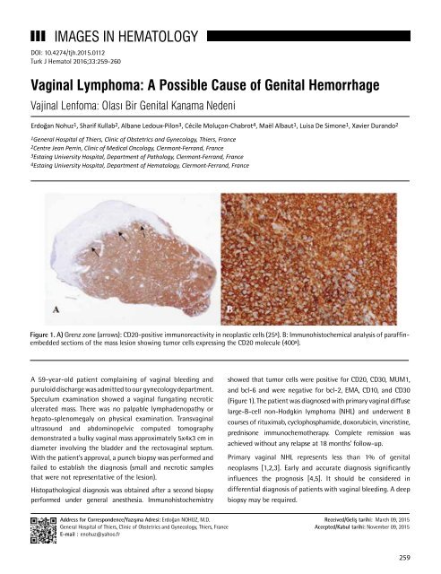
Figure 1. A) Grenz zone (arrows): CD20-positive immunoreactivity in neoplastic cells (25 x ). B: Immunohistochemical analysis <strong>of</strong> paraffinembedded<br />
sections <strong>of</strong> the mass lesion showing tumor cells expressing the CD20 molecule (400 x ).<br />
A 59-year-old patient complaining <strong>of</strong> vaginal bleeding and<br />
puruloid discharge was admitted to our gynecology department.<br />
Speculum examination showed a vaginal fungating necrotic<br />
ulcerated mass. There was no palpable lymphadenopathy or<br />
hepato-splenomegaly on physical examination. Transvaginal<br />
ultrasound and abdominopelvic computed tomography<br />
demonstrated a bulky vaginal mass approximately 5x4x3 cm in<br />
diameter involving the bladder and the rectovaginal septum.<br />
With the patient’s approval, a punch biopsy was performed and<br />
failed to establish the diagnosis (small and necrotic samples<br />
that were not representative <strong>of</strong> the lesion).<br />
Histopathological diagnosis was obtained after a second biopsy<br />
performed under general anesthesia. Immunohistochemistry<br />
showed that tumor cells were positive for CD20, CD30, MUM1,<br />
and bcl-6 and were negative for bcl-2, EMA, CD10, and CD30<br />
(Figure 1). The patient was diagnosed with primary vaginal diffuse<br />
large-B-cell non-Hodgkin lymphoma (NHL) and underwent 8<br />
courses <strong>of</strong> rituximab, cyclophosphamide, doxorubicin, vincristine,<br />
prednisone immunochemotherapy. Complete remission was<br />
achieved without any relapse at 18 months’ follow-up.<br />
Primary vaginal NHL represents less than 1% <strong>of</strong> genital<br />
neoplasms [1,2,3]. Early and accurate diagnosis significantly<br />
influences the prognosis [4,5]. It should be considered in<br />
differential diagnosis <strong>of</strong> patients with vaginal bleeding. A deep<br />
biopsy may be required.<br />
Address for Correspondence/Yazışma Adresi: Erdoğan NOHUZ, M.D.<br />
General Hospital <strong>of</strong> Thiers, Clinic <strong>of</strong> Obstetrics and Gynecology, Thiers, France<br />
E-mail : enohuz@yahoo.fr<br />
Received/Geliş tarihi: March 09, 2015<br />
Accepted/Kabul tarihi: November 09, 2015<br />
259


