

Turkish Journal of Hematology Volume: 33 - Issue: 3

Create successful ePaper yourself
Turn your PDF publications into a flip-book with our unique Google optimized e-Paper software.
Işıksaçan N, et al: Cytokines’ Association with ZAP70 in Chronic Lymphocytic Leukemia Turk J Hematol 2016;<strong>33</strong>:202-208<br />
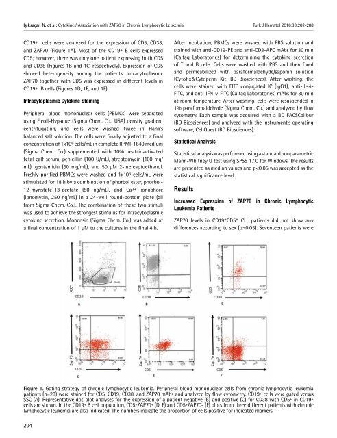
CD19+ cells were analyzed for the expression <strong>of</strong> CD5, CD38,<br />
and ZAP70 (Figure 1A). Most <strong>of</strong> the CD19+ B cells expressed<br />
CD5; however, there was only one patient expressing both CD5<br />
and CD38 (Figures 1B and 1C, respectively). Expression <strong>of</strong> CD5<br />
showed heterogeneity among the patients. Intracytoplasmic<br />
ZAP70 together with CD5 was expressed in different levels in<br />
CD19+ B cells (Figures 1D, 1E, and 1F).<br />
Intracytoplasmic Cytokine Staining<br />
Peripheral blood mononuclear cells (PBMCs) were separated<br />
using Ficoll-Hypaque (Sigma Chem. Co., USA) density gradient<br />
centrifugation, and cells were washed twice in Hank’s<br />
balanced salt solution. The cells were finally adjusted to a final<br />
concentration <strong>of</strong> 1x10 6 cells/mL in complete RPMI-1640 medium<br />
(Sigma Chem. Co.) supplemented with 10% heat-inactivated<br />
fetal calf serum, penicillin (100 U/mL), streptomycin (100 mg/<br />
mL), gentamicin (50 mg/mL), and 50 µM 2-mercaptoethanol.<br />
Freshly purified PBMCs were washed and 1x10⁶ cells/mL were<br />
stimulated for 18 h by a combination <strong>of</strong> phorbol ester, phorbol-<br />
12-myristate-13-acetate (50 ng/mL), and Ca2+ ionophore<br />
(ionomycin, 250 ng/mL) in a 24-well round-bottom plate (all<br />
from Sigma Chem. Co.). The combination <strong>of</strong> these two stimuli<br />
was used to achieve the strongest stimulus for intracytoplasmic<br />
cytokine secretion. Monensin (Sigma Chem. Co.) was added at<br />
a final concentration <strong>of</strong> 1 µM to the cultures in the final 4 h.<br />
After incubation, PBMCs were washed with PBS solution and<br />
stained with anti-CD19-PE and anti-CD3-APC mAbs for 30 min<br />
(Caltag Laboratories) for determining the cytokine secretion<br />
<strong>of</strong> T and B cells. Cells were washed with PBS and then fixed<br />
and permeabilized with paraformaldehyde/saponin solution<br />
(Cyt<strong>of</strong>ix&Cytoperm Kit, BD Biosciences). After washing, the<br />
cells were stained with FITC conjugated IC (IgG1), anti-IL-4-<br />
FITC, and anti-IFN-γ-FITC (Caltag Laboratories) mAbs for 30 min<br />
at room temperature. After washing, cells were resuspended in<br />
1% paraformaldehyde (Sigma Chem. Co.) and analyzed by flow<br />
cytometry. Each sample was acquired with a BD FACSCalibur<br />
(BD Biosciences) and analyzed with the instrument’s operating<br />
s<strong>of</strong>tware, CellQuest (BD Biosciences).<br />
Statistical Analysis<br />
Statistical analysis was performed using a standard nonparametric<br />
Mann-Whitney U test using SPSS 17.0 for Windows. The results<br />
are presented as median values and p0.05). Seventeen patients were<br />
SSC<br />
Figure 1. Gating strategy <strong>of</strong> chronic lymphocytic leukemia. Peripheral blood mononuclear cells from chronic lymphocytic leukemia<br />
patients (n=28) were stained for CD5, CD19, CD38, and ZAP70 mAbs and analyzed by flow cytometry. CD19 + cells were gated versus<br />
SSC (A). Representative dot-plot analyses for the expression <strong>of</strong> a patient negative (B) and positive (C) for CD38 with CD5 + in CD19 +<br />
cells are shown. In the CD19 + B cell population, CD5 + ZAP70 + (D, E) and CD5 + ZAP70- (F) plots from three different patients with chronic<br />
lymphocytic leukemia are also indicated. The numbers indicate the proportion <strong>of</strong> cells positive for indicated markers.<br />
204


