

Turkish Journal of Hematology Volume: 33 - Issue: 3

Create successful ePaper yourself
Turn your PDF publications into a flip-book with our unique Google optimized e-Paper software.
Turk J Hematol 2016;<strong>33</strong>:248-250<br />
Alkan A, et al: Radiation-Induced Tumor Lysis Syndrome<br />
lesions in the bilateral scapula, thoracic vertebral column, pelvis,<br />
bilateral humerus, and femur. For skeletal metastatic disease,<br />
zoledronic acid (4 mg intravenous, every 4 weeks) was started<br />
and palliative radiotherapy was planned for painful metastatic<br />
lesions in the thoracic vertebrae. Radiotherapy to T 3-6 and the<br />
right scapula with a total <strong>of</strong> 30 Gy divided into 10 fractions<br />
was planned. Pathological lymph nodes associated with CLL,<br />
ranging between 1 and 3 cm in diameter in the bilateral<br />
inguinal areas, axilla, neck, and hilum, were noted for followup.<br />
On day 7 <strong>of</strong> radiotherapy the patient complained about mild<br />
nausea, progressive malaise, and perioral numbness. Physical<br />
examination was normal except for pathological lymph nodes<br />
with minimal regression and paleness. The largest lymph node<br />
in the right axilla had regressed from 3 to 2 cm and the other<br />
pathological nodes were stable. The hilar fullness in chest<br />
X-ray was stable. The only medications used were drugs for<br />
prostate cancer (bicalutamide and goserelin), lansoprazole for<br />
dyspepsia, and dexamethasone (8 mg), which was planned with<br />
the radiotherapy. The laboratory evaluation revealed acute<br />
renal failure complicated with TLS (Table 1). The PSA value was<br />
stable. The patient was hospitalized and aggressively hydrated<br />
with 0.9% NaCl at 500 mL/h in the first 6 h <strong>of</strong> follow-up with<br />
monitorization <strong>of</strong> hourly urine output. In the initial evaluation,<br />
due to hypocalcemia (5.2 mg/dL) with neuromuscular symptoms<br />
and prolonged QT interval <strong>of</strong> 0.50 s, the patient was treated with<br />
calcium gluconate replacement under cardiac monitorization.<br />
Laboratory results were checked at 4-h intervals. Until we could<br />
use rasburicase, allopurinol was the initial specific therapy for<br />
TLS. The dosage was titrated according to glomerular filtration<br />
rate and a 0.2 mg/kg single dose <strong>of</strong> rasburicase could be added<br />
to therapy on day 3 <strong>of</strong> hospitalization. The patient’s symptoms<br />
and renal dysfunction progressively improved (Table 1). The<br />
patient was free <strong>of</strong> any findings <strong>of</strong> infection or septicemia. The<br />
leukocytosis was linked to the dexamethasone therapy, which<br />
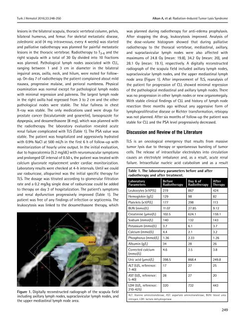
Figure 1. Digitally reconstructed radiograph <strong>of</strong> the scapula field<br />
including axillary lymph nodes, supraclavicular lymph nodes, and<br />
the upper mediastinal lymph node area.<br />
was planned during radiotherapy for anti-edema prophylaxis.<br />
After stopping the drug, leukocytosis improved. Analysis <strong>of</strong><br />
the dose-volume histogram showed that during palliative<br />
radiotherapy to the thoracal vertebrae, mediastinal, axillary,<br />
and supraclavicular lymph nodes were also affected with<br />
maximums <strong>of</strong> 24.8 Gy (mean: 19.8), 34.2 Gy (mean: 20), and<br />
28.1 Gy (mean: 19.1), respectively. A digitally reconstructed<br />
radiograph <strong>of</strong> the scapula field included axillary lymph nodes,<br />
supraclavicular lymph nodes, and the upper mediastinal lymph<br />
node area (Figure 1). After improvement <strong>of</strong> TLS, reanalysis <strong>of</strong><br />
the patient for progression <strong>of</strong> CLL showed minimal regression<br />
<strong>of</strong> the pathological mediastinal and axillary lymph nodes. There<br />
was no progression in other lymph nodes or new organomegaly.<br />
With stable clinical findings <strong>of</strong> CLL and history <strong>of</strong> lymph node<br />
resection three months ago without any aggressive form <strong>of</strong><br />
lymphoproliferative disease or Richter transformation, rebiopsy<br />
was not planned. After six months <strong>of</strong> follow-up the patient was<br />
stable for CLL and the PSA level progressively decreased.<br />
Discussion and Review <strong>of</strong> the Literature<br />
TLS is an oncological emergency that results from massive<br />
tumor lysis due to therapy or spontaneous bursting <strong>of</strong> tumor<br />
cells. The release <strong>of</strong> intracellular electrolytes into circulation<br />
causes an electrolyte imbalance and, as a result, acute renal<br />
failure. Intracellular nucleic acid catabolism and as a result<br />
Table 1. The laboratory parameters before and after<br />
radiotherapy and after treatment.<br />
Laboratory<br />
Parameters<br />
Before<br />
Radiotherapy<br />
Day 6 <strong>of</strong><br />
Radiotherapy<br />
Leukocytes (x10 9 /L) 219 867 424<br />
Hemoglobin (g/L) 129 98 82<br />
Platelets (x10 9 /L) 177 298 113<br />
After<br />
Therapy<br />
BUN (mmol/L) 11.07 27.85 9.12<br />
Creatinine (µmol/L) 102.5 624.1 159.1<br />
Sodium (mmol/L) 140 132 143<br />
Potassium (mmol/L) 3.7 6.1 3.7<br />
Calcium (mmol/L) 4.4 2.1 3.2<br />
Phosphorus (mmol/L) 1.36 2.<strong>33</strong> 1.26<br />
Albumin (g/L) 34 28 26<br />
Corrected calcium 4.6 2.5 3.8<br />
(mmol/L)<br />
Uric acid (µmol/L) 398.5 868.4 249.8<br />
ALT (U/L, reference: 17 27 25<br />
7-40)<br />
AST (U/L, reference: 28 27 20<br />
5-40)<br />
LDH (U/L, reference: 320 732 443<br />
210-425)<br />
ALT: Alanine aminotransferase, AST: aspartate aminotransferase, BUN: blood urea<br />
nitrogen, LDH: lactate dehydrogenase.<br />
249


