

Turkish Journal of Hematology Volume: 33 - Issue: 3

You also want an ePaper? Increase the reach of your titles
YUMPU automatically turns print PDFs into web optimized ePapers that Google loves.
LETTERS TO EDITOR<br />
Turk J Hematol 2016;<strong>33</strong>:254-258<br />
A Primary Bone Diffuse Large B-Cell Lymphoma with Ocular<br />
Adnexal Involvement<br />
Oküler Adneks Tutulumu Olan Kemiğin Primer Diffüz Büyük B Hücreli Lenfoması<br />
Rafet Eren 1 , Ceyda Aslan 1 , Cihan Gündoğan 2 , Osman Yokuş 1 , Mehmet Hilmi Doğu 1 , Elif Suyanı 1<br />
1İstanbul Training and Research Hospital, Clinic <strong>of</strong> <strong>Hematology</strong>, İstanbul, Turkey<br />
2İstanbul Training and Research Hospital, Clinic <strong>of</strong> Nuclear Medicine, İstanbul, Turkey<br />
To the Editor,<br />
Primary bone lymphomas (PBLs) [1,2] and ocular adnexal (OA)<br />
lymphomas [3,4] are rare types <strong>of</strong> extranodal lymphomas.<br />
Coexistence <strong>of</strong> these two rare entities without lymph node<br />
infiltration has not been reported previously.<br />
A 55-year-old man presented with left hip pain without a<br />
history <strong>of</strong> trauma. His medical history and physical examination<br />
did not reveal any remarkable findings. The X-ray radiographs<br />
<strong>of</strong> the pelvis and left hip showed multiple lytic lesions. Body<br />
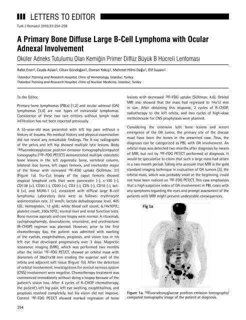
18fluorodeoxyglucose positron emission tomography/computed<br />
tomography (18F-FDG PET/CT) demonstrated multiple osteolytic<br />
bone lesions in the left zygomatic bone, vertebral column,<br />
bilateral iliac bones, left caput femoris, and trochanter major<br />
<strong>of</strong> the femur with increased 18F-FDG uptake (SUVmax: 31)<br />
(Figure 1a). Tru-Cut biopsy <strong>of</strong> the caput femoris showed<br />
atypical lymphoid cells that were pancreatin (-), s-100 (-),<br />
CD138 (+), CD30 (-), CD20 (+), CD3 (-), CD5 (-), CD10 (-), bcl-<br />
6 (+), and MUM-1 (+), consistent with diffuse large B-cell<br />
lymphoma. Laboratory data were as follows: erythrocyte<br />
sedimentation rate, 37 mm/h; lactate dehydrogenase level, 405<br />
U/L; hemoglobin, 12 g/dL; white blood cell count, 6.74x10⁹/L;<br />
platelet count, 250x109/L; normal liver and renal function tests.<br />
Bone marrow aspirate and core biopsy were normal. A rituximab,<br />
cyclophosphamide, doxorubicine, vincristine, and prednisolone<br />
(R-CHOP) regimen was planned. However, prior to the first<br />
chemotherapy day, the patient was admitted with swelling<br />
<strong>of</strong> the eyelids, exophthalmos, proptosis, and vision loss in his<br />
left eye that developed progressively over 3 days. Magnetic<br />
resonance imaging (MRI), which was performed two months<br />
after the initial 18F-FDG PET/CT, showed an orbital mass with<br />
diameters <strong>of</strong> 36x21x38 mm eroding the superior wall <strong>of</strong> the<br />
orbita and adjacent s<strong>of</strong>t tissue (Figure 1b). After the detection<br />
<strong>of</strong> orbital involvement, investigations for central nervous system<br />
(CNS) involvement were negative. Chemotherapy treatment was<br />
commenced immediately without doing a biopsy because <strong>of</strong> the<br />
patient’s vision loss. After 4 cycles <strong>of</strong> R-CHOP chemotherapy,<br />
the patient’s left hip pain, left eye swelling, exophthalmos, and<br />
proptosis resolved completely, but his vision did not improve.<br />
Control 18F-FDG PET/CT showed marked regression <strong>of</strong> bone<br />
lesions with decreased 18 F-FDG uptake (SUVmax: 4.6). Orbital<br />
MRI also showed that the mass had regressed to 14x12 mm<br />
in size. After obtaining this response, 2 cycles <strong>of</strong> R-CHOP,<br />
radiotherapy to the left orbita, and two cycles <strong>of</strong> high-dose<br />
methotrexate for CNS prophylaxis were planned.<br />
Considering the extensive lytic bone lesions and recent<br />
emergence <strong>of</strong> the OA tumor, the primary site <strong>of</strong> the disease<br />
must have been the bones in the presented case. Thus, the<br />
diagnosis can be categorized as PBL with OA involvement. An<br />
orbital mass was detected two months after diagnosis by means<br />
<strong>of</strong> MRI, but not by 18F-FDG PET/CT performed at diagnosis. It<br />
would be speculative to claim that such a large mass had arisen<br />
in a two month period. Taking into account that MRI is the gold<br />
standard imaging technique in evaluation <strong>of</strong> OA tumors [3], the<br />
orbital mass, which was probably small at the beginning, could<br />
not have been noticed on 18F-FDG PET/CT. This case emphasizes<br />
that a high suspicion index <strong>of</strong> OA involvement in PBL cases with<br />
any symptoms regarding the eyes and prompt assessment <strong>of</strong> the<br />
patients with MRI might prevent undesirable consequences.<br />
Figure 1a. 18 Fluorodeoxyglucose positron emission tomography/<br />
computed tomography image <strong>of</strong> the patient at diagnosis.<br />
254


