

Turkish Journal of Hematology Volume: 33 - Issue: 3

You also want an ePaper? Increase the reach of your titles
YUMPU automatically turns print PDFs into web optimized ePapers that Google loves.
Turk J Hematol 2016;<strong>33</strong>:244-247<br />
Aktürk H, et al: Vancomycin-Resistant Enterococci Infections in Pediatric Malignancies<br />
Introduction<br />
Children with cancer are at high risk <strong>of</strong> developing systemic<br />
infections by the microorganisms that colonize their own<br />
intestinal system [1,2]. Vancomycin-resistant enterococci (VRE)<br />
are health care-associated opportunistic pathogens. Limited data<br />
exist on the incidence <strong>of</strong> subsequent VRE infection development<br />
among VRE-colonized pediatric cancer patients and associated<br />
risk factors, which were investigated in this study.<br />
Materials and Methods<br />
All patients admitted to the pediatric hematology/oncology<br />
ward were sampled within 48-72 h after admission and weekly<br />
thereafter as part <strong>of</strong> institutional rectal VRE surveillance. An<br />
infection control nurse assigned by the Hospital Infection<br />
Control Committee (HICC) prospectively tracked the results <strong>of</strong><br />
rectal surveillance and all health care-associated infections<br />
occurring in the hematology/oncology ward. VRE-colonized<br />
and VRE-infected patients were identified from the HICC<br />
surveillance database retrospectively. Detailed clinical and<br />
laboratory features <strong>of</strong> these patients were collected from their<br />
medical records. The overall rate <strong>of</strong> VRE colonization and the<br />
subsequent infection occurrence throughout the study period<br />
were determined. To identify the risk factors associated with<br />
VRE infection occurrence in a colonized patient, a retrospective<br />
case-control study was performed. Patients were defined as VREcolonized<br />
(VRE-C) when the culture <strong>of</strong> the rectal swab yielded<br />
VRE in the absence <strong>of</strong> any clinical specimens positive for VRE<br />
[3]. Systemic VRE infection (VRE-I) was defined as isolation <strong>of</strong><br />
VRE from a clinical specimen together with signs and symptoms<br />
<strong>of</strong> infection. Statistical analysis was performed with SPSS 21.0<br />
for Windows. Parameters were compared between groups with<br />
the chi-square test, Fisher exact test, or Mann-Whitney U test.<br />
Variables with a p-value <strong>of</strong> ≤0.1 in univariate analysis were fitted<br />
to perform logistic regression analysis to identify independent<br />
risk factors associated with VRE infection occurrence.<br />
Results<br />
A total <strong>of</strong> 229 children were admitted to the hematology/<br />
oncology ward. Fecal VRE-C was documented in 72 <strong>of</strong> these<br />
patients (31.4%). Excluding eight patients who were transferred<br />
from the pediatric intensive care unit, 89% <strong>of</strong> the patients were<br />
colonized during their stay in the hematology/oncology ward.<br />
Species determination could be performed in 32 VRE-colonized<br />
patients: Enterococcus faecium was isolated in 28 patients,<br />
Enterococcus gallinarum in 2 patients, and nontypeable<br />
Enterococcus in 2 patients.<br />
VRE-I was detected in 7 patients, all <strong>of</strong> whom were previously<br />
colonized with VRE. The overall rate <strong>of</strong> VRE-I developing in<br />
patients with VRE-C was 9.7%. VRE bacteremia was detected<br />
in five patients (6.9%). Other VRE infections were urinary<br />
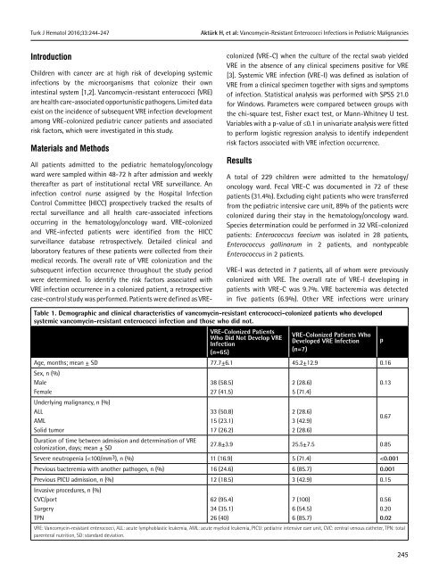
Table 1. Demographic and clinical characteristics <strong>of</strong> vancomycin-resistant enterococci-colonized patients who developed<br />
systemic vancomycin-resistant enterococci infection and those who did not.<br />
VRE-Colonized Patients<br />
Who Did Not Develop VRE<br />
Infection<br />
(n=65)<br />
VRE-Colonized Patients Who<br />
Developed VRE Infection<br />
(n=7)<br />
Age, months; mean ± SD 77.7±6.1 45.2±12.9 0.16<br />
Sex, n (%)<br />
Male<br />
Female<br />
Underlying malignancy, n (%)<br />
ALL<br />
AML<br />
Solid tumor<br />
Duration <strong>of</strong> time between admission and determination <strong>of</strong> VRE<br />
colonization, days; mean ± SD<br />
38 (58.5)<br />
27 (41.5)<br />
<strong>33</strong> (50.8)<br />
15 (23.1)<br />
17 (26.2)<br />
2 (28.6)<br />
5 (71.4)<br />
2 (28.6)<br />
3 (42.9)<br />
2 (28.6)<br />
p<br />
0.13<br />
0.67<br />
27.8±3.9 25.5±7.5 0.85<br />
Severe neutropenia (


