

Turkish Journal of Hematology Volume: 33 - Issue: 3

You also want an ePaper? Increase the reach of your titles
YUMPU automatically turns print PDFs into web optimized ePapers that Google loves.
Turk J Hematol 2016;<strong>33</strong>:202-208<br />
Işıksaçan N, et al: Cytokines’ Association with ZAP70 in Chronic Lymphocytic Leukemia<br />
Introduction<br />
Chronic lymphocytic leukemia (CLL) is a disease that shows<br />
varying clinical progression. The Rai and Binet classification<br />
systems are useful to predict treatment requirements and<br />
survival rates for CLL patients, but current classifications fail<br />
to distinguish patients who may develop aggressive disease<br />
[1,2,3,4]. The somatic mutations in the immunoglobulin heavy<br />
chain variable (IGVH) region are very valuable prognostic factors<br />
and, in CLL cases, IGVH-mutated patients have a better clinical<br />
outcome while nonmutated patients have a poorer prognosis<br />
and impaired response to chemotherapy [5,6,7].<br />
The protein tyrosine kinase zeta-associated protein (ZAP70) is a<br />
protein tyrosine kinase in the T cell receptor signal transduction<br />
system that can be detected in CLL cells. Studies have pointed<br />
out a correlation between ZAP70 expression in CLL cells and<br />
IGVH status, and showed that ZAP70 positive patients have an<br />
aggressive course, an immediate treatment requirement, longer<br />
therapy time, and lower survival rates [8,9,10]. Expression <strong>of</strong><br />
ZAP70 has been described as a very valuable prognostic factor<br />
[2,8].<br />
Many researchers have recently examined the cytokine content<br />
<strong>of</strong> the T cells in CLL patients and emphasized the significance<br />
<strong>of</strong> the T cell activity in the prognosis <strong>of</strong> the disease [11,12].<br />
Interleukin-4 (IL-4) production by T cells has been shown<br />
to significantly increase in patients with progressive disease<br />
[11,12]. In light <strong>of</strong> these data, it was suggested that in CLL, the<br />
type 1 T cell cytokine pr<strong>of</strong>ile is converted to the type 2 T cell<br />
pr<strong>of</strong>ile in the advanced stages <strong>of</strong> the disease [13]. The aim <strong>of</strong><br />
this study was to evaluate the expression <strong>of</strong> ZAP70 changing<br />
during disease progression, the intracellular interferon gamma<br />
(IFN-γ) and IL-4 content <strong>of</strong> T and B lymphocytes and the CLL cell<br />
subset (CD5 + CD19 + ) in CLL patients and healthy subjects, and<br />
ZAP70 correlation with cytokine production.<br />
Materials and Methods<br />
Study Population<br />
Twenty-eight patients in follow-up at the İstanbul University<br />
İstanbul Medical Faculty <strong>Hematology</strong> Department were included<br />
in the study. Patients were diagnosed according to the CLL<br />
diagnosis criteria published in 1996 by a study group supported<br />
by the National Cancer Institute. Clinical data and follow-up<br />
files <strong>of</strong> all the patients included in the study were gathered. All<br />
patients received written information about the study, including<br />
ethics committee approval, before the study was initiated.<br />
Twenty-eight CLL patients (18 males, 10 females) and 10 healthy<br />
individuals (3 males, 7 females) were involved in this study<br />
(Table 1). The ZAP70 expression was measured for all CLL cases<br />
and cytokine levels were measured for 12 CLL cases and the 10<br />
healthy volunteers. The ages <strong>of</strong> the patients were between 36<br />
and 81 (59±11) years, and the mean values were 60±12 years for<br />
males and 56±7 years for females.<br />
Peripheral Blood Mononuclear Cell Isolation and Flow Cytometric<br />
Analyses <strong>of</strong> Lymphocyte Subsets<br />
Samples were processed using a whole-blood lysis method<br />
to analyze lymphocyte subsets. Heparinized blood samples<br />
were collected from patient and healthy donors and stained<br />
with anti-CD19-fluorescein isothiocyanate (FITC), anti-CD3-<br />
allophycocyanin (APC), anti-CD5-TRI-COLOR (TC), anti-CD45-<br />
FITC, anti-CD14-phycoerythrin (PE), anti-CD23-PE, anti-CD38-<br />
PE, and PE, FITC, TC, or APC conjugated isotype control (IC)<br />
monoclonal antibodies (mAbs) (all from Caltag Laboratories,<br />
Austria) for 30 min at room temperature. Lysis was performed<br />
with FACS Lysing Solution (BD Biosciences, USA). After washing<br />
cells with phosphate-buffered saline (PBS), stained cells were<br />
fixed in 1% paraformaldehyde and the cells were analyzed with<br />
BD FACSCalibur with CellQuest S<strong>of</strong>tware (BD Biosciences).<br />
For the intracytoplasmic ZAP70 expression, the cells were labeled<br />
with anti-CD19-FITC and anti-CD5-TC mAbs (Caltag Laboratories)<br />
for 30 min at room temperature. After the lysing period, cells<br />
were washed with PBS and were fixed and permeabilized with<br />
paraformaldehyde/saponin solution (Cyt<strong>of</strong>ix&Cytoperm Kit, BD<br />
Biosciences), and then were stained with PE-conjugated-IgG1<br />
or PE-labeled ZAP70 (Caltag Laboratories) mAbs for 30 min<br />
at room temperature and analyzed by flow cytometry. Each<br />
analysis was performed using at least 5000 cells gated in the<br />
region <strong>of</strong> the B cell population, and the cells were analyzed<br />
with BD FACSCalibur with CellQuest S<strong>of</strong>tware (BD Biosciences).<br />
ZAP70 expression was investigated in CLL patients for positivity;<br />
20% was used as the cut-<strong>of</strong>f [9].<br />
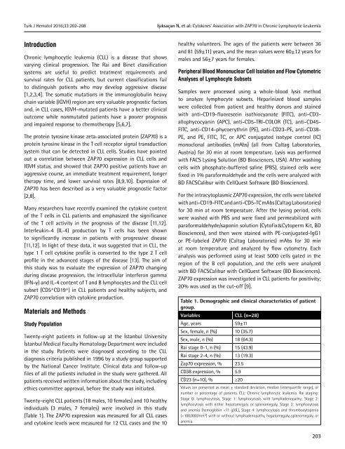
Table 1. Demographic and clinical characteristics <strong>of</strong> patient<br />
group.<br />
Variables<br />
CLL (n=28)<br />
Age, years 59±11<br />
Sex, female, n (%) 10 (35.7)<br />
Sex, male, n (%) 18 (64.3)<br />
Rai stage 0-1, n (%) 15 (43.9)<br />
Rai stage 2-4, n (%) 13 (19.3)<br />
Zap70 expression, % 23.5<br />
CD38 expression, % 5.9<br />
CD23 (n=10), % ≥20<br />
Values are presented as mean ± standard deviation, median (interquartile range), or<br />
number or percentage <strong>of</strong> patients. CLL: Chronic lymphocytic leukemia. Rai staging:<br />
Stage 0: lymphocytosis, Stage 1: lymphocytosis with lymphadenopathy; Stage 2:<br />
lymphocytosis with either hepatomegaly or splenomegaly, Stage 3: lymphocytosis<br />
and anemia (hemoglobin


