

Turkish Journal of Hematology Volume: 33 - Issue: 3

You also want an ePaper? Increase the reach of your titles
YUMPU automatically turns print PDFs into web optimized ePapers that Google loves.
LETTERS TO EDITOR<br />
Turk J Hematol 2016;<strong>33</strong>:254-258<br />
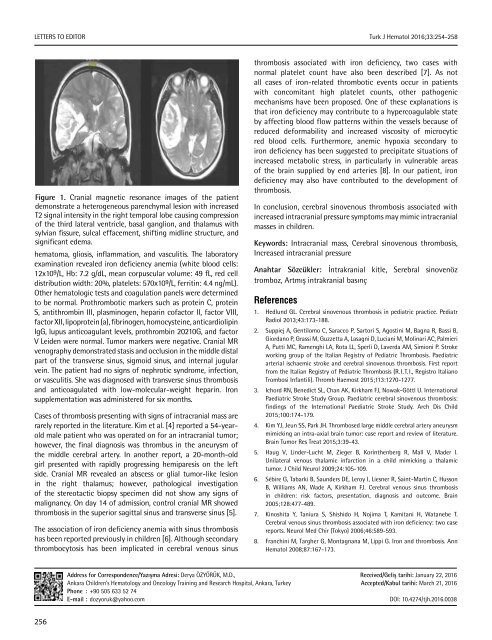
Figure 1. Cranial magnetic resonance images <strong>of</strong> the patient<br />
demonstrate a heterogeneous parenchymal lesion with increased<br />
T2 signal intensity in the right temporal lobe causing compression<br />
<strong>of</strong> the third lateral ventricle, basal ganglion, and thalamus with<br />
sylvian fissure, sulcal effacement, shifting midline structure, and<br />
significant edema.<br />
hematoma, gliosis, inflammation, and vasculitis. The laboratory<br />
examination revealed iron deficiency anemia (white blood cells:<br />
12x109/L, Hb: 7.2 g/dL, mean corpuscular volume: 49 fL, red cell<br />
distribution width: 20%, platelets: 570x10 9 /L, ferritin: 4.4 ng/mL).<br />
Other hematologic tests and coagulation panels were determined<br />
to be normal. Prothrombotic markers such as protein C, protein<br />
S, antithrombin III, plasminogen, heparin c<strong>of</strong>actor II, factor VIII,<br />
factor XII, lipoprotein (a), fibrinogen, homocysteine, anticardiolipin<br />
IgG, lupus anticoagulant levels, prothrombin 20210G, and factor<br />
V Leiden were normal. Tumor markers were negative. Cranial MR<br />
venography demonstrated stasis and occlusion in the middle distal<br />
part <strong>of</strong> the transverse sinus, sigmoid sinus, and internal jugular<br />
vein. The patient had no signs <strong>of</strong> nephrotic syndrome, infection,<br />
or vasculitis. She was diagnosed with transverse sinus thrombosis<br />
and anticoagulated with low-molecular-weight heparin. Iron<br />
supplementation was administered for six months.<br />
Cases <strong>of</strong> thrombosis presenting with signs <strong>of</strong> intracranial mass are<br />
rarely reported in the literature. Kim et al. [4] reported a 54-yearold<br />
male patient who was operated on for an intracranial tumor;<br />
however, the final diagnosis was thrombus in the aneurysm <strong>of</strong><br />
the middle cerebral artery. In another report, a 20-month-old<br />
girl presented with rapidly progressing hemiparesis on the left<br />
side. Cranial MR revealed an abscess or glial tumor-like lesion<br />
in the right thalamus; however, pathological investigation<br />
<strong>of</strong> the stereotactic biopsy specimen did not show any signs <strong>of</strong><br />
malignancy. On day 14 <strong>of</strong> admission, control cranial MR showed<br />
thrombosis in the superior sagittal sinus and transverse sinus [5].<br />
The association <strong>of</strong> iron deficiency anemia with sinus thrombosis<br />
has been reported previously in children [6]. Although secondary<br />
thrombocytosis has been implicated in cerebral venous sinus<br />
thrombosis associated with iron deficiency, two cases with<br />
normal platelet count have also been described [7]. As not<br />
all cases <strong>of</strong> iron-related thrombotic events occur in patients<br />
with concomitant high platelet counts, other pathogenic<br />
mechanisms have been proposed. One <strong>of</strong> these explanations is<br />
that iron deficiency may contribute to a hypercoagulable state<br />
by affecting blood flow patterns within the vessels because <strong>of</strong><br />
reduced deformability and increased viscosity <strong>of</strong> microcytic<br />
red blood cells. Furthermore, anemic hypoxia secondary to<br />
iron deficiency has been suggested to precipitate situations <strong>of</strong><br />
increased metabolic stress, in particularly in vulnerable areas<br />
<strong>of</strong> the brain supplied by end arteries [8]. In our patient, iron<br />
deficiency may also have contributed to the development <strong>of</strong><br />
thrombosis.<br />
In conclusion, cerebral sinovenous thrombosis associated with<br />
increased intracranial pressure symptoms may mimic intracranial<br />
masses in children.<br />
Keywords: Intracranial mass, Cerebral sinovenous thrombosis,<br />
Increased intracranial pressure<br />
Anahtar Sözcükler: İntrakranial kitle, Serebral sinovenöz<br />
tromboz, Artmış intrakranial basınç<br />
References<br />
1. Hedlund GL. Cerebral sinovenous thrombosis in pediatric practice. Pediatr<br />
Radiol 2013;43:173-188.<br />
2. Suppiej A, Gentilomo C, Saracco P, Sartori S, Agostini M, Bagna R, Bassi B,<br />
Giordano P, Grassi M, Guzzetta A, Lasagni D, Luciani M, Molinari AC, Palmieri<br />
A, Putti MC, Ramenghi LA, Rota LL, Sperlì D, Laverda AM, Simioni P. Stroke<br />
working group <strong>of</strong> the Italian Registry <strong>of</strong> Pediatric Thrombosis. Paediatric<br />
arterial ischaemic stroke and cerebral sinovenous thrombosis. First report<br />
from the Italian Registry <strong>of</strong> Pediatric Thrombosis (R. I. T. I., Registro Italiano<br />
Trombosi Infantili). Thromb Haemost 2015;113:1270-1277.<br />
3. Ichord RN, Benedict SL, Chan AK, Kirkham FJ, Nowak-Göttl U. International<br />
Paediatric Stroke Study Group. Paediatric cerebral sinovenous thrombosis:<br />
findings <strong>of</strong> the International Paediatric Stroke Study. Arch Dis Child<br />
2015;100:174-179.<br />
4. Kim YJ, Jeun SS, Park JH. Thrombosed large middle cerebral artery aneurysm<br />
mimicking an intra-axial brain tumor: case report and review <strong>of</strong> literature.<br />
Brain Tumor Res Treat 2015;3:39-43.<br />
5. Haug V, Linder-Lucht M, Zieger B, Korinthenberg R, Mall V, Mader I.<br />
Unilateral venous thalamic infarction in a child mimicking a thalamic<br />
tumor. J Child Neurol 2009;24:105-109.<br />
6. Sébire G, Tabarki B, Saunders DE, Leroy I, Liesner R, Saint-Martin C, Husson<br />
B, Williams AN, Wade A, Kirkham FJ. Cerebral venous sinus thrombosis<br />
in children: risk factors, presentation, diagnosis and outcome. Brain<br />
2005;128:477-489.<br />
7. Kinoshita Y, Taniura S, Shishido H, Nojima T, Kamitani H, Watanebe T.<br />
Cerebral venous sinus thrombosis associated with iron deficiency: two case<br />
reports. Neurol Med Chir (Tokyo) 2006;46:589-593.<br />
8. Franchini M, Targher G, Montagnana M, Lippi G. Iron and thrombosis. Ann<br />
Hematol 2008;87:167-173.<br />
Address for Correspondence/Yazışma Adresi: Derya ÖZYÖRÜK, M.D.,<br />
Ankara Children’s <strong>Hematology</strong> and Oncology Training and Research Hospital, Ankara, Turkey<br />
Phone : +90 505 6<strong>33</strong> 52 74<br />
E-mail : dozyoruk@yahoo.com<br />
Received/Geliş tarihi: January 22, 2016<br />
Accepted/Kabul tarihi: March 21, 2016<br />
DOI: 10.4274/tjh.2016.0038<br />
256


