

Turkish Journal of Hematology Volume: 33 - Issue: 3

Create successful ePaper yourself
Turn your PDF publications into a flip-book with our unique Google optimized e-Paper software.
Turk J Hematol 2016;<strong>33</strong>:254-258<br />
LETTERS TO EDITOR<br />
A Rare Cause <strong>of</strong> Unexplained Refractory Iron Deficiency Anemia:<br />
Unicentric Plasma-Cell Type Castleman’s Disease<br />
Tedaviye Dirençli Demir Eksikliği Anemisinin Nadir Bir Nedeni: Unisentrik Plazma Hücreli<br />
Tip Castleman Hastalığı<br />
Sevgi Kalayoğlu Beşışık 1 , İpek Yönal Hindilerden 1 , Fehmi Hindilerden 2 , İbrahim Öner Doğan 3 , Fatih Beşışık 4<br />
¹İstanbul University İstanbul Faculty <strong>of</strong> Medicine, Department <strong>of</strong> Internal Medicine, Division <strong>of</strong> <strong>Hematology</strong>, İstanbul, Turkey<br />
2İstanbul Bakırköy Sadi Konuk Training and Research Hospital, Clinic <strong>of</strong> <strong>Hematology</strong>, İstanbul, Turkey<br />
3İstanbul University İstanbul Faculty <strong>of</strong> Medicine, Department <strong>of</strong> Pathology, İstanbul, Turkey<br />
3İstanbul University İstanbul Faculty <strong>of</strong> Medicine, Department <strong>of</strong> Internal Medicine, Division <strong>of</strong> Gastroenterohepatology, İstanbul, Turkey<br />
To the Editor,<br />
Castleman’s disease (CD) is an uncommon benign<br />
lymphoproliferative disorder characterized by enlargement <strong>of</strong><br />
hyperplastic lymph nodes with abnormal interfollicular vascular<br />
growth [1]. It is clinically categorized as unicentric disease or<br />
multicentric disease. Histopathologic variants <strong>of</strong> CD include<br />
hyaline-vascular type, plasma-cell (PC) type, and mixed form<br />
[2]. CD presents with features ranging from asymptomatic<br />
lymphadenopathy to systemic manifestations such as serious<br />
infections, anemia, and nerve damage [3]. In CD, hepcidin<br />
secretion induced by IL-6 is the main cause <strong>of</strong> anemia [4,5].<br />
Anemia is more common in multicentric CD and is rarely reported<br />
in unicentric CD [3]. Few cases <strong>of</strong> unicentric PC type CD associated<br />
with iron deficiency anemia (IDA) have been reported [6,7,8,9]. To<br />
our knowledge, there is only one previous reported case <strong>of</strong> adult<br />
unicentric PC type CD located in the abdomen and presenting<br />
with IDA [9]. We describe an adult with occult unicentric PC type<br />
CD <strong>of</strong> the abdomen presenting with iron-refractory anemia (IRA)<br />
and achieving dramatic response to curative resection.<br />
A 47-year-old man was referred with a 5-year history <strong>of</strong> IRA.<br />
Blood analysis showed Hb <strong>of</strong> 8 g/dL, mean corpuscular volume<br />
<strong>of</strong> 70 fL, red cell distribution width <strong>of</strong> 17%, and platelet count<br />
<strong>of</strong> 478,000/mm 3 . Biochemical tests were as follows: serum<br />
ferritin, 371 µg/L; transferrin saturation, 8.6%; and erythrocyte<br />
sedimentation rate (ESR), 110 mm/h (reference range: 0-20).<br />
Serum protein electrophoresis revealed polyclonal gammopathy<br />
with gamma globulin <strong>of</strong> 2.16 g/dL. The soluble transferrin<br />
receptor/log10 ferritin index <strong>of</strong> 2.3 indicated the presence <strong>of</strong><br />
combined IDA and anemia <strong>of</strong> inflammation (AI) [10]. Underlying<br />
chronic inflammatory diseases were excluded. Upper and lower<br />
gastrointestinal endoscopic evaluations and bone marrow<br />
examination were normal. Positron emission tomographycomputed<br />
tomography showed a s<strong>of</strong>t tissue mass with diffuse<br />
fluorodeoxyglucose uptake with an SUVmax <strong>of</strong> 11.39 and a<br />
craniocaudal length <strong>of</strong> 8 cm extending from the hepatogastric<br />
ligament and with a maximal diameter <strong>of</strong> 4.4x4.3 cm in the axial<br />
plane at the portal region. He underwent exploratory laparotomy<br />
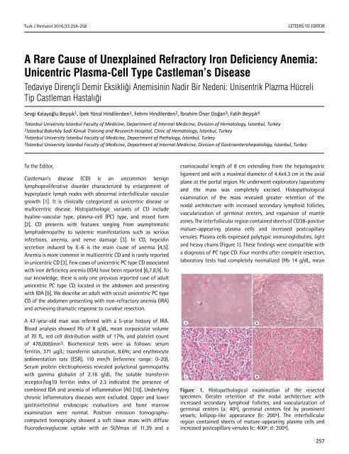
and the mass was completely excised. Histopathological<br />
examination <strong>of</strong> the mass revealed greater retention <strong>of</strong> the<br />
nodal architecture with increased secondary lymphoid follicles,<br />
vascularization <strong>of</strong> germinal centers, and expansion <strong>of</strong> mantle<br />
zones. The interfollicular region contained sheets <strong>of</strong> CD38-positive<br />
mature-appearing plasma cells and increased postcapillary<br />
venules. Plasma cells expressed polytypic immunoglobulins, light<br />
and heavy chains (Figure 1). These findings were compatible with<br />
a diagnosis <strong>of</strong> PC type CD. Four months after complete resection,<br />
laboratory tests had completely normalized (Hb 14 g/dL, mean<br />
Figure 1. Histopathological examination <strong>of</strong> the resected<br />
specimen. Greater retention <strong>of</strong> the nodal architecture with<br />
increased secondary lymphoid follicles, and vascularization <strong>of</strong><br />
germinal centers (a: 40 x ), germinal centers fed by prominent<br />
vessels; lollipop-like appearance (b: 200 x ). The interfollicular<br />
region contained sheets <strong>of</strong> mature-appearing plasma cells and<br />
increased postcapillary venules (c: 400 x ; d: 200 x ).<br />
257


