

Respircase Cilt: 4 - Sayı: 1 Yıl: 2015

You also want an ePaper? Increase the reach of your titles
YUMPU automatically turns print PDFs into web optimized ePapers that Google loves.
Pneumosiderosis in a Welder Masquerading as Hypersensitivity Pneumonitis Casued by Pigeons | Baççıoğlu et al.<br />
such as a desk job. On the other hand, his employer was<br />
informed about the use of respiratory protective equipment<br />
and local exhaust ventilation in the workplace. Written<br />
informed consent was obtained from the patient prior<br />
to publishing his story.<br />
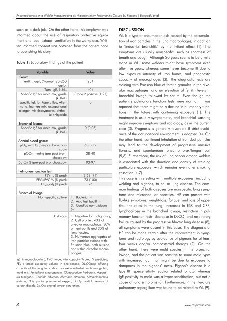
Table 1: Laboratory findings of the patient<br />
Variable<br />
Value<br />
Serum:<br />
Ferritin, ug/L (Normal: 20-250<br />
254<br />
ug/L).<br />
Total IgE, kU/L, 404<br />
Specific IgE for mold mix, grade Grade 2 positive (1.27)<br />
(kUA/L)<br />
Specific IgE for Aspergillus, Alternaria,<br />
feathers mix, occupational<br />
0<br />
allergen mix (Isocyanates, phthalic<br />
anhydride<br />
Bronchial lavage:<br />
Specific IgE for mold mix, grade<br />
(kUA/L)<br />
0 (0.05)<br />
Arterial blood gases:<br />
pO2, mmHg (pre-post bronchoscopy)<br />
63-80.9<br />
pCO2, mmHg (pre-post bronchoscopy)<br />
38-40<br />
Sa,O2 % (pre-post bronchoscopy) 93-97<br />
Pulmonary function test:<br />
FEV1 L (% pred) 3.53 (94)<br />
FEV1/FVC % (% pred) 73 (100)<br />
DL,COadj (% pred) 96<br />
Bronchial lavage:<br />
Non-specific culture 1. Bacteria (-)<br />
2. Acid fast bacilli (-)<br />
3. Candida non-albicans<br />
(+)<br />
Cytology<br />
1. Negative for malignancy,<br />
2. Cell profile : 40% of<br />
alveolar macrophage, 30%<br />
of neutrophils and 30% of<br />
lymphocytes,<br />
3. Numerous aggregates of<br />
iron particles stained with<br />
Prussian blue, both outside<br />
and within alveolar macrophages.<br />
IgE: immunoglobulin E; FVC: forced vital capacity; % pred: % predicted;<br />
FEV1: forced expiratory volume in one second; DL,COadj: diffusing<br />
capacity of the lung for carbon monoxide adjusted for haemoglobin;<br />
mold mix: Penicillium chrysogenum, Cladosporium herbarum, Aspergillus<br />
fumigatus, Candida albicans, Alternaria alternata, Setomelanomma<br />
rostrata, PO 2: partial pressure of oxygen; PCO 2: partial pressure of<br />
carbon dioxide; Sa,O 2: arterial oxygen saturation.<br />
DISCUSSION<br />
WL is a type of pneumoconiosis caused by the accumulation<br />
of iron particles in the lung macrophages, in addition<br />
to "industrial bronchitis" by the irritant effect (1). The<br />
symptoms are usually nonspecific, such as shortness of<br />
breath and cough. Although 20 years seems to be a mile<br />
stone in WL, some welders might have symptoms even<br />
after five years, whereas some never become ill due to<br />
low exposure intensity of iron fumes, and phagocytic<br />
capacity of macrophages (3). The diagnostic tests are<br />
staining with Prussian blue of ferritin granules in the alveolar<br />
macrophages, and an elevation of ferritin levels in<br />
bronchial lavage followed by serum. Even though the<br />
patient’s pulmonary function tests were normal, it was<br />
reported that there might be a decline in pulmonary functions<br />
in the future with continuing exposure (1). The<br />
treatment is usually symptomatic, and bronchial washing<br />
might improve symptoms and radiology, as in the current<br />
case (3). Prognosis is generally favorable if strict avoidance<br />
of the occupational environment is adopted (4). On<br />
the other hand, continued inhalation of iron dust particles<br />
may lead to the development of progressive massive<br />
fibrosis, and spontaneous pneumothorax/fungus ball<br />
(5,6). Furthermore, the risk of lung cancer among welders<br />
is associated with the duration and density of welding<br />
particulate exposure, which remains even after smoking<br />
cessation (4,7).<br />
This case is interesting with multiple exposures, including<br />
welding and pigeons, to cause lung disease. The common<br />
findings of both diseases are nonspecific lung symptoms<br />
and micronodular opacities. HP can present with<br />
flu-like symptoms, weight-loss, fatigue, and loss of appetite,<br />
fine rales in the lung, increases in ESR and CRP,<br />
lymphocytosis in the bronchial lavage, restriction in pulmonary<br />
function tests, decrease in DLCO, and respiratory<br />
failure caused by the progressive fibrotic lung disease (8);<br />
all symptoms were absent in this case. The diagnosis of<br />
HP can be made certain after the improvement in symptoms<br />
and radiology by avoidance of pigeons for at least<br />
four weeks and/or corticosteroid therapy (2). On the<br />
other hand, there were mold species in the bronchial<br />
lavage, and the patient was sensitive to some mold types<br />
with increased IgE, that might be due to exposure to<br />
dampness in the pigeons’ nests. Pigeon’s disease is a<br />
type III hypersensitivity reaction related to IgG, whereas<br />
IgE positivity to mold was a hyper-sensitization, but not a<br />
cause of lung symptoms (8). Furthermore, in the literature,<br />
pulmonary aspergillum was found to be related to WL (9).<br />
3 www.respircase.com


