

Respircase Cilt: 4 - Sayı: 1 Yıl: 2015

You also want an ePaper? Increase the reach of your titles
YUMPU automatically turns print PDFs into web optimized ePapers that Google loves.
A Review of Dental Technician’s Pneumoconiosis: Three Case Reports | Çiftçi et al.<br />
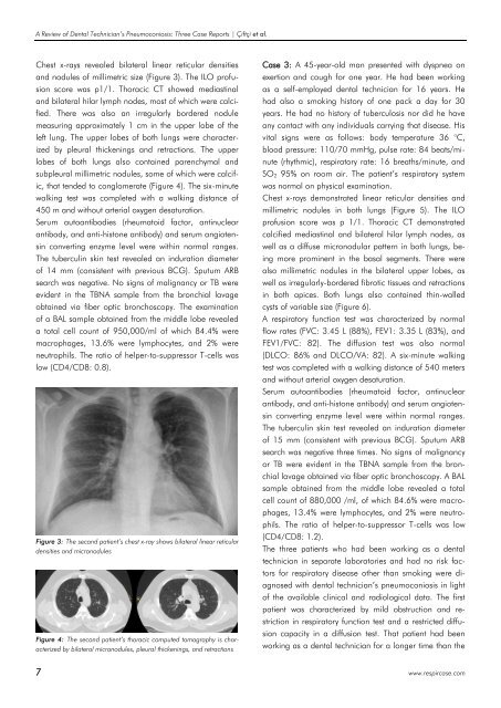
Chest x-rays revealed bilateral linear reticular densities<br />
and nodules of millimetric size (Figure 3). The ILO profusion<br />
score was p1/1. Thoracic CT showed mediastinal<br />
and bilateral hilar lymph nodes, most of which were calcified.<br />
There was also an irregularly bordered nodule<br />
measuring approximately 1 cm in the upper lobe of the<br />
left lung. The upper lobes of both lungs were characterized<br />
by pleural thickenings and retractions. The upper<br />
lobes of both lungs also contained parenchymal and<br />
subpleural millimetric nodules, some of which were calcific,<br />
that tended to conglomerate (Figure 4). The six-minute<br />
walking test was completed with a walking distance of<br />
450 m and without arterial oxygen desaturation.<br />
Serum autoantibodies (rheumatoid factor, antinuclear<br />
antibody, and anti-histone antibody) and serum angiotensin<br />
converting enzyme level were within normal ranges.<br />
The tuberculin skin test revealed an induration diameter<br />
of 14 mm (consistent with previous BCG). Sputum ARB<br />
search was negative. No signs of malignancy or TB were<br />
evident in the TBNA sample from the bronchial lavage<br />
obtained via fiber optic bronchoscopy. The examination<br />
of a BAL sample obtained from the middle lobe revealed<br />
a total cell count of 950,000/ml of which 84.4% were<br />
macrophages, 13.6% were lymphocytes, and 2% were<br />
neutrophils. The ratio of helper-to-suppressor T-cells was<br />
low (CD4/CD8: 0.8).<br />
Figure 3: The second patient’s chest x-ray shows bilateral linear reticular<br />
densities and micronodules<br />
Figure 4: The second patient’s thoracic computed tomography is characterized<br />
by bilateral micronodules, pleural thickenings, and retractions<br />
Case 3: A 45-year-old man presented with dyspnea on<br />
exertion and cough for one year. He had been working<br />
as a self-employed dental technician for 16 years. He<br />
had also a smoking history of one pack a day for 30<br />
years. He had no history of tuberculosis nor did he have<br />
any contact with any individuals carrying that disease. His<br />
vital signs were as follows: body temperature 36 °C,<br />
blood pressure: 110/70 mmHg, pulse rate: 84 beats/minute<br />
(rhythmic), respiratory rate: 16 breaths/minute, and<br />
SO 2 95% on room air. The patient’s respiratory system<br />
was normal on physical examination.<br />
Chest x-rays demonstrated linear reticular densities and<br />
millimetric nodules in both lungs (Figure 5). The ILO<br />
profusion score was p 1/1. Thoracic CT demonstrated<br />
calcified mediastinal and bilateral hilar lymph nodes, as<br />
well as a diffuse micronodular pattern in both lungs, being<br />
more prominent in the basal segments. There were<br />
also millimetric nodules in the bilateral upper lobes, as<br />
well as irregularly-bordered fibrotic tissues and retractions<br />
in both apices. Both lungs also contained thin-walled<br />
cysts of variable size (Figure 6).<br />
A respiratory function test was characterized by normal<br />
flow rates (FVC: 3.45 L (88%), FEV1: 3.35 L (83%), and<br />
FEV1/FVC: 82). The diffusion test was also normal<br />
(DLCO: 86% and DLCO/VA: 82). A six-minute walking<br />
test was completed with a walking distance of 540 meters<br />
and without arterial oxygen desaturation.<br />
Serum autoantibodies (rheumatoid factor, antinuclear<br />
antibody, and anti-histone antibody) and serum angiotensin<br />
converting enzyme level were within normal ranges.<br />
The tuberculin skin test revealed an induration diameter<br />
of 15 mm (consistent with previous BCG). Sputum ARB<br />
search was negative three times. No signs of malignancy<br />
or TB were evident in the TBNA sample from the bronchial<br />
lavage obtained via fiber optic bronchoscopy. A BAL<br />
sample obtained from the middle lobe revealed a total<br />
cell count of 880,000 /ml, of which 84.6% were macrophages,<br />
13.4% were lymphocytes, and 2% were neutrophils.<br />
The ratio of helper-to-suppressor T-cells was low<br />
(CD4/CD8: 1.2).<br />
The three patients who had been working as a dental<br />
technician in separate laboratories and had no risk factors<br />
for respiratory disease other than smoking were diagnosed<br />
with dental technician’s pneumoconiosis in light<br />
of the available clinical and radiological data. The first<br />
patient was characterized by mild obstruction and restriction<br />
in respiratory function test and a restricted diffusion<br />
capacity in a diffusion test. That patient had been<br />
working as a dental technician for a longer time than the<br />
7 www.respircase.com


