

Respircase Cilt: 4 - Sayı: 1 Yıl: 2015

You also want an ePaper? Increase the reach of your titles
YUMPU automatically turns print PDFs into web optimized ePapers that Google loves.
Pulmonary Hydatid Disease Mimicking Lung Cancer | <strong>Yıl</strong>maz et al.<br />
DISCUSSION<br />
Hydatid disease remains a common clinical problem in<br />
areas of the world endemic to echinococcal infestations,<br />
especially Australia, New Zealand, South America, the<br />
Middle East, and the Mediterranean region (6). The most<br />
common localization of the disease is the liver (65%),<br />
followed by the lungs (25%) and other organs such as the<br />
spleen, kidneys, brain, bones, and heart (1,7).<br />
The majority of pulmonary hydatid cyst cases can be<br />
diagnosed by the clinical findings and evaluation of data<br />
obtained through imaging techniques and serologic<br />
methods (4-6). Intact pulmonary hydatid cysts are frequently<br />
detected through chest radiography and typically<br />
appear as solitary or multiple, well-defined, round or oval<br />
masses with smooth borders surrounded by normal lung<br />
tissue (3,6,8). When a pulmonary hydatid cyst is infected<br />
or ruptured, the radiological appearance may become<br />
atypical (4,5,9). According to a previous report (4), typical<br />
radiological appearance was only seen in three of 24<br />
complicated pulmonary hydatid cysts. Complicated pulmonary<br />
hydatid cysts can mimic several pleural and pulmonary<br />
disease such as non-resolving pneumonia, tumors,<br />
tuberculosis, abscess, bronchiectasis, pleurisy, and<br />
empyema (3-8). There is no specific clinical finding except<br />
hydatidosis (4). Laboratory findings are non-specific<br />
(6). Serological tests support the diagnosis but they are<br />
positive in only 50 % of patients with pulmonary hydatid<br />
disease (6,9). Bronchoscopy is unnecessary in patients<br />
with a typical clinical and radiological picture. It can be<br />
performed when there is suspicion of a tumor or the<br />
presentation is atypical (3,5-8,10).<br />
Specific and non-specific bronchoscopic findings for pulmonary<br />
hydatid cysts were described in adults (4,5). Saygi<br />
et al. (4) detected a whitish-yellow gelatinous membrane<br />
in 12 of 24 cases. This feature was introduced as the<br />
single specific finding. Other bronchoscopic findings were<br />
hyperemia and edema in their study. Deshmukh et al. (11)<br />
reported that a white glistening membrane was observed<br />
in 9 of 14 patients during fiber optic bronchoscopy. A<br />
previous report presented three patients with pulmonary<br />
hydatid cyst diagnosed by bronchoscopy. None of these<br />
patients had a typical radiological picture of a hydatid<br />
cyst, and in one patient the serological tests were negative.<br />
The initial diagnoses were lung tumours or tuberculosis.<br />
Bronchoscopy revealed a whitish-yellow gelatinous<br />
membrane in all patients (5). Kilinc et al. (6) reported<br />
three cases of pulmonary hydatid cyst mimicking bronchial<br />
cancer. The authors found that serological tests<br />
were negative in two cases. It was reported that the patients<br />
were evaluated for non-resolving or recurrent<br />
pneumonia in whom complicated hydatid cysts were detected<br />
by bronchoscopy (7,12). Our patient had no history<br />
of close contact with dogs or other animals. She had<br />
no a typical radiological appearance of hydatid cyst. The<br />
patient was initially evaluated to have a lung tumor based<br />
on a radiological mass lesion on the chest radiograph<br />
and computed tomography of the thorax. Bronchoscopy<br />
revealed an endobronchial lesion. From this, a definitive<br />
diagnosis of pulmonary hydatid disease was obtained.<br />
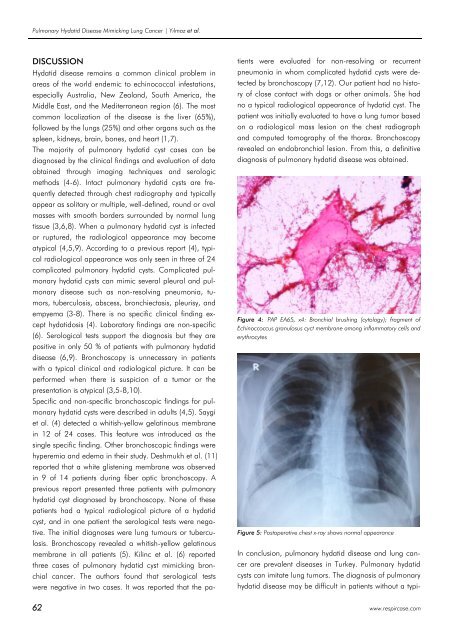
Figure 4: PAP EA65, x4: Bronchial brushing (cytology); fragment of<br />
Echinoccoccus granulosus cyct membrane among inflammatory cells and<br />
erythrocytes<br />
Figure 5: Postoperative chest x-ray shows normal appearance<br />
In conclusion, pulmonary hydatid disease and lung cancer<br />
are prevalent diseases in Turkey. Pulmonary hydatid<br />
cysts can imitate lung tumors. The diagnosis of pulmonary<br />
hydatid disease may be difficult in patients without a typi-<br />
62 www.respircase.com


