

Respircase Cilt: 4 - Sayı: 1 Yıl: 2015

You also want an ePaper? Increase the reach of your titles
YUMPU automatically turns print PDFs into web optimized ePapers that Google loves.
Respiratory Case Reports<br />
Benign fibrous histiocytoma (BFH) is a benign tumoral<br />
lesion consisting of fibroblastic and histiocytic cells, which<br />
is accompanied by varying numbers of inflammatory<br />
cells, foamy histiocytes and siderophages, and has sheet<br />
or short fascicle-type histomorphology (1). These tumors<br />
are generally localized in the dermis or the superficial<br />
subcutaneous tissue. Generally, they present during middle<br />
age, in the form of single, slow-growing nodules.<br />
Tumors with deep tissue localization and showing internal<br />
organ involvement (heart and eyes) and intracranial (2)<br />
involvement are rare compared to their cutaneous variants.<br />
There are only a few cases of primary fibrous histiocytoma<br />
of the lung in the literature (3). While the tumors<br />
usually emerge coincidentally, at times, symptoms such as<br />
coughing may arise.<br />
This study evaluated a case of a patient who was admitted<br />
with complaints of coughing. The chest x-ray showed<br />
a nodular opacity, which was subsequently defined by<br />
computerized tomography, and PET-CT. The formation<br />
was considered malignant due to the absence of a diagnosis,<br />
and the patient was operated on. Finally, after<br />
pathological examination, the patient was reported as a<br />
case of primary fibrous histiocytoma of the lung.<br />
CASE<br />
A 30-year-old female patient was admitted with a coughing<br />
complaint that had persisted for 15 days. The physical<br />
examination showed that the patient’s general condition<br />
was good and she had full cooperation; blood pressure<br />
was 120/70 mm Hg, heart rate was 90/min, and respiratory<br />
rate was 15/min. FEV1 was 2120 ml 95%. The physical<br />
examination showed no features. However, the chest<br />
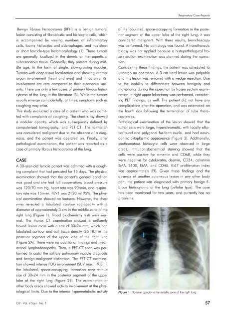
x-ray revealed a lobulated contour radiopacity with a<br />
diameter of approximately 3 cm in the middle zone of the<br />
right lung (Figure 1). Blood biochemistry tests were normal.<br />
The thorax CT examination showed a uniformly<br />
bound lesion mass with a size of 30x24 mm, which had<br />
lobulated contour and soft tissue density (26 HU) in the<br />
posterior segment of the upper lobe of the right lung<br />
(Figure 2A). There were no additional findings and mediastinal<br />
lymphadenopathy. Then, a PET-CT scan was performed<br />
to assist the solitary pulmonary nodule diagnosis<br />
and benign-malignant distinction. The PET-CT examination<br />
showed intense FDG involvement (SUV max: 19.3) in<br />
the lobulated, space-occupying, formation zone with a<br />
size of 30x24 mm in the posterior segment of the upper<br />
lobe of the right lung (Figure 2B). The examination of<br />
other body areas showed activity involvement at the physiological<br />
limits. Due to the intense hypermetabolic activity<br />
of the lobulated, space-occupying formation in the posterior<br />
segment of the upper lobe of the right lung, it was<br />
considered malignant. With these results, bronchoscopy<br />
was performed. No pathology was found. A transthoracic<br />
biopsy was not applied because a histopathological frozen<br />
section examination was planned during the operation.<br />
Considering these findings, the patient was scheduled to<br />
undergo an operation. A 3 cm hard lesion was palpable<br />
and this lesion was removed with a wedge resection. Due<br />
to the inability to differentiate between benignity and<br />
malignancy during the operation by frozen section examination,<br />
a right upper lobectomy was performed, considering<br />
PET findings, as well. The patient did not have any<br />
complications after the operation, and was externated on<br />
the fourth day following the termination of tube thoracostomies.<br />
Pathological examination of the lesion showed that the<br />
tumor cells were large, hyperchromatic, with locally elliptic/round<br />
and polygonal fusiform nuclei, and had eosinophilic<br />
cytoplasmic appearance (Figure 3). Additionally,<br />
xanthomatous histiocytic cells were observed in large<br />
areas. Immunohistochemical staining showed that the<br />
cells were positive for vimentin and CD68, while they<br />
were negative for cytokeratin, desmin, CD34, calretinin<br />
SMA, S100, EMA, and CD45. Ki67 proliferation index<br />
was approximately 5%. Given these findings and the<br />
absence of another cutaneous lesion in any other body<br />
part, the patient was diagnosed with primary benign fibrous<br />
histiocytoma of the lung (cellular type). The case<br />
has been monitored for two years, and currently has no<br />
problems.<br />
Figure 1: Nodular opacity in the middle zone of the right lung<br />
<strong>Cilt</strong> - Vol. 4 <strong>Sayı</strong> - No. 1 57


