

Respircase Cilt: 4 - Sayı: 1 Yıl: 2015

Create successful ePaper yourself
Turn your PDF publications into a flip-book with our unique Google optimized e-Paper software.
Respiratory Case Reports<br />
discovered incidentally on chest x-rays with typical radiographic<br />
appearances. Infection or rupture may change<br />
the typical radiographic appearance of a hydatid cyst,<br />
causing an incorrect diagnosis and delayed treatment (4).<br />
The diagnosis of complicated pulmonary hydatid disease<br />
is difficult because it can mimic many other pulmonary<br />
and pleural diseases, including pneumonia, bronchiectasis,<br />
tuberculosis, abscess, lung tumors, pleurisy, and empyema<br />
(3,4). Although bronchoscopy is unnecessary in<br />
patients with a typical clinical and radiological picture, it<br />
can be performed when there is suspicion of a tumor or<br />
when the presentation is atypical (3,5). We present a case<br />
evaluated for suspicion of lung cancer in which the diagnosis<br />
was made through bronchoscopy.<br />
CASE<br />
A 22-year-old Turkish woman was admitted to our hospital<br />
with hemoptysis. She had also a history of hemoptysis<br />
one year ago. The patient was a non-smoker and had no<br />
history of close contact with dogs or other animals. The<br />
physical examination, full blood count, and biochemical<br />
tests were normal. Erythrocyte sedimentation rate was 10<br />
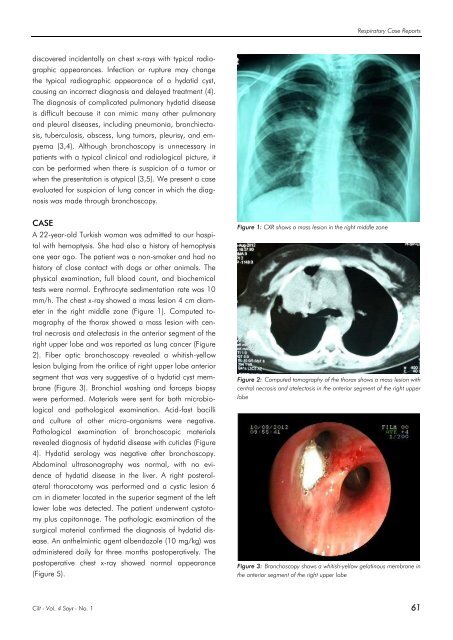
mm/h. The chest x-ray showed a mass lesion 4 cm diameter<br />
in the right middle zone (Figure 1). Computed tomography<br />
of the thorax showed a mass lesion with central<br />
necrosis and atelectasis in the anterior segment of the<br />
right upper lobe and was reported as lung cancer (Figure<br />
2). Fiber optic bronchoscopy revealed a whitish-yellow<br />
lesion bulging from the orifice of right upper lobe anterior<br />
segment that was very suggestive of a hydatid cyst membrane<br />
(Figure 3). Bronchial washing and forceps biopsy<br />
were performed. Materials were sent for both microbiological<br />
and pathological examination. Acid-fast bacilli<br />
and culture of other micro-organisms were negative.<br />
Pathological examination of bronchoscopic materials<br />
revealed diagnosis of hydatid disease with cuticles (Figure<br />
4). Hydatid serology was negative after bronchoscopy.<br />
Abdominal ultrasonography was normal, with no evidence<br />
of hydatid disease in the liver. A right posterolateral<br />
thoracotomy was performed and a cystic lesion 6<br />
cm in diameter located in the superior segment of the left<br />
lower lobe was detected. The patient underwent cystotomy<br />
plus capitonnage. The pathologic examination of the<br />
surgical material confirmed the diagnosis of hydatid disease.<br />
An anthelmintic agent albendazole (10 mg/kg) was<br />
administered daily for three months postoperatively. The<br />
postoperative chest x-ray showed normal appearance<br />
(Figure 5).<br />
Figure 1: CXR shows a mass lesion in the right middle zone<br />
Figure 2: Computed tomography of the thorax shows a mass lesion with<br />
central necrosis and atelectasis in the anterior segment of the right upper<br />
lobe<br />
Figure 3: Bronchoscopy shows a whitish-yellow gelatinous membrane in<br />
the anterior segment of the right upper lobe<br />
<strong>Cilt</strong> - Vol. 4 <strong>Sayı</strong> - No. 1 61


