

Respircase Cilt: 4 - Sayı: 1 Yıl: 2015

Create successful ePaper yourself
Turn your PDF publications into a flip-book with our unique Google optimized e-Paper software.
Respiratory Case Reports<br />
The most common symptoms are dyspnea, cough, and<br />
sputum expectoration. Respiratory function tests may be<br />
either normal or may show signs of minimal obstruction<br />
or reduced diffusion capacity. Cases with massive fibrosis<br />
are characterized by a loss in respiratory function (3). The<br />
respiratory impairment is worsened by smoking. X-rays<br />
and computerized tomography of chest are important<br />
diagnostic modalities.<br />
Pneumoconiosis among dental technicians has been<br />
reported since 1962 (4). The number of cases with dental<br />
technician’s pneumoconiosis has recently increased in<br />
Turkey. Herein, we report three affected patients to draw<br />
attention to this potentially preventable disorder.<br />
was negative three times. No signs of malignancy or TB<br />
were evident in the TBNA sample from the bronchial<br />
lavage obtained via fiber optic bronchoscopy. The examination<br />
of a BAL sample taken from the middle lobe of<br />
lung revealed a total cell count of 700,000/ml, of which<br />
87.5% were macrophages, 11.5% lymphocytes, and 1%<br />
neutrophils. The ratio of helper-to-suppressor T-cells was<br />
low (CD4/CD8: 0.2).<br />
CASE<br />
The current study reports three cases aged 45, 50, and<br />
53 years who presented to our clinic with dyspnea.<br />
Case 1: A 53-year-old man presented to the department<br />
of chest disease with dyspnea upon exertion and sputum<br />
expectoration for two years. His history was notable for<br />
working as a salaried dental technician in a private laboratory<br />
for 31 years. He had a smoking history of 25<br />
packs/year. He had no history of lung tuberculosis nor<br />
did he have any contact with infected persons.<br />
Upon physical examination, the patient’s vital signs were<br />
as follows: body temperature: 36°C, blood pressure:<br />
130/75 mmHg, pulse rate: 90 beats/minute (rhythmic),<br />
respiratory rate: 18 breaths/minute, SaO 2 95% (on room<br />
air). The respiratory system appeared normal on physical<br />
examination.<br />
Respiratory function tests revealed the following: FVC:<br />
3.15 L (80%), FEV1: 2.50 L (78%), and FEV1/FVC: 67. A<br />
diffusion test revealed a DLCO of 78% and a DLCO/VA<br />
of 80%.<br />
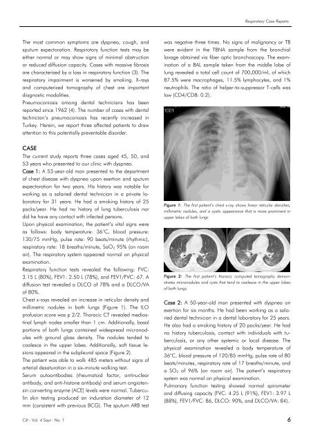
Chest x-rays revealed an increase in reticular density and<br />
millimetric nodules in both lungs (Figure 1). The ILO<br />
profusion score was p 2/2. Thoracic CT revealed mediastinal<br />
lymph nodes smaller than 1 cm. Additionally, basal<br />
portions of both lungs contained widespread micronodules<br />
with ground glass density. The nodules tended to<br />
coalesce in the upper lobes. Additionally, soft tissue lesions<br />
appeared in the subpleural space (Figure 2).<br />
The patient was able to walk 485 meters without signs of<br />
arterial desaturation in a six-minute walking test.<br />
Serum autoantibodies (rheumatoid factor, antinuclear<br />
antibody, and anti-histone antibody) and serum angiotensin<br />
converting enzyme (ACE) levels were normal. Tuberculin<br />
skin testing produced an induration diameter of 12<br />
mm (consistent with previous BCG). The sputum ARB test<br />
Figure 1: The first patient’s chest x-ray shows linear reticular densities,<br />
millimetric nodules, and a cystic appearance that is more prominent in<br />
upper lobes of both lungs<br />
Figure 2: The first patient’s thoracic computed tomography demonstrates<br />
micronodules and cysts that tend to coalesce in the upper lobes<br />
of both lungs<br />
Case 2: A 50-year-old man presented with dyspnea on<br />
exertion for six months. He had been working as a salaried<br />
dental technician in a dental laboratory for 25 years.<br />
He also had a smoking history of 20 packs/year. He had<br />
no history tuberculosis, contact with individuals with tuberculosis,<br />
or any other systemic or local disease. The<br />
physical examination revealed a body temperature of<br />
36°C, blood pressure of 120/85 mmHg, pulse rate of 80<br />
beats/minutes, respiratory rate of 17 breaths/minute, and<br />
a SO 2 of 96% (on room air). The patient’s respiratory<br />
system was normal on physical examination.<br />
Pulmonary function testing showed normal spirometer<br />
and diffusing capacity (FVC: 4.25 L (91%), FEV1: 3.97 L<br />
(88%), FEV1/FVC: 86, DLCO: 90%, and DLCO/VA: 84).<br />
<strong>Cilt</strong> - Vol. 4 <strong>Sayı</strong> - No. 1 6


