

Respircase Cilt: 4 - Sayı: 1 Yıl: 2015

You also want an ePaper? Increase the reach of your titles
YUMPU automatically turns print PDFs into web optimized ePapers that Google loves.
Primary Benign Fibrous Histiocytoma of the Lung with FDG Involvement | Akcam et al.<br />
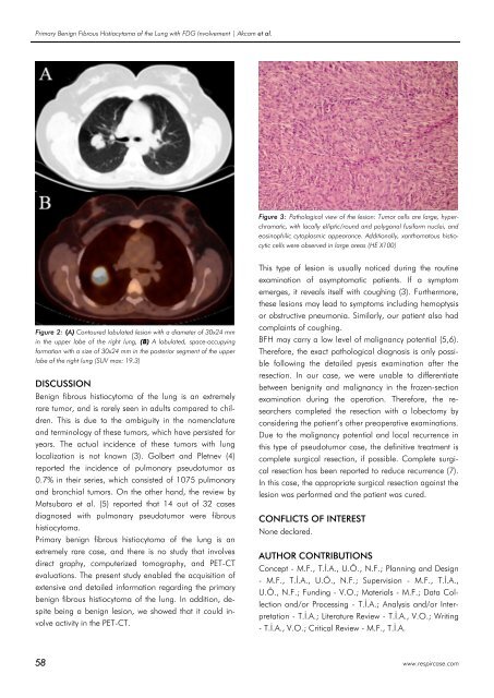
Figure 3: Pathological view of the lesion: Tumor cells are large, hyperchromatic,<br />
with locally elliptic/round and polygonal fusiform nuclei, and<br />
eosinophilic cytoplasmic appearance. Additionally, xanthomatous histiocytic<br />
cells were observed in large areas (HE X100)<br />
Figure 2: (A) Contoured lobulated lesion with a diameter of 30x24 mm<br />
in the upper lobe of the right lung, (B) A lobulated, space-occupying<br />
formation with a size of 30x24 mm in the posterior segment of the upper<br />
lobe of the right lung (SUV max: 19.3)<br />
DISCUSSION<br />
Benign fibrous histiocytoma of the lung is an extremely<br />
rare tumor, and is rarely seen in adults compared to children.<br />
This is due to the ambiguity in the nomenclature<br />
and terminology of these tumors, which have persisted for<br />
years. The actual incidence of these tumors with lung<br />
localization is not known (3). Golbert and Pletnev (4)<br />
reported the incidence of pulmonary pseudotumor as<br />
0.7% in their series, which consisted of 1075 pulmonary<br />
and bronchial tumors. On the other hand, the review by<br />
Matsubara et al. (5) reported that 14 out of 32 cases<br />
diagnosed with pulmonary pseudotumor were fibrous<br />
histiocytoma.<br />
Primary benign fibrous histiocytoma of the lung is an<br />
extremely rare case, and there is no study that involves<br />
direct graphy, computerized tomography, and PET-CT<br />
evaluations. The present study enabled the acquisition of<br />
extensive and detailed information regarding the primary<br />
benign fibrous histiocytoma of the lung. In addition, despite<br />
being a benign lesion, we showed that it could involve<br />
activity in the PET-CT.<br />
This type of lesion is usually noticed during the routine<br />
examination of asymptomatic patients. If a symptom<br />
emerges, it reveals itself with coughing (3). Furthermore,<br />
these lesions may lead to symptoms including hemoptysis<br />
or obstructive pneumonia. Similarly, our patient also had<br />
complaints of coughing.<br />
BFH may carry a low level of malignancy potential (5,6).<br />
Therefore, the exact pathological diagnosis is only possible<br />
following the detailed pyesis examination after the<br />
resection. In our case, we were unable to differentiate<br />
between benignity and malignancy in the frozen-section<br />
examination during the operation. Therefore, the researchers<br />
completed the resection with a lobectomy by<br />
considering the patient’s other preoperative examinations.<br />
Due to the malignancy potential and local recurrence in<br />
this type of pseudotumor case, the definitive treatment is<br />
complete surgical resection, if possible. Complete surgical<br />
resection has been reported to reduce recurrence (7).<br />
In this case, the appropriate surgical resection against the<br />
lesion was performed and the patient was cured.<br />
CONFLICTS OF INTEREST<br />
None declared.<br />
AUTHOR CONTRIBUTIONS<br />
Concept - M.F., T.İ.A., U.Ö., N.F.; Planning and Design<br />
- M.F., T.İ.A., U.Ö., N.F.; Supervision - M.F., T.İ.A.,<br />
U.Ö., N.F.; Funding - V.O.; Materials - M.F.; Data Collection<br />
and/or Processing - T.İ.A.; Analysis and/or Interpretation<br />
- T.İ.A.; Literature Review - T.İ.A., V.O.; Writing<br />
- T.İ.A., V.O.; Critical Review - M.F., T.İ.A.<br />
58 www.respircase.com


