3B MEDIZIN | Biologie | Bachmann Lehrmittel

Sie wollen auch ein ePaper? Erhöhen Sie die Reichweite Ihrer Titel.
YUMPU macht aus Druck-PDFs automatisch weboptimierte ePaper, die Google liebt.
VR1152/4006657/1001480<br />
Posteriorarch<br />
of atlas<br />
Spinous<br />
process<br />
Radiate ligament<br />
of head of rib<br />
Costotransverse<br />
ligament<br />
Lateral Vertebral<br />
costotransverse foramen<br />
ligament<br />
Verteberal arch<br />
Costal<br />
process<br />
Intervertebral<br />
disc<br />
Spinal nerve<br />
Auricular surface<br />
of sacrum<br />
Posterior rami<br />
of sacral nerves<br />
Osteoporosis is a disease characterized by a decrease<br />
in bone mass. Possible causes include inactivity,<br />
poor nutrition (protein, calcium, or vitamin<br />
defi ciency, alcoholism), or prolonged corticosteroid<br />
therapy. Postmenopausal women are particularly<br />
affected, as the reduction in estrogen production<br />
causes a predominance of osteoclast<br />
acti vity over that of the osteoblasts. This results in<br />
a loss bone stability leading to fractures, most<br />
commonly in the spinal column, femur, and radius.<br />
The collapse of thoracic vertebrae cause them<br />
to assume a wedge shape, resulting in the typical<br />
“dowager’s hump” and shortened statue of the<br />
osteo porosis patient.<br />
Printed in Germany<br />
VR1620/4006710/1001586<br />
Anterior cutaneous<br />
branches of intercostal<br />
nerves<br />
Intercostobrachial nerve<br />
Medial brachial cutaneous nerve<br />
Lateral cutaneous branches of<br />
intercostal nerves<br />
Medial antebrachial<br />
cutaneous nerve<br />
Posterior antebrachial cutaneous<br />
nerve (from radial nerve)<br />
Lateral antebrachial<br />
cutaneous nerve<br />
Superficial branch<br />
of radial nerve<br />
Femoral branch of<br />
genitofemoral nerve<br />
Ilioinguinal nerve<br />
Median branch<br />
of median nerve<br />
Dorsal<br />
nerve of<br />
penis<br />
Anterior<br />
cutaneous<br />
nerves (from<br />
femoral nerve)<br />
Great saphenous vein<br />
Lateral sural cutaneous nerve<br />
Superficial peroneal nerve<br />
Intermediate dorsal cutaneous nerve<br />
Medial dorsal cutaneous nerve<br />
Anterior arch of atlas<br />
Dens of axis<br />
Superior articular facet<br />
Foramen<br />
transversarium<br />
Atlas (C1)<br />
Auriculotemporal nerve<br />
Greater occipital nerve<br />
Trigeminal ganglion<br />
Greater petrosal nerve<br />
Chorda tympani<br />
Mandibular nerve<br />
Facial nerve<br />
Inferior alveolar nerve<br />
Lesser occipital nerve<br />
Great auricular nerve<br />
Accessory nerve<br />
Transverse cervical nerve<br />
Supraclavicular nerves<br />
Axis (C2)<br />
Body of vertebra<br />
Head of rib<br />
Joint of head of rib<br />
Rib<br />
Spinous process<br />
Costotransverse<br />
joint<br />
Spinal process<br />
Body of Healthy Vertebra<br />
Body of Osteoporitic<br />
Vertebra<br />
This is a disease of the thoracic spinal column<br />
caused by impaired bone growth. The vertebrae<br />
appear wedge-shaped due to defective positioning<br />
of the sympysis. The curvature of the thoracic vertebrae<br />
increases, and their mobility is limited. Boys<br />
are more often affected than girls.<br />
Infrapatellar<br />
branch of<br />
saphenous<br />
nerve<br />
Saphenous<br />
nerve<br />
Deep peroneal nerve<br />
Erector spinae muscle Medial branch of dorsal ramus<br />
Lateral branch of dorsal ramus<br />
Trapezius muscle<br />
Spinal ganglion<br />
Latissimus dorsi muscle<br />
Spinal cord<br />
External intercostal muscle<br />
Dorsal root of<br />
spinal nerve<br />
Internal intercostal muscle<br />
Ventral root of spinal<br />
Ventral ramus of spinal<br />
nerve<br />
nerve (intercostal nerve)<br />
Meningeal branch<br />
Sympathetic trunk<br />
Serratus anterior muscle<br />
Gray and white rami<br />
communicantes<br />
Lateral cutaneous branch<br />
Thoracic splanchnic<br />
Innermost<br />
nerves<br />
intercostal<br />
Ventral ramus of spinal nerve<br />
Subcostal muscles<br />
muscles<br />
External abdominal oblique muscle<br />
Vertebral column<br />
External intercostal membrane<br />
Rectus abdominus muscle<br />
Costal origin of diaphragm<br />
Anterior cutaneous branch of<br />
Linea alba<br />
ventral ramus<br />
Printed in Germany<br />
Femoral<br />
nerve<br />
Superficial<br />
branch<br />
of ulnar<br />
nerve<br />
Deep<br />
branch<br />
of ulnar<br />
nerve<br />
(II)<br />
C1<br />
C2<br />
C3<br />
C4<br />
C5<br />
C6<br />
C7<br />
D1<br />
D2<br />
D3<br />
D4<br />
D5<br />
D6<br />
D7<br />
D8<br />
D9<br />
D10<br />
D11<br />
D12<br />
L1<br />
L2<br />
L3<br />
L4<br />
L5<br />
Normal<br />
Posture<br />
Cervical<br />
vertebrae Vertebrae<br />
C1 cervicales = atlas<br />
C2 C1=Atlas, = axis<br />
C2=Axis<br />
Thoracic Vertebrae<br />
vertebrae thoracicae<br />
Lumbar Vertebrae<br />
vertebrae lumbales<br />
Sacrum Os sacrum<br />
Coccyx Os coccygis<br />
Lordosis<br />
Promontary of<br />
sacrum<br />
Auricular<br />
surface<br />
of sacrum<br />
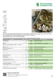
The double S-shaped curvature of the spinal column allows weight bearing on the<br />
vertical axis as needed for activities such as jumping. Four basic posture types<br />
are recognized. Individual posture is hereditary, but is also influenced by the<br />
growth process, age, illness, and condition of shoulder, back and hip muscles.<br />
Ulnar nerve<br />
Median nerve<br />
Cervical nerves<br />
1-8<br />
Thoracic nerves<br />
1-12<br />
Lumbar nerves<br />
1-5<br />
Severe<br />
lordosis<br />
Subcostal nerve<br />
Cauda equina<br />
Deep branch of radial nerve<br />
Rhomboid fossa<br />
Inferior colliculus<br />
Iliohypogastric nerve<br />
Arachnoid<br />
Cerebellar peduncle<br />
Ilioinguinal nerve<br />
Glossopharyngeal and<br />
Superficial branch of radial nerve<br />
vagus nerves<br />
Lateral cutaneous femoral nerve<br />
Genitofemoral nerve<br />
Femoral branch of<br />
genitofemoral nerve<br />
Genital branch of<br />
genitofemoral nerve<br />
Ulnar nerve<br />
Median nerve<br />
Obturator nerve<br />
Sacral plexus<br />
Spinal cord<br />
Common palmar<br />
digital nerves (from<br />
median nerve)<br />
1st, 2nd,<br />
4th ribs<br />
Proper palmar digital<br />
nerves (from median nerve)<br />
Proper palmar<br />
digital nerves<br />
(from ulnar nerve)<br />
Sacrum<br />
(opened)<br />
Arch<br />
of vertebra<br />
Spinal dura mater<br />
Coccygeal nerve<br />
Coccyx<br />
External filum<br />
terminale<br />
Sacral<br />
nerves 1-5<br />
l o r d<br />
k y p h<br />
u e<br />
i q<br />
Flat back<br />
Temporal branches of facial nerve<br />
Lateral branch of supraorbital nerve<br />
Medial branch of supraorbital nerve<br />
Longitudinal cerebral fissure<br />
Ophthalmic nerve<br />
Frontal pole of cerebrum<br />
Frontal lobe<br />
Frontal nerve<br />
Orbital gyri<br />
Olfactory bulb<br />
Maxillary nerve<br />
Pterygopalatine ganglion<br />
Temporal pole of cerebrum<br />
Olfactory tract [I]<br />
Posterior superior alveolar branches of infraorbital nerve<br />
Optic nerve [II]<br />
Infraorbital nerve<br />
Lingual nerve<br />
Anterior perforated substance<br />
Optic chiasm<br />
Ophthalmic nerve [V1]<br />
Pituitary gland (hypophysis)<br />
Maxillary nerve [V2]<br />
Oculomotor [III]<br />
nerve<br />
Mandibular nerve [V3]<br />
Mental nerve<br />
Mamillary body<br />
Trochlear nerve [IV]<br />
Trigeminal nerve [V]<br />
Pons<br />
Facial nerve [VII]<br />
4th cervical nerve<br />
5th cervical nerve<br />
Abducens nerve [VI]<br />
Vestibulocochlear nerve [VIII]<br />
6th cervical nerve<br />
Hypoglossal nerve [XII]<br />
Glossopharyngeal nerve [IX]<br />
7th cervical nerve<br />
8th cervical nerve<br />
Vagus nerve [X]<br />
Spinal cord<br />
Brachial plexus<br />
1st thoracic nerve<br />
Accessory nerve [XI]<br />
Vermis of cerebellum Olive Cerebellar hemisphere<br />
Axillary artery<br />
Thoracic portion of<br />
spinal cord<br />
Musculocutaneous nerve<br />
Sympathetic trunk<br />
Intercostal nerves<br />
Radial nerve<br />
Median nerve<br />
Ulnar nerve<br />
Saphenous<br />
nerve<br />
Vasoadductor<br />
membrane<br />
Cutaneous<br />
branch of<br />
obturator nerve<br />
Sartorius muscle<br />
Deep peroneal nerve<br />
Superficial<br />
peroneal nerve<br />
Ilium<br />
Hypoglossal nerve<br />
Filum terminale<br />
Femoral nerve<br />
Conus medullaris<br />
Neural plate<br />
Neural groove<br />
Future neural<br />
crest<br />
Ectoderm<br />
Ectoderm<br />
Neural fold<br />
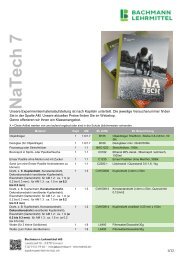
Cross Section of Nervous System Apparatus<br />
of a 20 Day-old Embryo<br />
After development of the neural plate, the midline neural groove is formed.<br />
It is bounded by both neural folds.<br />
Ectoderm<br />
Neural crest<br />
Sulcus limitans<br />
21st day<br />
24th day<br />
In the median area, the neural folds The neural groove closes and<br />
begin fusing to form the neural groove. is covered by surface ectoderm.<br />
Posterior<br />
femoral<br />
cutaneous nerve<br />
Dorsal digital nerves<br />
Common peroneal<br />
nerve<br />
Tibial nerve<br />
Vertebra<br />
prominens<br />
Transverse costal<br />
facet<br />
Intervertebral<br />
foramina<br />
Inferior and<br />
superior<br />
costal facets<br />
Spinal<br />
processes<br />
Lateral disc herniation<br />
Epidural space<br />
Subarachnoid space<br />
Arachnoid<br />
Posterior ramus<br />
of spinal nerve<br />
Anterior ramus<br />
of spinal nerve<br />
Medial disc herniation<br />
Degeneration or erosion of vertebrae and vertebral<br />
discs may cause part or all of the disc to protrude into<br />
the spinal canal, thereby compressing the spinal cord or<br />
spinal nerve roots. Disturbances in sensation, pain, and<br />
paralysis can result. Herniated discs often occur in the<br />
lower lumbar region, but can also be found in the cervical<br />
area.<br />
This hereditary disease, which affects nine times<br />
more men than women, is classified as an arthritis.<br />
The course of the illness can be chronic or episodic.<br />
Various joints show inflammatory changes with<br />
subsequent stiffening and ossification. The iliosacral,<br />
vertebral, and hip joints are most often affected.<br />
Cervical portion of<br />
spinal cord<br />
Dura mater<br />
Dorsal ramus of spinal nerve<br />
Ventral ramus of spinal nerve<br />
Spinal cord<br />
Ligamentum flavum<br />
Posterior<br />
root of<br />
spinal nerve<br />
Anterior<br />
root of<br />
spinal<br />
nerve<br />
Spinal<br />
ganglion<br />
Vertebral atery<br />
and vein<br />
a) b)<br />
a) Concentric pressure produces an evenly applied compression (C)<br />
on the nucleus pulposus and the anulus fibrosus.<br />
b) Eccentric pressure pushes the nucleus propulsus to the unaffected<br />
side; the anulus fibrosusis subjected to traction forces (T).<br />
Intervertebral foramen<br />
Superior articular process<br />
Costal<br />
Anterior<br />
Ligamentum flavum<br />
processes longitudinal<br />
ligament<br />
Supraspinous<br />
ligament<br />
Anulus<br />
fibrosis fibrosus<br />
Discus intervertebralis<br />
ligament<br />
Interspinous<br />
intervertebral<br />
disc Nucleus<br />
pulposus<br />
Spinous<br />
process<br />
Inferior articular<br />
Basi-vertebral vien canal<br />
process<br />
Posterior longitudinal ligament<br />
Posterior<br />
sacral<br />
foramina<br />
Epidural space<br />
Subarachnoid space<br />
Arachnoid<br />
Medial calcaneal<br />
branches of tibial<br />
nerve<br />
Cerebrum<br />
Cerebellum<br />
www.3bscientific.com<br />
Hamburg, Germany, 1995<br />
Design and text: Antje Gottberg<br />
Illustration: Heinrich Römisch<br />
Scoliosis is a term designating<br />
lateral curvature of the spinal<br />
column. Scoliotic deviations,<br />
which can result from anomalies<br />
such as differences in<br />
leg length, are differentiated<br />
from structual scoliosis which<br />
is caused by deformity of the<br />
vertebrae.<br />
Supraspinatus muscle<br />
Deltoid muscle<br />
Infraspinatus muscle<br />
Axillary nerve<br />
Teres major muscle<br />
Long head of triceps<br />
brachii muscle<br />
Lateral head of triceps<br />
brachii muscle<br />
Radial nerve<br />
Posterior brachial cutaneous nerve<br />
Latissimus dorsi muscle<br />
Intercostal nerves<br />
Subcostal nerve<br />
External abdominal oblique muscle<br />
Iliohypogastric nerve<br />
Supinator muscle<br />
Ilioinguinal nerve<br />
Extensor carpi radialis longus muscle<br />
Genitofemoral nerve<br />
Quadratus lumborum muscle<br />
Muscular branches of radial nerve<br />
Extensor carpi ulnaris<br />
muscle<br />
Abductor pollicis<br />
Superior<br />
longus muscle<br />
gluteal<br />
Extensor pollicis brevis<br />
nerve<br />
Extensor pollicis longus muscle<br />
Piriformis<br />
muscle<br />
Superficial branches of<br />
radial nerve<br />
Gluteus medius<br />
muscle<br />
Extensor indices muscle<br />
Extensor retinaculum<br />
Inferior gluteal<br />
nerve<br />
Tendon of<br />
extensor pollicis<br />
Dorsal branch<br />
muscles<br />
of ulnar nerve<br />
Dorsal<br />
Quadratus<br />
interosseous<br />
femoris muscle<br />
muscles<br />
Posterior femoral<br />
cutaneous nerve<br />
Gluteus maximus<br />
muscle<br />
Dorsal digital<br />
nerves<br />
Sciatic nerve<br />
Adductor magnus muscle<br />
Iliotibial tract<br />
Long head of biceps<br />
femoris muscle<br />
Short head of biceps<br />
femoris muscle<br />
Semitendinosus muscle<br />
Adductor magnus<br />
Vastus medialis muscle<br />
Long head of biceps<br />
femoris muscle<br />
Lateral sural cutaneous nerve<br />
Plantaris muscle<br />
Semimembranosus muscle<br />
Lateral head of<br />
gastrocnemius muscle<br />
Medial head of<br />
gastrocnemius muscle<br />
Soleus muscle<br />
Tibial nerve<br />
Flexor hallucis longus muscle<br />
Flexor digitorum longus muscle<br />
Tibialis posterior muscle<br />
Peroneus brevis muscle<br />
Spinal cord<br />
Ligamentum flavum<br />
Dorsal root<br />
Sural nerve<br />
Calcaneal (Achilles) tendon<br />
Lateral dorsal<br />
cutaneous nerve<br />
Ventral root<br />
Spinal ganglion<br />
Vertebral artery and vein<br />
www.3bscientific.com<br />
Hamburg, Germany, 1996<br />
Design and Text: Wilfried Hennig<br />
Tendon of<br />
extensor<br />
digitorum<br />
muscles<br />
Sternocleidomastoid<br />
muscle<br />
Scalene muscles<br />
Printed in Germany<br />
Clavicle<br />
Manubrium of<br />
sternum<br />
Right lung<br />
Sternum<br />
5th rib<br />
Xiphoid process<br />
External intercostal<br />
muscles<br />
Internal intercostal<br />
muscles<br />
Cartilage of 10th rib<br />
Heart<br />
Liver<br />
Stomach<br />
Gallbladder<br />
Duodenum<br />
Colon<br />
Frontal sinus<br />
Ethmoidal cells<br />
Maxillary sinus<br />
Hyoid bone<br />
Thyrohyoid membrane<br />
Larynx<br />
Cricothyroid muscle<br />
Thyroid gland<br />
Trachea<br />
Apex of the lung<br />
External intercostal muscles<br />
External abdominal muscle<br />
Internal abdominal muscle<br />
Rectus abdominis muscle<br />
Transversus abdominis muscle<br />
Superior recess<br />
Lateral recess<br />
Nasal recess<br />
Infraorbital recess<br />
Zygomatic recess<br />
Alveolar recess<br />
Clavicle<br />
1st rib<br />
Left lung<br />
The space inside the thorax is enlarged by contraction of the muscles<br />
responsible for inspiration. Air is breathed in as a result of the<br />
negative pressure generated. The most important muscle is the<br />
diaphragm which, on being flattened, enlarges the space inside<br />
the thorax. The muscles between the ribs create an enlarged<br />
space by raising the ribs. Intensified inspiration is underpinned<br />
by the ancillary respiratory muscles, which include<br />
the scalene muscles and the sternocleidomastoid muscle.<br />
Regular expiration is effected passively by virtue of the<br />
restoring force of the elastic pulmonary tissue. The<br />
internal intercostal muscles as well as the abdominal<br />
muscles facilitate intensified expiration.<br />
Apex of the lung<br />
Sulcus of subclavian artery<br />
Trachea<br />
Aortic arch<br />
Ribs<br />
Upper lobe<br />
Right pulmonary artery<br />
Horizontal fissure<br />
Right pulmonary veins<br />
Frontal section through<br />
left lung<br />
Heart<br />
Diaphragm<br />
Middle lobe<br />
Lower lobe<br />
Terminal bronchiolus<br />
Elastic fibers<br />
Bronchial branch<br />
Pulmonary vein<br />
Alveoli<br />
Alveolar wall<br />
Alveolar septum<br />
Alveolar pore<br />
Frontal sinus<br />
Middle nasal concha<br />
Inferior nasal concha<br />
Vestibule of nose<br />
Vestibule of mouth<br />
Horizontal fissure<br />
Right Lung<br />
Upper lobe<br />
1 Apical segment<br />
2 Posterior segment<br />
3 Anterior segment<br />
Middle lobe<br />
4 Lateral segment<br />
5 Medial segment<br />
Lower lobe<br />
6 Apical segment<br />
7 Medial basal segment<br />
8 Anterior basal segment<br />
9 Lateral basal segment<br />
10 Posterior basal segment<br />
Oblique fissure<br />
8<br />
4<br />
Left pulmonary artery<br />
Hilum of the lung<br />
Left principle bronchus<br />
Lower lobe<br />
Sulcus of esophagus<br />
2<br />
9<br />
1<br />
Base of the lung<br />
3<br />
5<br />
7<br />
10<br />
10<br />
Oblique fissure<br />
Right principle bronchus<br />
Bronchopulmonary lymph<br />
node<br />
Sulcus of azygos vein<br />
Pulmonary ligament<br />
Diaphragmatic surface<br />
3<br />
1<br />
9<br />
5<br />
4<br />
8<br />
Left lung<br />
3<br />
4<br />
Sphenoidal sinus<br />
Nasopharynx<br />
Oropharynx<br />
Epiglottis<br />
Laryngopharynx<br />
Apex of the lung<br />
Upper lobe<br />
Sulcus of aortic arch<br />
Left pulmonary veins<br />
Cardiac impression<br />
Oblique fissure<br />
Cardiac notch<br />
Lingula<br />
Respiratory bronchiolus<br />
Pulmonary vein<br />
Pulmonary artery<br />
Alveolar ductule<br />
Capillary network<br />
Sacculus<br />
Right lung<br />
Left lung<br />
Upper lobe<br />
1 Apical segment<br />
2 Posterior segment<br />
3 Anterior segment<br />
4 Superior lingual segment<br />
5 Inferior lingual segment<br />
Lower lobe<br />
6 Apical segment<br />
8 Anterior basal segment<br />
9 Lateral basal segment<br />
10 Posterior basal segment<br />
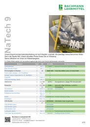
At the level of the 4th to 5th thoracic vertebra, the trachea divides into a<br />
left and a right principle bronchus. Together with the pulmonary vessels,<br />
the latter enter the pulmonary tissue at the hilum of the lung. Coursing further,<br />
the main bronchi divide into 2 or 3 lobar bronchi, with these in<br />
turn dividing to give rise to the segmental bronchi. The latter divide<br />
into increasingly smaller bronchi, called bronchioli. They lead into<br />
the so-called alveolar duct, which is surrounded by the alveoli.<br />
The individual alveoli are surrounded by a network of capillaries<br />
from the pulmonary circulation. It is here that the<br />
inspired oxygen passes from the alveoli into the pulmonary<br />
veins and carbon dioxide passes from the pulmonary<br />
arteries into the alveoli for expiration.<br />
6<br />
1<br />
1<br />
2<br />
9<br />
10<br />
10<br />
www.3bscientific.com<br />
Hamburg, Germany, 1998<br />
Design and text: Antje Gottberg, Wilfried Hennig<br />
Illustrations: Holger Vanselow<br />
6<br />
3<br />
4<br />
9<br />
Spinal Column<br />
Anatomy and Pathology<br />
r v<br />
c e<br />
i c a<br />
o s i s<br />
L o r d o s e<br />
C e r v i c a l<br />
l e<br />
Upper Cervical Vertebra<br />
Horizontal Section of 4th Cervical Vertebra<br />
o s i s<br />
Thoracic Vertebra with Costal Joints<br />
left side with ligaments<br />
T h o r a<br />
s e t h o<br />
C y p h o<br />
c i c<br />
r a c<br />
CD<br />
C D ZT CD<br />
Pressure on the Intervertebral Discs<br />
o m b<br />
s e l<br />
r d o<br />
a i r e<br />
r d o s<br />
r l o<br />
L u m b a<br />
i s<br />
L o<br />
Longitudinal Section of<br />
Lumbar Vertebral Segment<br />
Lumbar Vertebra, Sacrum and Coccyx<br />
with spinal nerves<br />
Osteoporosis<br />
Anterior View of Spinal Column<br />
Four Types of Posture<br />
Lateral View of Spinal Column<br />
Scoliosis<br />
Disc Herniation<br />
Scheuermann’s Disease<br />
(juvenile kyphosis)<br />
Bechterew’s Disease<br />
(ankylosing spondylitis)<br />
© <strong>3B</strong> Scientific GmbH<br />
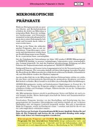
Die menschliche Wirbelsäule -<br />
Anatomie und Pathologie<br />
Laminiert M-1001314<br />
Papierversion M-4006574<br />
Der menschliche Schädel<br />
Laminiert M-1001312<br />
Papierversion M-4006573<br />
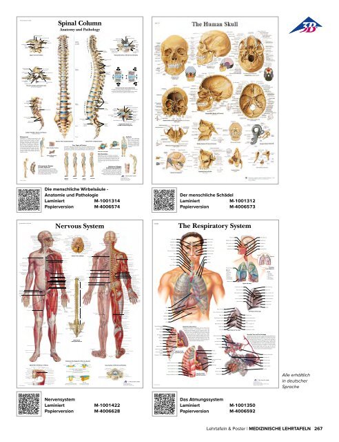
Nervous System<br />
VR1322<br />
The Respiratory System<br />
Inferior View of Brain<br />
Posterior<br />
Lung Segments<br />
Upper Airways<br />
Left hilum of the lung<br />
Spinal Cord<br />
with Spinal Nerves<br />
Muscles of the<br />
expiratory phase<br />
Respiratory Musculature<br />
Right hilum of the lung<br />
Bronchial Tree and Gas Exchange<br />
Embryonic Development of Nervous System<br />
Spinal Nerve Pathway in Thorax<br />
Cross Section of 4th Cervical Vertebra<br />
© <strong>3B</strong> Scientific GmbH<br />
Muscles of the<br />
inspiratory phase<br />
© <strong>3B</strong> Scientific GmbH<br />
Alle erhältlich<br />
in deutscher<br />
Sprache<br />
Nervensystem<br />
Laminiert M-1001422<br />
Papierversion M-4006628<br />
Das Atmungssystem<br />
Laminiert M-1001350<br />
Papierversion M-4006592<br />
Lehrtafeln & Poster | <strong>MEDIZIN</strong>ISCHE LEHRTAFELN 267