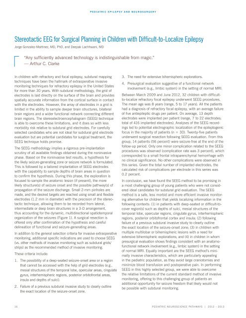
Pediatric Neuroscience Pathways Fall 2012 - Cleveland Clinic
Pediatric Neuroscience Pathways Fall 2012 - Cleveland Clinic
Pediatric Neuroscience Pathways Fall 2012 - Cleveland Clinic
Create successful ePaper yourself
Turn your PDF publications into a flip-book with our unique Google optimized e-Paper software.
In children with refractory and focal epilepsy, subdural mapping<br />
techniques have been the hallmark of extraoperative invasive<br />
monitoring techniques for refractory epilepsy in the united states<br />
for more than 30 years. With subdural methodology, the grid of<br />
electrodes is laid directly on the surface of the brain and provides<br />
spatially accurate information from the cortical surface in contact<br />
with the electrodes. However, the array of electrodes in a grid is<br />
limited in the ability to sample deeper brain structures, bilateral<br />
brain regions and a wider functional network connecting different<br />
brain regions. the stereoelectroencephalogram (seeg) technique<br />
is able to overcome these limitations, and it does so with less<br />
morbidity risk relative to subdural grid electrodes. For carefully<br />
selected candidates who are not ideal for subdural grid electrode<br />
evaluation but are potential candidates for surgical treatment, the<br />
seeg technique holds promise.<br />
The SEEG methodology implies a rigorous pre-implantation<br />
scrutiny of all available findings obtained during the noninvasive<br />
phase. Based on the noninvasive test results, a hypothesis for<br />
the likely seizure-generating zone or seizure network is formulated.<br />
this is followed by a tailored implantation of seeg electrodes<br />
with the capability to sample depths of brain areas in question<br />
to confirm the hypothesis. During this phase, the exploration is<br />
focused to sample the anatomic lesion (if present), the more<br />
likely structure(s) of seizure onset and the possible pathway(s) of<br />
propagation of the seizure discharge. Small 2-mm pinholes are<br />
made, and the desired targets are reached using small and flexible<br />
electrodes (1.2 mm in diameter) with the precision of the stereotactic<br />
technique, allowing them to be recorded from lateral,<br />
intermediate or deep brain structures in a 3-D arrangement,<br />
thus accounting for the dynamic, multidirectional spatiotemporal<br />
organization of the seizures (Figure 1). a surgical resection is<br />
offered only after confirmation of the hypothesis and careful<br />
delineation of functional and seizure-generating areas.<br />
in addition to the general selection criteria for invasive extraoperative<br />
monitoring, additional specific indications are used to choose SEEG<br />
(vs. other methods of invasive monitoring such as subdural grids/<br />
strips) as the recommended method of invasive monitoring.<br />
these criteria include:<br />
1. The possibility of a deep-seated seizure-onset area or a region<br />
that cannot be accessed with the help of grid electrodes (e.g.,<br />
mesial structures of the temporal lobe, opercular areas, cingulate<br />
gyrus, interhemispheric regions, posterior orbitofrontal areas,<br />
insula and depths of sulci).<br />
2. Failure of a previous subdural invasive study to clearly outline<br />
the exact location of the seizure-onset zone.<br />
<strong>Pediatric</strong> ePilePsy aNd Neurosurgery<br />
Stereotactic EEG for Surgical Planning in Children with Difficult-to-Localize Epilepsy<br />
Jorge Gonzalez-Martinez, MD, PhD, and Deepak Lachhwani, MD<br />
“Any sufficiently advanced technology is indistinguishable from magic.”<br />
— arthur c. clarke<br />
3. the need for extensive bihemispheric explorations.<br />
4. Presurgical evaluation suggestive of a functional network<br />
involvement (e.g., limbic system) in the setting of normal MRI.<br />
Between March 2009 and June <strong>2012</strong>, 32 children with difficultto-localize<br />
refractory focal epilepsy underwent SEEG procedures.<br />
The mean age was 8 years (range, 5 to 17 years). All the patients<br />
had a diagnosis of refractory focal epilepsy, with an average failure<br />
of five antiepileptic drugs per patient. On average, 13 depth<br />
electrodes were implanted per patient (range, 7 to 22 electrodes;<br />
total of 416 implanted electrodes). analyses of the seeg recordings<br />
led to potential electrographic localization of the epileptogenic<br />
focus in the majority of patients (n = 30). Twenty-five patients<br />
underwent surgical resection following seeg evaluation. From this<br />
group, 14 patients (56 percent) were seizure-free at the end of the<br />
follow-up period. Only one minor complication related to the SEEG<br />
procedures was observed (complication rate was 3 percent), which<br />
corresponded to a small frontal intraparenchymal hemorrhage with<br />
no clinical significance. No other complications were observed in<br />
this series. Given the total number of implanted electrodes, the<br />
calculated risk of complications per electrode in this series was<br />
0.2 percent.<br />
In conclusion, we have found the SEEG method to be promising in<br />
a most challenging group of young patients who were not considered<br />
ideal candidates for subdural grid evaluation. the seeg<br />
method is a safe, less morbid and more efficient invasive monitoring<br />
alternative for children that yields localizing information in the<br />
following contexts: (1) in patients with deep-seated or difficult-tocover<br />
region(s) such as depths of sulci, mesial structures of the<br />
temporal lobe, opercular regions, cingulate gyrus, interhemispheric<br />
regions, posterior orbitofrontal cortex and insula; (2) following<br />
failure of a previous subdural invasive study to clearly outline<br />
the exact location of the seizure-onset zone; (3) in children with<br />
multiple multilobar or bihemispheric lesions with a need for<br />
extensive bihemispheric explorations; and (4) in children in whom<br />
presurgical evaluation shows findings consistent with an anatomofunctional<br />
network involvement (e.g., limbic system) in the setting<br />
of normal mri. equally important are the seeg method’s minimally<br />
invasive characteristics, which are particularly appealing<br />
in the pediatric population, as they avoid large craniotomies and<br />
minimize blood transfusion and postoperative pain. in performing<br />
SEEG in this highly selected group, we were able to overcome<br />
the relative limitations of the current standard method of invasive<br />
monitoring, offering to this challenging group of patients an<br />
additional opportunity for seizure freedom that likely would not<br />
be possible with subdural monitoring.<br />
16 <strong>Pediatric</strong> NeuroscieNce <strong>Pathways</strong> | <strong>2012</strong> – 2013


