Pediatric Neuroscience Pathways Fall 2012 - Cleveland Clinic
Pediatric Neuroscience Pathways Fall 2012 - Cleveland Clinic
Pediatric Neuroscience Pathways Fall 2012 - Cleveland Clinic
You also want an ePaper? Increase the reach of your titles
YUMPU automatically turns print PDFs into web optimized ePapers that Google loves.
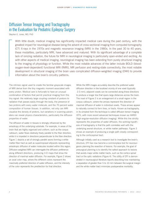
Diffusion Tensor Imaging and Tractography<br />
in the Evaluation for <strong>Pediatric</strong> Epilepsy Surgery<br />
Stephen E. Jones, MD, PhD<br />
the intrinsic signal used to create the familiar grayscale images<br />
of mri derive from the tiny magnetic moment associated with<br />
every proton. medical care is fortunate to have an unusual<br />
combination of factors that permit practical imaging from this<br />
tiny signal: the relatively large coupling constant of protons to<br />
radiation that passes easily through the body, the presence of<br />
two protons with every water molecule, and the 70 percent water<br />
composition of human tissues. In addition, not only can MRI<br />
visualize the density of protons, but variations in scanning parameters<br />
can reveal physics characteristics, particularly the diffusion<br />
properties of water.<br />
The diffusion of water in tissues is strongly influenced by the<br />
anisotropy of the underlying substrate. For example, in areas of the<br />
brain that are highly organized and uniform, such as the corpus<br />
callosum, water flows relatively freely parallel to the fiber direction,<br />
while it is impeded in directions perpendicular to the fiber direction.<br />
Figure 1 depicts a set of strongly parallel axons forming a white<br />
matter fiber tract as well as superimposed ellipsoids representing<br />
anisotropic diffusion of water molecules located within this region.<br />
Diffusion-weighted MRI can interrogate the diffusion preference<br />
along different directions, producing 3-D maps that show the<br />
direction of maximal water diffusion. For example, Figure 2 shows<br />
an axial color map, where the different colors represent the<br />
maximally preferred direction of water diffusion, and the intensity<br />
of the color represents the predilection for that direction.<br />
cover story | diagNostic radiology<br />
With little doubt, medical imaging has significantly impacted medical care during the past century, with the<br />
greatest impact for neurological disease being the advent of cross-sectional imaging from computed tomography<br />
(CT) X-rays in the 1970s and magnetic resonance imaging (MRI) in the 1980s. In the past 30 to 40 years,<br />
these modalities, particularly MRI, have advanced and matured. With its significant advantage of a complete<br />
lack of ionizing radiation, the future for MRI in neurological imaging is particularly open-ended and exciting. As<br />
with other aspects of medical imaging, neurological imaging has been extending from purely structural imaging<br />
to the imaging of physiology or function. While the most notable advances of the latter include BOLD (bloodoxygen-level-dependent)<br />
functional MRI (fMRI), MR perfusion and resting-state connectivity, the most notable<br />
development in structural imaging of the brain uses complicated diffusion-weighted imaging (DWI) to provide<br />
information about the brain’s circuitry patterns.<br />
while the mri images accurately describe the preferred water<br />
diffusion direction in the localized vicinity of one voxel (typically<br />
2-3 mm), adjacent voxels can be connected along these directions<br />
to produce a longer line that spans large distances across the brain.<br />
the inset of Figure 2 is an enlargement of a small region in the<br />
corpus callosum, where the arrows represent the direction of<br />
maximal diffusion of water in individual voxels. these arrows appear<br />
to naturally connect to form lines, or tracts. Known as tractography,<br />
in its simplest form the technique is called diffusion tensor imaging<br />
(DTI), with more recent advanced techniques known as HARDI<br />
(high-angular-resolution diffusion imaging). While this line strictly<br />
represents the properties of water diffusion, the working hypothesis<br />
of tractography is that the path correlates well with the<br />
underlying axonal structure, or white matter pathways. Figure 3<br />
shows an example of producing a single path closely corresponding<br />
to the corticospinal tract.<br />
although initially used as a research tool to investigate the brain’s<br />
structure, DTI has now become a commonplace tool for neurosurgeons<br />
planning the resection of lesions. For example, the goal of<br />
presurgical planning is to identify the safest approach to lesions<br />
that avoids major white matter tracts, such as the cortico-spinal<br />
tract or the optic radiations (Figure 4). the utility of dti is demonstrated<br />
in neurosurgical literature reports describing how maintaining<br />
a separation of greater than 5 to 10 mm between the surgical margin<br />
and the white matter tract minimizes postoperative morbidity.<br />
2 <strong>Pediatric</strong> NeuroscieNce <strong>Pathways</strong> | <strong>2012</strong> – 2013


