Pediatric Neuroscience Pathways Fall 2012 - Cleveland Clinic
Pediatric Neuroscience Pathways Fall 2012 - Cleveland Clinic
Pediatric Neuroscience Pathways Fall 2012 - Cleveland Clinic
Create successful ePaper yourself
Turn your PDF publications into a flip-book with our unique Google optimized e-Paper software.
<strong>Pediatric</strong><br />
<strong>Neuroscience</strong><br />
<strong>Pathways</strong><br />
<strong>2012</strong> – 2013
<strong>Pediatric</strong><br />
<strong>Neuroscience</strong><br />
<strong>Pathways</strong><br />
<strong>2012</strong> – 2013<br />
iN this issue:<br />
02 diagNostic radiology<br />
diffusion tensor imaging and tractography in the evaluation for<br />
<strong>Pediatric</strong> epilepsy surgery<br />
06 <strong>Pediatric</strong> ePilePsy<br />
appreciating New opportunities for epilepsy surgery<br />
10 <strong>Pediatric</strong> ePilePsy<br />
Project coPe — improving Knowledge and access to<br />
care for youth with epilepsy in ohio<br />
12 <strong>Pediatric</strong> ePilePsy<br />
hemispheric epilepsy and hemispherectomy in children<br />
14 <strong>Pediatric</strong> ePilePsy<br />
magnetoencephalography for localization of<br />
epileptogenic sources in children<br />
16 <strong>Pediatric</strong> ePilePsy aNd Neurosurgery<br />
stereotactic eeg for surgical Planning in children with<br />
Difficult-to-Localize Epilepsy<br />
18 <strong>Pediatric</strong> Neurology aNd NeurorestoratioN<br />
Intraoperative MRI or CT (O-arm ® ) guidance revolutionizes<br />
<strong>Pediatric</strong> deep Brain stimulation Program<br />
20 <strong>Pediatric</strong> Neurosurgery<br />
hydrocephalus: can the Brain adapt to a chronic squeeze?<br />
can we help?<br />
22 <strong>Pediatric</strong> Neurology<br />
<strong>Pediatric</strong> stroke: coming of age<br />
9<br />
3<br />
2 NeuroscieNce <strong>Pathways</strong> | sPriNg/summer <strong>Pediatric</strong> 2008 2007 NeuroscieNce clevelaNdcliNic.org/NeuroscieNce<br />
<strong>Pathways</strong> | <strong>2012</strong> – 2013<br />
21<br />
24 <strong>Pediatric</strong> Neurology<br />
Postural orthostatic tachycardia syndrome in children —<br />
an emerging entity<br />
26 <strong>Pediatric</strong> Neurology<br />
mitochondrial dysfunction and cyclic vomiting syndrome<br />
27 <strong>Pediatric</strong> sleeP disorders<br />
obstructive sleep apnea and unusual sleep Positioning<br />
in children with down syndrome<br />
28 <strong>Pediatric</strong> Psychiatry<br />
Biomarkers in youth with major depressive disorder<br />
30 <strong>Pediatric</strong> Psychiatry<br />
children with chronic Pain syndrome see improvement in anxiety<br />
and Depression After Hospital-Based Pain Rehabilitation Program<br />
31 develoPmeNtal aNd rehaBilitatioN <strong>Pediatric</strong>s<br />
Constraint-Induced Movement Therapy vs. Bimanual Therapy<br />
for children with hemiparesis<br />
32 <strong>Pediatric</strong> Behavioral health<br />
chronic Pain in children and adolescents: initial evaluation<br />
of an interdisciplinary Pain rehabilitation Program<br />
34 <strong>Pediatric</strong> Behavioral health<br />
comparing the effectiveness of stimulant and Behavioral<br />
Treatment in Youth with Attention-Deficit/Hyperactivity Disorder<br />
in a Quasi-Naturalistic Setting<br />
36 <strong>Pediatric</strong> Neurological staFF<br />
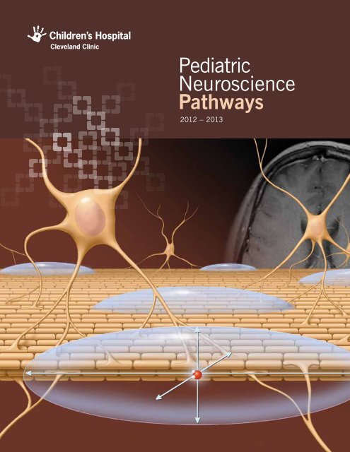
on the cover: cartoon diagram showing a tightly parallel bundle of axons representing a white matter tract. superimposed ellipsoids represent<br />
the anisotropic diffusion pattern of embedded water molecules. Note that the ellipsoids are elongated along the direction of the axons. Diffusionweighted<br />
mri has the ability to interrogate water diffusion and determine the direction of maximal diffusivity.<br />
R
Welcome from the Editors<br />
cleveland clinic’s pediatric neuroscience program includes a wide<br />
range of doctors in many specialties. integration of these teams<br />
in both the Neurological institute and children’s hospital offers<br />
seamless collaboration between adult and pediatric specialists in<br />
every discipline. Thanks to this integration, pediatric neuroscience<br />
physicians offer cutting-edge treatments for the widest imaginable<br />
spectrum of neurological disorders.<br />
welcome from the editors<br />
The reader will find several examples of this integration in these<br />
pages. For example, children with severe dystonia may receive<br />
relief through deep brain stimulation at cleveland clinic because the<br />
pediatric movement disorders team is integrated with a celebrated<br />
team of adult specialists in neurological restoration. Similarly,<br />
children with difficult-to-localize epilepsy may benefit from surgery<br />
planned with stereotactic-depth EEG because the pediatric epilepsy<br />
team works closely with prominent adult and pediatric neurosurgeons<br />
who have extensive experience with this technique. In addition,<br />
cleveland clinic’s pediatric neuroscience program has the honor and distinction<br />
of being ranked among the top three pediatric neurology and neurosurgery<br />
programs in the united states by U.S. News & World Report for <strong>2012</strong>-2013.<br />
The elements of this success are the world-renowned teams of physicians,<br />
scientists, nurses, therapists, technologists and other caregivers who collaborate<br />
to provide multidisciplinary care to children with neurological disorders.<br />
treatment of pediatric hydrocephalus is enhanced by the clinical and<br />
experimental study of its progression and consequences in adults.<br />
these synergistic collaborations provide opportunities for unparalleled<br />
innovations in pediatric clinical care, education and research.<br />
cleveland clinic’s premise is “Patients First.” whether the<br />
activity is providing today’s newest, most innovative treatments<br />
or researching and developing the treatments of the future, this<br />
focus on patient care is the compass for pediatric neuroscience.<br />
we proudly offer this compilation of research and innovations for<br />
use in your own cutting-edge practice for care of children with<br />
neurological disorders.<br />
elaine wyllie, md<br />
Professor, Epilepsy Center<br />
Co-Editor<br />
mark luciano, md, Phd<br />
Professor, Department of Neurosurgery<br />
Co-Editor<br />
visit clevelaNdcliNicchildreNs.org | 866.588.2264 1
Diffusion Tensor Imaging and Tractography<br />
in the Evaluation for <strong>Pediatric</strong> Epilepsy Surgery<br />
Stephen E. Jones, MD, PhD<br />
the intrinsic signal used to create the familiar grayscale images<br />
of mri derive from the tiny magnetic moment associated with<br />
every proton. medical care is fortunate to have an unusual<br />
combination of factors that permit practical imaging from this<br />
tiny signal: the relatively large coupling constant of protons to<br />
radiation that passes easily through the body, the presence of<br />
two protons with every water molecule, and the 70 percent water<br />
composition of human tissues. In addition, not only can MRI<br />
visualize the density of protons, but variations in scanning parameters<br />
can reveal physics characteristics, particularly the diffusion<br />
properties of water.<br />
The diffusion of water in tissues is strongly influenced by the<br />
anisotropy of the underlying substrate. For example, in areas of the<br />
brain that are highly organized and uniform, such as the corpus<br />
callosum, water flows relatively freely parallel to the fiber direction,<br />
while it is impeded in directions perpendicular to the fiber direction.<br />
Figure 1 depicts a set of strongly parallel axons forming a white<br />
matter fiber tract as well as superimposed ellipsoids representing<br />
anisotropic diffusion of water molecules located within this region.<br />
Diffusion-weighted MRI can interrogate the diffusion preference<br />
along different directions, producing 3-D maps that show the<br />
direction of maximal water diffusion. For example, Figure 2 shows<br />
an axial color map, where the different colors represent the<br />
maximally preferred direction of water diffusion, and the intensity<br />
of the color represents the predilection for that direction.<br />
cover story | diagNostic radiology<br />
With little doubt, medical imaging has significantly impacted medical care during the past century, with the<br />
greatest impact for neurological disease being the advent of cross-sectional imaging from computed tomography<br />
(CT) X-rays in the 1970s and magnetic resonance imaging (MRI) in the 1980s. In the past 30 to 40 years,<br />
these modalities, particularly MRI, have advanced and matured. With its significant advantage of a complete<br />
lack of ionizing radiation, the future for MRI in neurological imaging is particularly open-ended and exciting. As<br />
with other aspects of medical imaging, neurological imaging has been extending from purely structural imaging<br />
to the imaging of physiology or function. While the most notable advances of the latter include BOLD (bloodoxygen-level-dependent)<br />
functional MRI (fMRI), MR perfusion and resting-state connectivity, the most notable<br />
development in structural imaging of the brain uses complicated diffusion-weighted imaging (DWI) to provide<br />
information about the brain’s circuitry patterns.<br />
while the mri images accurately describe the preferred water<br />
diffusion direction in the localized vicinity of one voxel (typically<br />
2-3 mm), adjacent voxels can be connected along these directions<br />
to produce a longer line that spans large distances across the brain.<br />
the inset of Figure 2 is an enlargement of a small region in the<br />
corpus callosum, where the arrows represent the direction of<br />
maximal diffusion of water in individual voxels. these arrows appear<br />
to naturally connect to form lines, or tracts. Known as tractography,<br />
in its simplest form the technique is called diffusion tensor imaging<br />
(DTI), with more recent advanced techniques known as HARDI<br />
(high-angular-resolution diffusion imaging). While this line strictly<br />
represents the properties of water diffusion, the working hypothesis<br />
of tractography is that the path correlates well with the<br />
underlying axonal structure, or white matter pathways. Figure 3<br />
shows an example of producing a single path closely corresponding<br />
to the corticospinal tract.<br />
although initially used as a research tool to investigate the brain’s<br />
structure, DTI has now become a commonplace tool for neurosurgeons<br />
planning the resection of lesions. For example, the goal of<br />
presurgical planning is to identify the safest approach to lesions<br />
that avoids major white matter tracts, such as the cortico-spinal<br />
tract or the optic radiations (Figure 4). the utility of dti is demonstrated<br />
in neurosurgical literature reports describing how maintaining<br />
a separation of greater than 5 to 10 mm between the surgical margin<br />
and the white matter tract minimizes postoperative morbidity.<br />
2 <strong>Pediatric</strong> NeuroscieNce <strong>Pathways</strong> | <strong>2012</strong> – 2013
anticipated applications of dti and tractography<br />
cover story | diagNostic radiology<br />
Today, the utility of DTI and tractography for presurgical planning<br />
is well documented and applies to neurosurgical procedures in<br />
both the adult and pediatric populations. the anticipated application<br />
of dti and tractography is gestating in worldwide research<br />
efforts, including at <strong>Cleveland</strong> <strong>Clinic</strong>, which will have particular<br />
applications to pediatric neurology. First, the presence and<br />
progression of developmental neurological diseases can currently<br />
be visualized with track-density maps. Often this visualization<br />
is best appreciated with asymmetrical diseases, such as Parryromberg<br />
syndrome (Figure 5) and rasmussen encephalitis. a<br />
second application involves superior detection of epileptogenic foci<br />
in epilepsy patients, and in the pediatric population a majority of<br />
these are due to malformations of cortical development (mcd).<br />
many of these lesions have abnormal architecture not only within<br />
the cortex but also in the subjacent white matter, occasionally<br />
extending along radial-glial fibers toward the periventricular<br />
margins. many patients with mcd can be cured with proper<br />
resection of the lesion, but this requires visualization of the<br />
location, and a considerable proportion of MCD are “MRI invisible”<br />
using conventional techniques. Thus, there is great hope that<br />
advanced techniques, such as DTI and tractography, may help<br />
reveal the location of MCD. For example, high-resolution DTI<br />
focusing on the cortical-subcortical regions may reveal deranged<br />
architecture of water diffusion profiles, which could indicate cortex<br />
abnormalities, all of which appear normal on conventional MRI<br />
Figure 1 Figure 2<br />
Figure 1. Cartoon diagram showing a tightly parallel bundle of axons representing a white matter tract. Superimposed ellipsoids represent<br />
the anisotropic diffusion pattern of embedded water molecules. Note that the ellipsoids are elongated along the direction of the axons.<br />
Diffusion-weighted MRI has the ability to interrogate water diffusion and determine the direction of maximal diffusivity.<br />
Figure 2. DTI color map of an axial section of the brain. The colors encode the direction of maximal diffusion (green = anterior-posterior; red<br />
= left-right; blue = superior-inferior). The brightness of the color represents the predilection for water diffusion in that direction. The inset<br />
shows an enlargement of a small section of the corpus callosum with small superimposed arrows located at each voxel. Note how the arrows<br />
are easily visually connected to produce lines, or tracts.<br />
visit clevelaNdcliNicchildreNs.org | 866.588.2264 3
cover story | diagNostic radiology<br />
Figure 5<br />
Figure 3<br />
Figure 3. Example of DTI tractography. A single tract is produced from connecting the directions of maximal water<br />
diffusion in multiple voxels, starting from the corticospinal tract in the pons. It is essential to understand that the<br />
visualized tracts primarily represent the diffusion of water and secondarily may represent the underlying white<br />
matter pathways.<br />
Figure 4. Utility of DTI for presurgical planning. This young patient has a tumor in the right temporal-occipital<br />
lobe, near the expected location of the optic tracts. DTI tractography more accurately shows the location of the<br />
optic tracts and influenced the pathway for surgical resection.<br />
Figure 5. Deterministic tracking of bilateral corticospinal tracts in a patient with Parry-Romberg syndrome, which<br />
causes a progressive hemifacial atrophy with associated hemi-degenerative changes in the brain. Qualitatively, the<br />
image shows fewer DTI tracts on the patient’s right than on the left, which corresponds to the affected side.<br />
Figure 6. Sagittal view of the brain of an epilepsy patient who had numerous intracranial electrodes implanted after<br />
high-resolution DTI. The lines show tractography from an electrode in Broca’s area, connecting a subset of electrode<br />
contacts. The colors of the lines represent the magnitude of electrophysiological connectivity, as produced by<br />
cortico-cortical evoked potentials (CCEP). Such knowledge could reduce the degree of invasive evaluations for<br />
intractable epilepsy.<br />
4 <strong>Pediatric</strong> NeuroscieNce <strong>Pathways</strong> | <strong>2012</strong> – 2013<br />
Figure 4<br />
Figure 6
cover story | diagNostic radiology<br />
images. a third application of dti to pediatric neuroimaging regards<br />
brain connectivity. many diseases may manifest themselves as<br />
subtle alterations of diffusion properties along the pathways<br />
between portions of the brain. In epilepsy, for example, in addition<br />
to the primary epileptogenic focus, there is often a network of other<br />
lesser epileptogenic regions in the brain (sometimes far away). dti<br />
has the exciting potential application to measure the connectivity<br />
between cortical regions in addition to determining the location of<br />
the connecting pathway. an example of recent research at cleveland<br />
<strong>Clinic</strong> is shown in Figure 6, in which high-resolution tractography is<br />
compared with electrophysiological measurements obtained from an<br />
epilepsy patient with multiple intracranial electrodes. Not only are<br />
the connecting paths visualized in this sagittal image, but the color<br />
of each path corresponds to the DWI connectivity, which modestly<br />
correlates to the electrophysiological connectivity. the eventual goal<br />
would be to obviate the need for extensive invasive intracranial<br />
electrodes by using high-resolution DTI and tractography.<br />
conclusion<br />
mr tractography and dti are recently developed methods of<br />
advanced neurological imaging, clinically used today mainly<br />
for presurgical planning. However, a growing body of research<br />
indicates the capabilities of DTI for identification of (1) deranged<br />
white matter architecture subjacent to subtle MCD, (2) distal cortical<br />
regions involved in an epileptogenic network and (3) abnormal brain<br />
connectivity within epileptogenic zones.<br />
suggested readiNg<br />
Cauley KA, et al. Diffusion tensor imaging and tractography of<br />
rasmussen encephalitis. Pediatr Radiol. 2009;39:727–730.<br />
Hygino da Cruz LC Jr, ed. <strong>Clinic</strong>al applications of diffusion imaging<br />
of the brain. Neuroimag Clin N Am. 2011 Feb;21(1).<br />
Jellison BJ, et al. Diffusion tensor imaging of cerebral white matter:<br />
a pictorial review of physics, fiber tract anatomy, and tumor<br />
imaging patterns. Am J Neuroradiol. 2004 Mar;25:356–369.<br />
Kakisaka Y, So NK, Jones SE, et al. Intractable focal epilepsy<br />
contralateral to the side of facial atrophy in Parry-Romberg<br />
syndrome. Neurol Sci. <strong>2012</strong> Feb;33(1):165-168.<br />
Robin M, et al. K-space and q-space: Combining ultra-high spatial<br />
and angular resolution in diffusion imaging using ZooPPa at 7 t.<br />
NeuroImage. <strong>2012</strong>;60:967–978.<br />
Stephen E. Jones, MD, PhD, is a neuroradiologist and physicist<br />
whose specialty interests include advanced imaging, epilepsy,<br />
functional neuroimaging, MRI, neuroradiology and traumatic brain<br />
injury. He can be reached at 216.444.4454 or joness19@ccf.org.<br />
visit clevelaNdcliNicchildreNs.org | 866.588.2264 5
at cleveland clinic we are exploring the boundaries of possibility<br />
for epilepsy surgery to expand the pool of children who may<br />
benefit. Some of our most exciting results have been in children<br />
with early focal brain lesions and generalized eeg abnormalities.<br />
how can we understand this phenomenon when we are accustomed<br />
to thinking of a focal eeg seizure as the “gold standard”<br />
for identifying a focal epileptogenic zone? this takes us to the<br />
concept of plasticity, with downstream differences in the epilepsy<br />
and eeg based on whether the focal lesion interacted with a<br />
mature or developing brain.<br />
Focal brain lesions acquired later in life are imposed on a mature<br />
brain, which usually results in focal EEG discharges at the lesion<br />
location. But early focal brain lesions interact with a developing<br />
brain, and in these cases there may be aberrant circuitry<br />
resulting in generalized eeg. the most common epileptogenic<br />
lesions that occur early in life are cortical malformations or<br />
perinatal infarctions, but other early lesions may result in similar<br />
effects. the downstream generalized eeg patterns tend to be<br />
hypsarrhythmia in infants and generalized slow spike-wave<br />
complexes in older children.<br />
The mechanisms are unknown, but certainly the first year of life<br />
is characterized by processes leading to increased neuronal<br />
connectivity, such as myelination, axonal growth and synaptogenesis.<br />
disruption of these processes during infancy could lead to<br />
patterns of epileptogenicity that are different from those that<br />
start later in life.<br />
<strong>Pediatric</strong> ePilePsy<br />
Appreciating New Opportunities for Epilepsy Surgery<br />
Elaine Wyllie, MD<br />
In surgical candidates with focal lesions acquired after the brain is already mature, the EEG, seizure signs and<br />
symptoms, and MRI all typically give evidence of focal epilepsy. In patients with early brain lesions, however,<br />
the findings may be more complicated.<br />
we published our larger experience in Neurology (see suggested<br />
reading below). we blindly and retrospectively reviewed the<br />
preoperative video eeg from the 415 children who underwent<br />
epilepsy surgery at our institution between 1994 and 2004. We<br />
identified 50 children, median age of 8 years at surgery, who had<br />
abundant generalized ictal and interictal epileptiform discharges on<br />
their preoperative EEG, comprising 30 to 100 percent of all their<br />
recorded eeg abnormalities.<br />
the study group was selected based solely on the presence of<br />
abundant generalized epileptiform discharges on preoperative<br />
eeg. But when we unblinded ourselves and studied these 50<br />
patients in detail, we found another unifying feature: They all had<br />
a focal mri lesion that occurred early in life. Ninety percent of<br />
the lesions were prenatal, perinatal or acquired in the first two<br />
years of life. the latest lesion occurred by 5 years of age. the<br />
patients ranged in age from 6 months to 24 years at the time<br />
of their preoperative evaluations, but their focal epileptogenic<br />
lesions, usually cortical malformations or perinatal infarctions, all<br />
occurred during early brain development. Seventy-two percent of<br />
children had no seizures after surgery, at a median follow-up of<br />
two years. the results were similar to those in a comparison<br />
group of 159 children with early focal lesions and focal EEG<br />
findings who underwent epilepsy surgery at <strong>Cleveland</strong> <strong>Clinic</strong>.<br />
By appreciating the opportunities for successful epilepsy surgery<br />
in children with generalized eeg discharges and an early focal<br />
lesion, we expand the pool of candidates who may gain relief<br />
from seizures for a lifetime.<br />
6 <strong>Pediatric</strong> NeuroscieNce <strong>Pathways</strong> | <strong>2012</strong> – 2013
suggested readiNg<br />
Wyllie E, Lachhwani DK, Gupta A, et al. Successful surgery for<br />
epilepsy due to early brain lesions despite generalized eeg<br />
findings. Neurology. 2007;69:389-397.<br />
<strong>Pediatric</strong> ePilePsy<br />
Gupta A, Chirla A, Wyllie E, et al. <strong>Pediatric</strong> epilepsy surgery in focal<br />
lesions and generalized electroencephalogram abnormalities.<br />
Pediatr Neurol. 2007;37:8-15.<br />
Gupta A, Wyllie E. Epilepsy surgery: special considerations in<br />
children. In: Wyllie E, editor in chief; Cascino GD, Gidal BE,<br />
Goodkin HP, associate editors. Wyllie’s Treatment of Epilepsy.<br />
5th ed. Philadelphia, PA: Lippincott Williams & Wilkins;<br />
2011:993-1006.<br />
Loddenkemper T, Cosmo G, Kotagal P, et al. Epilepsy surgery in<br />
children with electrical status epilepticus in sleep. Neurosurgery.<br />
2009;64:328-337.<br />
wyllie e. The <strong>Cleveland</strong> <strong>Clinic</strong> Guide to Epilepsy. New York, NY:<br />
Kaplan Publishing; 2010.<br />
Elaine Wyllie, MD, is a Professor in the Epilepsy Center at <strong>Cleveland</strong><br />
<strong>Clinic</strong>. Her clinical and research interests include pediatric epilepsy,<br />
EEG, antiepileptic medications and epilepsy surgery. She can be<br />
contacted at 216.636.5860 or wylliee@ccf.org.<br />
See next two pages for a case study related to this article.<br />
visit clevelaNdcliNicchildreNs.org | 866.588.2264 7
Case Study: Successful Epilepsy Surgery Despite Complicated EEG<br />
the following case is illustrative. a girl presented to<br />
cleveland clinic at 4 years old. her development was on<br />
track and her neurological examination was normal, but<br />
at 3 years old she had developed severe epilepsy with<br />
daily clusters of facial tonic seizures often evolving to lip<br />
smacking, confused speaking and unprotected falls. She<br />
was on four antiepileptic medications and had failed a<br />
total of five.<br />
Her MRI showed a round, circumscribed lesion in the<br />
right parietal lobe, posterior to the sensorimotor cortex<br />
(Figure 1). The lesion had no significant mass effect<br />
and did not enhance with gadolinium, so it wasn’t clear<br />
whether it was a low-grade tumor or a malformation of<br />
cortical development.<br />
Whereas her MRI showed a focal lesion, her EEG<br />
showed only generalized epileptiform discharges (Figure<br />
2). These were abundant, nearly continuous during<br />
sleep, and lacked any focal features. EEG seizures also<br />
were generalized at the onset and maximal on the left<br />
(Figure 3). As seizures evolved, the left-sided predominance<br />
became even more apparent (Figure 4).<br />
In summary, this girl had medically intractable epilepsy,<br />
a right parietal lesion on MRI and generalized findings<br />
on EEG, sometimes maximum on the left. Would<br />
epilepsy surgery stop her seizures, or was her MRI<br />
figure 1. right parietal lesion<br />
<strong>Pediatric</strong> ePilePsy<br />
Right Parietal Lesion<br />
R<br />
abnormality just the “tip of the iceberg” of a more<br />
diffuse process?<br />
At <strong>Cleveland</strong> <strong>Clinic</strong>, each surgical candidate is discussed<br />
in detail at an epilepsy management conference, with<br />
input from all of the specialists. once a consensus has<br />
been reached, the recommendations are presented to<br />
the family for informed consent.<br />
In this case, the family was told that based on previous<br />
experience in similar cases, epilepsy surgery could be<br />
expected to resolve the epilepsy. However, this plan was<br />
clearly at the boundaries of epilepsy surgery because of<br />
the generalized eeg. after detailed informed consent<br />
with a full discussion of the possible risks and benefits,<br />
the parents elected to proceed.<br />
The lesion was resected at <strong>Cleveland</strong> <strong>Clinic</strong> in 2009,<br />
and histopathologic analysis showed that it was an<br />
oligoastrocytoma, World Health Organization grade II.<br />
the girl recovered from the surgery uneventfully and<br />
was discharged from the hospital after five days.<br />
Since surgery, she has had no seizures and has taken<br />
no antiepileptic medications, and her postoperative EEG<br />
shows no epileptiform discharges. this favorable result<br />
proves that the focal lesion was causing the epilepsy, even<br />
though the preoperative eeg features were generalized.<br />
Figure 1. Sagittal and axial<br />
MRI views of our patient’s<br />
right parietal lesion.<br />
8 <strong>Pediatric</strong> NeuroscieNce <strong>Pathways</strong> | <strong>2012</strong> – 2013
figure 2. interictal eeg<br />
FP1–F7<br />
F7-T7<br />
T7-P7<br />
P7-01<br />
FP2-F8<br />
F8-T8<br />
T8-P8<br />
P8-02<br />
FP1-F3<br />
F3-C3<br />
C3-P3<br />
P3-O1<br />
FP2-F4<br />
F4-C4<br />
C4-P4<br />
P4-02<br />
figure 3. eeg at seizure onset<br />
FP1–F7<br />
F7-T7<br />
T7-P7<br />
P7-01<br />
FP2-F8<br />
F8-T8<br />
T8-P8<br />
P8-02<br />
FP1-F3<br />
F3-C3<br />
C3-P3<br />
P3-O1<br />
FP2-F4<br />
F4-C4<br />
C4-P4<br />
P4-02<br />
FP1–F7<br />
F7-T7<br />
T7-P7<br />
P7-01<br />
FP2-F8<br />
F8-T8<br />
T8-P8<br />
P8-02<br />
FP1-F3<br />
F3-C3<br />
C3-P3<br />
P3-O1<br />
FP2-F4<br />
F4-C4<br />
C4-P4<br />
P4-02<br />
Interictal EEG<br />
EEG Seizure, Onset<br />
EEG Seizure, +10 Seconds<br />
figure 4. eeg 10 seconds after seizure onset<br />
<strong>Pediatric</strong> ePilePsy<br />
Figure 2. Interictal EEG showing our<br />
patient’s abundant generalized slow<br />
spike-wave complexes.<br />
Figure 3. EEG at onset of one of our<br />
patient’s seizures, showing a generalized<br />
pattern with fast activity higher on the left<br />
(highlighted in blue).<br />
Figure 4. EEG seizure 10 seconds after<br />
onset, with more pronounced left-side<br />
predominance (highlighted in blue).<br />
Despite the generalized and contralateral<br />
ictal and interictal EEG features, this child<br />
was free of seizures after resection of her<br />
right parietal oligoastrocytoma.<br />
VISIT CLEVELANDCLINICCHILDRENS.ORG | 866.588.2264 9
Project COPE – Improving Knowledge and<br />
Access to Care for Youth with Epilepsy in Ohio<br />
Tatiana Falcone, MD<br />
youth with epilepsy have increased mental health needs compared<br />
with the general population. we hypothesized that children<br />
with epilepsy will have barriers to accessing mental health care<br />
and an increased need for educational services. to further<br />
understand the needs of youth with epilepsy in Ohio, a needs<br />
assessment survey was conducted.<br />
A parent survey was administered to 359 families with children up<br />
to the age of 18 who were diagnosed with epilepsy. surveys also<br />
were distributed to epileptologists, school nurses, pediatricians<br />
and other key informants.<br />
The findings about epilepsy and mental health included:<br />
• Access to educational services is a major barrier for youth<br />
with epilepsy.<br />
• Parents lacked knowledge about the existing educational<br />
services available to their children.<br />
• Information about the resources available to these patients was<br />
poorly disseminated.<br />
• Ninety-eight percent of families felt empowered and involved in<br />
the decision-making for epilepsy and mental health services.<br />
seventy percent of families and patients felt they had very little<br />
knowledge about epilepsy.<br />
• Most concerns were related to quality of life, poor seizure control<br />
and psychiatric comorbidities.<br />
• Contrary to our belief, most families (76 percent) felt it was easy<br />
to access epilepsy services. the wait time for an epilepsy<br />
appointment was less than one week for 72 percent of the<br />
families. Nearly all (96 percent) of the families had a PCP, and<br />
58 percent reported receiving care from their PcP at least one<br />
time a year. Almost 92.6 percent reported receiving care from<br />
their PcP at least two times a year.<br />
• Most patients in our survey (76 percent) have to travel two<br />
hours or less to see their specialist (e.g., epileptologist, child<br />
psychiatrist). Only five families in our sample had to travel more<br />
than 10 hours for care.<br />
<strong>Pediatric</strong> ePilePsy<br />
the primary goal of Project coPe (collaboration for outreach and Prevention education) is to improve access and<br />
knowledge about mental healthcare for children and adolescents with epilepsy by facilitating development of a<br />
community-based, culturally and linguistically competent mental health model. The project’s objectives are to<br />
create a culturally and linguistically competent network of care that enhances the capacity of primary care providers<br />
(PCPs), pediatric neurologists and pediatric epileptologists to detect, refer and/or treat mental health problems in<br />
youth with epilepsy; to create an effective triage network to improve access to mental healthcare for youth with<br />
epilepsy; and to educate and assist families in accessing mental health services for their children with epilepsy.<br />
• Seventy percent of our patients felt that they had very poor<br />
knowledge about epilepsy and felt sad, frustrated and overwhelmed<br />
when they learned about the diagnosis.<br />
• Seventy-three percent of the families felt that they were given<br />
information about epilepsy and psychiatric comorbidities<br />
mostly by the specialist (e.g., pediatric epileptologist,<br />
child psychiatrist).<br />
• <strong>Pediatric</strong>ians in the <strong>Cleveland</strong> community felt that epileptologists<br />
provided most of the epilepsy care and that child psychiatrists<br />
provided most of the mental healthcare for youth with epilepsy.<br />
• Overwhelmingly, all PCPs in our sample felt the allotted time for<br />
appointments makes it difficult to support and guide the medical<br />
home model.<br />
• <strong>Pediatric</strong>ians providing hospital-based services felt closer to the<br />
medical home model because they had access to case management<br />
and specialists readily available during admission and after<br />
discharge to help coordinate care for youth with epilepsy.<br />
• Epileptologists, child psychiatrists and PCPs identified reimbursement<br />
as a barrier for epilepsy and mental healthcare for<br />
youth with epilepsy for as many as 75 percent of their patients.<br />
Additional survey findings are detailed in Figures 1 to 3.<br />
Psychoeducation is one of the underrecognized unmet needs of<br />
children. Therefore, it is important to engage pediatricians in it.<br />
stigma continues to be an important barrier for families and youth<br />
with epilepsy. Educating first responders will help us decrease<br />
stigma and improve access to mental health services for youth<br />
with epilepsy.<br />
the access issue in youth with special healthcare Needs<br />
In Ohio, it is estimated that there are 32,159 youths with epilepsy.<br />
the number of children with special healthcare needs (cshcN) is<br />
570,913. The federal government has estimated that around<br />
227,000 of these youths are uninsured.<br />
CSHCN are a vulnerable population, according to the Ohio Family<br />
10 <strong>Pediatric</strong> NeuroscieNce <strong>Pathways</strong> | <strong>2012</strong> – 2013
Health Survey. CSHCN have greater difficulty accessing the<br />
appropriate level of mental health services. Overall, these patients<br />
have higher unmet needs than does the general population. about<br />
6.2 percent of these patients who are eligible for medicaid services<br />
continue to be uninsured, perpetuating the unmet medical and<br />
psychiatric needs.<br />
<strong>Pediatric</strong> ePilePsy<br />
conclusions<br />
there are important and underrecognized unmet needs in youth<br />
with epilepsy.<br />
Psychoeducation is a key piece to help families of youth with<br />
epilepsy cope with some of the comorbidities these patients face.<br />
Although many services are provided in our community, parents and<br />
children do not know about these services and do not access them.<br />
it is important to engage PcPs in the psychoeducation of youth<br />
with epilepsy. stigma continues to be an important barrier for<br />
patients and families in accessing mental health services.<br />
Educating first responders (school nurses, parents, pediatricians)<br />
will help us decrease stigma and improve access to mental health<br />
services by youth with epilepsy.<br />
this project was developed with funding from the health<br />
resources services administration maternal and child health<br />
Bureau under grant H98MC2026n.<br />
suggested readiNg<br />
Plioplys S, Dunn DW, Caplan R. 10-year research update review:<br />
Psychiatric problems in children with epilepsy. J Am Acad Child<br />
Adolesc Psychiatry. 2007;46:1389-1402.<br />
Caplan R, Gillberg C, Dunn DW, Spence SJ. Psychiatric disorders<br />
in children with epilepsy. In: Engel J, Pedley TA, eds. Epilepsy:<br />
A Comprehensive Textbook. Philadelphia, PA: Lippincott<br />
Williams & Wilkins; 2008:2179-2193.<br />
Russ S, Larson K, Halfon N. A national profile of childhood epilepsy<br />
and seizure disorder. <strong>Pediatric</strong>s. <strong>2012</strong>;129:256-263.<br />
Tatiana Falcone, MD, is a staff physician in <strong>Cleveland</strong> <strong>Clinic</strong>’s<br />
Neurological Institute who focuses on child and adolescent<br />
psychiatry. Her specialty interests include anxiety and mood<br />
disorders, consultation-liaison psychiatry, emergency psychiatry,<br />
psycho-oncology, psychosis, schizophrenia, epilepsy and psychiatric<br />
issues in epilepsy. She can be contacted at 216.444.7459 or<br />
falcont1@ccf.org.<br />
figure 1. survey of 50 school nurses in Northeast ohio: what<br />
percentage of students in your school have a seizure action plan?<br />
■ 76-100%<br />
■ 51-75%<br />
■ 26-50%<br />
■ 11-25%<br />
■ 2-10%<br />
■ 1% or less<br />
Percentages shown on<br />
the pie chart indicate<br />
percent providing that<br />
response, e.g., 53<br />
percent responded that<br />
76-100% of their<br />
students had a plan.<br />
figure 2. surveys of 13 pediatricians in Northeast ohio:<br />
does your practice provide patient education about self-<br />
management and medication transition for youth with epilepsy<br />
and their parents?<br />
■ always<br />
■ usually<br />
■ rarely<br />
■ Never<br />
■ No response<br />
figure 3. survey of eight professionals in epilepsy care:<br />
Epileptologists identified the comorbidities as follows:<br />
visit clevelaNdcliNicchildreNs.org | 866.588.2264 11<br />
100<br />
80<br />
60<br />
40<br />
20<br />
0<br />
53.3%<br />
50%<br />
8.3%<br />
20%<br />
16.7%<br />
16.7%<br />
8.3%<br />
6.7%<br />
6.7%<br />
6.7%<br />
6.7%<br />
ADHD Behavior<br />
problems<br />
Learning<br />
problems<br />
Cognitive<br />
problems<br />
Mood<br />
issues
indications for hemispherectomy:<br />
common disorders with hemispheric epilepsy<br />
common disorders for which hemispherectomy is indicated are<br />
congenital malformations such as hemimegalencephaly, hemispheric<br />
or extensive multilobar cortical dysplasias, perinatal or<br />
acquired ischemic strokes, Sturge-Weber syndrome and Rasmussen<br />
syndrome. congenital malformations may occur as isolated<br />
disorders or in association with certain sporadic or inheritable<br />
genetic conditions. Figures 1 to 3 show brain MRI findings in<br />
children who are candidates for hemispherectomy.<br />
A thorough clinical evaluation — assessing age of seizure onset,<br />
course of the neurological symptoms, family history, presence of<br />
neurocutaneous markers, dysmorphic features or congenital<br />
anomalies involving multiple organ systems, and abnormal<br />
<strong>Pediatric</strong> ePilePsy<br />
Hemispheric Epilepsy and Hemispherectomy in Children<br />
Ajay Gupta, MD, and William E. Bingaman, MD<br />
Figure 1<br />
certain epilepsies are caused by congenital or acquired disorders that extensively involve all or most of one<br />
cerebral hemisphere. these disorders usually present with catastrophic epilepsy during infancy and require urgent<br />
diagnostic evaluation and treatment. After careful consideration of several complicating factors, hemispherectomy<br />
may be the most effective treatment for these children.<br />
Figure 2<br />
neurological findings — helps in making a specific diagnosis,<br />
determining disease progression and planning treatment. video<br />
eeg monitoring is the initial key tool in establishing the diagnosis<br />
of epilepsy, localizing the seizure-onset zone, and planning surgical<br />
strategy. Brain MRI, supplemented by FDG-PET, helps in determining<br />
the underlying epilepsy substrate and extent of hemispheric<br />
involvement as well as the anatomical and functional integrity of<br />
the unaffected hemisphere. other tests required may include an<br />
eye examination, neuropsychological assessment and metabolic/<br />
genetic consultation.<br />
timing and Planning of hemispherectomy<br />
hemispherectomy in children with catastrophic epilepsy is<br />
challenging and requires careful analysis of several complex<br />
age-related issues.<br />
Spectrum of brain MRI findings seen in children with hemispheric epilepsy syndromes. All these patients underwent<br />
hemispherectomy for intractable seizures.<br />
Figure 1: T2-weighted sagittal image of hemimegalencephaly showing diffuse right hemispheric enlargement and dysplasia. Midline shift with<br />
bulging of anterior falx to the left and compression of the right lateral ventricle suggest a mass effect due to increased volume of the brain<br />
parenchyma. Dysplastic changes are diffuse with a thick and disorganized cortex, poor gray-white matter differentiation, and abnormal signal in<br />
the white matter. Note the basal ganglia are also dysplastic with abnormal increased signal.<br />
Figure 2: Axial FLAIR brain MRI image of a child with Rasmussen syndrome with left hemispheric atrophy most prominent in the insular region,<br />
mildly dilated atrium of the left ventricle, and increased signal in the peri-insular subcortical and cortical ribbon.<br />
Figure 3: Axial FLAIR brain MRI image of a 5-year-old with a remote ischemic stroke in the distribution of the right middle cerebral artery with<br />
severe right hemispheric encephalomalacia and atrophy.<br />
12 <strong>Pediatric</strong> NeuroscieNce <strong>Pathways</strong> | <strong>2012</strong> – 2013<br />
Figure 3
<strong>Pediatric</strong> ePilePsy<br />
The first issue is the risk of performing surgery on a child. The<br />
lowest reported perioperative mortality rate in children is approximately<br />
1 percent from our and a few other experienced centers.<br />
mortality may be higher in infants undergoing hemispherectomy<br />
with weight < 10 kg (small blood volume). However, the mortality<br />
rate with epilepsy surgery must be weighed against the mortality of<br />
medically treated intractable seizures, which is approximately 0.3<br />
percent per year in children of all ages. another important risk of<br />
epilepsy surgery is the possibility of incurring new postoperative<br />
neurological deficits. In children with pre-existing motor, visual and<br />
language deficits undergoing hemispherectomy, the risk of new or<br />
worsening postoperative deficits is usually low. Young age confers<br />
distinct advantages because of developmental plasticity. the second<br />
issue is the timing of and goals for hemispherectomy. there is<br />
evidence that a shorter duration of frequent seizures before surgery<br />
may lead to better long-term cognitive outcomes in children under<br />
the age of 2 years. the third issue is the prediction of success after<br />
surgery in eliminating or controlling seizures. Prediction of success<br />
requires careful selection of candidates based on clinical, EEG and<br />
imaging tests. the majority of patients are expected to become<br />
seizure-free or show dramatic improvement, as outlined below.<br />
surgical approach, techniques and complications<br />
the most commonly employed hemispherectomy technique<br />
remains functional hemispherectomy. Benefits over anatomic<br />
removal of the hemisphere include less tissue removal, shorter<br />
operative time, less blood loss and perhaps less chance of<br />
hydrocephalus after surgery. More recently, less-invasive techniques<br />
such as peri-insular hemispherotomy and hemispheric<br />
deafferentation have been proposed as alternatives to large<br />
resections. Our practice, based on our experience over the past 15<br />
years, is to adapt the appropriate surgical hemispherectomy<br />
technique based on the underlying pathology. hemispherectomy is<br />
a major surgical procedure, and complications in the perioperative<br />
period include death, hemorrhage, aseptic meningitis, infection,<br />
coagulopathy, hypothermia and hydrocephalus requiring ventriculoperitoneal<br />
shunting. mortality reported in the literature is 0.5 to 3<br />
percent. However, the risks of perioperative mortality and morbidity<br />
could be further reduced if patients are carefully screened<br />
before surgery and the surgery is performed at a specialized<br />
pediatric epilepsy center with experience in critical care perioperative<br />
management.<br />
outcome after hemispherectomy<br />
At <strong>Cleveland</strong> <strong>Clinic</strong>, we capture longitudinal seizure outcome data<br />
after epilepsy surgery at every postoperative visit. Figure 4 shows<br />
long-term seizure outcomes in 222 patients after hemispherectomy<br />
(1997-2011). The rate of seizure freedom is 88 percent after<br />
one year, 83 percent after two years, 75 percent after five years,<br />
and 69 percent after eight years and beyond. Even among patients<br />
who do not become seizure-free after surgery, significant reduction<br />
(> 75 percent) in the frequency of seizures is usually seen in an<br />
additional 20 to 25 percent of patients. After surgery, children are<br />
figure 4. long-term seizure freedom following<br />
hemispherectomy (N = 222)<br />
surgical dates: 1997-2011<br />
seizure-free (%)<br />
100<br />
0<br />
0 1 2 3 4 5 6 7 8<br />
years from surgery<br />
years from surgery 1 2 5 8<br />
Seizure-free (%) 88% 83% 75% 69%<br />
often well-controlled on fewer medications and demonstrate<br />
improvement in their behavior and social interactions. our<br />
experience shows that global improvement in physical and<br />
intellectual function tends to go hand in hand with seizure<br />
outcome after hemispherectomy.<br />
the cleveland clinic hemispherectomy Program:<br />
our experience and organization<br />
cleveland clinic’s hemispherectomy program is one of the nation’s<br />
premier and most experienced programs in pediatric epilepsy<br />
surgery. our results and complication rates in more than 200<br />
procedures performed over the past 15 years are among the best in<br />
the country. <strong>Pediatric</strong> epileptologists, epilepsy neurosurgeons, and<br />
anesthesia and critical care specialists with interest and experience<br />
in taking care of children with catastrophic epilepsy form a core<br />
team for this program at cleveland clinic. active outcome and<br />
clinical research programs in pediatric epilepsy disorders supplement<br />
our clinical program to improve the lives of patients who are<br />
being considered for epilepsy surgery.<br />
Ajay Gupta, MD, is Head of the Section of <strong>Pediatric</strong> Epilepsy in<br />
<strong>Cleveland</strong> <strong>Clinic</strong>’s Epilepsy Center. His specialty interests include<br />
surgical and medical treatment of children with epilepsy, epilepsy<br />
treatment in neurocutaneous disorders such as tuberous sclerosis<br />
complex and Sturge-Weber syndrome, intra-operative monitoring<br />
and brain mapping for epilepsy surgery. He can be reached at<br />
216.445.7728 or guptaa1@ccf.org.<br />
William E. Bingaman, MD, is Vice Chairman of the Neurological<br />
Institute and Head of the Section of Epilepsy Surgery at <strong>Cleveland</strong><br />
<strong>Clinic</strong>. His specialty interests include epilepsy surgery and medical<br />
treatment of epilepsy in children, and general neurosurgery. He can<br />
be reached at 216.444.5670 or bingamb@ccf.org.<br />
visit clevelaNdcliNicchildreNs.org | 866.588.2264 13<br />
80<br />
60<br />
40<br />
20
MEG is a measure of the magnetic fields caused by neuronal<br />
activation. The instrument uses no X-ray radiation, generates no<br />
magnetic fields and requires no injections. The child simply lies<br />
quietly on a bed with his or her head resting in a special helmet.<br />
meg’s imaging capabilities offer a high spatial resolution along with<br />
a high temporal resolution — a combination that no other modality<br />
for studying the brain currently offers. The flow of electrical current<br />
through any conductor produces a magnetic field, which can then<br />
be recorded using sensitive magnetic sensors. Because the strength<br />
of the magnetic field produced by the brain is so small, very<br />
specialized instrumentation is required to pick up the signal. these<br />
sensing systems consist of small, high-resolution coils coupled to<br />
<strong>Pediatric</strong> ePilePsy<br />
Magnetoencephalography for Localization of Epileptogenic Sources in Children<br />
Patricia Klaas, PhD; Richard Burgess, MD, PhD; and John Mosher, PhD<br />
cleveland clinic began using magnetoencephalography (meg) in 2008 to aid in localizing epileptogenic sources.<br />
the procedure has been used with adults and children who have intractable epilepsy.<br />
Case Study: MEG Paves the Way for Successful Epilepsy Surgery<br />
A MEG was recorded at <strong>Cleveland</strong> <strong>Clinic</strong> of a 10-year-old boy with<br />
medically refractory epilepsy with daily seizures. video eeg evaluation<br />
suggested that the seizures arose from the left side of the brain<br />
but did not provide further localizing information. mri and neurological<br />
examination were normal. ictal sPect (single photon emission<br />
computed tomography) and interictal FDG-PET (fluorodeoxy-glucosepositron<br />
emission tomography) were not helpful.<br />
meg showed interictal epileptiform discharges from a restricted<br />
location in the deep gray matter of the posterior insula and inferior<br />
parietal cortex (Figure 1). all the interictal spikes were estimated to<br />
Time: 145596.0–146098.0 ms<br />
Grad: -400.0–400.0 fT/cm<br />
Scalebars: 200 ms, 200 fT/cm, 1000 fT<br />
t=14583.9 ms<br />
Copyright © 2010 The <strong>Cleveland</strong> <strong>Clinic</strong> Foundation. All rights reserved.<br />
Figure 1<br />
devices called superconducting quantum interference devices<br />
(SQUIDs), which must operate very close to absolute zero temperatures<br />
in order to achieve superconductivity. approximately 300 are<br />
arrayed around the head in a dewar containing liquid helium to<br />
provide whole-head coverage with high resolution. By analyzing the<br />
patterns of the signals recorded by all these sensors, the location,<br />
strength and orientation of the sources can be inferred.<br />
meg recordings are done both spontaneously and in response to<br />
specific external stimuli. MEG technology is similar to the electroencephalogram<br />
(EEG) in that it can record function over time. MEG,<br />
however, has a higher source resolution than EEG, its recordings are<br />
originate from the same region (Figure 2). careful scrutiny of the<br />
MRI in the region identified by the MEG revealed a subtle blurring<br />
of the gray-white border in the deep parietal lobe (Figure 3).<br />
Placement of intracranial electrodes, at 11 years of age, was<br />
guided by the results from the meg and mri. Based on the results<br />
from intracranial recording, it was possible to perform a limited<br />
resection of the seizure focus, sparing language and motor cortex<br />
(Figure 4). At 13 years of age, the boy remained free of seizures<br />
on reduced antiepileptic medication. In this case, MEG was very<br />
helpful in identifying the focal epileptogenic zone so that the child<br />
could have successful epilepsy surgery.<br />
14 <strong>Pediatric</strong> NeuroscieNce <strong>Pathways</strong> | <strong>2012</strong> – 2013<br />
Figure 2<br />
Figure 3 Figure 4
<strong>Pediatric</strong> ePilePsy<br />
reference-free, its signals are not attenuated by bone and scalp, and<br />
it is easy to obtain a multichannel, whole-head, high-spatial-density<br />
recording. As with PET and fMRI, the results of MEG are coregistered<br />
with the anatomic images and reconstructed in 3-D to show<br />
the exact areas of activity.<br />
while some metallic implants or other objects (such as dental<br />
orthotics) may cause interference, the preparation of the patient and<br />
the post-processing of the data can mitigate these sources of noise.<br />
cleveland clinic’s meg laboratory successfully records in children with<br />
orthopaedic implants, heart pacemakers, vagus nerve stimulators,<br />
implanted pumps and other devices. the increased sensitivity of meg<br />
means that even in some cases where there is no conclusive evidence<br />
of the epileptic source on scalp EEG, it is possible to pick up abnormal<br />
activity with MEG. For patients with epilepsy, MEG allows for<br />
better estimation of the origin of their epileptic discharges without<br />
intracranial insertion of electrodes. It also can help to better refine the<br />
exact implant location of electrodes when they are necessary.<br />
cleveland clinic’s meg lab can identify cortical functioning and<br />
lateralize language in children<br />
Since the MEG lab’s inception at <strong>Cleveland</strong> <strong>Clinic</strong>, nearly 200<br />
children, some as young as 8 months old, have had a MEG to help<br />
localize their seizures. In many cases, this assisted the planning for<br />
epilepsy surgery.<br />
The MEG may be used to help identify specific areas of cortical<br />
functioning. median nerve stimulation — the application of stimulation<br />
at the wrist to make the thumb twitch — may be used to<br />
delineate the primary somatosensory cortex in each hemisphere.<br />
The brain’s response to this stimulation is also used as a fiducial<br />
point during post-processing of the MEG data.<br />
In addition, the MEG may be used to help lateralize language in<br />
children who are being considered for epilepsy surgery. most people<br />
are left-hemisphere dominant for language, but early trauma or early<br />
onset of seizures may force reorganization of language into the right<br />
hemisphere. there is also a slightly higher incidence of atypical<br />
lateralization in people who are left-handed, especially if there is<br />
a strong family history of sinistrality or left-handedness. Language<br />
lateralization is not used in all cases, but if surgery is being considered<br />
for the left hemisphere and there is a question as to hemispheric<br />
dominance for language, it may be ordered. The language<br />
lateralization protocol is primarily a passive listening task. the<br />
children are asked to remember five words, and when they hear one<br />
of the five words, they indicate it by lifting their fingers. The children<br />
are asked to make an indication not as a test of memory but as a<br />
way of ensuring that they do not fall asleep during the task. the<br />
words are delivered via specialized earphones that fit inside the ear.<br />
After the children have heard the three blocks of words, the procedure<br />
is repeated, taking about 10 minutes overall. Analysis of the<br />
data allows for a determination of language lateralization.<br />
As with the other testing to evaluate young patients, the results<br />
of meg are utilized in concert with other diagnostic information.<br />
sending the patient for a meg recording is most often chosen<br />
for patients who are considered potential candidates for epilepsy<br />
surgery but who are MRI-negative, or where the activity from scalp<br />
eeg recordings is nonlocalizable or generalized.<br />
suggested readiNg<br />
Sutherling WW, Mamelak AN, Thyerlei D, Maleeva T, Minazad Y,<br />
Philpott L, Lopez N. Influence of magnetic source imaging for<br />
planning intracranial eeg in epilepsy. Neurology.<br />
2008;71(13):990-996.<br />
Wu JY, Salamon N, Kirsch HE, Mantle MM, Nagarajan SS,<br />
Kurelowech L, Aung MH, Sankar R, Shields WD, Mathern GW.<br />
Noninvasive testing, early surgery, and seizure freedom in<br />
tuberous sclerosis complex. Neurology. 2010;74(5):392-398.<br />
Burgess RC, Iwasaki M, Nair D. Localization and field determination<br />
in electroencephalography and magnetoencephalography. in:<br />
Wyllie E, Gupta A, Lachhwani D, eds. The Treatment of Epilepsy:<br />
Principles and Practice. 4th ed. Philadelphia, PA: Lippincott<br />
Williams & Wilkins; 2006.<br />
Iwasaki M, Burgess RC. Magnetoencephalography in the evaluation<br />
of the irritative zone. In: Luders HO, ed. Textbook of Epilepsy<br />
Surgery. London, UK: Informa Health Care; 2008.<br />
Burgess RC, Barkley GL, Bagic AL. Turning a new page in clinical<br />
magnetoencephalography: Practicing according to the first clinical<br />
practice guidelines. J Clin Neurophysiol. 2011;28(4):336-340.<br />
Jehi l. seizure freedom following epilepsy surgery. in: Neurological<br />
Institute Outcomes 2009. <strong>Cleveland</strong>, OH: The <strong>Cleveland</strong> <strong>Clinic</strong><br />
Foundation; 2010:54.<br />
Patricia Klaas, PhD, is an associate staff member in <strong>Cleveland</strong><br />
<strong>Clinic</strong>’s Center for Behavioral Health, Lou Ruvo Center for Brain<br />
Health, and Epilepsy Center, along with the departments of<br />
<strong>Neuroscience</strong>, Neurology, and Psychiatry and Psychology. Her<br />
specialty interests include epilepsy, magnetoencephalography,<br />
pediatric neuropsychology and evaluation of cognitive changes<br />
associated with epilepsy. She can be contacted at 216.636.5860<br />
or klaasp@ccf.org.<br />
Richard Burgess, MD, PhD, is a staff member in <strong>Cleveland</strong> <strong>Clinic</strong>’s<br />
Epilepsy Center. His specialty interests include clinical neurophysiology,<br />
computer processing of electrophysiologic signals, continuous<br />
computerized neurophysiologic assessment, dipole modeling,<br />
epilepsy, forward modeling of electrophysiological signals, magnetoencephalography<br />
and medical informatics. He can be contacted at<br />
216.444.7008 or burgesr@ccf.org.<br />
John Mosher, PhD, is a staff member in <strong>Cleveland</strong> <strong>Clinic</strong>’s<br />
Epilepsy Center. His specialty interests are electroencephalography,<br />
magnetoencephalography recording and analysis for<br />
detection of abnormal activity, localization of possible seizure<br />
onset zones, imaging analysis, and registration with MRI, fMRI,<br />
PET and SPECT images. He can be contacted at 216.444.3379<br />
or mosherj@ccf.org.<br />
visit clevelaNdcliNicchildreNs.org | 866.588.2264 15
In children with refractory and focal epilepsy, subdural mapping<br />
techniques have been the hallmark of extraoperative invasive<br />
monitoring techniques for refractory epilepsy in the united states<br />
for more than 30 years. With subdural methodology, the grid of<br />
electrodes is laid directly on the surface of the brain and provides<br />
spatially accurate information from the cortical surface in contact<br />
with the electrodes. However, the array of electrodes in a grid is<br />
limited in the ability to sample deeper brain structures, bilateral<br />
brain regions and a wider functional network connecting different<br />
brain regions. the stereoelectroencephalogram (seeg) technique<br />
is able to overcome these limitations, and it does so with less<br />
morbidity risk relative to subdural grid electrodes. For carefully<br />
selected candidates who are not ideal for subdural grid electrode<br />
evaluation but are potential candidates for surgical treatment, the<br />
seeg technique holds promise.<br />
The SEEG methodology implies a rigorous pre-implantation<br />
scrutiny of all available findings obtained during the noninvasive<br />
phase. Based on the noninvasive test results, a hypothesis for<br />
the likely seizure-generating zone or seizure network is formulated.<br />
this is followed by a tailored implantation of seeg electrodes<br />
with the capability to sample depths of brain areas in question<br />
to confirm the hypothesis. During this phase, the exploration is<br />
focused to sample the anatomic lesion (if present), the more<br />
likely structure(s) of seizure onset and the possible pathway(s) of<br />
propagation of the seizure discharge. Small 2-mm pinholes are<br />
made, and the desired targets are reached using small and flexible<br />
electrodes (1.2 mm in diameter) with the precision of the stereotactic<br />
technique, allowing them to be recorded from lateral,<br />
intermediate or deep brain structures in a 3-D arrangement,<br />
thus accounting for the dynamic, multidirectional spatiotemporal<br />
organization of the seizures (Figure 1). a surgical resection is<br />
offered only after confirmation of the hypothesis and careful<br />
delineation of functional and seizure-generating areas.<br />
in addition to the general selection criteria for invasive extraoperative<br />
monitoring, additional specific indications are used to choose SEEG<br />
(vs. other methods of invasive monitoring such as subdural grids/<br />
strips) as the recommended method of invasive monitoring.<br />
these criteria include:<br />
1. The possibility of a deep-seated seizure-onset area or a region<br />
that cannot be accessed with the help of grid electrodes (e.g.,<br />
mesial structures of the temporal lobe, opercular areas, cingulate<br />
gyrus, interhemispheric regions, posterior orbitofrontal areas,<br />
insula and depths of sulci).<br />
2. Failure of a previous subdural invasive study to clearly outline<br />
the exact location of the seizure-onset zone.<br />
<strong>Pediatric</strong> ePilePsy aNd Neurosurgery<br />
Stereotactic EEG for Surgical Planning in Children with Difficult-to-Localize Epilepsy<br />
Jorge Gonzalez-Martinez, MD, PhD, and Deepak Lachhwani, MD<br />
“Any sufficiently advanced technology is indistinguishable from magic.”<br />
— arthur c. clarke<br />
3. the need for extensive bihemispheric explorations.<br />
4. Presurgical evaluation suggestive of a functional network<br />
involvement (e.g., limbic system) in the setting of normal MRI.<br />
Between March 2009 and June <strong>2012</strong>, 32 children with difficultto-localize<br />
refractory focal epilepsy underwent SEEG procedures.<br />
The mean age was 8 years (range, 5 to 17 years). All the patients<br />
had a diagnosis of refractory focal epilepsy, with an average failure<br />
of five antiepileptic drugs per patient. On average, 13 depth<br />
electrodes were implanted per patient (range, 7 to 22 electrodes;<br />
total of 416 implanted electrodes). analyses of the seeg recordings<br />
led to potential electrographic localization of the epileptogenic<br />
focus in the majority of patients (n = 30). Twenty-five patients<br />
underwent surgical resection following seeg evaluation. From this<br />
group, 14 patients (56 percent) were seizure-free at the end of the<br />
follow-up period. Only one minor complication related to the SEEG<br />
procedures was observed (complication rate was 3 percent), which<br />
corresponded to a small frontal intraparenchymal hemorrhage with<br />
no clinical significance. No other complications were observed in<br />
this series. Given the total number of implanted electrodes, the<br />
calculated risk of complications per electrode in this series was<br />
0.2 percent.<br />
In conclusion, we have found the SEEG method to be promising in<br />
a most challenging group of young patients who were not considered<br />
ideal candidates for subdural grid evaluation. the seeg<br />
method is a safe, less morbid and more efficient invasive monitoring<br />
alternative for children that yields localizing information in the<br />
following contexts: (1) in patients with deep-seated or difficult-tocover<br />
region(s) such as depths of sulci, mesial structures of the<br />
temporal lobe, opercular regions, cingulate gyrus, interhemispheric<br />
regions, posterior orbitofrontal cortex and insula; (2) following<br />
failure of a previous subdural invasive study to clearly outline<br />
the exact location of the seizure-onset zone; (3) in children with<br />
multiple multilobar or bihemispheric lesions with a need for<br />
extensive bihemispheric explorations; and (4) in children in whom<br />
presurgical evaluation shows findings consistent with an anatomofunctional<br />
network involvement (e.g., limbic system) in the setting<br />
of normal mri. equally important are the seeg method’s minimally<br />
invasive characteristics, which are particularly appealing<br />
in the pediatric population, as they avoid large craniotomies and<br />
minimize blood transfusion and postoperative pain. in performing<br />
SEEG in this highly selected group, we were able to overcome<br />
the relative limitations of the current standard method of invasive<br />
monitoring, offering to this challenging group of patients an<br />
additional opportunity for seizure freedom that likely would not<br />
be possible with subdural monitoring.<br />
16 <strong>Pediatric</strong> NeuroscieNce <strong>Pathways</strong> | <strong>2012</strong> – 2013
<strong>Pediatric</strong> ePilePsy aNd Neurosurgery<br />
Jorge Gonzalez-Martinez, MD, PhD, is an associate staff member<br />
in <strong>Cleveland</strong> <strong>Clinic</strong>’s Epilepsy Center and the Neurological Surgery<br />
and Biomedical Engineering departments. His specialty interests<br />
include epilepsy and epilepsy surgery in children and adolescents<br />
and medical treatment of epilepsy in children. He can be reached<br />
at 216.636.5860 or gonzalj1@ccf.org.<br />
Deepak Lachhwani, MD, is a staff pediatric epileptologist in<br />
<strong>Cleveland</strong> <strong>Clinic</strong>’s Epilepsy Center. His specialty interests include<br />
children with complex epilepsies, children with epilepsy and<br />
Sturge-Weber syndrome, diagnostic video EEG for pediatric seizure<br />
disorders, interpretation of continuous EEG monitoring in the critical<br />
care setting, and invasive EEG monitoring for presurgical evaluations.<br />
He can be reached at 216.444.5559 or lachhwd@ccf.org.<br />
Figure 1 Figure 2<br />
Figure 3<br />
Figure 4<br />
ilustrative case. eight-year-old child with intractable focal epilepsy (bilateral asymmetric motor seizures with extension of<br />
the right hand followed by generalized tonic-clonic seizures). mri was normal.<br />
Figure 1. Ictal SPECT showing hyperperfusion in right frontal areas.<br />
Figure 2. Intraoperative picture, lateral view, showing bilateral frontal and left temporal implantation.<br />
Figure 3. Postoperative X-ray showing final aspect of the bilateral SEEG implantation.<br />
Figure 4. MRI reconstruction showing (red dots) the location of the contacts associated with the ictal onset zone and the early<br />
propagation of the epileptiform activity.<br />
suggested readiNg<br />
Cossu M, Chabardes S, Hoffmann D, Russo G. Presurgical evaluation<br />
of intractable epilepsy using stereo-electro-encephalography<br />
methodology: Principles, technique and morbidity. Neurochirurgie.<br />
2008;54(3):367-373.<br />
Engel J, Henry T, Risinger M, Mazziotta J, Sutherling W, Levesque M,<br />
Phelps m. Presurgical evaluation for partial epilepsy: relative<br />
contributions of chronic depth-electrode recordings versus<br />
FDG-PET and scalp-sphenoidal ictal EEG. Neurology.<br />
1990;40(11):1670-1677.<br />
Jayakar P. Invasive EEG monitoring in children: when, where, and<br />
what? J Clin Neurophysiol. 1999;16(5):408-418.<br />
Onal C, Otsubo H, Araki T, Chitoku S, Ochi A, Weiss S, Elliott I,<br />
Snead O, Rutka J, Logan W. Complications of invasive subdural<br />
grid monitoring in children with epilepsy. J Neurosurg. 2003;98<br />
(5):1017-1026.<br />
Widdess-Walsh P, Jeha L, Nair D, Kotagal P, Bingaman W, Najm I.<br />
subdural electrode analysis in focal cortical dysplasia: predictors<br />
of surgical outcome. Neurology. 2007;69(7):660-667.<br />
visit clevelaNdcliNicchildreNs.org | 866.588.2264 17
dBs has become an established treatment in adults for medically<br />
refractory Parkinson disease and essential tremor. dBs also has<br />
been found to be effective for the treatment of medically refractory<br />
primary dystonia and has been approved by the Fda under a<br />
humanitarian device exemption. dBs has been used in secondary<br />
dystonias as well, including those associated with cerebral palsy,<br />
stroke and heredodegenerative conditions, but with limited<br />
efficacy. Primary and secondary dystonias frequently affect<br />
children; however, only a few studies have focused on the role of<br />
dBs in dystonic children and adolescents. dystonia in children<br />
tends not to respond well to pharmacotherapy. Thus, DBS will<br />
have the greatest impact on children through improvement in<br />
quality of life, social integration, education and ability to work.<br />
the standard technique for lead placement during dBs is to use<br />
microelectrode recording after using a stereotactic head frame when<br />
the patient is awake. children do not tolerate the “awake surgery”<br />
well. They, and even more so their parents, are frightened by the<br />
idea of invasive brain surgery in the awake state. the advantage of<br />
intraoperative mri and ct guidance is that they allow lead placement<br />
under general anesthesia. cleveland clinic’s functional neurosurgeons<br />
recently used both these techniques to place the dBs leads in various<br />
targets, with excellent results.<br />
<strong>Pediatric</strong> Neurology aNd NeurorestoratioN<br />
Intraoperative MRI or CT (O-arm ® ) Guidance Revolutionizes<br />
<strong>Pediatric</strong> Deep Brain Stimulation Program<br />
Debabrata Ghosh, MD, DM; Andre Machado, MD, PhD; and Milind Deogaonkar, MD<br />
Figure 1<br />
<strong>Cleveland</strong> <strong>Clinic</strong>’s Center for Neurological Restoration, together with the Imaging Institute, has pioneered an<br />
innovative technique allowing brain surgery in children using real-time intraoperative MRI guidance (Figure 1).<br />
This adds another pediatric-friendly methodology, in addition to intraoperative CT (O-arm ® ) guidance (Figure 2),<br />
for placement of electrodes for deep brain stimluation (dBs) or treatment of severe primary dystonia.<br />
<strong>Pediatric</strong> dBs Before intraoperative<br />
mri or o-arm ct guidance<br />
cleveland clinic’s center for Neurological restoration performed<br />
eight pediatric dBs surgeries between 2003 and 2011. all were<br />
performed via the standard procedure of brain mapping by microelectrode<br />
recording during the awake state. six patients had primary<br />
dystonia, and two had secondary dystonia. All were refractory to<br />
multiple medications and severely disabled by dystonia. consistent<br />
with the literature, the outcome was excellent in the patients with<br />
primary cases (Figure 3) and modest in the patients with secondary<br />
cases (Figure 4).<br />
<strong>Pediatric</strong> dBs after intraoperative mri or o-arm ct guidance<br />
After successfully trying the real-time intraoperative MRI or<br />
CT guidance for DBS lead placement in adults, our functional<br />
neurosurgeons started using this technique in children in march<br />
<strong>2012</strong>. In the four months following, four children with primary<br />
dystonia underwent dBs using this new technology while under<br />
general anesthesia throughout the procedure. the lead placement<br />
is accurate in all of them. it is too early to report the clinical<br />
improvement in these patients.<br />
Figure 1. Intraoperative<br />
MRI-guided DBS lead<br />
placement showing the<br />
trajectory. This surgery<br />
is done in the intraoperative<br />
MRI suite. The<br />
patient is under general<br />
anesthesia throughout<br />
the procedure.<br />
Figure 2. Intraoperative<br />
CT (O-arm)-guided DBS<br />
lead placement. The<br />
whole procedure is<br />
done under general<br />
anesthesia.<br />
18 <strong>Pediatric</strong> NeuroscieNce <strong>Pathways</strong> | <strong>2012</strong> – 2013<br />
Figure 2
establishment of <strong>Pediatric</strong> deep Brain stimulation Program<br />
<strong>Pediatric</strong> Neurology aNd NeurorestoratioN<br />
cleveland clinic formed a multidisciplinary pediatric dBs program<br />
comprising the <strong>Pediatric</strong> Neurology Center, the Center for<br />
Neurological Restoration, Child Psychiatry, Neuropsychology, and<br />
Physical and occupational therapy. we have experts in each of<br />
the above centers dedicated to the care of children. all children<br />
with medically intractable dystonia are evaluated by the above<br />
team of doctors and specialists. these evaluations are presented<br />
during a multidisciplinary dBs conference to decide on candidacy.<br />
After the surgery, the same team follows the children periodically<br />
for assessment of clinical improvement when dBs programming<br />
is optimized. dBs surgery is an invasive brain surgery not without<br />
complications. so the decision to pursue this surgery should be<br />
reserved for the appropriate candidate during the proper window<br />
of opportunity, not too early and not too late. Delay in deciding to<br />
pursue the surgery may produce many irreversible changes such<br />
as contracture or fixed deformity. Similarly, it is not wise to do<br />
the surgery too early without trying other relatively noninvasive<br />
medical options.<br />
Primary generalized dystonia presents at an early age, is severely<br />
disabling and is usually resistant to medical therapy. globus pallidus<br />
internus dBs for patients with primary generalized dystonia has<br />
revolutionized the treatment of primary generalized dystonia, and<br />
its long-term benefit has been well documented. There has been<br />
a reluctance to consider dBs at an early stage in children with<br />
primary generalized dystonia despite the recent literature corroborating<br />
the efficacy and safety of early DBS in pediatric patients. This<br />
may be due to the lack of awareness or fear of brain surgery on the<br />
part of children, their parents and primary caregivers, and neurologists.<br />
the fear becomes further compounded by the idea of awake<br />
brain surgery. Preliminary observations in four children with dBs<br />
placement using intraoperative mri or ct guidance under general<br />
anesthesia show that all of them tolerated the procedure very well,<br />
the experience was very positive for both the children and their<br />
families, and lead placements also were accurate based on<br />
neurophysiological monitoring. This new technique will definitely<br />
boost the pediatric DBS program, helping many children suffering<br />
from disabling disorders such as dystonia.<br />
suggested readiNg<br />
Machado A, Rezai AR, Kopell BH, et al. Deep brain stimulation for<br />
Parkinson’s disease: surgical technique and perioperative<br />
management. Mov Disord. 2006;21(suppl 14):S247-S258.<br />
Rezai AR, Machado AG, Deogaonkar M, et al. Surgery for movement<br />
disorders. Neurosurgery. 2008;62(suppl 2):809-838.<br />
Marks WA, Honeycutt J, Acosta F Jr, et al. Dystonia due to cerebral<br />
palsy responds to deep brain stimulation of the globus pallidus<br />
internus. Mov Disord. 2011;26:1750-1751.<br />
Haridas A, Tagliati M, Osborn I, et al. Pallidal deep brain stimulation<br />
for primary dystonia in children. Neurosurgery. 2011;68:738-743.<br />
figure 3. Percentage improvement of Burke-fahn-marsden<br />
dystonia rating scale scores in children with primary<br />
dystonia. mean follow-up (fu): 5.8 ± 1.4 years.<br />
figure 4. Percentage improvement of Burke-fahn-marsden<br />
dystonia rating scale scores in children with secondary<br />
dystonia. mean follow-up (fu): 4.0 ± 2.8 years.<br />
Debabrata Ghosh, MD, DM, is a staff physician in the Center for<br />
<strong>Pediatric</strong> Neurology. His specialty interests include childhood<br />
movement disorders, EMG/electrodiagnostic studies in children,<br />
general pediatric neurology and spasticity management. He can<br />
be reached at 216.636.5860 or ghoshd2@ccf.org.<br />
Andre Machado, MD, PhD, is the Director of <strong>Cleveland</strong> <strong>Clinic</strong>’s<br />
Center for Neurological Restoration. His specialty interests include<br />
stereotactic and functional neurosurgery, surgery for Parkinson<br />
disease, movement disorders and deep brain stimulation. He can<br />
be reached at 216.444.4270 or machada@ccf.org.<br />
Milind Deogaonkar, MD, is an associate staff member in <strong>Cleveland</strong><br />
<strong>Clinic</strong>’s Center for Neurological Restoration. He can be reached at<br />
216.444.2210 or deoganm@ccf.org.<br />
VISIT CLEVELANDCLINICCHILDRENS.ORG | 866.588.2264 19<br />
Percent<br />
Percent<br />
70<br />
60<br />
50<br />
40<br />
30<br />
20<br />
10<br />
0<br />
40<br />
35<br />
30<br />
25<br />
20<br />
15<br />
10<br />
5<br />
0<br />
■ Series 1 - Motor score<br />
■ Series 2 - Disability<br />
6 months<br />
■ Series 1 - Motor score<br />
■ Series 2 - Disability<br />
6 months<br />
12 months last Fu<br />
12 months last Fu
We started by asking what is specifically compressed when the<br />
brain is squeezed by expanding ventricles. Previous work by others<br />
had indicated that the low-pressure vessels, such as capillaries<br />
and venules, were the most vulnerable and that neurons themselves<br />
would be resistant to the compression. In our first studies,<br />
we did indeed also find decreased vascularity and, specifically, a<br />
decreased density of capillaries in the early weeks of hydrocephalus<br />
development. 1 this reinforced a general hypothesis that<br />
hydrocephalus disrupted neuronal function in part by decreasing<br />
cerebral blood flow. However, our longer-term findings, after 12<br />
weeks of hydrocephalus, were a surprise: There was a dramatic<br />
two- to threefold increase in capillary density that suggested the<br />
creation of new vessels — angiogenesis. could new blood vessel<br />
growth in chronic hydrocephalus be an adaptive response to a<br />
low-blood-flow state and relative hypoxia?<br />
subsequent studies further suggested an angiogenic response<br />
to chronic hydrocephalus. Hypoxia, an established trigger for<br />
angiogenesis, was observed. CSF oxygen saturation was lower in<br />
chronic hydrocephalus, improved with shunting and decreased<br />
again with shunt removal. 2 receptors for vasoactive endothelial<br />
growth factor (VEGF), a promoter of angiogenesis, increase in<br />
the hydrocephalic hippocampus and caudate3,4 and decrease<br />
with shunting. Finally, VEGF levels are measurable in human and<br />
animal csF and are elevated in hydrocephalus. 5 in many but not<br />
all cases, increased VEGF receptor densities correlated with<br />
increased density of capillaries.<br />
although the story emerging is consistent with an adaptive<br />
angiogenic response to chronic hydrocephalus, the role of VEGF<br />
increases and angiogenesis is still unknown. vegF’s effects on<br />
blood vessel permeability and tissue edema also have been<br />
suggested to play a pathophysiological role in fluid accumulation.<br />
vegF may indeed be increasing capillary density in some<br />
<strong>Pediatric</strong> Neurosurgery<br />
Hydrocephalus: Can the Brain Adapt to a Chronic Squeeze? Can We Help?<br />
Mark Luciano MD, PhD, FACS, and Abhishek Deshpande, MD, PhD<br />
what happens to a child’s brain when it is compressed by expanding ventricles? it is well understood that<br />
when anything enlarges relatively quickly in the closed skull, whether a tumor or growing ventricles, pressure<br />
rises and blood flow to the brain is threatened. However, masses like the cerebral ventricles also may enlarge<br />
more slowly and, in these situations, the brain may anatomically and functionally accommodate to some<br />
extent. In chronic hydrocephalus, a dramatic thinning of the cortex may be observed but with a surprisingly<br />
high accompanying level of cognitive function. what is compressed or missing in that thinned cortical mantle?<br />
how is function preserved? can we learn how to help the brain cope even better with the compression of<br />
chronic hydrocephalus or any other chronic deforming force?<br />
situations, but whether this translates into more blood flow to<br />
tissue is uncertain. Other known effects of VEGF, such as neuroprotection,<br />
also may be pivotal in any attempt to mitigate the<br />
injury of hydrocephalus. this is especially intriguing since we have<br />
observed increased vegF receptors on neurons in the hydrocephalus-affected<br />
hippocampus. 4<br />
In order to explore the role of VEGF changes in hydrocephalus,<br />
current studies focus on the net effect of blocking vegF systems in<br />
experimentally induced hydrocephalus. does vegF stimulation in<br />
chronic hydrocephalus result in an adaptive response? can we use<br />
this physiological response as an avenue to improve function in<br />
children or adults with chronic ventriculomegaly? these are the<br />
questions, visions and hopes that will guide this research in the<br />
coming years.<br />
Mark Luciano, MD, PhD, FACS, is Head of Congenital and<br />
<strong>Pediatric</strong> Neurosurgery and Co-Director of the <strong>Pediatric</strong><br />
Neurology Center at <strong>Cleveland</strong> <strong>Clinic</strong>. His specialty interests<br />
include pediatric and adult neurocongenital anomalies, hydrocephalus,<br />
neuroendoscopy, neuro-oncology, pediatric neurosurgery<br />
and spasticity. He can be reached at 216.636.5860 or<br />
lucianm@ccf.org.<br />
Abhishek Deshpande, MD, PhD, is a neuroscience researcher.<br />
He received his medical degree from Manipal University in India<br />
and his doctorate in cell and molecular biology from Kent State<br />
University in Ohio. He was honored as a recipient of a Young<br />
Investigator Development Grant from the Hydrocephalus<br />
Association for the study of angiogenesis in hydrocephalus<br />
and is currently participating in a pilot study to block VEGF in<br />
hydrocephalus. He can be reached at deshpaa2@ccf.org.<br />
20 <strong>Pediatric</strong> NeuroscieNce <strong>Pathways</strong> | <strong>2012</strong> – 2013
<strong>Pediatric</strong> Neurosurgery<br />
figure 1. Bvd changes in hippocampus after ch figure 2. vegfr-2 expression in hippocampus<br />
reFereNces<br />
hydrocephalus surgical control hydrocephalus<br />
Normal control<br />
1. Luciano MG, Skarupa DJ, et al. Cerebrovascular adaptation in<br />
chronic hydrocephalus. J Cereb Blood Flow Metab. 2001;21(3):<br />
285-294.<br />
2. Fukuhara T, Luciano MG, et al. Effects of ventriculoperitoneal shunt<br />
removal on cerebral oxygenation and brain compliance in chronic<br />
obstructive hydrocephalus. J Neurosurg. 2001;94(4):573-581.<br />
3. Deshpande A, Dombrowski SM, et al. Dissociation between<br />
vascular endothelial growth factor receptor-2 and blood vessel<br />
density in the caudate nucleus after chronic hydrocephalus.<br />
J Cereb Blood Flow Metab. 2009;29(11):1806-1815.<br />
4. Dombrowski SM, Deshpande A, et al. Chronic hydrocephalusinduced<br />
hypoxia: increased expression of VEGFR-2+ and blood<br />
vessel density in hippocampus. <strong>Neuroscience</strong>. 2008;152(2):346-<br />
359.<br />
5. Yang J, Dombrowski SM, et al. VEGF/VEGFR-2 changes in frontal<br />
cortex, choroid plexus, and CSF after chronic obstructive<br />
hydrocephalus. J Neurol Sci. 2010;296(1-2):39-46.<br />
visit clevelaNdcliNicchildreNs.org | 866.588.2264 21
<strong>Pediatric</strong> Stroke: Coming of Age<br />
Neil Friedman, MBChB<br />
after more than a century of descriptive studies in pediatric<br />
stroke, the first interventional treatment trial for arterial ischemic<br />
stroke is taking place. the thrombolysis in <strong>Pediatric</strong> stroke<br />
(TIPS) Trial is a long-awaited five-year, multisite international<br />
study funded by the NIH. It is a safety and dose-finding study of<br />
intravenous tPa in children with acute arterial ischemic stroke<br />
(AIS) (clinicaltrials.gov/ct2/show/NCT01591096?term=pediatric+<br />
stroke&rank=1).<br />
the start of the 21st century saw the beginnings of a remarkable<br />
collaborative effort in pediatric stroke. the international Paediatric<br />
stroke study (iPss) was established in 2003 as an international<br />
registry with the long-term goal of developing a multicenter clinical<br />
research and trials network focused on pediatric stroke and<br />
outcomes. the iPss also has led to the development and establishment<br />
of pediatric stroke centers throughout the world with the aim<br />
of promoting an increasing awareness of pediatric stroke, more rapid<br />
and comprehensive evaluation of ais and the development of<br />
pediatric stroke protocols (app3.ccb.sickkids.ca/cstrokestudy/).<br />
The first funded trial utilizing the IPSS network investigated the<br />
application of a modified pediatric NIH stroke scale in acute AIS in<br />
children. it was funded by the Nih and was a multicenter prospective<br />
cohort study involving 15 North american sites between<br />
January 2007 and October 2009. 2 A second, more ambitious study<br />
investigating the association between infection and vasculopathy in<br />
arterial ischemic stroke in children is in the final year of enrollment. 3<br />
this too was funded by a grant through the Nih.<br />
<strong>Pediatric</strong> Neurology<br />
“a large number of cases of infantile cerebral palsy are caused by the same factors that bring about the majority<br />
of cases of cerebral paralysis of adults: tearing, embolism and thrombosis of cerebral vessels.”<br />
— Sigmund Freud, 1897 1<br />
the role of arteriopathy<br />
“No agreement has been reached so far as to the importance to be<br />
ascribed to general and special vascular factors.”<br />
—Sigmund Freud, 18971 Arteriopathies account for about one-third of childhood AIS and<br />
have been identified as an important target for research. Etiologies<br />
include genetic, infectious, inflammatory, traumatic and “iatrogenic”<br />
(e.g., postirradiation) causes (Table 1). In an elegant study<br />
by Fullerton et al, 4 the incidence of stroke recurrence risk was 66<br />
percent in the presence of an arteriopathy, as compared with<br />
approximately 20 percent in “all comers,” underscoring the need<br />
for appropriate vascular imaging and stroke prevention studies.<br />
the tiPs trial marks the coming of age of pediatric stroke and<br />
is a testament to the dedication of purpose and collaboration of<br />
such entities as the iPss and european collaborative groups.<br />
reFereNces<br />
1. Freud s. infantile cerebral paralysis (translated by la russin).<br />
Coral Gables, FL: University of Miami Press; 1968:181.<br />
2. Ichord RN, et al. Interrater reliability of the <strong>Pediatric</strong> National<br />
institutes of health stroke scale (PedNihss) in a multicenter<br />
study. Stroke. 2011;42:613-617.<br />
3. Fullerton HJ, et al. The Vascular Effects of Infection in <strong>Pediatric</strong><br />
stroke (viPs) study. J Child Neurol. 2011;26:1101-1110.<br />
4. Fullerton HJ, et al. Risk of recurrent childhood arterial ischemic<br />
stroke in a population-based cohort: the importance of<br />
cerebrovascular imaging. <strong>Pediatric</strong>s. 2007;119:495-501.<br />
22 <strong>Pediatric</strong> NeuroscieNce <strong>Pathways</strong> | <strong>2012</strong> – 2013
table 1: arteriopathies in pediatric stroke<br />
vasculitis<br />
Primary<br />
Primary angiitis of the central nervous system<br />
secondary<br />
Postinfectious<br />
varicella<br />
other<br />
infectious<br />
encephalitis<br />
meningitis<br />
associated with collagen vascular disease<br />
or systemic vasculitides<br />
vasospasms<br />
migraine<br />
reversible cerebral vasoconstriction syndrome<br />
dissection<br />
traumatic<br />
spontaneous<br />
vasculopathies<br />
transient/focal cerebral arteriopathy*<br />
* cause is uncertain<br />
Neil Friedman, MBChB, is on staff at <strong>Cleveland</strong> <strong>Clinic</strong>’s Center<br />
for <strong>Pediatric</strong> Neurology. His specialty interests include fetal<br />
and neonatal neurology, neuromuscular diseases, neurological<br />
complications of congenital heart disease, and pediatric stroke<br />
and cerebrovascular disease. He can be reached at<br />
216.636.5860 or friedmn@ccf.org.<br />
<strong>Pediatric</strong> Neurology<br />
monogenic disorders<br />
Fabry disease<br />
Neurofibromatosis type I<br />
down syndrome<br />
sickle cell disease<br />
acta 2 gene<br />
samhd1 gene<br />
moyamoya disease (primary)<br />
moyamoya syndrome (secondary)<br />
Neurofibromatosis type I<br />
down syndrome<br />
sickle cell disease<br />
williams syndrome<br />
Post-cranial irradiation<br />
acta 2 gene<br />
samhd1 gene<br />
Microcephalic osteodysplastic primordial dwarfism,<br />
type ii (moPd ii)<br />
Fibromuscular dysplasia<br />
Phace syndrome<br />
review articles<br />
Friedman NR. <strong>Pediatric</strong> stroke: past, present and future. Adv Pediatr.<br />
2009;56:271-299.<br />
Friedman N. <strong>Pediatric</strong> cardiovascular disease and stroke. J Pediatr<br />
Neurol. 2010;8:259-265.<br />
visit clevelaNdcliNicchildreNs.org | 866.588.2264 23
the scope of pediatric autonomic disorders is currently not well<br />
recognized as most publications on various aspects of autonomic<br />
function tend to concentrate mainly on adult disorders. in recent<br />
years, however, investigators have begun to appreciate the value of<br />
childhood genetic autonomic disorders as models to advance the<br />
understanding of pathophysiologic mechanisms involved in autonomic<br />
dysfunction. 1 it is therefore incumbent on pediatric caregivers<br />
to be more aware of the existence of this expanding spectrum of<br />
pediatric autonomic disorders, as early recognition is essential for<br />
appropriate investigation and management of this group of pediatric<br />
patients who may otherwise suffer debilitating symptoms and signs.<br />
symptoms of autonomic dysfunction in children<br />
Because the autonomic nervous system innervates multiple organ<br />
systems, the clinical manifestations of autonomic disorders are<br />
extremely varied and include abnormalities in the cardiovascular,<br />
respiratory, gastrointestinal, ophthalmologic, neurologic, sudomotor<br />
and urologic systems.<br />
<strong>Pediatric</strong> Neurology<br />
Postural Orthostatic Tachycardia Syndrome in Children – An Emerging Entity<br />
Manikum Moodley, MD<br />
At <strong>Cleveland</strong> <strong>Clinic</strong>, children and adolescents with postural orthostatic tachycardia syndrome (POTS) may<br />
receive state-of-the-art care from specialists dedicated to diagnosis and treatment of syncope and autonomic<br />
disorders. this multidisciplinary collaboration between pediatric neurology and cardiology offers the most<br />
modern approach to this complex group of disorders.<br />
table 1. symptoms of pediatric Pots<br />
orthostatic symptoms<br />
• Lightheadedness or dizziness<br />
• Presyncope or near faint<br />
• Blurred vision<br />
• Generalized bodily weakness<br />
• Shortness of breath<br />
• Headache<br />
• Nausea<br />
• Excessive fatigue<br />
sympathetic overactivity<br />
• Palpitation<br />
• Migraine<br />
• Chest pain<br />
• Diarrhea/constipation<br />
• Vomiting<br />
• Tremulousness<br />
• Anxiety or panic attacks<br />
• Bladder symptoms<br />
• Hyperhydrosis<br />
Postural orthostatic tachycardia syndrome in children<br />
The list of pediatric autonomic disorders is ever-expanding, with<br />
many presenting in early childhood as congenital/genetic disorders<br />
and others, like POTS, presenting in the adolescent/teen age<br />
group. POTS is a well-known entity in adult patients but less<br />
well-known in children. Fortunately, it has been increasingly<br />
recognized in children and adolescents as of late.<br />
POTS in adults is defined as the development of orthostatic<br />
symptoms associated with sustained increment of heart rate ≥ 30<br />
beats per minute (bpm) and/or an absolute heart rate ≥ 120 bpm,<br />
within 10 minutes of active standing or head-up tilt test. 2 in children<br />
and adolescents, a heart rate increment of 35 or 40 bpm is<br />
considered excessive. 3-6<br />
symptoms of <strong>Pediatric</strong> Pots<br />
the symptoms of orthostatic intolerance and sympathetic<br />
overactivation in POTS are listed in Table 1. In addition, a<br />
spectrum of somatic symptoms may occur, including fatigue,<br />
nausea, abdominal pain, migraine, sleep disturbance, widespread<br />
myofascial pain and cognitive dysfunction, as well as psychiatric<br />
symptoms of depression and anxiety. the most common<br />
adolescent presentation involves teenagers within one to three<br />
years of their growth spurt.<br />
diagnostic evaluation<br />
the most important part of the evaluation of patients with suspected<br />
Pots is a detailed history and clinical examination followed<br />
by several diagnostic studies, as listed in Table 2.<br />
treatment<br />
Both nonpharmacological and pharmacological interventions are<br />
useful in the management of Pots (table 3).<br />
conclusion<br />
POTS is common among adolescents, and its presence should be<br />
recognized by family physicians, pediatricians and child neurologists<br />
so that appropriate and timely interventions can be instituted.<br />
Its pathophysiological basis is complex, with multiple interacting<br />
models explaining its myriad manifestations. common mechanisms<br />
are denervation (neuropathic POTS), hyperadrenergic states<br />
and deconditioning. Many questions still surround pediatric POTS;<br />
24 <strong>Pediatric</strong> NeuroscieNce <strong>Pathways</strong> | <strong>2012</strong> – 2013
foremost among them are its diagnostic criteria, diagnostic<br />
evaluation and pharmacologic management. However, despite all<br />
the above limitations, recognition of this disorder, which is<br />
invariably very disabling, is important because some useful<br />
multidisciplinary treatment options exist.<br />
Manikum Moodley, MD, is a staff member in <strong>Cleveland</strong> <strong>Clinic</strong>’s<br />
Center for <strong>Pediatric</strong> Neurology. His specialty interests include<br />
general pediatric neurology, neonatal neurology, neuromuscular<br />
diseases, pediatric multiple sclerosis and other white matter<br />
disorders, neurofibromatosis, and pediatric autonomic disorders.<br />
He can be reached at 216.444.5559 or moodlem@ccf.org.<br />
reFereNces<br />
<strong>Pediatric</strong> Neurology<br />
1. Axelrod FB, Chelim Sky GG, Weese-Meyer DE. <strong>Pediatric</strong> autonomic<br />
disorders. <strong>Pediatric</strong>s. 2006;118:309-321.<br />
2. Low PA, Sandrone P, Joyner M, Shen W. Postural tachycardia<br />
syndrome (Pots). J Cardiovasc Electrophysiol. 2009 Mar;20(3):<br />
352-358.<br />
3. Mathias CJ, Low PA, Iodice V, et al. Postural tachycardia syndrome<br />
— current experience and concepts. Nat Rev Neurol. 2011 Dec 6;<br />
8(1):22-34.<br />
4. Yamaguchi H, Tanaka H, Adachi K, et al. Beat to beat blood<br />
pressure and heart rate responses to active standing in Japanese<br />
children. Acta Paediatr. 1996 May;85(5):577-583.<br />
5. Horam WJ, Roscelli JD. Establishing standards of orthostatic<br />
measurements in normovolemic adolescents. Am J Dis Child.<br />
1992 Jul;146(7):848-851.<br />
6. Singer W, Sletten DM, Opfer-Gehrking TL, Brands CK, Fischer PR,<br />
low Pa. Postural tachycardia in children and adolescents: what is<br />
normal? J Pediatr. <strong>2012</strong> Feb;160(2):222-226.<br />
table 2. diagnostic evaluation<br />
(1) hematological/biochemical<br />
• CBC<br />
• CMP<br />
• Serum ferritin<br />
• 25-OH vitamin D<br />
• TSH<br />
• Paraneoplastic antibodies (in neuropathic POTS,<br />
pandysautonomia and severe gi dysfunction)<br />
(2) cardiac evaluation<br />
• EKG<br />
• Echocardiography<br />
• Holter monitoring<br />
(3) autonomic function tests<br />
• Cardiovagal<br />
• Heart rate response to deep breathing<br />
• Valsalva ratio<br />
• Adrenergic<br />
• Head-up tilt test<br />
• Sudomotor<br />
• QSART/Q-Sweat<br />
• Thermoregulatory sweat test<br />
(4) genetic evaluation<br />
• Familial cases/genetic autonomic disorders (e.g., HSAN)<br />
• POTS with hypermobile joints (Ehlers-Danlos syndrome)<br />
(5) gastroenterology evaluation<br />
• Severe GI dysfunction including gastroparesis<br />
table 3. treatment<br />
summary of treatment options for pediatric Pots<br />
(1) Nonpharmacological<br />
• Water and salt supplementation<br />
• Compression stockings (20-30 mm Hg)<br />
• Avoid excessive standing and heat<br />
• Iron supplementation where necessary<br />
• Psychophysiologic training (pain, anxiety)<br />
• Family education<br />
(2) Pharmacological<br />
• Fludrocortisone<br />
• Beta blockers<br />
• Midodrine<br />
• Clonidine<br />
• Pyridostigmine<br />
• SSRIs<br />
• Methylphenidate<br />
• DDAVP<br />
• IVIG (in cases of autoimmune neuropathy – GBS)<br />
visit clevelaNdcliNicchildreNs.org | 866.588.2264 25
cleveland clinic’s cvs clinic is the third of its kind in the country<br />
and represents a multidisciplinary approach among pediatric<br />
headache, gastroenterology, neurometabolism and psychology<br />
specialists. three of the team members serve on the National cvs<br />
association (cvsa) medical advisory Board.<br />
While CVS can occur at any age, most patients are children and<br />
experience onset in their late preschool years. adults are affected by<br />
this condition, though true incidence and prevalence numbers are<br />
not known. Females are affected slightly more than males. the<br />
patient is prone to motion sickness, and there is often a family<br />
history of migraine headaches. In fact, most CVS patients transition<br />
to having migraines themselves as adolescents.<br />
The episodes can begin at any time, though most begin in the early<br />
morning hours. There can be associated triggers, such as positive<br />
or negative stress, certain foods, motion, and viral illness. There is<br />
associated pallor, anxiety and a decrease in the patient’s activity level.<br />
the individual is sensitive to the environment and might have<br />
photo- and phonophobia as during a migraine. There might be<br />
loosening of the stools or actual diarrhea. autonomic symptoms<br />
include low-grade fever and mild hypertension. Vomiting is often<br />
worse at the beginning of the cycle and gradually subsides. the lull<br />
in vomiting is followed by a period of sleepiness. there is often<br />
associated midline abdominal pain that is not typically severe and,<br />
less often, actual headache. The vomiting itself is usually bilious and<br />
infrequently bloody. gastric herniation or esophageal tears from<br />
frequent vomiting are rare complications. the spells usually end as<br />
abruptly as they started, and the child is “magically” well again.<br />
cvs is frequently misdiagnosed because it is still considered a novel<br />
diagnosis and there is no single test or procedure to aid diagnosis<br />
confirmation. Viral gastroenteritis and food poisoning are often<br />
invoked as the primary culprits — until the spells keep recurring.<br />
Despite increasing awareness of this condition, diagnosis is typically<br />
delayed two to three years after onset of symptoms. the diagnosis<br />
is now made after careful review of the patient’s history, along<br />
with the exclusion of other pathology including epilepsy, increased<br />
intracranial pressure, abdominal malrotation, gastrointestinal volvulus<br />
or obstruction, and renal colic due to hydronephrosis.<br />
Of recent interest are findings of mitochondrial dysfunction in a<br />
number of these patients and a clinical response to mitochondrial<br />
medications such as levocarnitine and coenzyme Q10 (coQ10).<br />
as part of our cvs clinic we have developed a standardized intake and<br />
evaluation protocol. the evaluation prior to diagnosis includes studies<br />
when well and during a bout of vomiting, including metabolic testing<br />
in blood and urine, amylase and lipase levels, an upper GI series, and<br />
abdominal ultrasound. Neuroimaging and eegs are obtained selectively.<br />
we have seen almost 200 patients with this condition and have<br />
reported on our first 100 or so. Our patients’ mean age at diagnosis<br />
<strong>Pediatric</strong> Neurology<br />
Mitochondrial Dysfunction and Cyclic Vomiting Syndrome<br />
Sumit Parikh, MD<br />
cleveland clinic now has the only dedicated clinic in the entire Northeast and eastern midwest united states for<br />
children with cyclic vomiting syndrome (CVS), a debilitating condition characterized by explosive, recurrent,<br />
prolonged and severe attacks of vomiting with no other underlying etiology. children with cvs vomit many times<br />
an hour for a period of hours to days. The episodes occur weekly or monthly and are self-limited and stereotypical<br />
in nature, with patients experiencing a complete return to normal health between episodes.<br />
was 8.9 ± 5.0 years, with 58 percent being males. The average<br />
duration of each vomiting cycle was 24 hours, with 18 episodes of<br />
vomiting per cycle and a peak vomiting intensity of five vomits per hour.<br />
Episode triggers were identified in 66 percent of patients and included<br />
stress and/or anxiety in many. Autonomic symptoms, including fever<br />
and hypertension, were observed in 25 percent of patients.<br />
Neuroimaging did not show an etiology of vomiting in any of these<br />
patients. Abdominal ultrasounds, however, showed abnormalities in 15<br />
percent of patients during an acute episode and in 7 percent of patients<br />
when well, most commonly affecting the renal system. Sixty-one<br />
patients had an upper gastrointestinal series, all of which were normal.<br />
Ninety percent of patients completed metabolic testing in blood<br />
and urine during a bout of vomiting, a period of wellness or both,<br />
with 38 percent showing abnormalities suggestive of mitochondrial<br />
dysfunction.<br />
These findings have been reported at meetings of the North<br />
American Society for <strong>Pediatric</strong> Gastroenterology, the Child<br />
Neurology society and the united mitochondrial disease<br />
Foundation. a publication is pending.<br />
treatment of these conditions is available and includes both abortive<br />
and prophylactic therapy. Acutely, anti-nausea and anti-migraine<br />
medications are used along with a sedative. Chronically, the patient is<br />
started on a medication such as amitriptyline, cyproheptadine and/or<br />
coQ10. a protocol for appropriate management has been developed by<br />
the cvsa medical advisory Board and is available at cvsaonline.org.<br />
Sumit Parikh, MD, is a staff member in <strong>Cleveland</strong> <strong>Clinic</strong>’s Center<br />
for <strong>Pediatric</strong> Neurology. His specialty interests include the diagnosis<br />
and treatment of patients with mitochondrial cytopathies, inborn<br />
errors of metabolism, cognitive and developmental regression,<br />
autism and developmental delays. He can be reached at<br />
216.444.5559 or parikhs@ccf.org.<br />
suggested readiNg<br />
Li BUK, Lefevre F, Chelimsky GG, et al. North American Society<br />
for <strong>Pediatric</strong> Gastroenterology, Hepatology, and Nutrition. North<br />
American Society for <strong>Pediatric</strong> Gastroenterology, Hepatology, and<br />
Nutrition consensus statement on the diagnosis and management<br />
of cyclic vomiting syndrome. J Pediatr Gastroenterol Nutr.<br />
2008;47(3):379-393.<br />
Olson A, Li BUK. The diagnostic evaluation of children with<br />
cyclic vomiting: A cost-effectiveness assessment. J Pediatr.<br />
2002;141(5):724-728.<br />
26 <strong>Pediatric</strong> NeuroscieNce <strong>Pathways</strong> | <strong>2012</strong> – 2013
<strong>Pediatric</strong> sleeP disorders<br />
Obstructive Sleep Apnea and Unusual Sleep Positioning in Children with Down Syndrome<br />
Jyoti Krishna, MD<br />
Children with Down syndrome commonly have sleep apnea, especially obstructive sleep apnea (OSA), with<br />
prevalence rates as high as 50 to 75 percent. Not unexpectedly, parents of these children frequently report<br />
sleep-related concerns. While classic symptoms include snoring, witnessed apnea or other respiratory difficulty,<br />
other common observations include restless sleep or unusual sleeping positions, such as a hyperextended neck<br />
posture. Some children may be observed sleeping in a peculiar body position that involves sitting cross-legged<br />
with the trunk flopped forward such that the head is resting on the bed. It has been suggested that these<br />
peculiar positions are assumed in order to maintain a patent airway.<br />
we recently conducted a retrospective study using polysomnogram<br />
(Psg) data from cleveland clinic’s sleep disorders center to<br />
evaluate body position in 17 children with down syndrome. these<br />
children were compared with 17 age- and gender-matched nonsyndromic,<br />
neurologically intact controls. We found no difference<br />
between the two groups with regard to total sleep time, sleep<br />
efficiency, rapid eye movement time, supine sleep time, oxygenation<br />
or apnea-hypopnea index. Despite this, we confirmed that a<br />
significant number (53 percent) of children with Down syndrome<br />
slept sitting up bent forward at the waist for part of the total sleep<br />
time. since this posture was not seen more frequently in children<br />
with or without OSA, it does not appear to be a marker for sleep<br />
apnea. these results provide helpful information for physicians who<br />
counsel parents of children with down syndrome.<br />
although the unusual body positioning in sleep need not in itself<br />
raise concerns about OSA, there are cogent reasons to perform OSA<br />
screening in all children with down syndrome. Parental reports are<br />
not known to be very reliable in screening for OSA. As a result, the<br />
american academy of <strong>Pediatric</strong>s recently published guidelines that<br />
recommend Psg in all children with down syndrome by the age of<br />
4 years, or sooner if the child has typical symptoms.<br />
Once PSG confirms OSA, the treatment depends on the area of<br />
obstruction. This sleep-related breathing disorder in children with<br />
down syndrome stems from a myriad of anatomical and metabolic<br />
factors (Table 1). Thus, the reason for airflow obstruction may be<br />
anatomically located in more than one part of the airway or be<br />
related to factors outside the airway itself. investigations to help<br />
with localization include upper airway endoscopy and, in some<br />
cases, radiography including cephalometry and cine MRI of the<br />
upper airway. otolaryngology opinion is needed to help with these<br />
investigations and management.<br />
Most often the first line of therapy for OSA is adenotonsillectomy.<br />
Due to higher risk of perioperative complications, day surgery is<br />
avoided and overnight observation is advised. it is generally prudent<br />
to repeat the Psg a few weeks after surgery to look for residual<br />
OSA. If OSA persists, medical management choices may include use<br />
of positive airway pressure or oxygen support during sleep. surgical<br />
options need to be individualized to include lingual tonsillectomy,<br />
uvulopalatopharyngoplasty, tongue reduction surgery, hyoid<br />
suspension and craniofacial advancement. orthodontia may help<br />
in some cases. tracheotomy is a last resort.<br />
table 1. Possible causes of sleep-related breathing disorder<br />
in children with down syndrome<br />
(1) anatomic<br />
• Midfacial hypoplasia<br />
• Narrow palatal arch<br />
• Mandibular hypoplasia<br />
• Relative macroglossia and glossoptosis<br />
• Hypertrophy of adenoids and tonsils<br />
(both palatine and lingual)<br />
• Laryngotracheal abnormalities<br />
• Nasal obstruction from sinusitis or deviated septum<br />
(2) metabolic<br />
• Obesity • Hypothyroidism<br />
(3) central<br />
• Hypotonia • Abnormal hypoxic ventilatory drive<br />
Jyoti Krishna, MD, is Head of the Section of <strong>Pediatric</strong> Sleep at<br />
<strong>Cleveland</strong> <strong>Clinic</strong> and a staff member in the Center for <strong>Pediatric</strong><br />
Neurology. His specialty interests include pediatric sleep apnea<br />
and sleep disordered breathing. He can be reached at<br />
216.445.1352 or krishnj@ccf.org.<br />
suggested readiNg<br />
National down syndrome society: ndss.org<br />
Senthilvel E, Krishna J. Body position and obstructive sleep apnea in<br />
children with down syndrome. J Clin Sleep Med. 2011;7:158-162.<br />
american academy of <strong>Pediatric</strong>s. health supervision for children with<br />
down syndrome. <strong>Pediatric</strong>s. 2011;128:393-406.http://pediatrics.<br />
aappublications.org/content/early/2011/07/21/peds.2011-1605.<br />
Bandla H, Splaingard M. Sleep problems in children with common<br />
medical disorders. Pediatr Clin N Am. 2004;51(1):203-227.<br />
visit clevelaNdcliNicchildreNs.org | 866.588.2264 27
depression is already the leading cause of disability as measured by<br />
the years lived with a disability, and among 15- to 44-year-olds,<br />
depression is the second cause of disability-adjusted life years. The<br />
point prevalence of depression is approximately 1 to 2.5 percent in<br />
school-age children and 2 to 8.3 percent in adolescents. Among<br />
adolescents, 14 to 25 percent experience at least one episode of<br />
depression before adulthood. the lifetime prevalence of depression in<br />
adolescents of 15 to 20 percent is similar to the lifetime prevalence<br />
found in adult populations with depression, suggesting that depression<br />
in adults often begins in adolescence. individuals born in the<br />
20th century are at a greater risk of developing depression, and<br />
depressive disorders are manifesting at a younger age in children.<br />
<strong>Pediatric</strong> Psychiatry<br />
Biomarkers in Youth with Major Depressive Disorder<br />
Tatiana Falcone, MD<br />
depression has an enormous impact on individuals and society. about 121 million people worldwide suffer<br />
from depression, and fewer than 25 percent of those have access to effective treatments. With a lifetime<br />
prevalence of more than 15 percent in the year 2020, depression will be the second most disabling illness in<br />
the world as projected by the world health organization.<br />
Table 1. Two inflammatory markers in a group of 29<br />
adolescents with mdd vs. 29 healthy controls<br />
marker f value P value<br />
IL-1α 0.71 0.40<br />
IL-1β 0.18 0.67<br />
IL-2 0.02 0.90<br />
IL-4 0.89 0.35<br />
IL-6 0.58 0.45<br />
IL-8 12.38 0.0009<br />
IL-10 1.44 0.23<br />
IFN-γ 0.01 0.91<br />
TNF-α 0.08 0.78<br />
s100B 6.46 0.0139<br />
To evaluate the role of inflammation in pediatric patients with<br />
mood disorders, a pilot study was conducted, and patients ages<br />
12-18 were recruited; 29 adolescents with MDD and 29 healthy<br />
controls (matched by age, gender and ethnicity) were enrolled.<br />
Patients with MDD were diagnosed by two child psychiatrists<br />
and recruited in an outpatient child psychiatry clinic. Patients<br />
included in this study had scores on the Child Depression<br />
Inventory higher than 80. All patients or controls underwent<br />
serum evaluation of S100B and nine inflammatory cytokines<br />
(IL-1α, IL-1β, IL-2, IL-4, IL-6, IL-8, IL-10, IFN-γ and TNF-α).<br />
Depression in youth, on average, lasts 7 to 9 months and recurs<br />
in up to 70 percent. multiple negative sequelae are related to<br />
depression, including impairment in school performance and<br />
interpersonal relationships. suicidal ideation and attempts are<br />
frequent in adolescents. a survey of adolescents showed that<br />
19 percent had seriously considered suicide in the past year, 15<br />
percent had made a plan and 8 percent reported that they had<br />
made an attempt. Since 1950, the suicide rate among adolescents<br />
has quadrupled and represents 12 percent of the mortality rate for<br />
this age group. Suicide is now the third-leading cause of death<br />
among adolescents in the united states.<br />
The Link Between Inflammation and Depression<br />
adults with depression often have immunological alterations that<br />
can be detected in blood samples by clinical detection methods. a<br />
recent meta-analysis of more than 180 studies and more than 40<br />
immune measures provides overwhelming evidence that immunological<br />
abnormalities are associated with mood disorders in adults.<br />
This evidence points to activation of the innate immune inflammatory<br />
response and alteration in the ability of the immune cells to<br />
express inflammatory cytokines. Inflammation is an important<br />
biological factor that might increase the risk of major depression.<br />
astrocytes and microglia are the major immunocompetent cells in<br />
the brain, and their activation points toward inflammation and<br />
immune reactions indicated by the release of cytokines and S100B,<br />
an astrocytic protein that has been measured in serum at increased<br />
levels in adults with severe mood disorders and depression.<br />
there is strong evidence linking major depressive disorder (mdd)<br />
with immunological abnormalities, inflammation and cytokine<br />
production. Likewise, strong evidence suggests that inflammation<br />
may contribute to the pathophysiology of MDD, and this is supported<br />
by multiple studies.<br />
Biomarkers have the potential to help us understand the development,<br />
diagnosis and prognosis of depression and to monitor treatment<br />
response. The episodic course of mood disorders, particularly in<br />
adolescents, makes biomarker validation a very important goal to<br />
further our understanding of the pathophysiology of this disease.<br />
Studying the association between serum markers of inflammation<br />
and symptoms of depression in youth with mood disorders is<br />
advantageous because studies of adults are constrained by years<br />
28 <strong>Pediatric</strong> NeuroscieNce <strong>Pathways</strong> | <strong>2012</strong> – 2013
<strong>Pediatric</strong> Psychiatry<br />
of chronic symptoms and multiple medical comorbidities that can<br />
affect the levels of certain biomarkers. In addition, early interventions<br />
are important to prevent progression to full-blown disease.<br />
there are important immunological differences in patients with<br />
mood disorders. cytokines may prove to be important biomarkers<br />
in the course of pediatric patients with mood disorders. Further<br />
studies are needed.<br />
Tatiana Falcone, MD, is a staff physician in <strong>Cleveland</strong> <strong>Clinic</strong>’s<br />
Neurological Institute who focuses on child and adolescent<br />
psychiatry. Her specialty interests include anxiety and mood<br />
disorders, consultation-liaison psychiatry, emergency psychiatry,<br />
psycho-oncology, psychosis, schizophrenia, epilepsy and psychiatric<br />
issues in epilepsy. She can be contacted at 216.444.7459 or<br />
falcont1@ccf.org.<br />
suggested readiNg<br />
Segerstrom SC, Miller GE. Psychological stress and the human<br />
immune system: A meta-analytic study of 30 years of inquiry.<br />
Psychol Bull. 2004 Jul;130(4):601-630.<br />
Falcone T, Fazio V, Lee C, Simon B, Franco K, Marchi N, Janigro D.<br />
serum s100B: a potential biomarker for suicidality in adolescents?<br />
PLoS ONE. 2010;5(6):e11089.<br />
Gabbay V, Klein RG, Guttman LE, Babb JS, Alonso CM, Nishawala<br />
M, Katz Y, Gaite MR, Gonzalez CJ. A preliminary study of cytokines<br />
in suicidal and nonsuicidal adolescents with major depression.<br />
J Child Adolesc Psychopharmacol. 2009 Aug;19(4):423-430.<br />
Steiner J, Bielau H, Brisch R, Danos P, Ullrich O, Mawrin C,<br />
Bernstein HG, Bogerts B. Immunological aspects in the<br />
neurobiology of suicide: elevated microglial density in<br />
schizophrenia and depression is associated with suicide.<br />
J Psychiatr Res. 2008 Jan;42(2):151-157.<br />
figure 1. Biomarker levels in a group of adolescents with mdd<br />
(n = 29) and healthy age-matched controls (n = 29)<br />
Levels of both markers are significantly increased in adolescents<br />
with MDD compared with healthy controls (P = .0139 for S100B;<br />
P = .0009 for IL-8). In contrast with adults, IL-8 is elevated in<br />
depressed youth while IL-1β is not.<br />
VISIT CLEVELANDCLINICCHILDRENS.ORG | 866.588.2264 29<br />
s100B<br />
il-8<br />
1.0<br />
0.8<br />
0.6<br />
0.4<br />
0.2<br />
0.0<br />
70<br />
60<br />
50<br />
40<br />
30<br />
20<br />
10<br />
0<br />
levels of s100B<br />
Control Patients<br />
depression<br />
levels of il-8<br />
Control Patients<br />
depression
although anxiety and depression are well characterized in the adult<br />
chronic pain literature, few studies have examined these psychological<br />
factors in the pediatric population. even less is known about the<br />
best management approach for anxiety and depression in relation to<br />
chronic pain in children. <strong>Clinic</strong>ally, we have discovered that anxiety<br />
and depression play an integral part in pediatric chronic pain, similar<br />
to what has been characterized in adult chronic pain syndrome.<br />
However, little is known about the impact in overall functioning of<br />
managing anxiety and depression in children with chronic pain. to<br />
this end, we have studied the severity of anxiety and depressive<br />
symptoms in a pediatric chronic pain cohort and then determined<br />
the effectiveness of an interdisciplinary intensive pain rehabilitation<br />
program on pain, anxiety and depression.<br />
Children aged 9 to 18 (N = 49) were admitted to an intensive<br />
interdisciplinary, hospital-based pain rehabilitation program for three<br />
weeks of inpatient treatment and one week of day hospital treatment.<br />
The treatment team consisted of a psychologist, a psychiatrist, social<br />
workers, teachers, occupational therapists, physical therapists,<br />
physiatrists, general pediatricians and recreational therapists. The<br />
children were assessed for anxiety using screen for child anxiety<br />
Related Emotional Disorders (SCARED), for depression using the<br />
Center for Epidemiological Studies Depression Scale (CES-D) and<br />
for pain using the Pain rating scale. all assessments were done<br />
at admission and at discharge from the rehabilitation program.<br />
At entry, the mean score for the SCARED was 29.73, which is<br />
indicative of clinical anxiety. At discharge, the mean score showed<br />
a statistically significant reduction to 23.93 (Figure 1). The mean<br />
score for the CES-D at admission was 26.42, which is indicative of<br />
clinical depression. It showed a statistically significant reduction to<br />
21.44 at discharge. the mean score on the Pain rating scale at<br />
admission was 7.30 and at discharge was 7.06, which does not<br />
represent a statically significant change in pain ratings.<br />
Based on normative data from the SCARED and CES-D, this<br />
pediatric chronic pain population displays a higher degree of<br />
anxiety and depression than the general population. there were<br />
significant reductions in both anxiety and depression upon<br />
discharge from this interdisciplinary intensive treatment model.<br />
Surprisingly, these reductions in scores occurred despite no change<br />
<strong>Pediatric</strong> Psychiatry<br />
Children with Chronic Pain Syndrome See Improvement in Anxiety and Depression<br />
After Hospital-Based Pain Rehabilitation Program<br />
Joseph Austerman, DO<br />
chronic pain syndromes in children are gaining increased recognition with estimated prevalence rates ranging from<br />
11 to 38 percent. 1 Chronic pain syndromes place a significant morbidity burden on both children and society. 2<br />
However, there is evidence that an intensive interdisciplinary, hospital-based pain rehabilitation program that includes<br />
both somatic and psychological treatments is effective in the management of pediatric chronic pain. 3 cleveland clinic<br />
has one of the first accredited chronic pain treatment programs for children. The function of this program is to accept<br />
children who have undergone — without success — all possible medical interventions to reduce pain. the goal of<br />
the program is to improve global functioning of severely debilitated children with chronic pain.<br />
figure 1. comparison of symptoms at admission and discharge<br />
in the reported pain intensity. However, patients undergoing this<br />
management approach find significant improvements in global<br />
functioning. this indicates that managing anxiety and depression<br />
is essential in treating chronic pain syndromes in children.<br />
Joseph Austerman, DO, is Head of <strong>Cleveland</strong> <strong>Clinic</strong>’s Department<br />
of Psychiatry and Psychology and a staff member in the Center for<br />
<strong>Pediatric</strong> Behavioral Health. His specialty interests include chronic<br />
pain rehabilitation, pain management and psychosomatic medicine.<br />
He can be contacted at 216.445.7656 or austerj@ccf.org.<br />
reFereNces<br />
1. King S, Chambers CT, Huguet A, et al. The epidemiology of chronic<br />
pain in children and adolescents revisited: a systematic review.<br />
Pain. 2011;152(12):2729-2738.<br />
2. Sleed M, Eccleston C, Beecham J, et al. The economic impact of<br />
chronic pain in adolescence: methodological considerations and a<br />
preliminary costs-of-illness study. Pain. 2005;119(1-3):183-190.<br />
3. Maynard CS, Amari A, Wieczorek B, et al. Interdisciplinary<br />
behavioral rehabilitation of pediatric pain-associated disability:<br />
retrospective review of an inpatient treatment protocol. J Pediatr<br />
Psychol. 2010;35(2):128-137.<br />
30 <strong>Pediatric</strong> NeuroscieNce <strong>Pathways</strong> | <strong>2012</strong> – 2013<br />
30<br />
25<br />
20<br />
15<br />
10<br />
5<br />
0<br />
29.73<br />
23.93<br />
anxiety<br />
26.42<br />
21.44<br />
depression<br />
■ admission<br />
■ discharge<br />
7.3 7.06<br />
Pain
develoPmeNtal aNd rehaBilitatioN <strong>Pediatric</strong>s<br />
Constraint-Induced Movement Therapy vs. Bimanual Therapy<br />
for Children with Hemiparesis<br />
Douglas Henry, MD<br />
Constraint-induced movement therapy (CIMT) has been used for the past decade to improve the active movement<br />
and functional abilities in an upper extremity (ue) that is weak secondary to stroke or cerebral palsy. many centers<br />
now offer intensive therapy sessions lasting two to six weeks with three hours of therapy per day, several days per<br />
week, for children with hemiparesis. The unaffected or dominant UE is constrained, often with a cast, while the<br />
weaker limb is subjected to intensive forced use and sensory interventions, with an attempt to activate and strengthen<br />
specific muscle groups and to improve coordination, motor planning and functional abilities. Multiple studies show<br />
that this technique improves motor outcomes in children with hemiparetic cerebral palsy, with lasting benefit.<br />
more recently there has been interest in bimanual therapy for<br />
hemiparesis that focuses more on functional tasks requiring the<br />
use of both hands. the thinking is that this may result in more<br />
practical, or functional, gains. One randomized trial has shown<br />
improvements in the bimanual group compared to a control group.<br />
so an obvious question is whether one method is better than the<br />
other. a few studies have sought to compare cimt to bimanual<br />
therapy. One multicenter study (Facchin et al, 2011) enrolled 39<br />
patients in CIMT, 33 in bimanual therapy and 33 in standard<br />
occupational therapy. Not surprisingly, the CIMT group improved<br />
more in grasp, whereas the bimanual group increased in bimanual<br />
spontaneous use of play and in activities of daily living. in another<br />
comparison study (Gordon et al, 2011), both constraint and<br />
bimanual groups showed improvements that persisted at six months,<br />
although the bimanual group showed more progress toward goals on<br />
the goal attainment scale than did the cimt group.<br />
We see benefits of both strategies and incorporate them into our<br />
management of children with hemiparesis. Certainly, children should<br />
begin some intervention as soon as their hemiparesis is detected.<br />
However, we do not think they are appropriate for an intensive CIMT<br />
program until at least 18 months, preferably 24 months, of age,<br />
depending on their developmental level. Before that age we begin<br />
a modified constraint program that incorporates a fair amount of<br />
bimanual activity, thus promoting developmental progress. When<br />
we feel they are ready for an intensive program, we bring them<br />
into the cimt program and focus strictly on that philosophy.<br />
In the CIMT program, children receive occupational therapy for<br />
three consecutive hours, five days a week, for three weeks. This<br />
includes pool therapy three times a week. they wear a cast on the<br />
unaffected arm until the last two days of the program. at the end of<br />
this program we see improvements in isolated movements, strength<br />
and coordination in the affected ue. then we return the children to<br />
standard outpatient therapy and a daily home activity program. the<br />
focus then is on both independent use of the affected limb as well<br />
as bimanual activities. For example, they are taught to open doors<br />
and turn on light switches only with their affected limb. they also<br />
practice using both extremities in functional tasks such as dressing.<br />
we often use a similar approach in patients with unilateral ue<br />
weakness secondary to other conditions, such as brachial plexus<br />
injury and hemispherectomy, with good success.<br />
our feeling is that bimanual training may be more relevant for<br />
lifelong functioning, but CIMT can boost a child’s bimanual abilities.<br />
Regardless of the approach, in order to see long-term improvements<br />
in UE functioning, parents need to see their commitment<br />
not as a several-week process but as a several-year process. With<br />
our current knowledge of central nervous system plasticity, we<br />
know that the more an activity is practiced, the more the responsible<br />
neurologic pathways are strengthened, both functionally<br />
and structurally. limited functional mri studies before and after<br />
constraint therapy support this. But parents must also consider<br />
the tradeoffs involved in focusing so much time on a weak limb at<br />
the expense of other developmental, recreational, and educational<br />
activities as well as family time. As with any intervention, the<br />
clinician’s role is to help the parents make an educated decision.<br />
Douglas Henry, MD, is Director of Developmental and Rehabilitative<br />
<strong>Pediatric</strong>s, <strong>Cleveland</strong> <strong>Clinic</strong> Children’s Hospital for Rehabilitation. His<br />
specialty interests include gait abnormalities, toe walking, complex<br />
regional pain syndrome, fibromyalgia, cerebral palsy and traumatic<br />
brain injury. He can be reached at 216.448.6179 or henryd@ccf.org.<br />
suggested readiNg<br />
Facchin P, Rosa-Rizzotto M, Visonà Dalla Pozza L, Turconi AC,<br />
Pagliano E, Signorini S, Tornetta L, Trabacca A, Fedrizzi E; GIPCI<br />
Study Group. Multisite trial comparing the efficacy of constraintinduced<br />
movement therapy with that of bimanual intensive training<br />
in children with hemiplegic cerebral palsy: postintervention results.<br />
Am J Phys Med Rehabil. 2011 Jul;90(7):539-553.<br />
Gordon AM, Hung YC, Brandao M, Ferre CL, Kuo HC, Friel K,<br />
Petra E, Chinnan A, Charles JR. Bimanual training and<br />
constraint-induced movement therapy in children with<br />
hemiplegic cerebral palsy: a randomized trial. Neurorehabil<br />
Neural Repair. 2011 Oct; 25(8):692-702.<br />
Hoare BJ, Wasiak J, Imms C, Carey L. Constraint-induced movement<br />
therapy in the treatment of the upper limb in children with<br />
hemiplegic cerebral palsy. Cochrane Database Syst Rev.<br />
2007 Apr 18;(2):CD004149.<br />
visit clevelaNdcliNicchildreNs.org | 866.588.2264 31
cleveland clinic’s <strong>Pediatric</strong> Pain rehabilitation Program was<br />
designed to assist children and adolescents with chronic pain<br />
that interferes with their normal activities. the program blends<br />
pediatric subspecialty care, behavioral health and rehabilitation<br />
therapies in an individualized but coordinated manner. the primary<br />
goals of the program are to help children manage their pain<br />
effectively and to restore daily activity. Patients referred to the<br />
program have experienced little to no improvement with past<br />
outpatient services (medical, psychological, rehabilitative) of<br />
appropriate length, frequency and/or intensity.<br />
inpatient and day treatment<br />
In the first two weeks of the program, children and adolescents<br />
are admitted to an inpatient rehabilitation unit. this provides the<br />
environment and structure necessary to begin the improvement<br />
process. children are engaged in therapies and other services that<br />
address the physical, psychological and social aspects of their pain<br />
and functional impairment. In their third week, children participate<br />
as outpatients and return home with their parents at the end of<br />
each day. they continue the activities of the prior two weeks but<br />
have opportunities to apply their new skills in real-world situations.<br />
interdisciplinary therapies and services<br />
<strong>Pediatric</strong> medicine<br />
Upon admission, pediatric hospitalists and medical subspecialists<br />
provide consultation, assisting with treatment planning and<br />
supervision of medical care. they monitor each patient’s progress<br />
and revise treatment as needed. When appropriate, they request<br />
and coordinate additional subspecialty assessment, although<br />
completion of medical evaluations prior to admission is preferred.<br />
<strong>Pediatric</strong> Behavioral health<br />
Chronic Pain in Children and Adolescents:<br />
Initial Evaluation of an Interdisciplinary Pain Rehabilitation Program<br />
Gerard A. Banez, PhD<br />
Chronic pain in children and adolescents is common, with a prevalence of at least 15 percent. A significant subset<br />
of these patients experiences a downward spiral of increasing functional disability. children and adolescents in this<br />
subset do not attend school, interact with peers, and/or participate in sports, extracurricular activities and other<br />
personal/family activities. For severely affected patients, an interdisciplinary rehabilitation approach provides an<br />
understandable and useful model of care. this approach accepts pain as a symptom that may or may not be<br />
eradicated and focuses on independent functioning, improved coping and increased self-efficacy.<br />
table 1. Pain ratings and school and workdays sissed<br />
school days<br />
missed/wk<br />
workdays<br />
missed/wk<br />
hospitalization<br />
days/mo<br />
admission 3.32 2.51 2.47 6.76<br />
two years<br />
post-discharge<br />
three years<br />
post-discharge<br />
0.22 0.16 0.13 3.79<br />
0.32 0.00 0.31 3.0<br />
Pain ratings<br />
(0-10)<br />
Behavioral health care<br />
Psychological or behavioral health services are provided to support<br />
patients as they participate in their rehabilitation therapies, enhance<br />
pain management skills and resources, and facilitate improved<br />
emotional adjustment and familial functioning. Patients participate in<br />
individual and/or family therapy as well as a mind-body skills training<br />
group, which provides training in evidence-based pain management<br />
strategies (e.g., progressive muscle relaxation, diaphragmatic<br />
breathing, cognitive self-statements), and are exposed to complementary/alternative<br />
techniques (e.g., aromatherapy, acupressure).<br />
each patient in the program has a written individualized functional<br />
plan with specific health and wellness goals.<br />
rehabilitation therapies<br />
children in the program are involved in intensive physical therapy<br />
(Pt) and occupational therapy (ot) on a daily basis. Pt and ot<br />
sessions focus on endurance and strengthening exercises, sensory<br />
desensitization, and restoration of functioning. During the program,<br />
each patient is given evening exercises to perform independent of<br />
his or her therapy sessions. Prior to discharge, an individualized<br />
home exercise program is developed. all patients participate in<br />
morning exercise and aquatic therapy groups.<br />
leisure education and recreation therapy<br />
leisure education and recreation therapy help patients develop<br />
leisure awareness and activity skills, learn to use leisure resources,<br />
and improve social interaction skills during recreational activities.<br />
These services play an important role in facilitating a return to ageappropriate<br />
activities.<br />
Parent/family education<br />
Parent/family educational activities address the physical, psychological<br />
and social aspects of pain, disability and treatment. Guidelines<br />
that emphasize the importance of encouraging normal activity and<br />
discouraging pain behaviors are presented. Family members are<br />
provided with assistance in preventing regression upon discharge.<br />
school re-entry Process<br />
to help children keep pace during their time away from school and<br />
to facilitate re-entry into the classroom environment, all patients<br />
receive classroom services throughout the three-week program.<br />
they are asked to bring work and assignments from their home<br />
schools, and a teacher from the <strong>Cleveland</strong> Metropolitan School<br />
32 <strong>Pediatric</strong> NeuroscieNce <strong>Pathways</strong> | <strong>2012</strong> – 2013
table 2<br />
school days missed (week prior)<br />
table 4<br />
hospitalization days (month prior)<br />
5<br />
4<br />
3<br />
2<br />
1<br />
0<br />
5<br />
4<br />
3<br />
2<br />
1<br />
0<br />
Pain chronicity<br />
■ ≤ 12 months<br />
■ > 12 months<br />
Admission 1 month 12 month 24-42 month<br />
Pain chronicity<br />
■ ≤ 12 months<br />
■ > 12 months<br />
Admission 1 month 12 month 24-42 month<br />
table 3<br />
table 5<br />
District provides individualized assistance. During the final week<br />
of each patient’s program, a school re-entry meeting is held. This<br />
conference call provides the home school with information about<br />
the child’s diagnosis and functioning and the importance of<br />
focusing on the child’s abilities and activities, not on pain.<br />
<strong>Pediatric</strong> Behavioral health<br />
Program impact<br />
evaluation of the initial 177 adolescents (mean age = 15.12<br />
years) treated in our three-week, combined inpatient and day<br />
hospital program suggests that the program is accomplishing its<br />
goals, particularly in the area of improved functioning. Complex<br />
regional pain syndrome (n = 62) was the most common referral<br />
diagnosis, followed by headache (n = 38), abdominal pain (n =<br />
22), fibromyalgia (n = 22) and other pain conditions (n = 33).<br />
the mean chronicity of pain was 2.16 years.<br />
Program impact was assessed with a 12-item outcomes measure<br />
completed by patients and parents by phone post-discharge.<br />
Mixed-effects regression models examined changes over time in<br />
school days and workdays missed by children and their parents in<br />
the preceding week, number of days hospitalized in the preceding<br />
month, and pain ratings (0-10).<br />
Follow-up analyses revealed clinically significant improvements both<br />
during and after the program (table 1). treatment resulted in<br />
significant reductions in school days missed by children and workdays<br />
missed by parents and the number of days hospitalized (all P values<br />
< .001), with reductions evident at one-month follow-up and<br />
workdays missed (week prior)<br />
Pain rating<br />
5<br />
4<br />
3<br />
2<br />
1<br />
0<br />
10<br />
8<br />
6<br />
4<br />
2<br />
0<br />
Pain chronicity<br />
■ ≤ 12 months<br />
■ > 12 months<br />
Admission 1 month 12 month 24-42 month<br />
Pain chronicity<br />
■ ≤ 12 months<br />
■ > 12 months<br />
Admission Discharge 1 month 12 months 24-42 months<br />
Table 2. School days (M ± 95% CI) missed from<br />
admission through follow-up, separately by low<br />
and high pain chronicity<br />
Table 3. Workdays (M ± 95% CI) missed from<br />
admission through follow-up, separately by low<br />
and high pain chronicity<br />
Table 4. Hospitalization days (M ± 95% CI) in<br />
prior month from admission through follow-up,<br />
separately by low and high pain chronicity<br />
Table 5. Pain ratings (M ± 95% CI) from<br />
admission through follow-up, separately by low<br />
and high pain chronicity<br />
maintained through 24- to 42-month follow-up (Table 2 and Table 3).<br />
Patients also reported a significant decline in pain from admission to<br />
42-month follow-up (F(4, 358) = 12.19, P < .001) (table 4). longer<br />
baseline chronicity was significantly associated with smaller reductions<br />
over time in pain ratings (t(10) = 3.96, P < .001) (table 5).<br />
results<br />
Children and adolescents with pain-associated disability syndrome<br />
are among the most physically complex and psychologically challenging<br />
patients to treat. For those who have not responded to unidisciplinary<br />
and symptom-focused treatments, an interdisciplinary<br />
rehabilitation approach, which is focused on independent functioning,<br />
may be warranted. our results support the utility of our treatment<br />
approach, with patients showing enduring improvement on real-world<br />
indices of pain and functioning 24 to 42 months following program<br />
completion. Future investigations will be aimed at determining which<br />
patients respond best to the rehabilitation approach, what combinations<br />
of services are most beneficial, and the mechanisms that<br />
underlie the improvements that result from these services.<br />
Gerard A. Banez, PhD, is Program Director of the <strong>Pediatric</strong> Pain<br />
Rehabilitation Program and a staff member in <strong>Cleveland</strong> <strong>Clinic</strong>’s<br />
Center for <strong>Pediatric</strong> Behavioral Health. His specialty interests<br />
are pediatric chronic pain, pain-associated disability syndrome,<br />
interdisciplinary pain rehabilitation and pediatric behavioral<br />
medicine. He can be reached at 216.448.6253 or banezg@ccf.org.<br />
visit clevelaNdcliNicchildreNs.org | 866.588.2264 33
we studied 25 children ages 6 to 12 years who participated in a<br />
seven-week summer treatment program. 1 the study used a unique<br />
experimental design that alternated treatments by day with<br />
children acting as their own controls. ratings and behavioral<br />
observations were conducted across the length of the day. three<br />
dependent measures were investigated: ratings of symptom<br />
reduction as measured by a scale commonly used in clinical trials,<br />
parent ratings of emotional dysregulation (i.e., frustration intolerance)<br />
and the ability of the child to follow instructions. the results<br />
indicated a unique set of findings: COM and MED conditions<br />
showed improvement over Beh treatment beginning three hours<br />
post-dose and maintained 12.5 hours post-dose for two of the<br />
dependent measures: overall symptom reduction and following<br />
instructions. Results for frustration tolerance, however, indicated<br />
significant improvement in all three conditions until nine hours<br />
post-dose. Medicine sustained benefit across the day for two of<br />
three measures, and behavior modification had an additive effect<br />
on frustration tolerance.<br />
the study addressed an important gap in the literature in that it<br />
examined the duration of effect of a long-acting medication alone<br />
relative to intensive behavior therapy alone relative to the combined<br />
effect of behavior therapy and long-acting medication. In contrast to<br />
randomized clinical trials, we used a small-N alternating treatments<br />
design2,3 in a quasi-naturalistic setting to compare active treatments<br />
rather than treatment vs. placebo alone. we were interested in<br />
when the effect of behavior therapy ends, and what, if any,<br />
augmentative effects emerge when behavior therapy is combined<br />
with long-acting medication. Most previous comparative<br />
<strong>Pediatric</strong> Behavioral health<br />
Comparing the Effectiveness of Stimulant and Behavioral Treatment in Youth<br />
with Attention-Deficit/Hyperactivity Disorder in a Quasi-Naturalistic Setting<br />
Michael J. Manos, PhD; Donald A. Caserta, MSSA; Elizabeth J. Short, PhD; Kimberly C. Giuliano, MD; and Thomas W. Frazier, PhD<br />
Attention-deficit/hyperactivity disorder (ADHD) affects 9.5 percent of children and about 4.5 percent of adults. As<br />
such, effective interventions are sought to assist people in managing symptoms to improve their quality of life.<br />
cleveland clinic’s center for <strong>Pediatric</strong> Behavioral health conducted a study to compare the relative effects of three<br />
treatment conditions: long-acting stimulant medication (MED), intensive behavior modification (BEH) and medication/<br />
behavioral treatments combined (com) in children with adhd. the study was designed to answer two questions:<br />
1. What is the relative effectiveness of behavioral intervention and long-acting medication in the treatment of ADHD?<br />
2. What is the duration of effect of extended-release medication and behavior therapy across the day in<br />
a quasi-naturalistic setting?<br />
effectiveness studies focused exclusively on global improvement of<br />
symptoms using short-acting stimulants, and they seldom measured<br />
real-world behaviors using long-acting stimulants. Thus, little<br />
information is available on the relative effectiveness of MED, BEH<br />
and com. treatments in this study were implemented in a camp<br />
environment that approximates daily settings children experience<br />
in play and classrooms. As such, the present study represents an<br />
effectiveness extension to previously conducted efficacy trials. 4,5<br />
Behavioral interventions are the other side of the adage “pills don’t<br />
teach skills.” From an ecological perspective, most would agree<br />
that psychosocial interventions are important to treating adhd<br />
in everyday settings such as classrooms, playgrounds and sports<br />
events. Previous effectiveness studies provide evidence that<br />
behavioral intervention enhances medical intervention by impacting<br />
co-occurring conditions such as anxiety. 6 we extend this to<br />
show that behavioral treatment also contributes to managing<br />
frustration in children with adhd.<br />
the observation that applied behavioral intervention as monotherapy<br />
sustained frustration tolerance as effectively as medicine suggests<br />
interesting differential effects of behavioral treatment. Frustration<br />
intolerance, a form of emotional dysregulation, is a free operant in<br />
that it occurs at irregular, unpredictable intervals. Because in the<br />
summer treatment program it is met with very consistent aversive<br />
consequences (labeling the behavior and loss of points), it can come<br />
under more rigorous and vigilant self-control by the child. This may<br />
indicate possible selective effects of behavioral intervention as a<br />
monotherapy for some children with adhd.<br />
34 <strong>Pediatric</strong> NeuroscieNce <strong>Pathways</strong> | <strong>2012</strong> – 2013
figure 1. sKamP ratings by extended time and treatment<br />
mean sKamP: clinician item score by time by treatment condition<br />
mean item score<br />
1.2<br />
1.0<br />
0.8<br />
0.6<br />
0.4<br />
0.2<br />
0.0<br />
<strong>Pediatric</strong> Behavioral health<br />
Figure 1. Counselor SKAMP ratings across all time periods, separately for each treatment condition. Values represent mean item ratings.<br />
SKAMP = Swanson, Kotkin, Agler, M-Flynn, and Pelham rating scale; MED = lisdexamfetamine; BEH = behavioral modification;<br />
COM = combined MED + BEH<br />
Figure 2. Counselor Behavior Monitoring Scale-Parent ratings across all time periods, separately for each treatment condition. Values<br />
represent mean item ratings. MED = lisdexamfetamine; BEH = behavioral modification; COM = combined MED + BEH<br />
Michael J. Manos, PhD, is Head of <strong>Cleveland</strong> <strong>Clinic</strong>’s Center<br />
for <strong>Pediatric</strong> Behavioral Health. His specialty interests include<br />
attention-deficit/hyperactivity disorder, behavioral pediatrics,<br />
clinical behavioral pediatrics and pharmacotherapy research.<br />
He can be reached at 216.445.7574 or manosm@ccf.org.<br />
Donald A. Caserta, MSSA, is a staff member in <strong>Cleveland</strong> <strong>Clinic</strong>’s<br />
Center for <strong>Pediatric</strong> Behavioral Health; Elizabeth J. Short, PhD,<br />
is a professor in the Department of Psychology at Case Western<br />
Reserve University; Kimberly C. Giuliano, MD, is a <strong>Cleveland</strong><br />
<strong>Clinic</strong> pediatrician; and Thomas W. Frazier, PhD, is a staff<br />
member in <strong>Cleveland</strong> <strong>Clinic</strong>’s Center for <strong>Pediatric</strong> Behavioral<br />
Health Autism Center.<br />
reFereNces<br />
9:00 AM<br />
Beh<br />
com<br />
med<br />
9:30 AM 11:30 AM 1:30 PM 3:30 PM 5:30 PM 7:00 PM 9:00 PM<br />
7:00 PM<br />
time<br />
1. Pelham WE, Gnagy EM, Greiner AR, et al. Behavioral versus<br />
behavioral and pharmacological treatment in adhd children<br />
attending a summer treatment program. J Abnorm Child Psychol.<br />
2000 Dec;28(6):507-525.<br />
figure 2. following instructions<br />
BmsP: following instructions by treatment condition and time<br />
2. Hersen M, Barlow D. Single-Case Experimental Designs: Strategies<br />
for Studying Behavior Change. New York, NY: Pergamon Press; 1976.<br />
3. Kazdin ae. Single-Case Research Designs. New York, NY: Oxford<br />
University Press; 1982.<br />
4. Gartlehner G. Hansen RA, Nissman D, Lohr KN, Carey TS.<br />
A simple and valid tool distinguished efficacy from effectiveness<br />
studies. J Clin Epidemiol. 2006;59:1040-1048.<br />
5. Frazier TW, Weiss M, Hodgkins P, Manos MJ, Landgraf JM,<br />
Gibbins C. Time course and predictors of health-related quality<br />
of life improvement and medication satisfaction in children<br />
diagnosed with attention-deficit/hyperactivity disorder treated<br />
with the methylphenidate transdermal system. J Child Adolesc<br />
Psychopharmacol. 2010;20:355-364.<br />
6. Jensen Ps. introduction — adhd comorbidity and treatment<br />
outcomes in the mta. J Am Acad Child Adolesc Psychiatry.<br />
2001;40:134-136.<br />
visit clevelaNdcliNicchildreNs.org | 866.588.2264 35<br />
mean cgi-s rating<br />
2.4<br />
2.2<br />
2.0<br />
1.8<br />
1.6<br />
time<br />
9:00 PM<br />
com<br />
Beh<br />
med
<strong>Pediatric</strong> Neurology<br />
Kerry levin, md<br />
Interim Director,<br />
Center for <strong>Pediatric</strong> Neurology<br />
Specialty Interests: clinical trials, electromyography,<br />
EMG, general and neuromuscular<br />
neurology, immune-mediated neuropathies,<br />
metabolic myopathies, neuromuscular<br />
disorders, peripheral neuropathy<br />
216.444.5551 Phone<br />
216.445.4653 Fax<br />
gerald erenberg, md<br />
Specialty Interests: Tourette syndrome,<br />
tic and other movement disorders<br />
216.444.2375 Phone<br />
216.445.9139 Fax<br />
Neil friedman, mBchB<br />
specialty interests: pediatric neuromuscular<br />
disease, pediatric stroke and cerebrovascular<br />
disease, neurocardiology, fetal<br />
and neonatal neurology<br />
216.444.6772 Phone<br />
216.445.9139 Fax<br />
debabrata ghosh, md, dm<br />
specialty interests: pediatric movement<br />
disorders, spasticity and cerebral palsy,<br />
myelomeningocele, pediatric neurology<br />
216.444.1109 Phone<br />
216.445.9139 Fax<br />
gary hsich, md<br />
specialty interests: general pediatric<br />
neurology, genetic metabolic disorders,<br />
leukodystrophies, lysosomal storage<br />
disorders, headache<br />
216.444.4899 Phone<br />
216.445.9139 Fax<br />
irwin Jacobs, md<br />
specialty interests: neuromuscular<br />
disorders, general child neurology including<br />
epilepsy, headache, tics and ADHD<br />
216.476.7600 Phone<br />
216.476.7675 Fax<br />
<strong>Pediatric</strong> Neurological staff<br />
<strong>Pediatric</strong> Neurosurgery<br />
General and Congenital Neurosurgery<br />
sudeshna mitra, md<br />
Specialty Interests: pediatric neurology,<br />
neuromuscular disorders<br />
216.444.1844 Phone<br />
216.445.9139 Fax<br />
manikum moodley, mBchB, fcP, frcP<br />
specialty interests: pediatric multiple<br />
sclerosis and white matter disorders,<br />
pediatric neuromuscular diseases, neurofibromatosis,<br />
neonatal neurology, pediatric<br />
autonomic disorders<br />
216.444.3135 Phone<br />
216.445.9139 Fax<br />
sumit Parikh, md<br />
Specialty Interests: child neurology,<br />
neurometabolism, neurogenetics,<br />
mitochondrial disease<br />
216.444.1994 Phone<br />
216.445.9139 Fax<br />
a. david rothner, md<br />
Specialty Interests: pediatric headache,<br />
pediatric neurology, neurofibromatosis<br />
216.444.5514 Phone<br />
216.445.9139 Fax<br />
mark luciano, md, Phd<br />
Director, <strong>Pediatric</strong> Neurosurgery<br />
Specialty Interests: pediatric neurosurgery,<br />
neuroendoscopy, hydrocephalus, adult<br />
and pediatric brain and spinal cord<br />
tumors, Chiari malformation, craniofacial<br />
surgery, spasticity<br />
216.444.5747 Phone<br />
216.445.6878 Fax<br />
36 <strong>Pediatric</strong> NeuroscieNce <strong>Pathways</strong> | <strong>2012</strong> – 2013
<strong>Pediatric</strong> Neurosurgery<br />
Epilepsy Surgery<br />
stephen dombrowski, Phd<br />
Specialty Interests: hydrocephalus research,<br />
neurohydrodynamics, cerebral blood flow<br />
and autoregulation<br />
216.444.9923 Phone<br />
216.445.6878 Fax<br />
Kambiz Kamian, md<br />
Specialty Interests: pediatric neurosurgery,<br />
brain tumors, hydrocephalus, congenital spine<br />
and brain malformation, skull base tumors,<br />
pediatric spine surgery<br />
216.529.7110 Phone<br />
216.529.8682 Fax<br />
violette recinos, md<br />
Specialty Interests: pediatric neurosurgery,<br />
adult and pediatric brain and spinal cord<br />
tumors, Chiari malformation, craniofacial<br />
surgery, pediatric hydrocephalus and<br />
neuroendoscopy, pediatric spina bifida and<br />
tethered cord<br />
216.444.4549 Phone<br />
216.445.6878 Fax<br />
william Bingaman, md<br />
Vice Chairman, <strong>Clinic</strong>al Areas,<br />
Neurological Institute<br />
Specialty Interests: epilepsy surgery,<br />
complex spinal disorders<br />
216.444.9058 Phone<br />
216.444.0343 Fax<br />
Jorge gonzalez-martinez, md, Phd<br />
Specialty Interests: epilepsy surgery, general<br />
neurosurgery, stereoelectroencephalography<br />
(seeg)<br />
216.445.4425 Phone<br />
216.445.0343 Fax<br />
<strong>Pediatric</strong> Neurological staff<br />
<strong>Pediatric</strong> Epilepsy<br />
erin murphy, md<br />
Specialty Interests: radiation oncology, adult<br />
brain tumors, pediatric brain tumors, gamma<br />
knife, sarcoma, late effects, neurocognitive<br />
outcomes, quality of life<br />
216.445.4895 Phone<br />
216.445.1068 Fax<br />
violette recinos, md<br />
Specialty Interests: pediatric neurosurgery,<br />
adult and pediatric brain and spinal cord<br />
tumors, Chiari malformation, craniofacial<br />
surgery, pediatric hydrocephalus and<br />
neuroendoscopy, pediatric spina bifida and<br />
tethered cord<br />
216.444.4549 Phone<br />
216.445.6878 Fax<br />
tanya tekautz, md<br />
Specialty Interests: brain tumors in infants,<br />
children and young adults; the treatment of<br />
progressive/recurrent glioma; atypical teratoid/<br />
rhabdoid tumors; school and/or work re-entry<br />
and cognitive retraining for children and young<br />
adults with brain tumors<br />
216.444.9532 Phone<br />
216.444.3577 Fax<br />
ajay gupta, md<br />
Section Head, <strong>Pediatric</strong> Epilepsy<br />
specialty interests: epilepsy and epilepsy<br />
surgery in children and adolescents, tuberous<br />
sclerosis, brain malformations causing<br />
epilepsy, intraoperative brain mapping,<br />
neurophysiology<br />
216.445.7728 Phone<br />
216.445.6813 Fax<br />
william Bingaman, md<br />
Vice Chairman, <strong>Clinic</strong>al Areas,<br />
Neurological Institute<br />
Specialty Interests: epilepsy surgery, complex<br />
spinal disorders<br />
216.444.9058 Phone<br />
216.444.0343 Fax<br />
visit clevelaNdcliNicchildreNs.org | 866.588.2264 37
tatiana falcone, md<br />
Specialty Interests: first-episode psychosis,<br />
epilepsy, the role of inflammation in<br />
schizophrenia, research and education, child<br />
psychiatry, consultation-liaison psychiatry<br />
216.444.7459 Phone<br />
216.444.9054 Fax<br />
Jorge gonzalez-martinez, md, Phd<br />
Specialty Interests: epilepsy surgery,<br />
general neurosurgery, stereoelectroencephalography<br />
(seeg)<br />
216.445.4425 Phone<br />
216.445.0343 Fax<br />
Jennifer w. haut, Phd, aBPP-cN<br />
specialty interest: pediatric neuropsychology<br />
216.444.2454 Phone<br />
216.444.4525 Fax<br />
Patricia Klaas, Phd<br />
specialty interests: pediatric neuropsychology,<br />
neurodevelopmental disorders,<br />
epilepsy, epilepsy surgery, head injury<br />
216.444.2450 Phone<br />
216.444.4525 Fax<br />
Prakash Kotagal, md<br />
specialty interests: pediatric epilepsy and<br />
epilepsy surgery, pediatric sleep disorders<br />
216.444.9083 Phone<br />
216.445.6813 Fax<br />
deepak lachhwani, mBBs, md<br />
Specialty Interests: pediatric epilepsy; EEG;<br />
surgical treatment of refractory epilepsy due<br />
to cortical malfunctions, Rasmussen<br />
syndrome, tuberous sclerosis and other<br />
causes; outcome following epilepsy surgery<br />
216.445.9818 Phone<br />
216.445.6813 Fax<br />
<strong>Pediatric</strong> Neurological staff<br />
<strong>Pediatric</strong> Psychiatry<br />
elaine wyllie, md<br />
Specialty Interests: pediatric epilepsy, EEG,<br />
epilepsy surgery, brain malformations and<br />
early brain injuries causing epilepsy<br />
216.444.2095 Phone<br />
216.445.6813 Fax<br />
Joseph m. austerman, do<br />
specialty interests: pediatric and adult<br />
ADHD, children and adolescents with<br />
comorbid physical and mental illness,<br />
eating disorders, pediatric anxiety disorders,<br />
acute care psychiatry<br />
216.445.7656 Phone<br />
216.445.9054 Fax<br />
tatiana falcone, md<br />
Specialty Interests: first-episode psychosis,<br />
epilepsy, the role of inflammation in<br />
schizophrenia, research and education, child<br />
psychiatry, consultation-liaison psychiatry<br />
216.444.7459 Phone<br />
216.444.9054 Fax<br />
Kathleen franco, md<br />
Specialty Interests: consultation-liaison<br />
psychiatry<br />
216.444.2671 Phone<br />
216.445.7032 Fax<br />
Kathleen Quinn, md<br />
Specialty Interests: ADHD, anxiety<br />
disorders and autistic spectrum disorders,<br />
residency and medical school education<br />
216.444.5950 Phone<br />
216.444.9054 Fax<br />
Barry simon, do<br />
specialty interests: crisis management and<br />
inpatient psychiatry<br />
216.445.1954 Phone<br />
216.444.9054 Fax<br />
38 <strong>Pediatric</strong> NeuroscieNce <strong>Pathways</strong> | <strong>2012</strong> – 2013
<strong>Pediatric</strong> Neuropsychology<br />
<strong>Pediatric</strong> Behavioral Health<br />
mackenzie varkula, do<br />
specialty interests: inpatient child and<br />
adolescent psychiatry<br />
216.476.6958 Phone<br />
216.476.4845 Fax<br />
molly wimbiscus, md<br />
specialty interests: child and adolescent<br />
psychiatry, community psychiatry, medical<br />
humanities<br />
216.444.8674 Phone<br />
216.444.9054 Fax<br />
Jennifer w. haut, Phd, aBPP-cN<br />
specialty interest: pediatric neuropsychology<br />
216.444.2454 Phone<br />
216.444.4525 Fax<br />
Patricia Klaas, Phd<br />
Specialty Interests: pediatric neuropsychology,<br />
neurodevelopmental disorders, epilepsy,<br />
epilepsy surgery, head injury<br />
216.444.2450 Phone<br />
216.444.4525 Fax<br />
michael manos, Phd<br />
Head, Center for <strong>Pediatric</strong><br />
Behavioral Health<br />
specialty interests: adhd in children and<br />
adults, parent guidance and training, Summer<br />
Treatment Program, social skills training,<br />
medication monitoring clinic<br />
216.445.7574 Phone<br />
216.448.6087 Fax<br />
<strong>Pediatric</strong> Neurological staff<br />
gerard a. Banez, Phd<br />
Program Director, <strong>Pediatric</strong> Pain<br />
Rehabilitation<br />
specialty interests: chronic pain/functional<br />
disability, pediatric functional gastrointestinal<br />
disorders, elimination disorders<br />
216.448.6253 Phone<br />
216.448.6126 Fax<br />
ethan Benore, Phd, BcB, aBPP<br />
Specialty Interests: headache, chronic pain,<br />
biofeedback, sleep disorders<br />
216.448.6253 Phone<br />
216.448.6207 Fax<br />
cara cuddy, Phd<br />
specialty interests: elimination disorders<br />
(toileting), headaches, feeding disorders<br />
216.448.6024 Phone<br />
216.448.6026 Fax<br />
wendy cunningham, Psyd<br />
Specialty Interests: adjustment disorders,<br />
anorexia, anxiety disorders, depression,<br />
emotional and behavioral problems in<br />
children and adolescents, parenting support,<br />
pediatric stress<br />
440.878.2500 Phone<br />
440.878.3225 Fax<br />
Kristen eastman, Psyd<br />
specialty interests: general child psychology<br />
(anxiety, depression, OCD, ADHD and<br />
evaluation of developmental concerns), social<br />
anxiety, selective mutism<br />
440.835.1445 Phone<br />
440.835.1537 Fax<br />
Kate eshleman, Psyd<br />
specialty interests: tic disorders (tourette<br />
syndrome, HRT treatment); developmental,<br />
behavioral and emotional concerns associated<br />
with chronic physical health problems<br />
216.444.9323 Phone<br />
216.445.7792 Fax<br />
VISIT CLEVELANDCLINICCHILDRENS.ORG | 866.588.2264 39
thomas frazier ii, Phd<br />
specialty interests: adhd in children and<br />
adults, autism spectrum disorder in children<br />
4 years and older, parent guidance and<br />
training, bipolar disorder<br />
216.448.6440 Phone<br />
216.448.6341 Fax<br />
catherine gaw, Psyd<br />
Specialty Interests: anxiety/OCD/panic;<br />
depression; autism spectrum disorders;<br />
social skills group: autism spectrum<br />
disorder (ages 6-17); loss and bereavement;<br />
divorce; family therapy<br />
440.516.8690 Phone<br />
440.516.8695 Fax<br />
vanessa K. Jensen, Psyd<br />
Specialty Interests: diagnostic evaluation,<br />
psychological testing/assessment and<br />
consultation, infant to age 18; focused<br />
primarily on autism and related disorders<br />
(Asperger syndrome, PDD, autism)<br />
440.516.8690 Phone<br />
440.516.8695 Fax<br />
eileen Kennedy, Phd<br />
Specialty Interests: depression,<br />
anxiety disorders, emotional and<br />
behavioral difficulties in young children,<br />
adjustment disorders, fit youth —<br />
group obesity treatment<br />
216.986.4000 Phone<br />
216.986.4920 Fax<br />
Kathleen laing, Phd<br />
Specialty Interests: trichotillomania, anxiety<br />
disorders, depression, emotional and<br />
behavioral difficulties in young children,<br />
adjustment disorders, headache, abdominal<br />
pain, chronic illness<br />
440.519.3003 Phone<br />
440.519.3004 Fax<br />
<strong>Pediatric</strong> Neurological staff<br />
Katherine lamparyk, Psyd<br />
specialty interests: pediatric<br />
behavioral health<br />
216.448.6253 Phone<br />
216.448.6026 Fax<br />
amy lee, Phd<br />
Specialty Interests: new evaluations, ADHD,<br />
behavioral problems in young children,<br />
behavior disorders, depression, autism<br />
spectrum disorders, somatization disorders<br />
440.835.1445 Phone<br />
440.835.1537 Fax<br />
Beth anne martin, Phd<br />
specialty interests: pervasive development<br />
disorders – evaluation and treatment,<br />
anxiety disorders (obsessive-compulsive<br />
disorder), behavior disorders, ADHD,<br />
toileting control<br />
440.519.3003 Phone<br />
440.519.3004 Fax<br />
ted raddell, Phd<br />
Specialty Interests: trauma recovery,<br />
depression/anxiety conditions, grief<br />
recovery, stress management, marital<br />
therapy, health psychology/behavioral<br />
medicine, codependency<br />
216.839.3900 Phone<br />
216.839.3330 Fax<br />
Pamela senders, Phd<br />
specialty interests: pediatric inpatient<br />
rehabilitation, traumatic brain injury,<br />
psychoeducational evaluation of learning<br />
and school problems, school re-entry,<br />
chronic pain, fibromyalgia, dialysis<br />
216.448.6282 Phone<br />
216.795.0740 Fax<br />
40 <strong>Pediatric</strong> NeuroscieNce <strong>Pathways</strong> | <strong>2012</strong> – 2013
sandra sommers, Phd<br />
Consulting Staff<br />
specialty interests: general pediatric and<br />
adult psychology, psychoeducational<br />
evaluation of learning and school problems,<br />
anxiety disorder spectrum (anxiety, OCD,<br />
panic attacks)<br />
216.986.4000 Phone<br />
216.986.4920 Fax<br />
leslie speer, Phd<br />
specialty interests: evaluations of autism<br />
spectrum disorders (autistic disorder,<br />
asperger disorder and pervasive developmental<br />
disorder, not otherwise specified),<br />
treatment of autism spectrum disorders,<br />
consultations regarding educational placements<br />
and services (e.g., special education,<br />
IEP, 504 accommodations), behavior<br />
management<br />
216.448.6440 Phone<br />
216.448.6445 Fax<br />
Developmental and Rehabilitative <strong>Pediatric</strong>s<br />
douglas henry, md<br />
Director, Center for Developmental and<br />
Rehabilitative <strong>Pediatric</strong>s<br />
Specialty Interests: spasticity management,<br />
chronic pain disorders<br />
216.448.6179 Phone<br />
216.448.6180 Fax<br />
roberta Bauer, md<br />
Specialty Interests: Developmental pediatrics,<br />
autism, aversive feeding disorders<br />
216.448.6196 Phone<br />
216.448.6180 Fax<br />
Jeffrey Bolek, Phd<br />
Specialty Interests: biofeedback,<br />
motor control in rehabilitation<br />
216.448.6362 Phone<br />
216.448.6126 Fax<br />
<strong>Pediatric</strong> Neurological staff<br />
<strong>Pediatric</strong> Sleep Disorders<br />
carol delahunty, md<br />
Specialty Interests: developmental pediatrics,<br />
autism, fragile X syndrome, pharmacologic<br />
management of behavior problems<br />
440.878.2500 Phone<br />
440.878.3185 Fax<br />
Benjamin Katholi, md<br />
Specialty Interests: spinal cord injuries,<br />
cerebral palsy, wheelchair evaluations,<br />
spasticity management, nerve blocks, spina<br />
bifida, amputee care, neuromuscular disorders,<br />
acupuncture<br />
216.448.6179 Phone<br />
216.448.6061 Fax<br />
virmarie Quinones-Pagan, md<br />
Specialty Interests: traumatic brain injury,<br />
spinal cord injury, rheumatologic and<br />
musculoskeletal disorders, torticollis,<br />
plagiocephaly, spina bifida, gait abnormalities,<br />
limb deficiencies, spasticity management,<br />
spine disorders, neuromuscular diseases<br />
216.448.6179 Phone<br />
216.448.6065 Fax<br />
Jyoti Krishna, md<br />
Section Head, <strong>Pediatric</strong> Sleep<br />
specialty interests: pediatric sleep disorders<br />
including snoring, sleep apnea, excessive<br />
daytime sleepiness, parasomnias, behavioral<br />
insomnia and circadian rhythm disorders<br />
216.445.8308 Phone<br />
216.636.0090 Fax<br />
sally ibrahim, md<br />
specialty interests: pediatric and adult sleep<br />
apnea, restless legs syndrome, circadian<br />
disorders, parasomnias, pediatrics and<br />
internal medicine<br />
216.444.7719 Phone<br />
216.636.0090 Fax<br />
visit clevelaNdcliNicchildreNs.org | 866.588.2264 41
<strong>Pediatric</strong> Radiologists and Neuroradiologists<br />
Paul ruggieri, md<br />
Director, Center for Neuroimaging<br />
specialty interests: pediatric neuroradiology<br />
and advanced epilepsy neuroimaging<br />
216.445.7035 Phone<br />
216.636.5194 Fax<br />
ramin hamidi, do<br />
specialty interests: pediatric neuroradiology<br />
216.448.0212 Phone<br />
216.448.0220 Fax<br />
stephen Jones, md, Phd<br />
Specialty Interests: pediatric neuroradiology,<br />
advanced epilepsy neuroimaging<br />
216.444.4454 Phone<br />
216.445.3558 Fax<br />
doksu moon, md<br />
Specialty Interests: pediatric neuroradiology,<br />
advanced epilepsy neuroimaging<br />
216.444.0988 Phone<br />
216.636.5194 Fax<br />
stuart morrison, md<br />
Specialty Interests: pediatric radiology,<br />
pediatric brain and spinal mri and<br />
ultrasound<br />
216.445.2983 Phone<br />
216.445.4432 Fax<br />
<strong>Pediatric</strong> Neurological staff<br />
ellen Park, md<br />
Specialty Interests: pediatric radiology,<br />
pediatric brain and spinal mri and<br />
ultrasound<br />
216.445.4236 Phone<br />
216.445.4432 Fax<br />
unni udayasankar, md<br />
Specialty Interests: pediatric radiology,<br />
pediatric neuroradiology<br />
216.445.0441 Phone<br />
216.445.4432 Fax<br />
Neil vachhani, md<br />
Specialty Interests: pediatric radiology,<br />
pediatric brain and spinal mri and<br />
ultrasound<br />
216.444.4778 Phone<br />
216.445.4432 Fax<br />
42 <strong>Pediatric</strong> NeuroscieNce <strong>Pathways</strong> | <strong>2012</strong> – 2013
<strong>Pediatric</strong> <strong>Neuroscience</strong> <strong>Pathways</strong> is published<br />
by cleveland clinic to provide the latest<br />
information about our diverse services and<br />
research related to pediatric neurological care.<br />
<strong>Pediatric</strong> <strong>Neuroscience</strong> <strong>Pathways</strong> is written for<br />
physicians and should be relied on for medical<br />
education purposes only. it does not provide<br />
a complete overview of the topics covered and<br />
should not replace the independent judgment of<br />
a physician about the appropriateness or risks<br />
of a treatment or procedure for a given patient.<br />
<strong>Cleveland</strong> <strong>Clinic</strong> Children’s Hospital & <strong>Pediatric</strong><br />
Institute offer comprehensive medical, surgical<br />
and rehabilitative care for infants, children and<br />
adolescents. more than 300 pediatric physicians<br />
accommodate 600,000 patient visits annually at<br />
our main campus, Shaker campus, community<br />
hospitals and family health centers. cleveland<br />
clinic children’s hospital was recognized for top<br />
care in 10 out of 10 specialties by U.S. News &<br />
World Report in its “america’s Best children’s<br />
Hospitals” survey for <strong>2012</strong>-2013.<br />
clevelandclinic.org<br />
© the cleveland clinic Foundation <strong>2012</strong>
the cleveland clinic Foundation<br />
9500 Euclid Avenue / AC311<br />
<strong>Cleveland</strong>, OH 44195<br />
24/7 referrals<br />
Referring Physician Hotline<br />
855.reFer.123 (855.733.3712)<br />
Hospital Transfers<br />
800.553.5056<br />
On the Web at<br />
clevelandclinic.org/refer123<br />
Stay connected with us on …<br />
about cleveland clinic<br />
cleveland clinic is an integrated healthcare<br />
delivery system with local, national and<br />
international reach. At <strong>Cleveland</strong> <strong>Clinic</strong>,<br />
2,800 physicians represent 120 medical<br />
specialties and subspecialties. we are a main<br />
campus, 18 family health centers, eight<br />
community hospitals, <strong>Cleveland</strong> <strong>Clinic</strong><br />
Florida, the <strong>Cleveland</strong> <strong>Clinic</strong> Lou Ruvo Center<br />
for Brain Health in Las Vegas, <strong>Cleveland</strong><br />
<strong>Clinic</strong> Canada, Sheikh Khalifa Medical City,<br />
and cleveland clinic abu dhabi.<br />
In <strong>2012</strong>, <strong>Cleveland</strong> <strong>Clinic</strong> was ranked one<br />
of america’s top 4 hospitals in U.S. News<br />
& World Report’s annual “america’s Best<br />
hospitals” survey. the survey ranks cleveland<br />
clinic among the nation’s top 10 hospitals<br />
in 14 specialty areas, and the top hospital<br />
in three of those areas.<br />
appropriate logo to be<br />
inserted by printer.<br />
resources for Physicians<br />
referring Physician center and hotline<br />
cleveland clinic’s referring Physician center has<br />
established a 24/7 hotline — 855.refer.123<br />
(855.733.3712) — to streamline access to our<br />
array of medical services. contact the referring<br />
Physician hotline for information on our clinical<br />
specialties and services, to schedule and<br />
confirm patient appointments, for assistance in<br />
resolving service-related issues, and to connect<br />
with cleveland clinic specialists.<br />
Physician directory<br />
view all cleveland clinic staff online at<br />
clevelandclinic.org/staff.<br />
track your Patient’s care online<br />
drconnect is a secure online service providing<br />
real-time information about the treatment<br />
your patient receives at cleveland clinic.<br />
establish a drconnect account at<br />
clevelandclinic.org/drconnect.<br />
critical care transport worldwide<br />
cleveland clinic’s critical care transport teams<br />
and fleet of vehicles are available to serve<br />
patients across the globe.<br />
• To arrange for a critical care transfer,<br />
call 216.448.7000 or 866.547.1467 (see<br />
clevelandclinic.org/criticalcaretransport).<br />
• For STEMI (ST elevated myocardial infarction),<br />
acute stroke, ICH (intracerebral hemorrhage),<br />
sah (subarachnoid hemorrhage) or aortic<br />
syndrome transfers, call 877.379.code (2633).<br />
outcomes data<br />
view clinical outcomes Books from<br />
all cleveland clinic institutes at<br />
clevelandclinic.org/outcomes.<br />
clinical trials<br />
we offer thousands of clinical trials for qualifying<br />
patients. visit clevelandclinic.org/clinicaltrials.<br />
cme opportunities: live and online<br />
the cleveland clinic center for continuing<br />
Education’s website offers convenient,<br />
complimentary learning opportunities. visit<br />
ccfcme.org to learn more, and use <strong>Cleveland</strong><br />
clinic’s mycme portal (available on the site) to<br />
manage your cme credits.<br />
executive education<br />
cleveland clinic has two education programs for<br />
healthcare executive leaders — the executive<br />
Visitors’ Program and the two-week Samson<br />
global leadership academy immersion program.<br />
visit clevelandclinic.org/executiveeducation.<br />
same-day appointments<br />
<strong>Cleveland</strong> <strong>Clinic</strong> offers same-day appointments<br />
to help your patients get the care they need,<br />
right away. Have your patients call our same-day<br />
appointment line, 216.444.care (2273) or<br />
800.223.care (2273).<br />
resources for Patients<br />
medical concierge<br />
For complimentary assistance for out-of-state<br />
patients and families, call 800.223.2273, ext.<br />
55580, or email medicalconcierge@ccf.org.<br />
global Patient services<br />
For complimentary assistance for national<br />
and international patients and families, call<br />
001.216.444.8184 or visit clevelandclinic.org/gps.<br />
mychart<br />
cleveland clinic mychart ® is a secure, online<br />
personal healthcare management tool that<br />
connects patients to their medical record.<br />
Patients can register for mychart through<br />
their physician’s office or by going online to<br />
clevelandclinic.org/mychart.<br />
myconsult<br />
cleveland clinic offers online medical second<br />
opinions for more than 1,000 life-threatening<br />
and life-altering diagnoses. For more information,<br />
visit clevelandclinic.org/myconsult or call<br />
800.223.2237, ext. 43223.<br />
12-NEU-929
clevelaNd cliNic | Pedicatric NeuroscieNce <strong>Pathways</strong> | <strong>2012</strong> – 2013<br />
spine art ...<br />
width tBd!


