

Gene Cloning and DNA Analysis: An Introduction, Sixth Edition ...
Gene Cloning and DNA Analysis: An Introduction, Sixth Edition ...
Gene Cloning and DNA Analysis: An Introduction, Sixth Edition ...

Create successful ePaper yourself
Turn your PDF publications into a flip-book with our unique Google optimized e-Paper software.
Chapter 11 Studying <strong>Gene</strong> Expression <strong>and</strong> Function 187<br />
Ribosomal<br />
RNA b<strong>and</strong>s<br />
Hybridizing<br />
b<strong>and</strong><br />
1 2 3<br />
Smears<br />
of RNA<br />
1 2 3<br />
Northern hybridization<br />
RNA gel<br />
Autoradiograph<br />
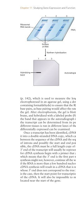
Figure 11.2<br />
Northern hybridization. Three RNA extracts from<br />
different tissues have been electrophoresed in an<br />
agarose gel. The extracts are made up of many<br />
RNAs of different lengths so each gives a smear<br />
of RNA, but two distinct b<strong>and</strong>s are seen, one for<br />
each of the abundant ribosomal RNAs. The sizes<br />
of these rRNAs are known (e.g. 4718 <strong>and</strong> 1874<br />
nucleotides in mammals), so they can be used as<br />
internal size markers. The gel is transferred to a<br />
membrane, probed with a cloned gene, <strong>and</strong> the<br />
results visualized, for example by autoradiography<br />
if the probe has been radioactively labeled. Only<br />
lane 1 gives a b<strong>and</strong>, showing that the cloned gene<br />
is expressed only in the tissue from which this<br />
RNA extract was obtained.<br />
(p. 142), which is used to measure the length of a transcript. <strong>An</strong> RNA extract is<br />
electrophoresed in an agarose gel, using a denaturing electrophoresis buffer (e.g., one<br />
containing formaldehyde) to ensure that the RNAs do not form inter- or intramolecular<br />
base pairs, as base pairing would affect the rate at which the molecules migrate through<br />
the gel. After electrophoresis, the gel is blotted onto a nylon or nitrocellulose membrane,<br />
<strong>and</strong> hybridized with a labeled probe (Figure 11.2). If the probe is a cloned gene,<br />
the b<strong>and</strong> that appears in the autoradiograph is the transcript of that gene. The size of<br />
the transcript can be determined from its position within the gel, <strong>and</strong> if RNA from<br />
different tissues is run in different lanes of the gel, then the possibility that the gene is<br />
differentially expressed can be examined.<br />
Once a transcript has been identified, c<strong>DNA</strong> synthesis (p. 133) can be used to convert<br />
it into a double-str<strong>and</strong>ed <strong>DNA</strong> copy, which can be cloned <strong>and</strong> sequenced. Comparison<br />
between the sequence of the c<strong>DNA</strong> <strong>and</strong> the sequence of its gene will reveal the positions<br />
of introns <strong>and</strong> possibly the start <strong>and</strong> end points of the transcript. For this to be possible,<br />
the c<strong>DNA</strong> must be a full length copy of the mRNA from which it is derived. The<br />
3′ end of the transcript will usually be represented in the c<strong>DNA</strong>, because most methods<br />
for c<strong>DNA</strong> synthesis begin with a primer that anneals to the poly(A) tail of the mRNA,<br />
which means that the 3′ end is the first part to be copied (see Figure 8.7). The c<strong>DNA</strong><br />
synthesis might not, however, continue all the way to the 5′ end of the transcript, especially<br />
if the RNA is more than a few hundred nucleotides in length. Premature termination of<br />
c<strong>DNA</strong> synthesis will result in a c<strong>DNA</strong> that is not a full length copy of its transcript, <strong>and</strong><br />
whose 3′ end does not correspond to the true 5′ end of the mRNA (Figure 11.3). If this<br />
is the case, then the start point for transcription cannot be identified from the sequence<br />
of the c<strong>DNA</strong>. It will also be impossible to map the positions of any introns that are<br />
located near the start of the gene.