VectorCatalog2012.pdf? - Vector Laboratories
VectorCatalog2012.pdf? - Vector Laboratories
VectorCatalog2012.pdf? - Vector Laboratories

Create successful ePaper yourself
Turn your PDF publications into a flip-book with our unique Google optimized e-Paper software.
C<br />
C<br />
2<br />
V E C T O R L A B O R A T O R I E S<br />
Introduction<br />
Fluorescence and enzyme-based detection reagents from<br />
<strong>Vector</strong> <strong>Laboratories</strong> are ideal for in situ hybridization (ISH)<br />
applications, because of their high affinity, high sensitivity, and<br />
low background.<br />
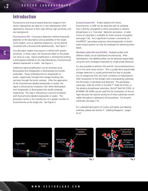
Fluorescence ISH. Choosing a detection method frequently<br />
depends on the abundance and accessibility of the target.<br />
Some targets, such as repetitive sequences, can be directly<br />
visualized with a fluorescently labeled probe. See Figure 1.<br />
Less abundant targets may require a method with greater<br />
sensitivity. In these cases, the fluorescent label on the probe<br />
can serve as a tag. Signal amplification is achieved by binding<br />
a biotinylated antibody to this tag followed by a fluorochromelabeled<br />
streptavidin or avidin. See Figure 2.<br />
Additional signal amplification can be achieved using<br />
Biotinylated Anti-Streptavidin or Biotinylated Anti-Avidin<br />
antibodies. These antibodies bind to streptavidin or<br />
avidin, respectively, through their antigen binding sites<br />
and also through the biotin residues. After the application<br />
of the fluorochrome-labeled streptavidin or avidin, the<br />
signal is enhanced by incubation with either Biotinylated<br />
Anti-Streptavidin or Biotinylated Anti-Avidin antibody,<br />
respectively. This step is followed by a second incubation<br />
with fluorochrome-labeled streptavidin or avidin. This<br />
procedure results in the introduction of a greater number of<br />
fluorochromes at the target site. See Figure 3.<br />
Target DNA Fluorescent<br />
Probe<br />
Biotinylated<br />
Antibody<br />
w w w . V E C T O R L A B S . C O m<br />
Enzyme-based ISH. Probes labeled with biotin,<br />
fluorochrome, or DNP can be detected with an antibody<br />
that is directly conjugated to either peroxidase or alkaline<br />
phosphatase in a “one-step” detection procedure. A wide<br />
choice of substrates is available for these enzyme conjugates<br />
(see page C16). For a significant increase in sensitivity, an<br />
ImmPRESS peroxidase polymer, biotin/streptavidin or biotin/<br />
avidin based systems can also be employed for detecting these<br />
labels.<br />
Multiple Label ISH and IHC/ISH. Multiple probes with<br />
different labels can be hybridized simultaneously. After<br />
hybridization, the labeled probes can be detected sequentially<br />
using the same strategies employed for single probe detection.<br />
It is also possible to perform ISH and IHC (immunohistochemistry)<br />
in the same tissue section. This is usually done sequentially.<br />
Antigen detection is usually performed first due to possible<br />
loss of antigenicity from the harsh conditions of hybridization.<br />
After localization of the antigen with a precipitating substrate,<br />
the ISH probe is hybridized and detected. The peroxidase<br />
substrates, DAB (SK-4100) or ImmPACT DAB (SK-4105), or<br />
the alkaline phosphatase substrates, <strong>Vector</strong> ® Red (SK-5100) or<br />
BCIP/NBT (SK-5400) can be used first for localization of the antigen<br />
because the reaction products of these substrates remain<br />
stable throughout subsequent ISH procedures. For enzyme<br />
substrates see page C16.<br />
For a detailed description of nucleic acid labels and labeling<br />
methods, please see Section F, “Labeling Reagents”, pages<br />
F2-F7.<br />
Fluorochrome-<br />
Labeled<br />
(Strept)avidin<br />
Second Layer of<br />
Fluorescent<br />
Strept(avidin)<br />
Biotinylated<br />
Anti-<br />
Streptavidin<br />
or Anti-Avidin<br />
Fig. 1 Fig. 2 Fig. 3