ABSTRACTS OF THE 21st ANNUAL MEETING OF THE ITALIAN ...
ABSTRACTS OF THE 21st ANNUAL MEETING OF THE ITALIAN ...
ABSTRACTS OF THE 21st ANNUAL MEETING OF THE ITALIAN ...
Create successful ePaper yourself
Turn your PDF publications into a flip-book with our unique Google optimized e-Paper software.
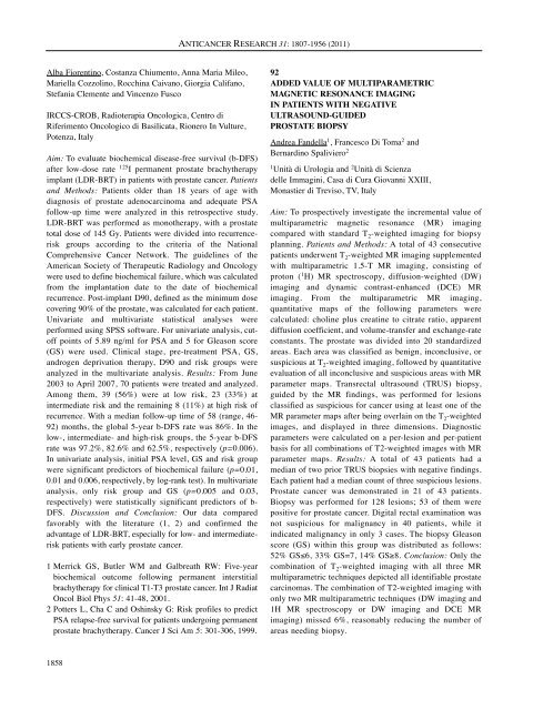
Alba Fiorentino, Costanza Chiumento, Anna Maria Mileo,<br />
Mariella Cozzolino, Rocchina Caivano, Giorgia Califano,<br />
Stefania Clemente and Vincenzo Fusco<br />
IRCCS-CROB, Radioterapia Oncologica, Centro di<br />
Riferimento Oncologico di Basilicata, Rionero In Vulture,<br />
Potenza, Italy<br />
Aim: To evaluate biochemical disease-free survival (b-DFS)<br />
after low-dose rate 125 I permanent prostate brachytherapy<br />
implant (LDR-BRT) in patients with prostate cancer. Patients<br />
and Methods: Patients older than 18 years of age with<br />
diagnosis of prostate adenocarcinoma and adequate PSA<br />
follow-up time were analyzed in this retrospective study.<br />
LDR-BRT was performed as monotherapy, with a prostate<br />
total dose of 145 Gy. Patients were divided into recurrencerisk<br />
groups according to the criteria of the National<br />
Comprehensive Cancer Network. The guidelines of the<br />
American Society of Therapeutic Radiology and Oncology<br />
were used to define biochemical failure, which was calculated<br />
from the implantation date to the date of biochemical<br />
recurrence. Post-implant D90, defined as the minimum dose<br />
covering 90% of the prostate, was calculated for each patient.<br />
Univariate and multivariate statistical analyses were<br />
performed using SPSS software. For univariate analysis, cutoff<br />
points of 5.89 ng/ml for PSA and 5 for Gleason score<br />
(GS) were used. Clinical stage, pre-treatment PSA, GS,<br />
androgen deprivation therapy, D90 and risk groups were<br />
analyzed in the multivariate analysis. Results: From June<br />
2003 to April 2007, 70 patients were treated and analyzed.<br />
Among them, 39 (56%) were at low risk, 23 (33%) at<br />
intermediate risk and the remaining 8 (11%) at high risk of<br />
recurrence. With a median follow-up time of 58 (range, 46-<br />
92) months, the global 5-year b-DFS rate was 86%. In the<br />
low-, intermediate- and high-risk groups, the 5-year b-DFS<br />
rate was 97.2%, 82.6% and 62.5%, respectively (p=0.006).<br />
In univariate analysis, initial PSA level, GS and risk group<br />
were significant predictors of biochemical failure (p=0.01,<br />
0.01 and 0.006, respectively, by log-rank test). In multivariate<br />
analysis, only risk group and GS (p=0.005 and 0.03,<br />
respectively) were statistically significant predictors of b-<br />
DFS. Discussion and Conclusion: Our data compared<br />
favorably with the literature (1, 2) and confirmed the<br />
advantage of LDR-BRT, especially for low- and intermediaterisk<br />
patients with early prostate cancer.<br />
1 Merrick GS, Butler WM and Galbreath RW: Five-year<br />
biochemical outcome following permanent interstitial<br />
brachytherapy for clinical T1-T3 prostate cancer. Int J Radiat<br />
Oncol Biol Phys 51: 41-48, 2001.<br />
2 Potters L, Cha C and Oshinsky G: Risk profiles to predict<br />
PSA relapse-free survival for patients undergoing permanent<br />
prostate brachytherapy. Cancer J Sci Am 5: 301-306, 1999.<br />
1858<br />
ANTICANCER RESEARCH 31: 1807-1956 (2011)<br />
92<br />
ADDED VALUE <strong>OF</strong> MULTIPARAMETRIC<br />
MAGNETIC RESONANCE IMAGING<br />
IN PATIENTS WITH NEGATIVE<br />
ULTRASOUND-GUIDED<br />
PROSTATE BIOPSY<br />
Andrea Fandella1 , Francesco Di Toma2 and<br />
Bernardino Spaliviero2 1Unità di Urologia and 2Unità di Scienza<br />
delle Immagini, Casa di Cura Giovanni XXIII,<br />
Monastier di Treviso, TV, Italy<br />
Aim: To prospectively investigate the incremental value of<br />
multiparametric magnetic resonance (MR) imaging<br />
compared with standard T 2-weighted imaging for biopsy<br />
planning. Patients and Methods: A total of 43 consecutive<br />
patients underwent T 2-weighted MR imaging supplemented<br />
with multiparametric 1.5-T MR imaging, consisting of<br />
proton ( 1 H) MR spectroscopy, diffusion-weighted (DW)<br />
imaging and dynamic contrast-enhanced (DCE) MR<br />
imaging. From the multiparametric MR imaging,<br />
quantitative maps of the following parameters were<br />
calculated: choline plus creatine to citrate ratio, apparent<br />
diffusion coefficient, and volume-transfer and exchange-rate<br />
constants. The prostate was divided into 20 standardized<br />
areas. Each area was classified as benign, inconclusive, or<br />
suspicious at T 2-weighted imaging, followed by quantitative<br />
evaluation of all inconclusive and suspicious areas with MR<br />
parameter maps. Transrectal ultrasound (TRUS) biopsy,<br />
guided by the MR findings, was performed for lesions<br />
classified as suspicious for cancer using at least one of the<br />
MR parameter maps after being overlain on the T 2-weighted<br />
images, and displayed in three dimensions. Diagnostic<br />
parameters were calculated on a per-lesion and per-patient<br />
basis for all combinations of T2-weighted images with MR<br />
parameter maps. Results: A total of 43 patients had a<br />
median of two prior TRUS biopsies with negative findings.<br />
Each patient had a median count of three suspicious lesions.<br />
Prostate cancer was demonstrated in 21 of 43 patients.<br />
Biopsy was performed for 128 lesions; 53 of them were<br />
positive for prostate cancer. Digital rectal examination was<br />
not suspicious for malignancy in 40 patients, while it<br />
indicated malignancy in only 3 cases. The biopsy Gleason<br />
score (GS) within this group was distributed as follows:<br />
52% GS≤6, 33% GS=7, 14% GS≥8. Conclusion: Only the<br />
combination of T 2-weighted imaging with all three MR<br />
multiparametric techniques depicted all identifiable prostate<br />
carcinomas. The combination of T2-weighted imaging with<br />
only two MR multiparametric techniques (DW imaging and<br />
1H MR spectroscopy or DW imaging and DCE MR<br />
imaging) missed 6%, reasonably reducing the number of<br />
areas needing biopsy.


