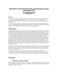
Simulation - ANSYS
Simulation - ANSYS
Simulation - ANSYS

Create successful ePaper yourself
Turn your PDF publications into a flip-book with our unique Google optimized e-Paper software.
BIOMEDICAL<br />
Understanding the<br />
Dangers of Aneurysms<br />
<strong>Simulation</strong> is used to validate measured<br />
blood flow in cerebral aneurysms.<br />
By Christof Karmonik, Research Scientist, Richard Klucznik, Director of Interventional Neuroradiology,<br />
Hani Haykal, Director of Neuroradiology and Goetz Benndorf, Director of Interventional Neuroradiology Research,<br />
The Methodist Hospital Research Institute, Department of Radiology, Houston, Texas, U.S.A.<br />
Cerebral aneurysms are arterial outpouchings in the<br />
brain that result from weak spots in the vessel wall. Rupture<br />
of a cerebral aneurysm can be dangerous for a patient and<br />
occurs most commonly between 40 and 60 years of age.<br />
When an aneurysm ruptures, blood leaks from the ruptured<br />
wall into the subarachnoid space, or the brain itself, potentially<br />
causing serious damage. This may lead to major<br />
permanent neurological deficits or even death. While it is<br />
estimated that approximately 3 percent to 6 percent of the<br />
general population have cerebral aneurysms, there is a rupture<br />
in only eight in approximately 100,000 person-years.<br />
The overall prognosis is poor for the patients with aneurysm<br />
rupture — 50 percent will die one month after rupture and<br />
an additional 20 percent will be unable to live on their own.<br />
Aneurysm growth and rupture depends on multiple<br />
factors: geometrical factors such as aneurysm size and<br />
shape or the ratio of the aneurysm dome height to the neck<br />
width; biological factors such as decreased concentration<br />
of structural proteins of the extracellular matrix in the<br />
intracranial arterial wall; and hemodynamic factors, especially<br />
wall shear stresses. While patients with unruptured<br />
aneurysms may have symptoms such as headache,<br />
The 3-D model created by the NOVA system of the cerebral vasculature using the MRI TOF images<br />
(bottom) shows the anatomical location of the aneurysm. The yellow plane illustrates a scan plane for<br />
recording the inflow waveform in the left anterior cerebral artery. The DSA model was then used to<br />
create a surface model used by the GAMBIT tool.<br />
38<br />
<strong>ANSYS</strong> Advantage • Volume II, Issue 2, 2008<br />
peripheral visual deficits, loss of balance and coordination,<br />
or other neurological deficits (depending on the location of<br />
the aneurysm), often cerebral aneurysms are asymptomatic,<br />
especially if they are small in size.<br />
When an aneurysm is detected, treatment options other<br />
than conservative management include surgical clipping or<br />
minimally invasive therapy that approaches and occludes, or<br />
blocks, the aneurysm using endovascular techniques. Both<br />
treatment options are associated with an overall morbidity<br />
and mortality of about 11 percent. Because not all aneurysms<br />
will rupture and bleed, it would be highly beneficial to understand<br />
the underlying mechanisms that lead to rupture in<br />
order to minimize risk to the patient. Currently, there is<br />
no medical imaging modality that can provide complete<br />
quantitative information about hemodynamic parameters,<br />
such as wall shear stresses and dynamic pressure.<br />
In order to better understand the dynamic forces of blood<br />
flow within an aneurysm and the conditions that may cause<br />
rupture, an interdisciplinary team of researchers at The<br />
Methodist Hospital Research Institute (TMHRI) in Houston,<br />
Texas, is studying hemodynamics using computational fluid<br />
dynamics (CFD) simulations with FLUENT software. More<br />
Pathlines of blood flow during time of average inflow<br />
www.ansys.com