Hypoplasia-Hyperplasia
Hypoplasia-Hyperplasia
Hypoplasia-Hyperplasia

You also want an ePaper? Increase the reach of your titles
YUMPU automatically turns print PDFs into web optimized ePapers that Google loves.
ע"<br />
שת/<br />
תבט/<br />
ד"<br />
י•<br />
21•<br />
Vertical Distraction<br />
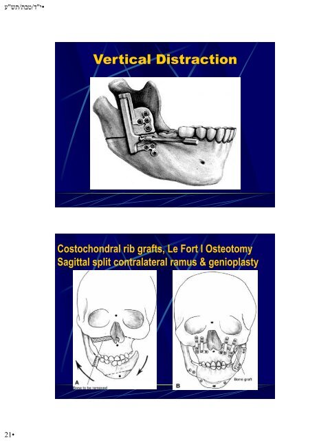
Costochondral rib grafts, Le Fort I Osteotomy<br />
Sagittal split contralateral ramus & genioplasty<br />
A: The skeletal deformity in unilateral<br />
(left) craniofacial microsomia. Note the<br />
absent ramus with deviation of the chin to<br />
the affected side and an occlusal cant<br />
upward toward the left side. The arrows<br />
designate the proposed skeletal movements<br />
of the mandible and maxilla. The dots<br />
designate the midpoints of the upper face,<br />
midface and chin. The dotted area<br />
represents the area where bone is removed<br />
from the maxilla and the interrupted lines<br />
designate the proposed maxillary and chin<br />
osteotomy sites. (Modified from Munro, I.<br />
R., Lauritzen C. G. Classification and<br />
treatment of hemifacial microsomia. In:<br />
Caronni, E. P., (ed.). Craniofacial Surgery.<br />
Boston: Little, Brown & Co., 391–400,<br />
1985.)<br />
B: At the completion of the<br />
osteotomies with plate and screw<br />
fixation applied. Note that the dots<br />
have recreated the midsagittal<br />
plane of the craniofacial skeleton.<br />
The ramus has been reconstructed<br />
with costochondral rib grafts. The<br />
Le Fort I segment has been<br />
impacted on the right side and a<br />
bone graft has been inserted on the<br />
left side. The sagittal split<br />
osteotomy has been fixated with<br />
screws and the genioplasty has<br />
been secured with lag screws.