Hypoplasia-Hyperplasia
Hypoplasia-Hyperplasia
Hypoplasia-Hyperplasia

Create successful ePaper yourself
Turn your PDF publications into a flip-book with our unique Google optimized e-Paper software.
ע"<br />
שת/<br />
תבט/<br />
ד"<br />
י•<br />
48•<br />
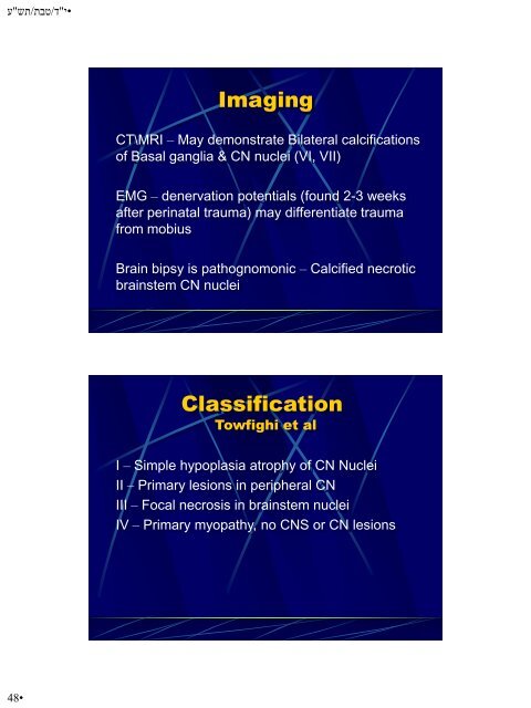
Imaging<br />
CT\MRI – May demonstrate Bilateral calcifications<br />
of Basal ganglia & CN nuclei (VI, VII)<br />
EMG – denervation potentials (found 2-3 weeks<br />
after perinatal trauma) may differentiate trauma<br />
from mobius<br />
Brain bipsy is pathognomonic – Calcified necrotic<br />
brainstem CN nuclei<br />
Classification<br />
Towfighi et al<br />
I – Simple hypoplasia atrophy of CN Nuclei<br />
II – Primary lesions in peripheral CN<br />
III – Focal necrosis in brainstem nuclei<br />
IV – Primary myopathy, no CNS or CN lesions